
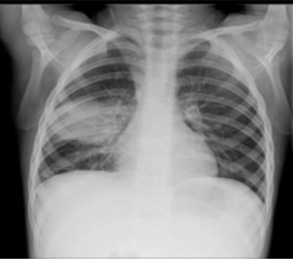
Respiratory symptoms are among the most frightening and distressing experiences for palliative care patients. Unlike pain, which a patient can often hide, breathlessness is visible and terrifying — for the patient, the family, and the nurse.
Breathlessness is a subjective, frightening sensation of difficult or uncomfortable breathing. It is not the same as low oxygen levels — a patient can have normal oxygen saturation but still feel they are suffocating.
The patient's words: "I felt like I was suffocating," "I couldn't get enough air," "It felt like I was about to die."
Physiological Expansion (The "Air Hunger" Mechanism): Dyspnoea occurs when there is a mismatch between the brain's motor command to breathe (respiratory drive from the medulla) and the mechanical response of the respiratory system. When chemoreceptors (sensing CO2/O2) or mechanoreceptors (in the lungs/chest wall) send signals that the breathing effort is insufficient, the brain registers this as life-threatening "air hunger," triggering massive sympathetic nervous system panic.
| System | Causes |
|---|---|
| Respiratory | Primary or secondary lung cancers, pleural effusion, pulmonary embolism, tracheal tumours, airway collapse, infections (pneumonia, TB), lymphangitis carcinomatosa (cancer spread to lymphatic vessels), COPD, weak respiratory muscles. |
| Cardiac | Superior vena cava obstruction (SVCO), anaemia, cardiac failure, cardiomyopathy, pericardial effusion. |
| Other | Ascites (pressure on diaphragm), radiotherapy/chemotherapy side effects, pneumonectomy, anxiety. |
- R - Respiratory (cancer, effusion, embolism, infection, COPD)
- E - Effusion (pleural, pericardial)
- S - SVCO (Superior Vena Cava Obstruction)
- P - Pulmonary embolism
- I - Infection (pneumonia, TB)
- R - Radiotherapy / treatment effects
- E - Emotional (anxiety, panic)
| Question to Ask | Why It Matters |
|---|---|
| "When did it start? Sudden or gradual?" | Sudden = embolism, pneumothorax, acute infection. |
| "Is it worse lying down?" (Orthopnoea) | Suggests cardiac failure, pleural effusion, SVCO. |
| "Is it worse on exertion?" | Suggests cardiac or respiratory limitation. |
| "Any chest pain?" | Pleuritic pain = infection, embolism, tumour. |
| "Any blood in sputum?" | Haemoptysis — see Section 5. |
| "What makes it better or worse?" | Guides positioning and intervention. |
| "How does it make you feel emotionally?" | Identifies anxiety and panic as contributors. |
| Intervention | Details | Rationale |
|---|---|---|
| Positioning | Usually sitting upright with pillows for support. | Gravity pulls abdominal organs down, allowing maximum diaphragmatic excursion. |
| Pleural effusion positioning | Lie on affected side with good lung upwards. | Maximises ventilation and perfusion matching (V/Q) of the healthy lung. |
| Ventilation | Open windows, use a fan, or fan with newspaper. | Cool air stimulates trigeminal nerve → reduces sensation of breathlessness. |
| Breathing techniques | Slow, deep breathing; pursed-lip breathing. | Increases positive end-expiratory pressure (PEEP), keeping airways open and reducing panic. |
| Activity pacing | Rest between activities; avoid overexertion. | Conserves cellular energy and reduces oxygen demand. |
| Suction secretions | Gently suction excessive secretions if present. | Clears mechanical airway obstruction. |
| Reassurance and presence | Stay with the patient; hold their hand; speak calmly. | Reduces sympathetic nervous system panic, which worsens breathlessness. |
| Drug | Dose | Indication | Notes / Mechanism |
|---|---|---|---|
| Morphine | 2.5–5 mg PO every 4 hours | Reduces the sensation of breathlessness | If already on morphine for pain, increase by 2.5 mg. Mechanism: Binds to Mu-receptors in the medulla, altering the brain's response to high CO2, making the brain "ignore" the air hunger. Does NOT dangerously suppress respiration at these low doses. |
| Diazepam | 2–5 mg at night | Anxiety and panic associated with breathlessness | Also helps sleep. Enhances GABA (inhibitory neurotransmitter). |
| Dexamethasone | 8–12 mg daily | SVCO, lymphangitis carcinomatosa, airway compression | Potent corticosteroid. Reduces peritumoral oedema and inflammation, mechanically opening the airway. |
| Bronchodilators | Salbutamol, ipratropium | Reversible airway obstruction (COPD, asthma) | Nebulised or inhaler. Relaxes bronchial smooth muscle. |
| Diuretics | Frusemide 40 mg IV | Cardiac failure, pleural effusion, ascites | Reduces fluid overload and pulmonary congestion. |
| Oxygen | 2–4 L/min via nasal cannula | If hypoxic (SpO2 < 90%) and available | May not help the sensation of dyspnoea if SpO2 is already normal. |
| Population | Incidence of Cough |
|---|---|
| All cancer patients | ~30% |
| Lung / bronchus cancer patients | ~80% |
| HIV/AIDS patients with cough | Any duration of cough = high suspicion of TB |
Uganda : In any patient living with HIV/AIDS, cough should always raise suspicion of tuberculosis. Refer for GeneXpert (MTB/RIF) testing immediately.
Anatomy of the Cough Reflex: Receptors in the airway detect irritation → Vagus nerve (afferent) sends signal to Medulla → Medulla sends efferent signal via Phrenic and Spinal nerves → Diaphragm and intercostal muscles contract forcefully against a closed glottis, which then snaps open to expel air.
| Cause | Explanation |
|---|---|
| Bronchial obstruction | Primary tumour or enlarged mediastinal lymph nodes — most common cause in cancer. |
| Infection | TB, pneumonia — especially in immunosuppressed patients. |
| Left ventricular failure | Dyspnoea and cough that wakes the patient at night (paroxysmal nocturnal dyspnoea) due to fluid backing up into the lungs. |
| Vocal cord paralysis | Due to hilar tumour or lymphadenopathy compressing the Recurrent Laryngeal Nerve, making the vocal cords unable to close properly for an effective cough. |
| Unrelated causes | Smoking, common cold, asthma, congestive heart failure. |
| Feature to Assess | What to Look For |
|---|---|
| Type of cough | Productive (with phlegm) or dry? |
| Ability to cough effectively | Weak cough = severe risk of aspiration and retained secretions leading to pneumonia. |
| Sputum characteristics | Colour (yellow/green = infection; blood = haemoptysis); amount; consistency. |
| Precipitating factors | Worse at night? After eating? On exertion? In certain positions? |
| Associated symptoms | Fever (infection), weight loss (TB, cancer), chest pain, dyspnoea. |
| Physical examination | Mouth, throat, lungs (auscultation), heart. |
| Intervention | Details |
|---|---|
| Postural drainage | Position patient to allow gravity to drain secretions from affected lung segments. |
| Steam inhalation | Helps liquefy thick sputum; add menthol or eucalyptus if available. |
| Antibiotics | For confirmed or suspected infection (e.g., TB, pneumonia). |
| Bronchodilators | Salbutamol in cough mixture if bronchospasm present. |
| Hydration | Adequate fluids thin secretions (if not contraindicated by heart failure). |
| Drug | Dose | Notes |
|---|---|---|
| Codeine linctus | 10 ml every 4 hours (1 mg/ml) | Suppresses the medullary cough reflex; highly useful at night to allow sleep. |
| Morphine | 2.5 mg, increase usual dose by 2.5 mg every 4 hours | More potent medullary cough suppressant; also helps if pain coexists. |
- Positioning: Propped up with 2–3 pillows in the most comfortable position.
- Pleural effusion: Lie on side of effusion in semi-recumbent position.
- Humidification: Steam inhalation or humidified oxygen.
- Encourage expectoration: Provide tissues, emesis basin; assist weak patients.
- Monitor for haemoptysis: See Section 5.
- Infection control: If TB suspected, wear a mask; isolate if confirmed.
Death rattles — also called terminal secretions or noisy breathing — occur when a dying patient loses the ability to cough or swallow, and saliva and bronchial secretions accumulate in the back of the throat and upper airways. This creates a gurgling, rattling sound with each breath.
| Mechanism | Explanation |
|---|---|
| Loss of swallowing reflex | The brainstem (Glossopharyngeal IX and Vagus X nerves) functions that control swallowing fail. |
| Loss of cough reflex | Secretions cannot be cleared from the airway due to severe muscle weakness and neurological decline. |
| Pooling of secretions | Saliva and bronchial secretions accumulate in the oropharynx and trachea. Air bubbling through this fluid creates the sound. |
| Relaxation of muscles | The jaw and airway muscles relax, allowing secretions to pool further. |
- Level of consciousness: Usually reduced or unconscious.
- Airway sounds: Gurgling, rattling, bubbling — usually louder on inspiration.
- Secretions in mouth: Pooling of saliva; may dribble from the mouth.
- Respiratory pattern: Often irregular (Cheyne-Stokes breathing or agonal gasps).
| Intervention | How to Do It / Rationale |
|---|---|
| Repositioning | Turn patient onto their side (lateral position). Allows gravity to drain secretions from the mouth rather than pooling in the throat. |
| Oral suctioning | Gentle suction of mouth and oropharynx ONLY. Clears visible secretions. Do NOT deep suction (causes severe distress, bleeding, and trauma). |
| Mouth care | Swab mouth with moistened gauze or sponge. Keeps mouth comfortable; removes excess thick saliva. |
| Elevate head of bed | 30–45° if possible. Assists postural drainage. |
| Reassure family | Explain that this sound is normal, not distressing to the patient, and a sign that death is near. This is your most important intervention! |
Anticholinergic drugs block the parasympathetic nervous system, drastically reducing salivary and bronchial secretions ("drying them up"), thereby reducing the rattling sound.
| Drug | Dose / Route | Notes & Blood-Brain Barrier (BBB) Effect |
|---|---|---|
| Hyoscine butylbromide (Buscopan) | 20 mg SC or IV every 4–6 hours | Reduces secretions; also heavily smooth muscle relaxant (helps with colic). |
| Hyoscine hydrobromide | 0.4 mg SC every 4 hours or via syringe driver | CROSSES the blood-brain barrier. This causes central sedation. Highly effective for secretions. Preferred if the patient is agitated or unconscious. |
| Glycopyrronium bromide | 0.2–0.4 mg SC every 4 hours or via syringe driver | DOES NOT cross the blood-brain barrier. Causes zero central sedation. Preferred if the patient is still somewhat conscious and wants to interact with family. |
| Atropine | 1% eye drops — 2 drops sublingually Q4H | Sublingual route is an excellent alternative if injections are unavailable. |
Your explanation and reassurance are often more therapeutic than any drug. Families remember how you made them feel during this time.
- "Is he choking?" ➔ "No, he is not choking. The sound is from saliva pooling in the throat because he is too weak to swallow. He is not in distress."
- "Is she suffering?" ➔ "She is unconscious and not aware of the sound. We are keeping her comfortable and her mouth moist."
- "Can't you do something to stop it?" ➔ "We are giving medicine to reduce the secretions and turning her to help drainage. The sound may lessen but may not stop completely. This is a natural part of the dying process."
- "How long does this last?" ➔ "It usually means death is hours to a few days away. We will stay with you and keep her comfortable."
Haemoptysis is the coughing up of blood from the respiratory tract — ranging from blood-streaked sputum to massive, life-threatening bleeding.
- Mild: Blood-streaked sputum; small amounts.
- Moderate: Frank blood in sputum; several tablespoons.
- Massive: >100–600 ml in 24 hours. Can be fatal rapidly due to asphyxiation.
| Cause | Explanation |
|---|---|
| Lung cancer | Tumour erosion directly into blood vessels; most common cause in oncology. |
| Tuberculosis (TB) | Cavitary TB erodes into pulmonary arteries, sometimes forming a fragile aneurysm (Rasmussen's aneurysm) that bursts. |
| Pulmonary embolism | Infarction causes necrosis and bleeding into alveoli. |
| Infection / Bronchiectasis | Severe pneumonia, lung abscess, or dilated damaged airways with fragile neovascularized vessels. |
| Coagulopathy | Low platelets, anticoagulant medications, liver failure. |
| Aspergilloma | Fungus ball growing inside a pre-existing lung cavity (very common in healed TB). |
Uganda Context: In HIV-positive patients, TB and fungal infections (aspergilloma) are incredibly important causes of haemoptysis. Always consider TB!
It is vital to distinguish coughing up blood (lungs) from vomiting blood (stomach).
| Feature | Haemoptysis (Lungs) | Haematemesis (Stomach) |
|---|---|---|
| Colour | Bright red, frothy (mixed with air) | Dark red or coffee-ground (digested by stomach acid) |
| pH | Alkaline | Acidic |
| Associated with | Cough, dyspnoea, chest symptoms | Nausea, vomiting, abdominal pain |
| History | Lung disease, TB, cancer, smoking | Peptic ulcer, liver disease, NSAID use |
- Reassurance: Explain that small amounts are common and not immediately dangerous.
- Treat underlying cause: Antibiotics for infection, anti-TB if confirmed.
- Cough suppression: Codeine or morphine to reduce coughing (vigorous coughing can dislodge clots and worsen bleeding).
- Monitor: Watch for increase in amount or frequency.
- Call for help immediately: This is life-threatening.
- Position patient: Lie the patient on the side of the BLEEDING lung (if known) or semi-prone. Rationale: Gravity keeps the blood in the diseased lung, preventing it from spilling over and drowning the healthy "good" lung.
- Keep calm and reassure: Panic spikes heart rate and blood pressure, which forcefully increases the bleeding.
- Suction & Oxygen: Keep airway clear of blood; provide high-flow O2.
- IV access: Large-bore cannula; fluids for shock.
- Medications: Tranexamic acid (1 g IV — antifibrinolytic), Vitamin K / FFP for coagulopathy, Morphine for severe distress and cough suppression.
- Definitive treatment: Bronchial artery embolisation if available.
- Stay with the patient: Reduces panic; allows instant monitoring for deterioration.
- Dark-coloured towels/bowls: Blood is highly visible and terrifying on white sheets. Using dark green/blue towels hides the visual impact of the blood, significantly reducing panic for the patient and family. Prepare these in advance for at-risk patients!
- Monitor vital signs: Tachycardia and hypotension indicate hypovolemic shock. Document estimated blood loss.
- Advance Care Planning: In advanced incurable disease where massive bleeding is expected, clarify DNR (Do Not Resuscitate) wishes. Have a terminal sedation protocol ready (e.g., Midazolam 5–10 mg SC/IV) to rapidly relieve terror if a terminal bleed occurs. Allow family to be present if they wish, or leave if it is too traumatic.
| Symptom | Key Feature | Most Common Cause | First-Line Management | Nursing Priority | Red Flag |
|---|---|---|---|---|---|
| Breathlessness | Frightening sensation of suffocation | Lung cancer, effusion, COPD, anxiety | Morphine 2.5–5 mg; fan; upright positioning | Stay with patient; reassurance | Stridor = airway emergency |
| Cough (productive) | Cough with phlegm | Bronchial obstruction, infection, TB | Postural drainage, antibiotics, bronchodilators | Positioning; infection control | Haemoptysis |
| Cough (dry) | Harsh, non-productive cough | Tumour irritation, post-nasal drip | Codeine linctus 10 ml Q4H; morphine | Night-time sedation; comfort | Increasing frequency |
| Death Rattles | Gurgling, rattling sound in dying patient | Loss of swallow/cough reflex | Hyoscine or glycopyrronium; repositioning | Reassure family | Family distress — manage this actively |
| Haemoptysis (massive) | Large-volume fresh blood | Eroded vessel, aspergilloma, TB | Position on bleeding side down; suction; O2 | Airway protection; calm presence | Asphyxiation risk — emergency |
- F - Fan (Cool air across the face / Trigeminal nerve)
- A - Anxiolytics (Diazepam for panic)
- N - Narcotic (Morphine reduces sensation of breathlessness)
- S - Steroids (Dexamethasone for SVCO, lymphangitis)
- R - Reposition (side-lying allows drainage)
- A - Anticholinergics (Hyoscine, glycopyrronium)
- T - Tell the family (Explain this is normal)
- T - Turn regularly (Prevents pooling)
- L - Listen and reassure (Your presence matters)
- E - Explain (Education reduces fear)
- B - Bleeding side down (Protect the good lung)
- L - Large-bore IV (For fluids/drugs)
- E - Emergency call (Get help immediately)
- E - Endotracheal suction (Keep airway clear)
- D - Dark towels (Reduce visual panic)
Q1: A patient with lung cancer becomes increasingly breathless. Oxygen saturation is 94% on room air. What is your first nursing intervention?
Answer: Position upright and use a fan. Oxygen may not help the sensation if SpO2 is adequate. The fan stimulates the trigeminal nerve and reduces the perception of breathlessness. Reassure the patient and stay with them.
Q2: A dying patient has loud, gurgling breathing. The family is distressed and asks if the patient is drowning. How do you respond?
Answer: Explain that this is terminal secretions — a normal part of the dying process. The patient is unconscious and not in distress. Turn the patient onto their side, give anticholinergics, and provide continuous reassurance to the family. Never deep suction.
Q3: An HIV-positive patient has had a cough for 3 weeks with night sweats and weight loss. What is your priority action?
Answer: Refer for TB investigation (GeneXpert). In Uganda, any cough in an HIV-positive patient must raise high suspicion of TB. Isolate if TB is confirmed.
Q4: A patient with lung cancer suddenly coughs up 200 ml of bright red blood. What is your immediate action?
Answer: This is massive haemoptysis (medical emergency). Position on the bleeding side down, call for help, suction airway gently, give oxygen, stay calm, and monitor for shock.
- Breathlessness is frightening — your calm presence is as important as any drug.
- A fan is free, safe, and effective for breathlessness — never forget it.
- Morphine relieves the sensation of breathlessness at low doses — it does not kill the patient.
- In HIV-positive patients, cough = think TB — refer for GeneXpert.
- Productive cough should not be heavily suppressed — the body needs to clear secretions.
- Death rattles are distressing to families, not the patient — your explanation is therapeutic.
- Never deep suction a dying patient — gentle oral suction and repositioning are sufficient.
- Glycopyrronium is preferred over hyoscine if the patient is still somewhat alert (less sedation).
- Massive haemoptysis is an airway emergency — position on the bleeding side down and protect the airway.
- Dark towels reduce panic during haemoptysis — prepare them in advance for at-risk patients.
- World Health Organization (WHO) Guidelines for the Pharmacological and Radiotherapeutic Management of Cancer Pain in Adults and Adolescents.
- Oxford Textbook of Palliative Nursing.
- National Guidelines for Palliative Care in Uganda.
- American Academy of Hospice and Palliative Medicine (AAHPM) Guidelines on Symptom Management.
Quick Quiz
Respiratory Symptoms Quiz
Palliative Care - mobile-friendly and focused practice.
Privacy: Your details are used only for quiz tracking and certificates.
Respiratory Symptoms Quiz
Palliative Care
Preparing questions...
Choose your answer and keep your streak alive.
Great effort.
Here is your quick performance summary.