
Pathology: Congenital & Developmental Anomalies of the Lung
I. Introduction to Lung Anomalies
Congenital and developmental anomalies of the lung refer to a broad spectrum of structural malformations that occur during embryonic and fetal lung development. The respiratory system begins developing early in gestation (around week 4) as an outpouching (respiratory diverticulum) from the ventral wall of the primitive foregut. Any disruption in the delicate sequence of branching, separation from the esophagus, vascularization, or alveolar differentiation can result in significant anatomical defects.
To understand these anomalies, one must recognize the 5 Stages of Fetal Lung Development:
- Embryonic (Weeks 4-7): Lung bud formation and initial branching. Errors here cause agenesis or tracheoesophageal fistulas.
- Pseudoglandular (Weeks 5-17): Formation of the bronchial tree up to terminal bronchioles.
- Canalicular (Weeks 16-25): Respiratory bronchioles and alveolar ducts form; primitive capillary networks develop. Viability becomes possible.
- Saccular (Week 26-Birth): Terminal sacs (primitive alveoli) form, and surfactant production begins.
- Alveolar (Week 36-8 Years): Maturation and massive multiplication of functional alveoli.
These anomalies vary widely in severity. Some are incompatible with extrauterine life and present as immediate respiratory failure in the delivery room, while others may remain asymptomatic and are only discovered incidentally in adulthood. Understanding these conditions requires a thorough grasp of both the structural defect and the resulting physiological compromise.
II. Classification of Congenital Lung Anomalies
Congenital anomalies of the respiratory system are broadly classified into three main categories based on the embryological structures involved:
- Congenital lobar emphysema
- Congenital bronchial atresia
- Congenital pulmonary airway malformation (CPAM)
- Bronchogenic cyst (Foregut duplications)
- Tracheal bronchus & Accessory cardiac bronchus
- Tracheomalacia & Tracheal stenosis
- Pulmonary underdevelopment/agenesis
- Hypogenetic lung (Scimitar) syndrome
- Bronchopulmonary sequestration (Intralobar and Extralobar)
- Absence of a main pulmonary artery
- Anomalous origin of the left pulmonary artery from the right (Pulmonary Sling)
- Anomalous pulmonary venous drainage
- Pulmonary arteriovenous malformation (AVM)
III. Detailed Bronchopulmonary Anomalies
Tracheal Stenosis
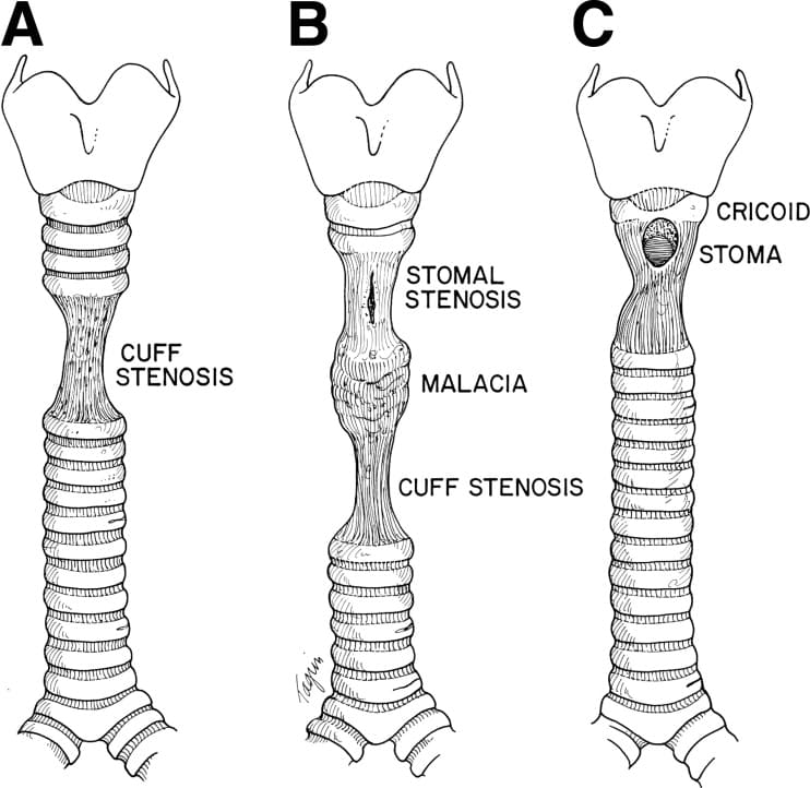
Tracheal stenosis is a congenital narrowing of the windpipe. Normally, the trachea is supported by C-shaped cartilage rings with a membranous, flexible posterior wall. In this anomaly, there is a focal or diffuse presence of complete (O-shaped) tracheal cartilage rings, resulting in a fixed, non-distensible tracheal narrowing.
- Presentation Patterns: It can occur as an isolated defect or in association with other congenital anomalies (like cardiovascular defects). Structurally, it presents as:
- ~ 50% Focal: Localized narrowing (often web-like).
- ~ 30% Generalized: Involving long segments of the trachea.
- ~ 20% Funnel-shaped: Tapering narrowing, resembling an inverted carrot.
- Clinical Features: 90% of cases present during the 1st year of life. Due to the fixed airway obstruction, it often presents with biphasic stridor (a harsh, high-pitched respiratory sound heard during both inspiration and expiration) and "washing machine" breathing sounds.
- Prognosis: Infants who present with severe symptoms and are diagnosed early in life generally have a worse prognosis. Importantly, the degree (severity) of the stenosis is more critical to survival than the length of the stenotic segment.
Tracheoesophageal Fistula (TEF)
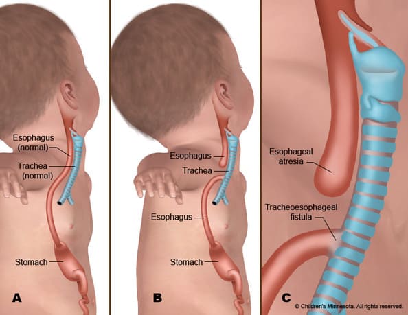
This is an abnormal connection (fistula) between the respiratory tract (trachea or bronchi) and the gastrointestinal tract (esophagus), often resulting from a failure of the primitive foregut to separate completely during weeks 4-5 of gestation.
The most common variant (accounting for ~85% of cases) is Type C: Proximal esophageal atresia (blind pouch) with a distal tracheoesophageal fistula. TEF is highly associated with VACTERL syndrome (Vertebral, Anorectal, Cardiac, Tracheoesophageal, Renal, and Limb anomalies).
- Clinical Impact: It is highly life-threatening and requires prompt surgical repair when possible.
- Diagnosis: The diagnosis is typically made shortly after birth. When the infant attempts to feed, there is an immediate inhalation of food or milk via the fistula directly into the lung parenchyma. This leads to profound choking, coughing, cyanosis, and severe chemical pneumonitis (lung inflammation). Polyhydramnios (excess amniotic fluid) is often noted on prenatal ultrasounds because the fetus cannot swallow the fluid.
Congenital Lobar Emphysema (CLE)
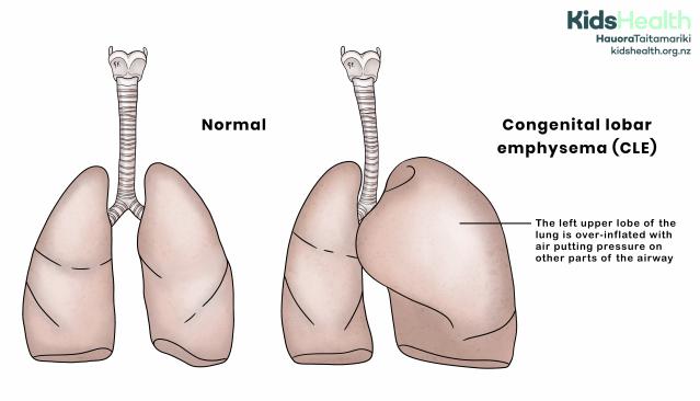
This condition involves the massive hyperinflation of a single lobe of the lung. The Left Upper Lobe is most commonly affected (40-50%), followed by the Right Middle Lobe (30%). It is not an "emphysema" caused by smoking or alveolar destruction, but rather a mechanical airway issue.
- Pathophysiology: When there is a failure of development (hypoplasia or dysplasia) of the cartilage plates in the main or large bronchus, the entire bronchus becomes "floppy" and collapses.
- The Ball-Valve Mechanism: This floppy airway creates a one-way valve. It allows air to enter the respiratory tree during inspiration (when negative chest pressure pulls the airway open), but it collapses during expiration, preventing air from exiting.
- Result: The trapping of air in the distal area of the pulmonary system causes severe inflation of the acini (air sacs) and eventual over-distension. This over-inflated lobe can compress the surrounding healthy lung and shift the mediastinum/heart (mediastinal shift), causing severe respiratory distress and hypoxia in a neonate.
Foregut Duplication Cysts
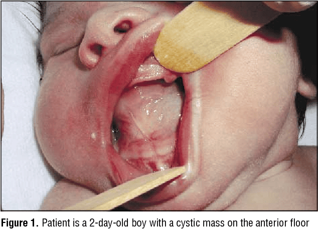
These are fluid-filled sacs that arise from the abnormal detachment of the primitive foregut during early embryonic development. They are most often located in the hilum of the lung, along the tracheobronchial tree, or the middle mediastinum.
They are classified according to their microscopic wall structure into three types: Bronchogenic cysts (most common), Esophageal cysts, and Enteric cysts.
Bronchogenic Cysts (Detailed)
- Anatomy: They are rarely connected directly to the functioning tracheobronchial tree. They form closed, blind sacs, most often near the carina.
- Presentation: They often present in children and young adults as an incidental finding on a chest X-ray. If symptomatic, symptoms relate to mass effect (compressing airways or the esophagus causing dysphagia or stridor), secondary infection, hemorrhage within the cyst, or rupture.
- Histology: The cyst is lined by ciliated pseudostratified columnar epithelium (classic respiratory lining). A definitive diagnosis is made because the wall contains bronchial mucosal glands, cartilage plates, and smooth muscle.
- Size & Complications: Size varies typically from 1 to 4 cm in diameter, but they can grow quite large. Complications include infection, internal hemorrhage, or erosion into adjacent critical structures. Rarely, malignant transformation can occur, leading to Rhabdomyosarcoma, pulmonary blastoma, anaplastic carcinoma, leiomyosarcoma, and adenocarcinoma.
Pulmonary or Peripheral Cysts
Unlike central bronchogenic cysts, these cystic lesions of the lung are often multiple and are located at the extreme periphery of the lungs. They are not in communication with a bronchus.
- Pneumothorax: A common complication is the rupture of these peripheral cysts directly into the pleural spaces. If a large amount of air accumulates, it results in a pneumothorax (collapsed lung) with hemodynamic compromise.
- Respiratory Distress: They may compress adjacent areas of the lungs, causing a loss of functional pulmonary air space.
- Infection & Hemorrhage: Secondary bacterial infection can lead to profound abscess formation. Erosion of an enlarging cyst inside a blood vessel may result in massive, life-threatening hemoptysis.
- Interstitial Emphysema: Infiltration of the leaked air into the surrounding soft tissues.
Other Bronchopulmonary Anomalies
- Congenital Bronchial Atresia: A focal obliteration of a proximal segmental bronchus. The distal lung tissue develops normally but becomes hyperinflated via collateral air drift (through the Pores of Kohn). Most commonly affects the apicoposterior segment of the left upper lobe.
- Tracheal Bronchus: An anomalous bronchus originating directly from the trachea (rather than the main bronchi), typically supplying the right upper lobe. Colloquially known as a "pig bronchus."
- Accessory Cardiac Bronchus: A rare anomalous bronchus arising from the inner wall of the intermediate bronchus, growing towards the heart.
- Tracheomalacia: Extreme weakness and flaccidity of the tracheal cartilage, leading to airway collapse during expiration. Often associated with prolonged intubation or vascular rings.
- Pulmonary Underdevelopment: Ranging from total agenesis (complete absence of lung tissue, bronchi, and vessels) to mild hypoplasia.
IV. Combined Lung and Vascular Anomalies
Pulmonary Sequestration
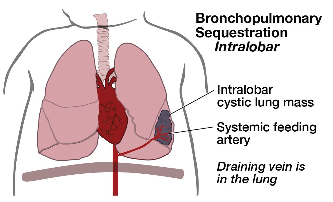
Pulmonary sequestration is a discrete mass of non-functioning lung tissue that completely lacks any normal connection to the airway system, and paradoxically receives an abnormal systemic blood supply (arising either directly from the aorta or its branches) rather than the pulmonary circulation.
Because this part of the lung is isolated from the rest of the organ, it cannot participate in gas exchange. It is classified into two distinct types:
| Feature | Intralobar Sequestration | Extralobar Sequestration |
|---|---|---|
| Location & Pleura | Located inside the normal lung tissue, sharing the same visceral pleural covering as the rest of the lobe. | Located outside the normal lung tissue, enveloped in its own separate visceral pleural sac. |
| Venous Drainage | Drains via normal pulmonary veins (can cause left-to-right shunting). | Drains via systemic veins (azygos/hemiazygos system or IVC). |
| Presentation Age | Usually presents in older children, adolescents, or adults. Often thought to be an acquired lesion following infection. | Most commonly presents in neonates and infants as a congenital mass lesion. |
| Clinical Signs | Often discovered due to recurrent localized pneumonias or bronchiectasis in the same lung segment. | May present with respiratory distress, cyanosis, or feeding difficulties shortly after birth. |
| Demographics / Specifics | Typically lower lobes. Less associated with other anomalies. | 90% occur on the left side (left base). 4:1 Male-to-Female ratio. Highly associated with other severe congenital anomalies (e.g., congenital diaphragmatic hernia, cardiac defects). |
Hypogenetic Lung Syndrome (Scimitar Syndrome)
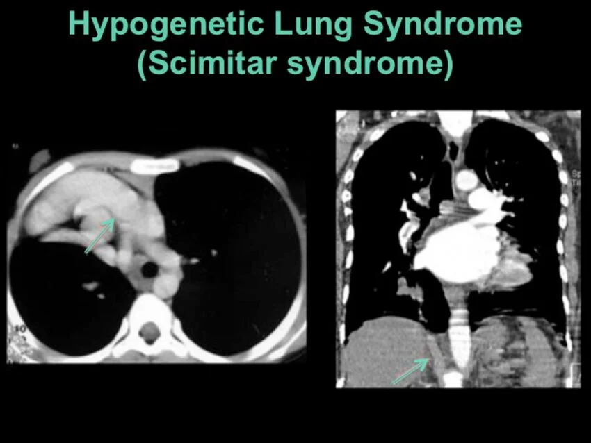
This is a rare and complex congenital condition characterized by the underdevelopment (hypoplasia) of the right lung and an anomalous pulmonary venous drainage system.
- Anomalous Venous Drainage: Instead of draining oxygenated blood into the left atrium, the pulmonary veins from the right lung drain abnormally directly into the inferior vena cava (IVC) below the diaphragm. This creates a left-to-right cardiovascular shunt.
- Other Drainage Sites: On occasion, the anomalous vein may drain into the hepatic veins, portal veins, azygous vein, coronary sinus, or directly into the right atrium.
- Anatomical Anomalies: The right lung may have abnormal lobation (frequently having only two lobes instead of three), and the bronchographic branching pattern may mirror or mimic that of the left lung (left isomerism). Right pulmonary artery hypoplasia is common.
- Associated Defects: About one-fourth (25%) of affected patients have associated congenital heart disease, most often a sinus venosus Atrial Septal Defect (ASD). Other reported associated anomalies include bronchogenic cysts, horseshoe lung (fusion of the bases of the lungs behind the heart), accessory diaphragm, and diaphragmatic hernia.
- Rationale for the name: It is called "Scimitar" syndrome because the anomalous vein draining down to the IVC creates a curved, sword-like (Turkish scimitar) shadow parallel to the right heart border on an AP chest X-ray.
V. Vascular Malformations of the Lung
The extent of vascular anomalies of the lungs varies drastically. It ranges from a small, insignificant arteriovenous fistula at the level of the capillaries to a complete absence of a major blood vessel (which is often associated with severe cardiac malformations that are not compatible with extra-uterine life).
- Absence of a main pulmonary artery: Leads to unilateral lack of perfusion.
- Anomalous origin of the left pulmonary artery: Forms a "pulmonary sling" that loops around and can strictly compress the trachea/esophagus.
- Anomalous pulmonary venous drainage: (Partial or Total - TAPVR) where pulmonary veins return oxygenated blood to the right heart instead of the left.
Arteriovenous Malformation (AVM): Abnormal direct connections between the pulmonary artery and pulmonary vein, bypassing the capillary bed.
- Genetic Association: 30 to 60% of pulmonary AVMs have a strong genetic association with Osler-Weber-Rendu syndrome (Hereditary Hemorrhagic Telangiectasia, linked to mutations in ENG or ACVRL1 genes).
Clinical Manifestations of AVM: Because unoxygenated venous blood shunts directly into the systemic circulation without passing through oxygen-exchanging capillaries (right-to-left shunt), patients develop profound systemic effects. Symptoms include:
- Dyspnea (shortness of breath) and Cyanosis.
- Hemoptysis (coughing up blood) and Hemothorax (blood in the pleural cavity) due to vessel rupture.
- Polycythemia: Abnormally high red blood cell count as the kidneys release EPO to compensate for chronic hypoxia.
- Clubbing of the fingers.
Normally, the lung capillary bed acts as a filter, trapping small venous blood clots originating from the deep veins (DVT). In an AVM, a venous clot can pass directly through the abnormal large vessel into the left heart and shoot up to the brain, causing a severe paradoxical stroke or brain abscess.
VI. Other Significant Lung Malformations
Pulmonary Hypoplasia
Pulmonary hypoplasia is the defective, incomplete development of both lungs (though one may be significantly more affected than the other), resulting in drastically decreased lung weight, lung volume, and number of acini compared to what is expected for the body weight and gestational age.
- Incidence & Prognosis: It is a surprisingly common anomaly, seen in up to 10% of all neonatal autopsies. Severe hypoplasia is uniformly fatal in the early neonatal period due to the absolute inability to oxygenate.
- Etiology (Causes): It is primarily caused by extrinsic abnormalities that physically compress the lung or impede lung expansion in utero. The fetal lungs require physical space in the thorax and internal fluid distension to grow normally. Causes include:
- Congenital Diaphragmatic Hernia: Abdominal organs (stomach, intestines, liver) herniate through a congenital hole in the diaphragm (usually the posterolateral Foramen of Bochdalek on the left side) into the chest cavity, physically squashing the developing lung tissue.
- Oligohydramnios (Potter Sequence): A critical lack of amniotic fluid (often due to bilateral renal agenesis or urinary tract obstruction). Fetal breathing of amniotic fluid provides the necessary internal distending pressure for the lungs to branch and grow. Without it, the lungs remain severely stunted.
- Thoracic Cage Abnormalities: Severe skeletal dysplasias (e.g., thanatophoric dysplasia) that restrict chest wall expansion.
Congenital Pulmonary Airway Malformation (CPAM)
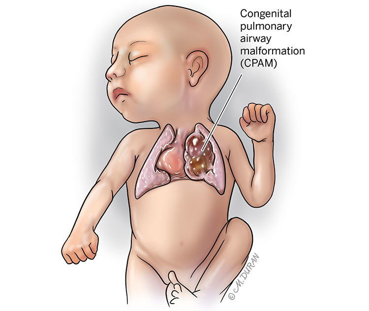
Formerly known as Congenital Cystic Adenomatoid Malformation (CCAM), CPAM is a hamartomatous lesion of the lung (a disorganized, tumor-like overgrowth of respiratory tissues that are normally present in the lung). It has an incidence of about 1 in 5,000 live births.
It can be separated into five highly specific types (Types 0 to 4) based on clinical, developmental timing, and microscopic pathologic features. The most clinically relevant are:
- CPAM Type 1 (Macrocystic): The most common form (65% of cases). It features a few very large cysts (2 to 10 cm in diameter) lined by ciliated pseudostratified epithelium. Fortunately, it carries a good prognosis and can often be surgically resected (lobectomy) with excellent long-term outcomes.
- CPAM Type 2 (Microcystic): Features multiple medium-sized cysts (< 2 cm). It accounts for 10-15% of cases. It often carries a poor prognosis, not strictly because of the lung lesion itself, but owing to its frequent association with other highly significant and fatal congenital anomalies (e.g., renal agenesis, severe cardiac defects, pulmonary sequestration).
- Other types (Types 0, 3, 4): These are rare.
- Type 0 (Acinar Dysplasia): Incompatible with life.
- Type 3 (Solid): Bulky, non-cystic adenomatoid overgrowth causing massive mediastinal shift and hydrops fetalis; very poor prognosis.
- Type 4: Distal acinar/peripheral cysts, notoriously associated with malignant transformation to Pleuropulmonary Blastoma.
VII. Neonatal Atelectasis & RDS
Atelectasis literally means "incomplete expansion" (from Greek ateles = incomplete, ektasis = stretching). In the context of neonatology, neonatal atelectasis refers to the failure of the lungs to expand adequately at or shortly after birth. It is a major cause of perinatal death and morbidity. Making a precise distinction between the primary and secondary forms is clinically vital, and especially useful in medico-legal/forensic cases.
Primary Atelectasis
In this form, there is absolutely no initial inflation of the lung tissue. No air has ever penetrated the respiratory tract of the newborn.
- Demographics: Seen mainly in severe premature infants, and in babies who have suffered profound intrauterine hypoxia (e.g., due to the kinking or knotting of the umbilical cord, placental abruption) resulting in a depression of the respiratory center and failure to take an initial breath.
- Infants of Diabetic Mothers: Maternal hyperglycemia causes fetal hyperglycemia, triggering massive compensatory hypersecretion of insulin by the fetal pancreas. While insulin promotes fetal growth (macrosomia), it actively inhibits the maturation of Type II pneumocytes. This results in decreased production of surfactant (dipalmitoylphosphatidylcholine), keeping the alveoli glued shut.
- Pathology: The lungs are completely collapsed. Grossly, they are red-blue, meaty, and have a distinct, tough, rubbery consistency because they contain no air. Microscopically, the alveolar spaces are tiny, unexpanded, and alveolar walls appear very thick.
- Forensic Relevance: In a flotation test, if a deceased infant's lungs sink entirely in water, it implies the infant was never born alive (stillbirth) or never took a breath.
Secondary Atelectasis (Resorption)
In this form, there is initial inflation of the lungs during vaginal delivery or shortly after. The baby breathes, but the lungs subsequently collapse.
- Pathophysiology (Aspiration): The infant aspirates massive amounts of amniotic fluid, meconium, or maternal blood (Meconium Aspiration Syndrome). This causes a physical obliteration and plugging of the terminal bronchioles, preventing further aeration of the distal lung tissue. The trapped air is eventually resorbed into the blood, leading to collapse.
- Chemical & Hypoxic Damage (NRDS): The resulting severe hypoxia and lack of surfactant cause necrosis of the alveolar epithelial cells. Endothelial damage causes protein-rich fluid (fibrin) to leak into the alveoli. This forms thick, glassy Hyaline Membranes that line the alveolar spaces, creating a rigid barrier to gas exchange (Neonatal Respiratory Distress Syndrome).
- Forensic Relevance: If a deceased infant shows secondary atelectasis (the lungs show some areas of expansion, hyaline membranes, and pieces of the lung float in water), it proves definitively that the infant was born alive, took independent breaths, and subsequently died of respiratory failure.
References & Further Reading
- Kumar, V., Abbas, A. K., & Aster, J. C. (2021). Robbins & Cotran Pathologic Basis of Disease (10th ed.). Elsevier. (Pathology of congenital lung anomalies and Neonatal Respiratory Distress Syndrome).
- Sadler, T. W. (2018). Langman's Medical Embryology (14th ed.). Wolters Kluwer. (Fetal lung development stages and foregut partitioning).
- Kliegman, R. M., et al. (2019). Nelson Textbook of Pediatrics (21st ed.). Elsevier. (Clinical presentations of CPAM, Sequestrations, and TEF).
- Stocker, J. T. (2002). Congenital pulmonary airway malformation: a new name for and an expanded classification of congenital cystic adenomatoid malformation of the lung. Histopathology, 41(Suppl 2), 424-431.
- Guttentag, S. H. (2020). Surfactant biology and Neonatal Respiratory Distress Syndrome. Clinics in Perinatology.