
Atelectasis & Pulmonary Edema
Atelectasis
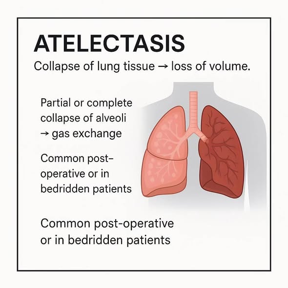
The term atelectasis is derived from the Greek words ateles (incomplete) and ektasis (expansion). It refers to either the incomplete expansion of the lungs (typically in neonates) or the collapse of a previously inflated lung in older patients. This results in areas of poorly aerated, airless pulmonary parenchyma.
Significant atelectasis drastically reduces oxygenation by creating a ventilation-perfusion (V/Q) mismatch (blood flows to the collapsed area but receives no oxygen, acting as a right-to-left shunt). Furthermore, the collapsed, poorly cleared airways strongly predispose the patient to secondary bacterial infections (pneumonia). Except in cases caused by fibrosis, atelectasis is generally a reversible disorder.
Classification & Types of Atelectasis
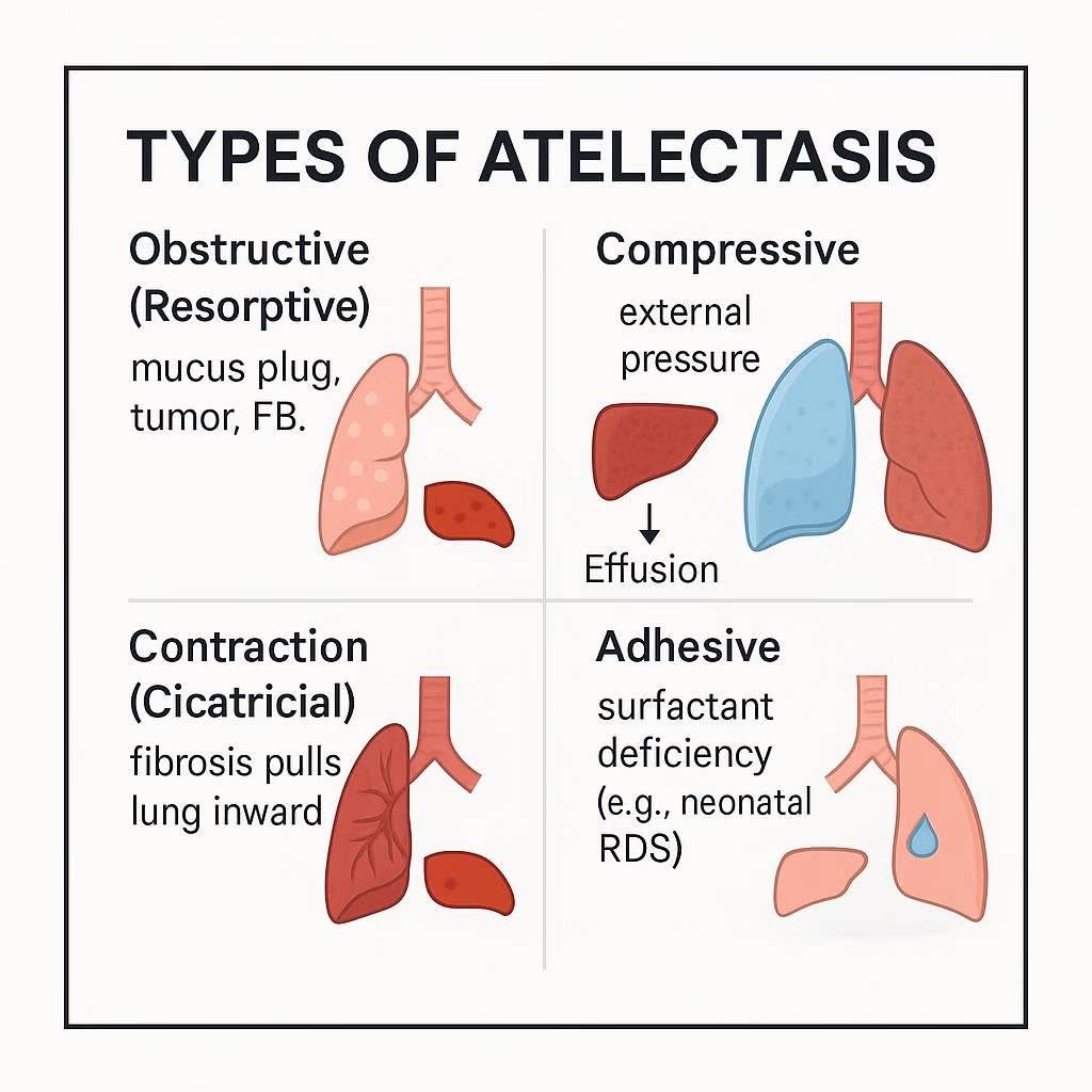
Acquired atelectasis in adults is primarily classified into three major mechanisms based on the underlying pathology:
Stems from a complete obstruction of an airway. Over time, the trapped air distal to the obstruction is resorbed into the pulmonary circulation, causing the alveoli to collapse.
- Mediastinal Shift: Because lung volume is lost, the negative intrapleural pressure pulls the mediastinum/trachea TOWARD the affected/atelectatic lung.
- Common Causes: Excessive secretions or mucus plugs (e.g., Bronchial asthma, chronic bronchitis, bronchiectasis, and critically in postoperative states where coughing is suppressed by pain).
- Other Causes: Aspiration of foreign bodies (common in children) and intrabronchial tumors.
Results whenever significant volumes of material accumulate within the pleural cavity, physically pushing against and collapsing the underlying fragile lung tissue.
- Mediastinal Shift: The accumulated mass or fluid pushes the mediastinum/trachea AWAY from the affected lung.
- Common Causes:
- Fluid: Pleural effusion (transudate in heart failure, exudate in infection), hemothorax (blood).
- Air: Tension pneumothorax.
- Tissue: Large intrathoracic tumors.
Occurs when focal or generalized pulmonary or pleural fibrosis (scar tissue) prevents full lung expansion. The scar tissue shrinks and mechanically restricts the alveoli.
- Reversibility: Unlike the other types, contraction atelectasis is irreversible because the lung parenchyma has been permanently replaced by stiff fibrotic tissue.
- Common Causes: Severe ARDS recovery, pulmonary fibrosis, chronic tuberculosis, or radiation-induced scarring.
Note: Neonatal atelectasis (Primary and Secondary) involves surfactant deficiency and amniotic fluid aspiration, covered comprehensively in pediatric developmental anomalies.
Pulmonary Edema
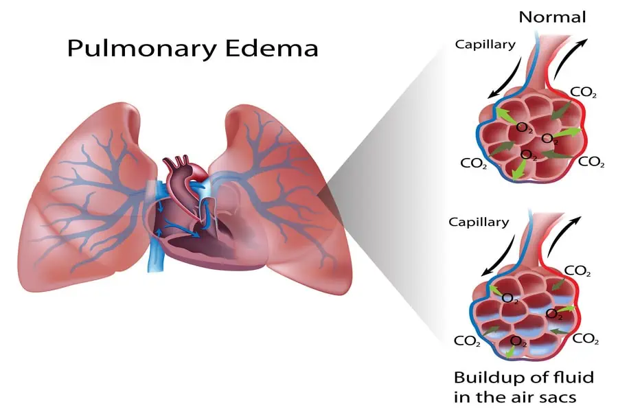
Pulmonary edema represents the abnormal transference of fluids from the intravascular compartment (pulmonary capillaries) into the lung interstitium and eventually into the alveolar spaces. This severe fluid accumulation creates a physical barrier to gas exchange, causing profound hypoxia (low oxygen) and hypercapnia (retention of carbon dioxide).
Pathophysiology & Starling Forces
The mechanism of pulmonary edema is governed by the same physiological principles as edema anywhere else in the body: Starling Forces.
Under normal conditions, a delicate balance keeps the interstitium and alveoli dry for optimal gas exchange:
- Plasma Oncotic Pressure: (Usually ~25 mmHg) The proteins in the blood (mostly albumin) act like a sponge, holding fluid inside the blood vessels.
- Pulmonary Capillary Hydrostatic Pressure: (Usually low, ~7-12 mmHg) The physical blood pressure pushing fluid out of the vessels.
- Tissue Barriers: The connective tissue and cellular barriers (tight junctions of alveolar epithelium) are relatively impermeable to plasma proteins.
- Lymphatic Drainage: An extensive pulmonary lymphatic system constantly vacuums up any minor physiological fluid leakage.
Edema occurs when this balance is broken (i.e., Hydrostatic pressure exceeds Oncotic pressure, or the barrier is physically destroyed).
The 3 Stages of Pulmonary Edema
Fluid accumulation does not happen all at once; it progresses through three distinct pathophysiological stages:
- Stage 1 (Compensated): Fluid transfer is increased into the lung interstitium. However, lymphatic flow also increases proportionately to drain it. There is no net increase in interstitial volume. The patient is usually asymptomatic.
- Stage 2 (Interstitial Edema): The capacity of the lymphatics to drain excess fluid is finally exceeded. Liquid begins to accumulate in the loose interstitial spaces that surround the bronchioles and lung vasculature (perivascular cuffing).
- Stage 3 (Alveolar Flooding): Fluid continues to build up. The increased pressure causes the fluid to track into the interstitial space around the thin alveoli. It finally disrupts and bursts through the tight junctions of the alveolar epithelial membranes. Fluid first builds up in the periphery of the alveolar-capillary membranes and finally floods the alveoli entirely. Gas exchange is heavily impaired, leading to respiratory failure.
Gravity plays a massive influence on the fluid mechanics of the lung. Under normal circumstances, more perfusion (blood flow) occurs at the lung bases than the apices due to gravity. However, when fluid accumulates heavily at the lung bases (creating increased local pressure), the pulmonary blood flow begins to be redistributed upward toward the apices. This phenomenon is known radiologically as Cephalization of pulmonary vessels.
Etiology (Causes of Pulmonary Edema)
The causes of pulmonary edema are broadly divided into hemodynamic (cardiogenic) and microvascular injury (non-cardiogenic) categories.
| Mechanism | Pathophysiology | Specific Causes & Examples |
|---|---|---|
| Increased Capillary Hydrostatic Pressure | The most common hemodynamic mechanism. High pressure backs up into the lungs, physically forcing fluid out. |
Cardiac Causes: Left ventricular failure (CHF), Mitral stenosis, Subacute bacterial endocarditis. Non-Cardiac Causes: Pulmonary venous fibrosis, Congenital stenosis of pulmonary veins, Pulmonary veno-occlusive disease, Iatrogenic fluid overload (over-infusion of IV fluids). |
| Altered Capillary Permeability | Primary injury to the endothelium or alveolar epithelium. Proteins leak out, drawing fluid with them (exudative). | Infections (Severe Pneumonia), Inhaled toxins (smoke, toxic gases), Vasoactive substances (histamine in anaphylaxis), DIC, Immunologic reactions, Radiation pneumonitis, Uremia, Near-drowning, Aspiration of gastric acid, and classically, ARDS (Acute Respiratory Distress Syndrome). |
| Decreased Oncotic Pressure | Lack of blood proteins (albumin) means there is no osmotic force to hold fluid in the vessels. | Hypoalbuminemia from: Renal failure (Nephrotic syndrome), Hepatic failure (Cirrhosis), Nutritional starvation (Kwashiorkor), Protein-losing enteropathies. |
| Lymphatic Insufficiency | Filtered interstitial fluid cannot be cleared from the connective tissue spaces. | Lymphatic obstruction by massive tumors (Lymphangitic carcinomatosis) or fibrosing diseases. |
Mixed / Unknown Mechanisms
- High Altitude Pulmonary Edema (HAPE): An acute, life-threatening form of non-cardiogenic edema that develops in the first 48 hours after reaching high altitudes (usually >10,000 ft) in unacclimatized people without preexisting heart/lung disease. Thought to be caused by patchy, severe hypoxic pulmonary vasoconstriction causing capillary stress failure in non-constricted areas. Symptoms resolve rapidly upon descent to sea level or oxygen administration.
- Neurogenic Pulmonary Edema: Occurs after severe central nervous system injury (head trauma, seizures, intracranial hemorrhage). A massive sympathetic nervous system discharge causes intense systemic vasoconstriction, rapidly shifting blood volume to the pulmonary circulation.
- Narcotic Overdose: (e.g., Heroin overdose). Exact mechanism is heavily debated but involves hypoxia-induced capillary damage and neurogenic factors.
- Other triggers: Pulmonary embolism, Eclampsia, Post-anesthetic states, and post-cardiopulmonary bypass.
Clinical Features & Diagnosis
1. Clinical Presentation
Patients presenting with acute pulmonary edema are often in severe distress. Classical signs include:
- Dyspnea, Tachypnea, & Orthopnea: Extreme shortness of breath, rapid breathing, and inability to breathe while lying flat (fluid spreads out over the lungs when supine).
- Cardiovascular Signs: Tachycardia (fast heart rate) and hypertension (compensatory sympathetic activation).
- Thoracic Oppression & Cyanosis: Feeling of a heavy chest, with cold extremities and bluish discoloration due to severe hypoxia.
- Cough with Frothy/Pink Sputum: A hallmark sign. The fluid in the alveoli mixes with air and surfactant to become frothy. It is pink because of ruptured capillaries leaking red blood cells into the mixture.
- Auscultation: Extensive use of accessory muscles of respiration. Stethoscopes reveal moist rales (crackles) at the lung bases, often accompanied by wheezing (sometimes called "cardiac asthma" due to peribronchial edema narrowing the airways).
2. Diagnostic Imaging (Chest X-Ray)
A CXR will typically show a bilateral reticulonodular pattern, more marked in the lung bases. Characteristic features include:
- Kerley B Lines: Short, horizontal linear shadows seen at the lung periphery (subpleurally) at the bases. They represent edematous, thickened interlobular septa.
- Kerley A Lines: Longer, unbranching lines pointing toward the hilum. They represent thickened connective tissue planes deeper in the lung.
- Cephalization: Prominent upper lobe vessels due to blood diversion from the edematous bases.
- Bat-wing/Butterfly appearance: In severe alveolar edema, fluffy central infiltrates spread outward from the hila.
Pathology & Autopsy Findings
Macroscopic (Gross) Findings
- The lungs are tremendously heavy and wet (a normal lung weighs ~300-400g; edematous lungs can weigh 2 to 3 times that).
- Upon slicing the lung tissue, a massive amount of frothy, blood-tinged (pink) fluid exudes freely from the cut surface.
Microscopic (Histological) Findings
- Severely congested alveolar capillaries (engorged with blood).
- Alveoli are completely filled with a homogeneous, pale pink-staining fluid (proteinaceous exudate or transudate).
- In ARDS specifically, you will see Hyaline Membranes (thick, waxy pink bands of dead cells and fibrin lining the alveolar walls).
- Chronic interstitial edema (e.g., in chronic heart failure) leads to permanent fibrosis of the alveolar and interlobular septa, and thickening of the basement membranes.
- Heart Failure Cells: Persistent high pressure causes alveolar microhemorrhages. Alveolar macrophages phagocytose the leaked Red Blood Cells and convert the iron into brown hemosiderin. These hemosiderin-laden macrophages are pathognomonic.
- The combination of fibrosis (firmness) and hemosiderin (brown pigment) causes the soggy lungs to eventually become firm and brown, a gross morphological finding called Brown Induration of the Lung.
References & Further Reading
- Kumar, V., Abbas, A. K., & Aster, J. C. (2021). Robbins & Cotran Pathologic Basis of Disease (10th ed.). Elsevier. (Hemodynamic disorders, pulmonary edema, and Atelectasis mechanics).
- West, J. B., & Luks, A. (2015). West's Respiratory Physiology: The Essentials (10th ed.). Wolters Kluwer. (Starling forces in the pulmonary capillary bed, V/Q mismatch).
- Bärtsch, P., & Swenson, E. R. (2013). Acute high-altitude illnesses. The New England Journal of Medicine, 368(24), 2294-2302. (Pathophysiology of HAPE).
- Ware, L. B., & Matthay, M. A. (2005). Clinical practice. Acute pulmonary edema. The New England Journal of Medicine, 353(26), 2788-2796.