
Table of Contents
TogglePharmacology of Cardiac Arrhythmias
I. Introduction to Cardiac Arrhythmias
Before diving into the drugs, we must understand the disease. An arrhythmia (or dysrhythmia) is an abnormal rhythm of the heart that causes the heart to pump too slowly (bradycardia), too fast (tachycardia), or irregularly. This disrupts the heart's ability to maintain adequate Cardiac Output (CO), potentially leading to syncope (fainting) or death.
Normally, electrical impulses flow in a one-way, orderly manner through the atria and down to the ventricles. Arrhythmias occur under three main conditions:
- The heart’s natural pacemaker (the Sinoatrial/SA Node) develops an abnormal rate or rhythm.
- The normal conduction pathway is interrupted or blocked.
- Another part of the heart (an ectopic focus) takes over as the primary pacemaker.
All arrhythmias essentially boil down to two physiological glitches:
- Ectopic Pacemakers (Altered Automaticity): The activity of the SA node may be suppressed (e.g., due to hypoxia or ischemia). A new region within the heart that is capable of automaticity "wakes up" and fires its own impulses, taking over as the primary pacemaker.
- Reentry Impulses (Conduction Defect): This is the most common cause of arrhythmias. It occurs when a conduction flow is blocked or slowed down in one specific area (a unidirectional block). Instead of the electrical signal dying out normally, it loops back around and re-excites tissue it just passed through, creating a continuous "racetrack" of electricity.
II. Types of Cardiac Arrhythmias
Arrhythmias are classified anatomically according to their site of origin:
The rhythm originates normally from the SA node, but the rate is off.
- Sinus Bradycardia: Less than 60 beats per minute (bpm). Can happen normally during deep sleep or in highly trained athletes.
- Sinus Tachycardia: More than 100 bpm. Normal during physical exercise or stress (sympathetic response).
The glitch is located in the upper chambers.
- Premature Atrial Contraction (PAC): Contraction before the normal beat, caused by ectopic impulses (premature beats/extra systole).
- Paroxysmal Atrial Tachycardia (PAT): Sudden increase in atrial contraction rates to ~150 bpm.
- Atrial Flutter: Atria beat at approx. 300 bpm (sawtooth pattern on ECG).
- Atrial Fibrillation (A-Fib): Atria quiver rather than contract due to rapid, irregular signals. High risk of blood clots.
3. Ventricular Arrhythmias
Ventricular arrhythmias are far more dangerous because the ventricles are the main pumping chambers. If they fail, Cardiac Output (CO) drops to zero.
- Ventricular Premature Beats (PVCs): Very common; can progress into VT or VF.
- Ventricular Tachycardia (VT): Excessive contraction rates severely reducing ventricular filling time and CO.
- Ventricular Fibrillation (V-Fib): The most serious arrhythmia. The heart ceases to beat regularly and instead "quivers." Complete loss of coordination; rapid death ensues if not shocked (defibrillated).
The AV node acts as a "gatekeeper" or tollbooth between the atria and ventricles. The absolute maximum rate at which the AV node can conduct impulses is 180 bpm. Therefore, even if the atria are fluttering at 300 bpm, the AV node protects the ventricles from matching that deadly speed. However, if the arrhythmia originates below the AV node (in the ventricles), there is no gatekeeper to protect the heart, leading to fatal rates.
III. Electrophysiology 101: The Cardiac Action Potential
To understand how antiarrhythmic drugs work, we must intimately understand the electrical cycle of a single heart cell. The heart has two distinct types of electrical profiles: Fast-Response Fibers (working muscle) and Slow-Response Fibers (pacemakers).
1. Fast-Response Fibers (Cardiac Muscle & His-Purkinje System)
These cells are responsible for the forceful contraction of the heart. Their action potential has 5 distinct phases (0 through 4).
| Phase | Name | Ion Movement & Physiological Event | Drug Target |
|---|---|---|---|
| Phase 0 | Rapid Depolarization | When the stimulus reaches the threshold potential, Fast Na+ channels open. Sodium rushes into the cell down its concentration gradient, making the inside sharply positive. (A slow influx of Ca²⁺ also begins). Rate of depolarization depends on the number of open Na+ channels. | Class I drugs block this phase. |
| Phase 1 | Initial Repolarization | Inactivation of Na+ channels occurs rapidly. K+ channels open (K+ flows out) and Cl- channels open (Cl- flows in). This causes a brief "notch" or drop in voltage. (Drugs have no significant effect here). | None |
| Phase 2 | Plateau Phase | A slow influx of Ca²⁺ is "balanced" by a late-appearing outward K+ current (delayed rectifier current). The voltage flatlines. This calcium entry is what triggers the actual muscle contraction. | Minimal drug effects on the electrical current here. |
| Phase 3 | Rapid Repolarization | Ca²⁺ channels close. Massive, rapid outward movement of K+ occurs, making the inside of the cell negative again, returning the voltage to baseline. Note: A slow Na+ "window current" exists here which prolongs the action potential. | Class III drugs block this phase. |
| Phase 4 | Resting Potential | The cell is resting. However, Na+ and K+ are on the wrong sides of the membrane. The Na+/K+ ATPase pump actively restores the normal balance (3 Na+ out, 2 K+ in). | Digoxin blocks the ATPase pump. |
2. Slow-Response Fibers (SA Node & AV Node)
These are the specialized pacemaker cells. They do not contract; their sole job is to generate and conduct electricity. They lack fast Na+ channels!
- Phase 0 (Depolarization): There is no appreciable Na+ current. Depolarization depends entirely on the activation of slow Ca²⁺ channels. Because they are slow, the slope of Phase 0 is much gentler. (Class IV drugs block this).
- Phase 3 (Repolarization): Ca²⁺ channels close, and delayed K+ outward currents take over to repolarize the cell.
- Phase 4 (Spontaneous Depolarization - The Pacemaker Current): This is the hallmark of pacemaker cells. Phase 4 is not flat; it slowly creeps upward. This is driven by a unique composite of inward Na+ (the "Funny" current, If) and Ca²⁺, and outward K+. When it hits threshold, it fires automatically.
Autonomic Regulation of Pacemakers (The role of cAMP)
The SA and AV nodes are heavily innervated by the Parasympathetic (PANS) and Sympathetic (SANS) nervous systems via M2 and β1 receptors, respectively.
- Increase in cAMP (Sympathetic / β1 activation): Increases upstroke velocity by increasing Ca²⁺ current. It specifically increases the "Funny" current (If), making the slope of Phase 4 steeper. The cell hits threshold faster, leading to an increased heart rate.
- Decrease in cAMP (Parasympathetic / M2 activation / Beta-blockers): Slows the rate of diastolic depolarization (flattens Phase 4) and produces a K+ current that hyperpolarizes the cell, decreasing heart rate.
IV. Critical Electrophysiological Concepts
1. The State of Sodium Channels
The Fast Na+ channel is voltage-gated and exists in three strict conformations, controlled by two gates: M (activating) and h (inactivating).
- Resting / Ready State: M gate closed, h gate open. Ready to fire.
- Open / Active State: (Phase 0). Both gates open. Na+ rushes in.
- Inactivated / Refractory State: The h gate slowly closes. The channel cannot be stimulated at all until the membrane repolarizes and resets it to the "Ready" state.
2. Refractory Periods
The refractory period is a safety mechanism preventing the heart from firing too rapidly and going into tetany (sustained cramp).
- Effective Refractory Period (ERP): No stimulus, of any magnitude, can elicit a response. It lasts into late Phase 3 because Na+ channels are effectively inactivated (h gate closed) and not yet "ready." Blockers of K+ channels prolong the ERP.
- Relative Refractory Period (RRP): A very strong stimulus can elicit a response. However, if an impulse hits here, the timing will be out of sync with the rest of the heart, triggering lethal arrhythmias.
V. Principles of Antiarrhythmic Drugs
The ultimate goal of these drugs is to suppress ectopic pacemakers and alter impaired conduction pathways to break reentry circuits. The general Mechanisms of Action (MOA) include:
- Reducing conduction velocity (blocking Na+ or Ca²⁺).
- Increasing the Effective Refractory Period (blocking K+).
- Increasing the overall duration of the cardiac Action Potential (APD).
- Decreasing sympathetic activity on the heart (Beta-blockers).
VI. The Vaughan Williams Classification
Drugs are classified based on the primary ion channel they block.
Mnemonic: Some Beta Potassium Channels (Sodium, Beta, Potassium, Calcium).
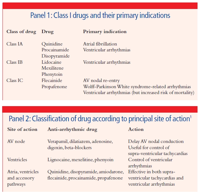
Class I: Sodium Channel Blockers
These block the fast Na+ channels (Phase 0), mostly in fast-response fibers.
Quinidine, Procainamide, Disopyramide
MOA: Block Na+ channels in the open/activated state. They also block K+ channels, which delays repolarization, thereby increasing Action Potential Duration (APD) and ERP.
- Quinidine: Causes muscarinic blockade (anticholinergic) leading to ↑HR, dry mouth, urinary retention. Also blocks alpha receptors (vasodilation + reflex tachycardia). Adverse: Cinchonism (tinnitus, ocular dysfunction, CNS excitation), Torsades. Digoxin toxicity risk.
- Procainamide: Less anticholinergic/alpha block. Metabolized by N-acetyltransferase to NAPA (active). Adverse: Drug-induced SLE (Lupus-like syndrome) in 30% of patients, especially slow acetylators. Hematotoxicity.
- Disopyramide: Heavy anticholinergic effects.
Lidocaine, Mexiletine, Tocainide
MOA: Rapidly associates/dissociates. Blocks Na+ channels in the inactivated state. Has a preference for partially depolarized tissue (i.e., hypoxic and ischemic tissue post-MI). Decreases APD by blocking the slow Na+ "window" current, extending diastole recovery time.
- Lidocaine: Given IV (high first-pass metabolism). Used for Post-MI arrhythmias and Digoxin toxicity. Adverse: CNS toxicity (seizures), but it is the least cardiotoxic of conventional antiarrhythmics.
- Mexiletine/Tocainide: Oral analogs of Lidocaine.
Flecainide, Propafenone
MOA: Block Na+ channels in all states (activated, inactivated, resting), especially in His-Purkinje tissue. No effect on APD (no K+ block). No ANS effects.
- Flecainide: Heavily restricted use because of severe pro-arrhythmogenic and negative inotropic effects. Causes increased sudden death post-MI.
- Propafenone: Used for rhythm control of A-Fib and paroxysmal SVTs.
Class II: Beta-Blockers
MOA: Prevent β-receptor activation. This prevents the increase of cAMP, which decreases SA and AV nodal activity (depresses automaticity) and flattens the slope of Phase 4 in pacemakers.
- Propranolol: Non-selective. Risk of bronchospasm (β2 blockade).
- Cardioselective (β1): Metoprolol (most used), Acebutolol, and Esmolol (Very short half-life, given IV for surgical emergencies).
- Uses: Prophylaxis post-MI to prevent ventricular arrhythmias, treating SVTs caused by high sympathetic tone, atrial flutter/fibrillation.
Class III: Potassium Channel Blockers
MOA: Decrease outward K+ currents, slowing down Phase 3 (repolarization). This massively increases the Action Potential Duration (APD) and prolongs the Effective Refractory Period (ERP), especially in Purkinje and ventricular fibers.
ALL Class III drugs significantly prolong the QT interval on an ECG (because they delay repolarization), meaning they all have a high potential to induce deadly arrhythmias, specifically Torsades de Pointes.
- Amiodarone: The most commonly used, despite side effects, because it actually has the least proarrhythmic effect. It structurally mimics thyroxine and iodine. Interestingly, it exhibits Class I, II, III, and IV actions. Very large Volume of Distribution (Vd) and a half-life of >80 days (binds heavily in adipose tissue).
- Side Effects: Highly toxic over time. Causes Pulmonary fibrosis, blue-gray skin pigmentation (Smurf syndrome), hepatotoxicity, corneal deposits, and profound thyroid dysfunction (hyper- or hypothyroidism).
- Dronedarone: An Amiodarone analog lacking iodine (no thyroid toxicity) and less lipophilic (shorter half-life). However, it is contraindicated in symptomatic heart failure due to increased risk of death.
- Sotalol: A unique drug. It is a non-selective Beta-blocker (l-isomer) AND a Class III K+ channel blocker (d-isomer). High risk of Torsades. Monitor renal function.
- Dofetilide & Ibutilide: Pure K+ blockers. High risk of pro-arrhythmias, so they are restricted strictly to inpatient hospital use for chemical cardioversion of A-Fib.
Class IV: Calcium Channel Blockers (Non-Dihydropyridines)
MOA: Bind only to open/depolarized voltage-sensitive Ca²⁺ channels. They block slow cardiac Ca²⁺ channels, decreasing Phase 0 and Phase 4 upstrokes in slow-response fibers (SA/AV nodes). This slows AV conduction heavily.
- Verapamil: Shows greater action on the heart muscle itself than on vasculature. High incidence of constipation and AV block.
- Diltiazem: Intermediate action on both the heart and vascular smooth muscle.
- Uses & Interactions: Highly effective for Atrial arrhythmias (reentrant SVT, controlling ventricular rate in A-Fib). Do not combine with Beta-blockers (causes additive/fatal AV block). Verapamil displaces Digoxin from tissue, causing toxicity.
Unclassified Antiarrhythmics
MOA: Activates adenosine receptors (Gi coupled), sharply decreasing cAMP. This massively decreases SA and AV nodal activity, temporarily stopping the heart's electrical conduction to "reset" it.
- Drug of Choice: For paroxysmal supraventricular tachycardias (SVT).
- Kinetics: Given IV rapid push. Half-life is < 10 seconds.
- Side Effects: Intense flushing, sense of impending doom/chest pressure, dyspnea.
- Interaction: Antagonized by methylxanthines (Caffeine and Theophylline block the receptor).
Use: The absolute Drug of Choice for treating Torsades de Pointes.
- What is Torsades? A specific, lethal polymorphic ventricular tachycardia associated with a prolonged QT interval on an ECG (the waveform looks like a twisting ribbon).
- Causes of Torsades: Potassium-channel blockers (Class IA and Class III), Antipsychotics (thioridazine), and Tricyclic antidepressants. Magnesium stabilizes the membrane to break the cycle.
Quick Quiz
Cardiac Arrythmias Pharmacology Quiz
Pharmacology - mobile-friendly and focused practice.
Privacy: Your details are used only for quiz tracking and certificates.
Cardiac Arrythmias Pharmacology Quiz
Pharmacology
Preparing questions...
Choose your answer and keep your streak alive.
Great effort.
Here is your quick performance summary.