
The Cell Cycle
The cell cycle describes the entire, highly regulated lifespan of a cell, from the exact moment of its formation after one division until it inevitably divides again. It is a continuous, dynamic journey that ensures tissue growth, repair, and genetic continuity.
It consists of two main, over-arching stages:
- Interphase: The prolonged period of cell growth, DNA replication, and meticulous preparation for division. This is by far the longest phase of the cell's life (occupying up to 90% of the cycle).
- M Phase (Mitotic Phase): The period of actual, physical cell division, which includes mitosis (the precise division of the nucleus) and cytokinesis (the physical division of the cytoplasm).
Part I: Interphase – The Preparation Phase
Historically, early microscopists called interphase a "resting phase" because the cell wasn't actively splitting. However, we now know that Interphase is not a resting phase at all. It is a highly active, metabolically intense period of growth, protein synthesis, and genetic replication. It is absolutely crucial for preparing the cell for successful division. It is divided into several distinct sub-phases:
1. G₀ Phase (Gap 0 / Quiescent Phase)
This is an optional phase where cells exit the active cell cycle and stop dividing, entering a state of dormancy or terminal differentiation. While they remain metabolically active (they are still doing their daily jobs), they are completely completely halted from preparing for division.
- Terminally Differentiated (Permanent G₀): Highly specialized cells that have lost the ability to ever divide again.
Examples: Mature skeletal muscle cells, cardiac myocytes (heart muscle cells), and mature nerve cells (neurons) often enter G₀ permanently. This is why spinal cord injuries or heart attacks are so devastating—the cells cannot divide to replace the dead tissue. - Reversible G₀ (Quiescent): Cells that are dormant but retain the capacity to re-enter the active cell cycle if they receive the right chemical stimulus.
Examples: Hepatocytes (liver cells) usually sit in G₀, but if a portion of the liver is surgically removed, they rapidly re-enter G₁ to regenerate the tissue. Naive lymphocytes (immune T-cells and B-cells) sit in G₀ until they encounter an antigen, which triggers explosive division to fight the infection. - Significance: The G₀ phase is a vital protective mechanism. It prevents uncontrolled cell growth, conserves bodily energy, and allows cells to devote all their resources to performing their specialized, mature roles.
2. G₁ Phase (Gap 1 / First Growth)
This is the first true growth phase immediately following a successful cell division. The cell is actively growing, "bulking up" to reach its normal mature size.
- Key Activities: The cell rapidly synthesizes massive amounts of mRNA and proteins. It physically expands its cytoplasm and begins duplicating its organelles (like mitochondria and ribosomes) to ensure there is enough machinery for two future cells.
- Critical "Decision Point" (The Restriction Point): Near the end of G₁, the cell faces the most important checkpoint in its life. The cell assesses internal factors (DNA integrity, energy reserves) and external factors (growth signals). Here, it "decides" whether to absolutely commit to division and proceed to the S phase, or to exit the cycle and retreat into the G₀ phase.
3. S Phase (Synthesis Phase)
The "synthesis" phase is the point of no return. Here, the most crucial and vulnerable event for cell division occurs: DNA replication.
- Key Activities: The cell unzips its double helix using enzymes (like DNA helicase and DNA polymerase). Each of the 46 chromosomes is perfectly duplicated, resulting in two identical copies called sister chromatids (attached at a central point called the centromere).
- Histone Production: Massive amounts of new histone proteins are synthesized to safely package and coil the newly replicated, fragile DNA.
- Outcome: By the end of the S phase, the cell still has 46 chromosomes, but it contains exactly double the amount of actual DNA material (92 chromatids).
4. G₂ Phase (Gap 2 / Second Growth)
The second growth phase and the final preparatory stage before the cell dives into the violent process of mitosis.
- Key Activities: The cell synthesizes the final proteins necessary for cell division, particularly tubulin, which will be used to build the mitotic spindle (the microscopic cables that will pull the chromosomes apart).
- "Quality Control" Checkpoint: Before entering mitosis, the cell strictly checks the newly replicated DNA for errors, missing sequences, or damage. If damage is found, it pauses the cycle and attempts repairs using DNA repair enzymes.
- The Ultimate Failsafe: If the genetic damage is too severe and irreparable, the cell takes a heroic protective measure. It triggers programmed cell death (apoptosis), literally committing suicide to prevent passing on dangerous, potentially cancer-causing mutations to the next generation.
Part II: Cell Division (Mitosis vs. Meiosis)
Cells reproduce through a fundamental, ancient process called cell division. This is absolutely essential for the growth of an organism, the repair of injured tissues, and the reproduction of the species. There are two primary types of cell division in the human body:
| Feature | Mitotic Cell Division (Mitosis) | Meiotic Cell Division (Meiosis) |
|---|---|---|
| Primary Role | Growth, maintenance, and repair of tissues. | Production of sex cells (gametes: sperm and ova). |
| Occurs In | Somatic cells (e.g., neurons, epithelial cells, muscle cells, hepatocytes, keratinocytes). | Reproductive organs only (Testes in males, Ovaries in females). |
| Outcome (Daughter Cells) | Two (2) genetically identical daughter cells. | Four (4) genetically unique daughter cells. |
| Chromosome Number | 46 chromosomes (Diploid - exactly the same as the parent cell). | 23 chromosomes (Haploid - exactly half of the parent cell, ready to combine during fertilization). |
Part III: Mitotic Cell Division – The Basis of Growth and Repair
Mitotic cell division is a continuous, highly choreographed process crucial for increasing the number of cells for bodily growth and replacing worn out, damaged, or dead cells. However, not all cells divide at the same rate. For example, epithelial cells (like those lining the skin or gut) divide almost continuously to replace shed cells, while mature muscle and nerve cells largely lose the ability to divide.
Key Processes in Mitotic Cell Division:
- Replication of Chromosomes: Creating exact copies of the genetic material (this strictly occurs earlier, in the S phase of interphase).
- Mitosis: The physical division of the nucleus and its genetic contents.
- Cytokinesis: The physical division of the cytoplasm and cell membrane.
Mechanism
During mitosis, the cell's previously loose, diffuse DNA (chromatin) condenses into tightly packed, visible chromosomes to prevent tangling. The centrosome (an organelle) duplicates, and each copy moves to opposite ends (poles) of the cell. They act as anchors, creating spindle fibers (microtubules) that reach out, grab onto the center of the chromosomes, and pull them apart. This ensures that when the cell finally divides, each new daughter cell receives its own flawless, identical copy of the genetic material.
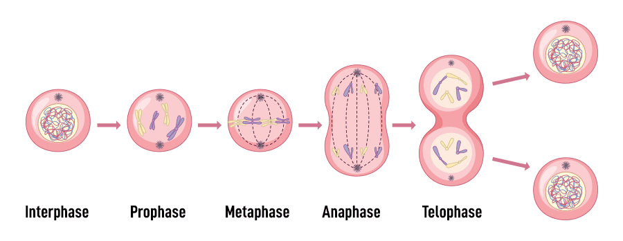
The Four Sequential Phases of Mitosis
Once interphase is complete, the cell enters mitosis. While it is a continuous, fluid process, biologists divide it into four sequential phases for easier understanding:
The Condensation Phase
- Replicated, loose chromatin tightly coils and condenses, becoming visible under a microscope as X-shaped structures consisting of two identical sister chromatids joined at a central pinch point called the centromere.
- The nuclear envelope (membrane) dissolves and completely disappears, spilling the chromosomes into the open cytoplasm.
- Centrioles migrate to opposite poles of the cell, and the intricate microtubule framework of the mitotic spindle begins to form.
The Alignment Phase
- The mitotic spindle fibers engage in a cellular "tug-of-war."
- The replicated chromosomes are pulled and line up precisely at the cell's exact equator (an imaginary line called the metaphase plate).
- The centromere of each chromosome is securely attached to the spindle fibers via special protein patches called kinetochores.
The Separation Phase
- An enzyme (separase) rapidly cleaves the glue holding the chromatids together. The centromeres divide, and the sister chromatids violently separate.
- Once separated, each individual chromatid is now officially considered its own individual chromosome.
- The spindle fibers reel in, pulling the newly separated chromosomes towards the opposite poles of the cell.
The Reconstruction Phase
- The chromosomes safely reach the opposite poles, and the spindle fibers completely disassemble.
- A brand new nuclear envelope forms around each of the two sets of chromosomes at the poles.
- The chromosomes relax and uncoil back into their original, thread-like chromatin form, ready to begin gene expression again.
Cytokinesis: Division of the Cytoplasm
Usually initiating during late anaphase and finalizing after telophase, cytokinesis is the very last step. In human (animal) cells, a cleavage furrow forms in the plasma membrane (driven by a contractile ring of actin and myosin filaments). This furrow deepens and eventually pinches the parent cell completely into two separate, genetically identical daughter cells, each with its own distinct nucleus and cytoplasm. (Extra detail: In plant cells, because of the rigid cell wall, a "cell plate" forms down the middle instead of a pinching furrow).
Part IV: Cell Cycle Disorders – When Regulation Fails
The cell cycle is a tightly regulated, beautifully orchestrated sequence of events with a strict series of internal checkpoints that monitor the cell's health, energy, and DNA integrity. When these regulatory mechanisms fail, the cell cycle can become dangerously dysregulated, leading to various disorders, most notably cancer.
Cells have strict checks and balances. Special proteins called cyclins constantly monitor the cell's health. Unhealthy cells normally self-destruct via apoptosis. Cancer cells, however, lose this critical ability. For many cells, the G₁ checkpoint is the most important; if a cell receives a specific "go-ahead" signal here, it will usually complete the entire division process. If it does not receive the signal, it enters the non-dividing state called the G₀ phase.
Key Regulators of the Cell Cycle
Before discussing disorders, it's essential to understand the main biochemical players that normally control the cell cycle. Think of the cell cycle like driving a car:
- Cyclins and CDKs (The Engine): These are the "engine" of the cell cycle. Cyclin-Dependent Kinases (CDKs) are enzymes that remain inactive until they are activated by binding to specific proteins called Cyclins. Different Cyclin-CDK complexes drive the cell through each specific phase of the cycle.
- Cell Cycle Checkpoints (The Traffic Lights): Critical control points that monitor internal and external conditions. The main ones are the G₁ Checkpoint (the "start" point that checks for DNA damage before replication), the G₂ Checkpoint (checks if DNA replication was flawless), and the M Checkpoint (checks if the spindle is perfectly attached before pulling chromosomes apart).
- Proto-oncogenes & Oncogenes (The Accelerator): Proto-oncogenes are normal genes that promote standard, healthy cell growth and division. However, when they are mutated, they become Oncogenes. An oncogene is like a car accelerator pedal that is permanently stuck to the floor, causing uncontrolled, rapid growth.
- Tumor Suppressor Genes (The Brakes): These genes encode proteins that inhibit cell division, halt the cycle to repair DNA, or force the cell into apoptosis if damage is too severe.
Key Examples:- p53 (Known famously as the "Guardian of the Genome"). If p53 detects DNA damage, it halts the cycle. If the damage is unfixable, p53 orders the cell to commit suicide.
- Rb (Retinoblastoma protein), which actively prevents the cell from entering the S phase until the cell is truly ready.
Causes of Cell Cycle Disorders
Disorders arise when the delicate balance of these activators and inhibitors is disrupted, often due to:
- Genetic Mutations: Physically altering the DNA code to either inactivate "brake" genes (tumor suppressors) or hyper-activate "accelerator" genes (proto-oncogenes).
- Epigenetic Changes: Altering gene expression without changing the actual DNA sequence, such as chemically silencing a tumor suppressor gene so it can no longer be read by the cell.
- Viral Infections: Viruses are notorious hijackers. For example, the Human Papillomavirus (HPV) produces highly destructive viral proteins (E6 and E7). The E6 protein specifically hunts down and destroys the cell's p53, while E7 destroys Rb. With the brakes completely removed, the cell divides wildly, leading to cervical cancer.
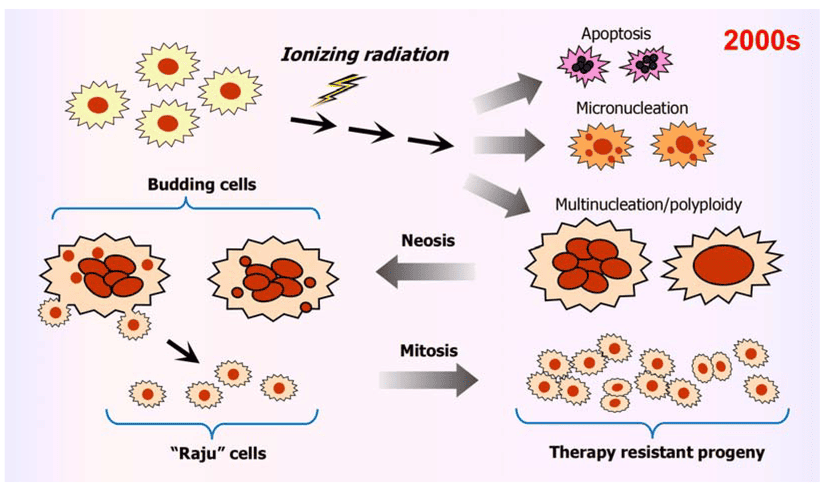
- Environmental Factors: Exposure to powerful carcinogens (like tobacco smoke chemicals) and ionizing radiation (like UV rays or X-rays) that physically shatter the DNA, leading to catastrophic mutations.
Consequences & Types of Cell Cycle Disorders
1. Cancer (Malignancy)
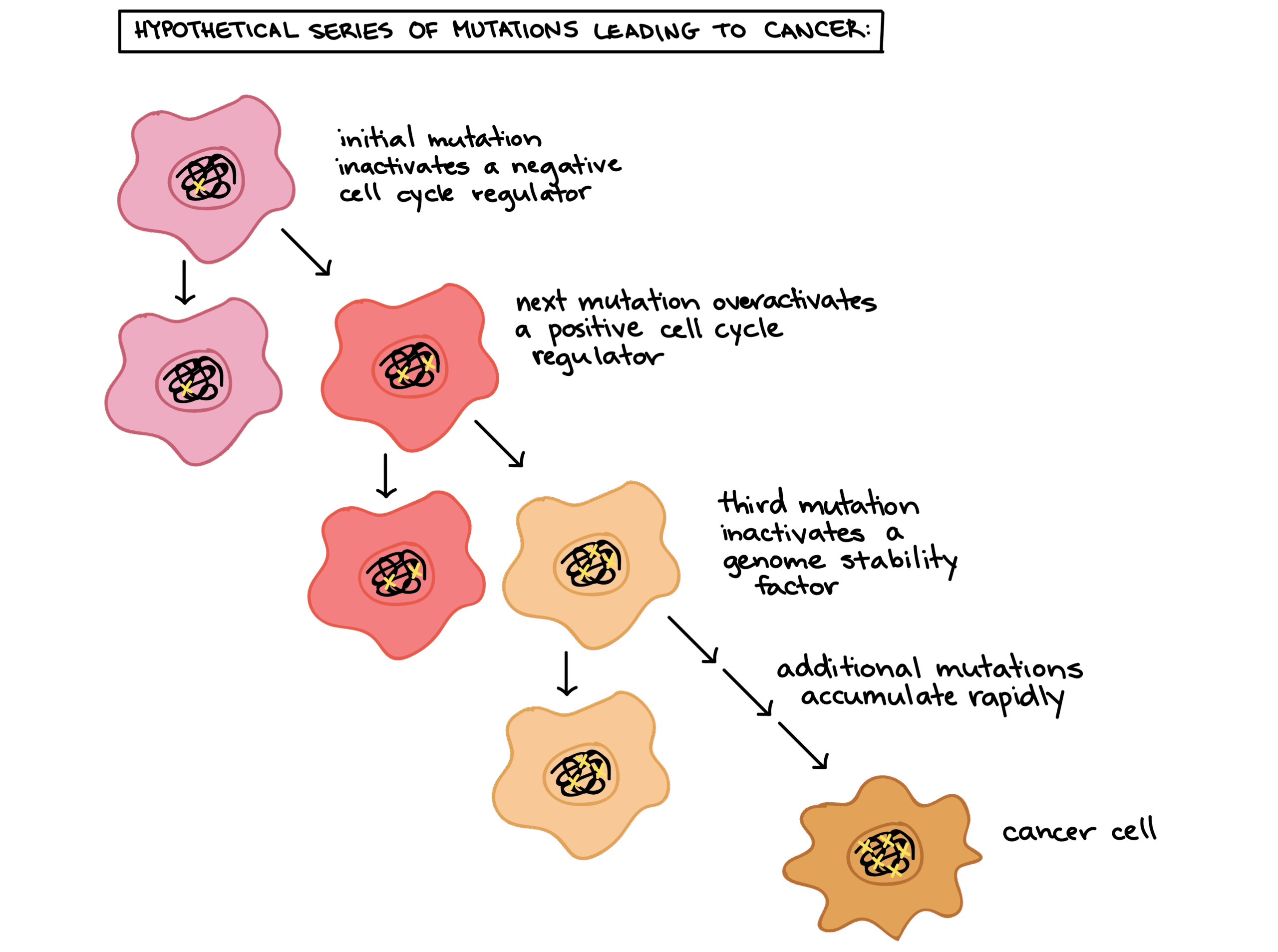
Cancer is the primary disease of uncontrolled cell division. Cancer cells completely ignore the normal signals that control the cell cycle. They enter the S phase without waiting for a signal, and they become functionally "immortal," escaping the normal biological limit on how many times a cell can divide. This is typically caused by the accumulation of multiple mutations that activate oncogenes and inactivate tumor suppressor genes.
The Hallmarks of Cancer Cells:
- Sustained proliferative signaling: They create their own growth factors.
- Evasion of growth suppressors: They ignore "stop" signals from neighbors.
- Resistance to cell death: They disable apoptosis pathways (like mutating p53).
- Enabling replicative immortality: They reactivate an enzyme called telomerase to prevent their DNA from degrading over time.
- Inducing angiogenesis: They secrete chemicals (like VEGF) to force the body to build new blood vessels to feed the growing tumor.
- Activating invasion & metastasis: They break loose from tissue boundaries and spread through the blood to distant organs.
2. Aneuploidy (Incorrect Chromosome Number)
A catastrophic failure of the M checkpoint (failure to attach the spindle correctly) can lead to an unequal distribution of chromosomes during cell division, a phenomenon known as nondisjunction. While most aneuploid cells die instantly, some survive and can lead to severe genetic disorders like Down Syndrome (Trisomy 21), where a child inherits three copies of chromosome 21 instead of two. Severe, chaotic aneuploidy is also a fundamental feature of advanced cancer cells.
3. Developmental & Premature Aging Disorders
Precise, timed control of the cell cycle is critical during embryonic development. Errors during gestation can lead to severe underdevelopment (e.g., microcephaly, a condition resulting in an abnormally small brain and head) or chaotic overgrowth syndromes. Similarly, some premature aging syndromes (like Progeria) are tightly linked to deep genetic defects in DNA repair mechanisms that impact cell cycle checkpoints, causing cells to age and die far too rapidly.
Therapeutic Implications
Understanding the intricate biochemistry of these disorders is fundamental to modern medicine. Many cutting-edge therapies are explicitly designed to target the cell cycle:
- Chemotherapy: Uses highly toxic, systemic drugs that intentionally damage DNA or physically disrupt the mitotic spindle (e.g., the drug Paclitaxel prevents the spindle from breaking down, trapping the cell in mitosis until it dies). This preferentially kills rapidly dividing cells (which is why cancer patients lose their hair—hair follicle cells divide rapidly).
- Targeted Therapies: Newer, smarter drugs that specifically seek out and inhibit mutated or overactive molecules, such as CDK inhibitors (like Palbociclib for breast cancer) that jam the "engine" of the cell cycle.
- Immunotherapy: Harnessing the body's own immune system (using drugs like Pembrolizumab) to recognize, unmask, and aggressively destroy cancer cells that have learned to evade normal cell cycle and immune controls.
Part V: Chromosomal Mutations – Large-Scale Genetic Changes
While gene mutations (point mutations) involve tiny changes to individual DNA base pairs within a single gene, chromosomal mutations are massive, large-scale changes affecting the structure or number of entire chromosomes. These alterations involve millions of base pairs and multiple genes at once. Such sweeping structural changes often arise from devastating errors during the crossover phase of meiosis, or from heavy exposure to severe mutagens (like gamma radiation).
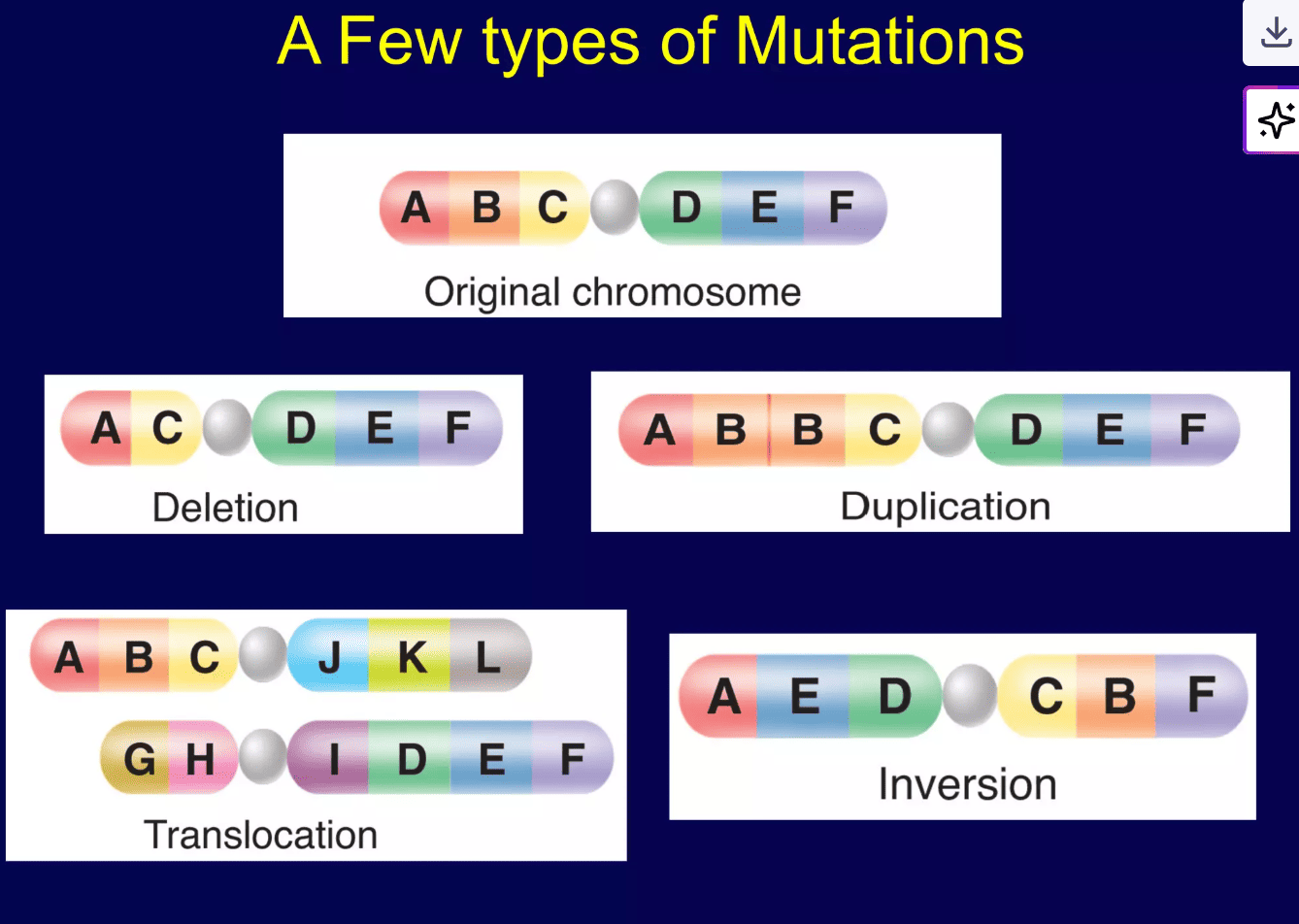
Types of Chromosomal Mutations:
Loss of Information
A segment of the chromosome, containing one or more entire genes, is physically lost, broken off, or excised during division.
- Example Concept: A chromosome originally containing gene segments [A-B-C-D-E-F] loses the [C] segment, resulting in a shortened chromosome[A-B-D-E-F].
- Impact: Results in a permanent loss of vital genetic information. The consequences can range from mild to extremely severe, depending on the size and exact function of the deleted genes.
- Clinical Example: Cri-du-chat syndrome (Cry of the Cat syndrome) is caused by a massive deletion on the short arm of chromosome 5, leading to severe intellectual disability and a characteristic high-pitched cry in infants.
Copying Errors
A segment of the chromosome is accidentally copied and repeated, resulting in extra, redundant copies of genes.
- Example Concept: The[B-C] segment is erroneously repeated, resulting in an elongated chromosome[A-B-C-B-C-D-E-F].
- Impact: While sometimes benign (and over millions of years, an engine of evolution by creating gene families), sudden duplications can disrupt normal "gene dosage" and overwhelm cellular processes with too much protein, leading to developmental problems.
- Clinical Example: Charcot-Marie-Tooth disease type 1A is caused by a duplication on chromosome 17, leading to progressive muscle weakness and nerve damage.
Flipped Sequence
A segment of a chromosome violently breaks off, flips 180 degrees in the opposite direction, and reattaches backwards onto the very same chromosome.
- Example Concept: The[B-C-D] segment is inverted, resulting in a jumbled sequence [A-D-C-B-E-F].
- Impact: The genetic material is still technically present, so the individual carrying it may appear completely normal. However, inversions (whether paracentric or pericentric) can cause massive alignment issues during meiosis when they try to mate, potentially leading to nonviable gametes (infertility) or offspring with unbalanced, broken chromosomes.
- Clinical Example: Severe forms of Hemophilia A (a blood clotting disorder) are frequently caused by an inversion disrupting the Factor VIII gene on the X chromosome.
Wrong Address
A segment of one chromosome breaks off and illegally attaches to an entirely different, non-homologous chromosome.
- Example Concept: A segment from chromosome 8 breaks off and attaches to chromosome 14. This is an inappropriate exchange of genetic material between two vastly different chromosomes.
- Impact: Balanced translocations (where two chromosomes swap pieces perfectly with no net loss/gain of DNA) may not immediately affect the individual but can lead to severe fertility issues and miscarriages. Unbalanced translocations in offspring, where there is extra or missing genetic material, typically cause significant, often fatal health problems.
- Clinical Example: The Philadelphia Chromosome is a famous reciprocal translocation between chromosome 9 and chromosome 22 [t(9;22)]. This accidental fusion fuses two genes together to create a powerful, permanent oncogene (BCR-ABL), which is the primary cause of Chronic Myeloid Leukemia (CML).