
Body Fluid and Electrolyte Physiology
By the conclusion of this exhaustive master guide, you will be deeply conversant with:
- The precise composition, volume, and distribution of Total Body Water (TBW) and its sub-compartments.
- The fundamental principles of Solutes, Solvents, Diffusion, and Osmosis.
- The exact application of Starling Forces in governing capillary fluid exchange.
- The complex neurohormonal mechanisms regulating Volume and Osmolarity (RAAS, ADH, ANP).
- The clinical methodology for Measuring Fluid Compartments using the Indicator-Dilution method.
- The pathophysiology of fluid imbalances and the evidence-based application of Intravenous (IV) Fluid Therapy based on tonicity.
I. Solutes, Solvents, and the Principles of Fluid Movement
At the absolute heart of all physiological processes involving body fluids is the complex biochemical interaction between solutes and solvents, and their dynamic movement across semipermeable biological membranes.
1. Solutes and Solvents: The Basics
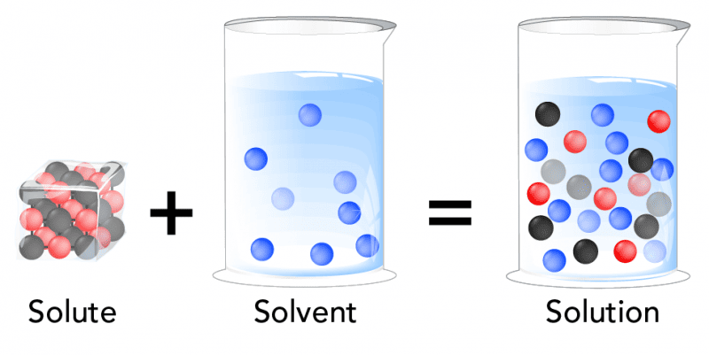
- Solution: A perfectly homogeneous mixture composed of two or more substances.
- Solvent: The dissolving medium; the substance present in the greatest amount.
- Solute: The substance(s) present in a lesser amount that gets dissolved by the solvent.
Water: The Universal Biological Solvent
The primary and overwhelmingly abundant solvent in all body fluids is WATER (H&sub2;O). Water possesses unique biochemical properties that make it the ideal medium for life:
- Polarity: Due to its bent shape and uneven electron distribution, water is a highly polar dipole. This allows it to dissolve a massive variety of other polar molecules and ionic compounds (like sodium chloride) by forming hydration shells around them.
- High Heat Capacity: Water can absorb or release vast amounts of heat with only a modest change in its own temperature, which is essential for regulating core body temperature.
- High Heat of Vaporization: Evaporating a small amount of water takes a large amount of heat, allowing for highly efficient cooling through sweating and insensible respiratory fluid loss.
2. Common Solutes in Body Fluids
Body fluids are not just water; they are highly complex "soups" containing a vast array of life-sustaining solutes:
- Electrolytes: Chemical compounds that dissociate into ions in water and can conduct an electrical current.
- Cations (positively charged): Sodium (Na+), Potassium (K+), Calcium (Ca²+), Magnesium (Mg²+).
- Anions (negatively charged): Chloride (Cl−), Bicarbonate (HCO&sub3;−), Phosphate (HPO&sub4;²−), Sulfate (SO&sub4;²−).
- Non-electrolytes: Substances with covalent bonds that do not dissociate in solution (they carry no electrical charge).
- Nutrients: Glucose, amino acids, fatty acids, vitamins.
- Metabolic Wastes: Urea, creatinine, uric acid, bilirubin.
- Proteins: Massive molecules like Albumin, globulins, and fibrinogen, which act as polyanions (carrying multiple negative charges).
- Gases: Dissolved Oxygen (O&sub2;) and Carbon Dioxide (CO&sub2;).
3. Simple Movement: Diffusion vs. Osmosis
The movement of these substances is primarily governed by passive, physical processes that strictly do not require the expenditure of cellular energy (ATP).
Diffusion
- Definition: The net movement of solute particles from an area of higher solute concentration to an area of lower solute concentration (moving "down" the concentration gradient).
- Mechanism: Driven purely by the inherent, random kinetic energy (Brownian motion) of molecules.
- Influencing Factors (Fick's Law): The rate of diffusion increases with a larger concentration gradient, higher temperature, smaller molecular size, a shorter travel distance, and a massively larger surface area (e.g., the alveoli in the lungs).
- Simple Diffusion: Lipid-soluble (non-polar) solutes pass directly through the phospholipid bilayer (e.g., O&sub2;, CO&sub2;, steroid hormones).
- Facilitated Diffusion: Water-soluble or charged solutes move down their gradient but require the help of specific membrane proteins, either channel proteins (for ions) or carrier proteins (for glucose).
Osmosis
- Definition: The net movement of water (the solvent) across a selectively permeable membrane from an area of higher water concentration (lower solute concentration) to an area of lower water concentration (higher solute concentration).
- Mechanism: Water molecules move down their own concentration gradient, largely through specialized protein channels called Aquaporins.
- Selectively Permeable Membrane: Crucial for osmosis. The membrane must allow water to pass freely but strictly restrict the passage of the osmotically active solutes.
- Osmotic Pressure: The exact amount of hydrostatic pressure that must be applied to prevent the inward flow of water across a semipermeable membrane. The higher the trapped solute concentration, the higher the osmotic "pull" or pressure.
Summary of Movement Principles:
Rule 1: Solutes move by Diffusion (from high solute to low solute).
Rule 2: Water moves by Osmosis (from low solute to high solute). In clinical terms: "Water follows solutes."
These passive movements are absolutely essential for delivering oxygen and nutrients to tissues, removing metabolic wastes, and maintaining precise cell volume and shape.
II. The Body Fluid Compartments
To truly appreciate the clinical dynamics of body fluids, we must map exactly where this fluid resides. Imagine the human body as a complex system of interconnected containers. This meticulous compartmentalization is the ultimate key to maintaining cellular and systemic homeostasis.
1. Total Body Water (TBW)
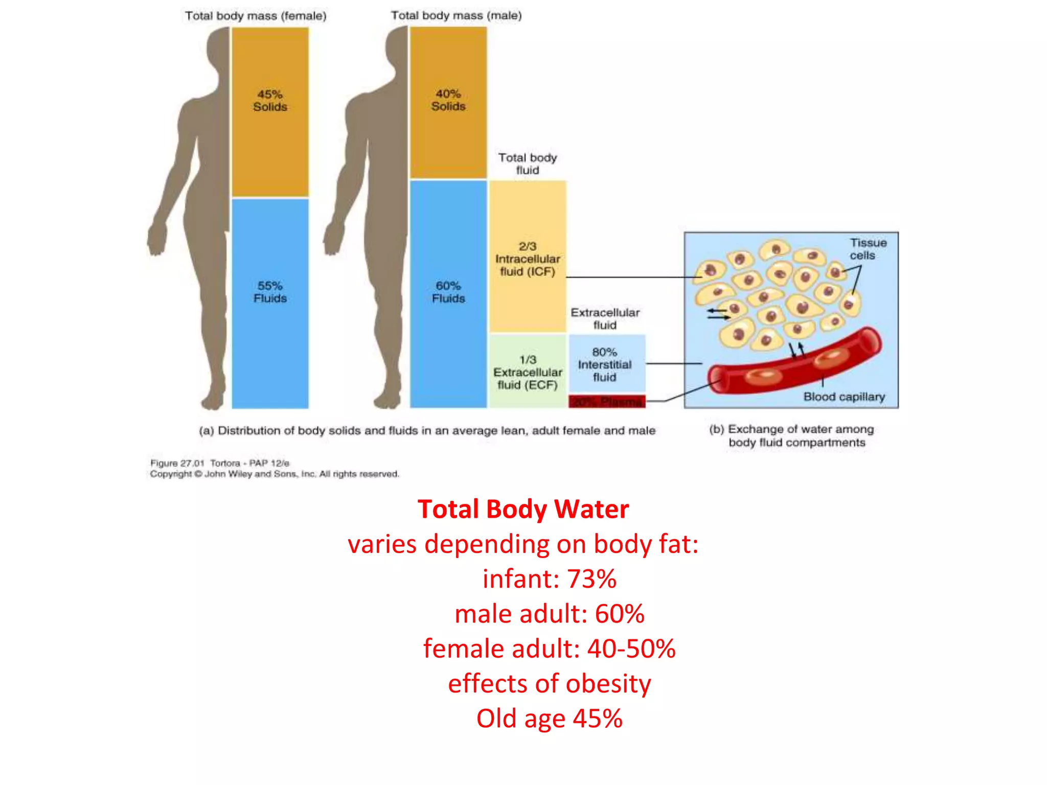
TBW refers to the absolute sum of all water contained within the body. It represents a massively significant proportion of human body mass.
- Standard Reference Volume: In a standard, healthy 70 kg (154 lb) adult male, TBW is roughly 42 Liters.
- Proportion & Variations: Approximately 60% of an adult male's body weight is water. However, this percentage fluctuates heavily based on physiological factors:
- Age: Premature infants can be up to 80% water. Full-term infants are ~70-75%. As humans age, muscle mass decreases and connective tissue/fat increases, meaning the elderly can drop to a dangerous 45-50% TBW (putting them at exceptionally high risk for rapid, fatal dehydration).
- Sex: Females generally have a slightly lower TBW percentage (~50-55%) than males. This is because women naturally have a higher physiological percentage of adipose tissue (fat).
- Body Fat Content: Fat cells (adipocytes) contain almost zero water. Therefore, individuals with higher body fat percentages (obesity) will have significantly lower TBW percentages relative to their total weight compared to highly muscular individuals.
The total 42 Liters of body water is not uniformly distributed but is strictly divided into two primary compartments by the cell membrane:
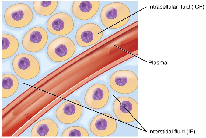
A. Intracellular Fluid (ICF)
The ICF is the fluid locked inside the trillions of cells in the body. It is the immediate, highly regulated aqueous environment where the vast majority of metabolic and biochemical activities occur.
- Proportion: The ICF constitutes the largest single fluid compartment, accounting for exactly two-thirds (2/3) of the TBW. In a 70 kg adult, this is roughly 28 Liters (40% of total body weight).
- Composition (The Cell's Internal Environment):
- Major Cations: Potassium (K+) is the undisputed king of the ICF (concentration ~140 mEq/L). Its high internal concentration is crucial for resting membrane potential, nerve impulse transmission, and muscle contraction. Magnesium (Mg²+) is the second most abundant, vital as a cofactor for all ATP-dependent enzymatic reactions.
- Major Anions: Phosphate (PO&sub4;³−) is a critical component of energy currency (ATP) and intracellular buffering. Proteins are heavily concentrated inside the cell, carrying strong negative charges, contributing to osmolarity, and acting as intracellular buffers.
- Low Concentrations: Sodium (Na+) is kept extremely low inside the cell (~14 mEq/L), as is Chloride (Cl−).
- Key Characteristics:
- Selective Permeability: The phospholipid bilayer plasma membrane is the ultimate barrier separating the ICF from the outside world.
- Osmotic Equilibrium: Despite having completely different chemical ingredients than the outside fluid, the total number of particles (osmolarity) of the ICF is normally in perfect, dynamic equilibrium with the outside.
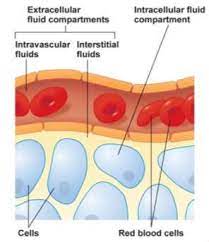
B. Extracellular Fluid (ECF)
The ECF is all the fluid found outside the cells. It acts as the body's vast "internal sea" that bathes, feeds, and cleanses all cells.
- Proportion: The ECF constitutes approximately one-third (1/3) of the TBW, which is roughly 14 Liters (20% of total body weight).
- Composition (The Body's Transport Medium):
- Major Cations: Sodium (Na+) dominates the ECF (~140 mEq/L). It is the absolute primary determinant of ECF osmolarity and overall fluid volume.
- Major Anions: Chloride (Cl−) (~100 mEq/L) and Bicarbonate (HCO&sub3;−) (~24 mEq/L), which acts as the body's primary acid-base buffer system.
- Other Components: A rich, circulating soup of glucose, amino acids, circulating hormones, oxygen, and metabolic waste (urea).
The 3 Sub-compartments of the ECF:
This is the literal "tissue fluid" filling the microscopic spaces between the cells. It is the largest component of the ECF, comprising about 80% of ECF volume (~10.5 Liters). Its ionic composition is nearly identical to blood plasma, but it lacks large proteins. The ISF is the critical "middle-man" medium for exchanging nutrients and gases between the blood capillaries and the cell membranes.
This is the pale-yellow fluid component of blood, circulating violently within the cardiovascular system. It accounts for 20% of ECF volume (~3.5 Liters). Its defining characteristic, compared to ISF, is a massive concentration of large plasma proteins (like Albumin). Plasma is the primary transport highway for red/white blood cells, nutrients, and waste.
A highly specialized, trapped component of the ECF, representing only 1-2% of body weight (approx. 1 to 2 Liters). It consists of fluids actively secreted by specific epithelial cells into distinct, enclosed body cavities. Examples include: Cerebrospinal Fluid (CSF), Intraocular Fluid (aqueous humor), Synovial Fluid in joints, Serous Fluids (pleural, pericardial, peritoneal fluid), and massive amounts of Gastrointestinal secretions.
III. Measurement of Fluid Compartments (The Indicator Dilution Method)
Physicians and physiologists cannot simply drain a human to measure fluid volumes. Instead, they use a highly accurate mathematical principle called the Indicator-Dilution Method.
The Principle: You inject a known mass (amount) of a non-toxic marker substance (indicator) into the blood. You allow it time to distribute evenly through a specific compartment. You then draw a blood sample and measure the final concentration.
Formula: Volume = Mass of Indicator Injected / Concentration of Indicator in Sample.
Choosing the Right Indicator:
The entire validity of the test relies on choosing an indicator that distributes only in the target compartment you wish to measure.
- Total Body Water (TBW): Measured using isotopes of water, like heavy water (D&sub2;O) or tritiated water (HTO), or Antipyrine. Because these are literally water molecules, they go everywhere water goes, crossing all membranes perfectly.
- Extracellular Fluid (ECF) Volume: Measured using Inulin, Mannitol, or radioactive Sodium/Thiosulfate. These molecules easily cross the capillary wall into the interstitial space but are physically too large to cross the cell membrane, so they remain trapped perfectly inside the ECF.
- Plasma Volume: Measured using Evans blue dye (T-1824) or Radioactive Iodine-125 tagged Albumin. These are massive molecules that bind instantly to plasma proteins. They are completely physically trapped inside the blood vessels and cannot even cross into the interstitial space.
Calculating the Hidden Compartments:
You cannot directly measure the Interstitial Fluid or the Intracellular Fluid because there is no chemical tracer that exclusively targets them without first going through plasma. Therefore, they are derived via simple subtraction:
- Interstitial Fluid (ISF) Volume = ECF Volume − Plasma Volume.
- Intracellular Fluid (ICF) Volume = Total Body Water (TBW) − ECF Volume.
IV. Fluid Movement Between Compartments and Regulatory Mechanisms
The precise, uninterrupted movement of water and solutes between the body's fluid compartments is the absolute cornerstone of physiological survival. This dynamic equilibrium is meticulously regulated by intense physical forces and membrane properties.
A. Fluid Movement Between Plasma and Interstitial Fluid (Capillary Exchange)
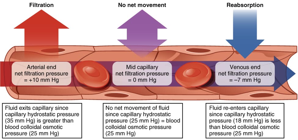
The exchange of massive amounts of fluid, nutrients, and waste between the blood plasma and the tissue cells (via the ISF) occurs primarily across the microscopically thin, porous walls of the capillaries. This movement is governed by Starling Forces.
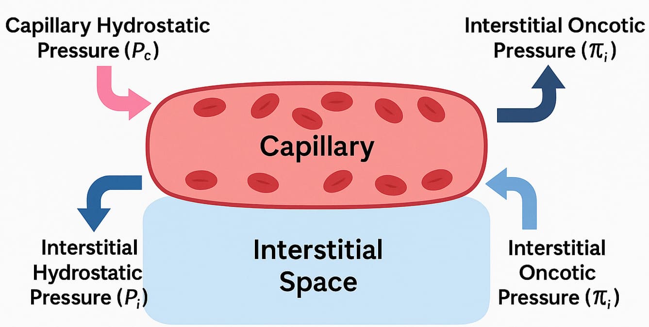
Starling Forces: The Four Drivers of Capillary Exchange
- Capillary Hydrostatic Pressure (Pc): This is the physical blood pressure exerted by the fluid within the capillaries, acting as the "pushing" force against the capillary wall.
- Effect: It violently forces fluid OUT of the capillary and into the interstitial space (Filtration).
- Dynamics: Pc is highest at the arterial end of the capillary (~30-35 mmHg) due to heart pumping, and progressively drops as blood flows to the venous end (~10-15 mmHg).
- Interstitial Fluid Hydrostatic Pressure (Pif): The pressure exerted by the physical fluid already sitting in the interstitial space outside the capillary.
- Effect: It tends to push fluid BACK INTO the capillary.
- Dynamics: Pif is usually very low, often close to zero or even slightly negative (due to lymphatic suction), creating a slight vacuum effect.
- Capillary Oncotic (Colloid Osmotic) Pressure (πc): The specialized osmotic pressure exerted by the massive, non-diffusible plasma proteins (primarily Albumin) permanently trapped within the blood.
- Effect: Acts as a massive chemical sponge, PULLING fluid back into the capillary from the interstitial space (Reabsorption).
- Dynamics: πc remains highly constant along the entire length of the capillary (typically around 25-28 mmHg).
- Interstitial Fluid Oncotic Pressure (πif): The osmotic pull exerted by the tiny amount of proteins that manage to leak into the interstitial fluid.
- Effect: Tends to pull fluid OUT of the capillary.
- Dynamics: πif is normally very low (typically 2-8 mmHg) because healthy capillaries restrict protein leakage.
Net Filtration Pressure (NFP)
The absolute net movement of fluid is determined by a mathematical balance of these forces, expressed by the Starling equation:
NFP = (Pc - Pif) - (πc - πif)
- At the arterial end: NFP = (35 - 0) - (26 - 2) = +11 mmHg. A positive NFP means outward pressure wins, indicating robust Net Filtration (nutrient-rich fluid moves OUT to feed tissues).
- At the venous end: NFP = (15 - 0) - (26 - 2) = -9 mmHg. A negative NFP means the inward pull of albumin wins, indicating robust Net Reabsorption (waste-filled fluid moves IN to go back to the heart/kidneys).
The Vital Role of the Lymphatic System
Notice the math: +11 out, but only -9 back in. There is a continuous, slight physiological imbalance where filtration constantly exceeds reabsorption by about 2-3 Liters a day. This excess fluid, along with any escaped proteins, is swept up by the Lymphatic System. The lymphatics act as an emergency drainage sewer, returning this "lymph" fluid back into the venous circulation at the subclavian veins.
Clinical Pathology: If the lymph nodes are blocked (e.g., by a tumor, radiation, or parasitic elephantiasis), the fluid has nowhere to go, resulting in massive, disfiguring tissue swelling called Lymphedema.
B. Fluid Movement Between ECF and ICF (Across Cell Membranes)
While capillary walls are leaky, the cell membrane is highly restrictive. Exchange between the ISF and the ICF is driven exclusively by Osmosis. The cell membrane is highly permeable to water (via aquaporins) but highly impermeable to most solutes (like Sodium).
Osmolarity vs. Tonicity (Crucial Distinction)
- Osmolarity: A strict chemical measurement. It quantifies the total absolute concentration of ALL solute particles present in a solution, regardless of whether they can cross a cell membrane. Expressed as milliosmoles per liter (mOsm/L). Normal blood plasma is strictly regulated at 280-300 mOsm/L.
- Effective Osmoles: Solutes that CANNOT cross the cell membrane (like Na+, Cl−, Mannitol). Because they are trapped on one side, they exert a permanent osmotic "pull" on water.
- Ineffective Osmoles: Solutes that freely slip right through the cell membrane (like Urea and Ethanol). Because they move freely, they balance out instantly and cause zero permanent water shifting.
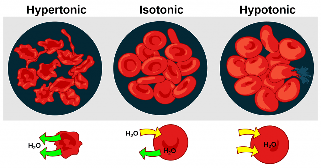
- Tonicity: A strictly biological, functional term. It describes the physical effect a fluid has on actual cell volume. Tonicity is determined SOLELY by the concentration of Effective (non-penetrating) osmoles.
The extracellular fluid has the exact same concentration of non-penetrating solutes as the inside of the cell. There is zero net movement of water. Cell volume remains perfectly stable and healthy.
The extracellular fluid is diluted (has fewer trapped solutes than the inside of the cell). Water aggressively rushes INTO the hyper-concentrated cells to dilute them. Cells swell massively, and may rupture (lyse). Can cause deadly cerebral edema.
The extracellular fluid is highly concentrated (has too much sodium/solutes). The strong ECF aggressively sucks water OUT of the cells. Cells shrink and shrivel violently (crenation). Can cause severe neurological damage and coma.
Active Transport's Essential Role: The Na+/K+ Pump
While water movement is passive, maintaining these osmotic gradients requires massive energy. Cells are packed with highly concentrated, trapped negative proteins that constantly try to suck water into the cell. To prevent all human cells from swelling and exploding, the Na+/K+ ATPase pump runs continuously. By burning ATP to forcefully kick 3 Na+ ions OUT of the cell for every 2 K+ ions it brings in, it creates a net outward osmotic pull that exactly counters the inward pull of the proteins. If a cell loses oxygen (hypoxia) and runs out of ATP, the pump fails, sodium rushes in, water follows, and the cell undergoes fatal swelling (hydropic degeneration).
V. Neurohormonal Regulation of Body Fluid Volume and Osmolarity
The human body uses incredibly sophisticated feedback loops involving the brain, heart, and kidneys to ensure fluid balance remains absolute.
A. Regulation of ECF Volume (Primarily Sodium Balance)
The overall volume of the blood and ECF is dictated entirely by its total Sodium (Na+) content. The golden rule of physiology is: "Where Sodium goes, water is forced to follow."
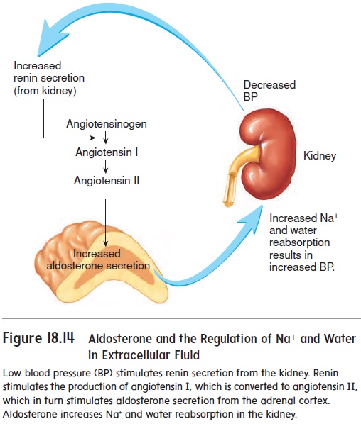
- Renin-Angiotensin-Aldosterone System (RAAS): The body's primary volume-preservation system.
- Trigger: The kidneys detect low blood pressure, low blood volume, or low sodium delivery.
- Action: Kidneys secrete the enzyme Renin into the blood. Renin converts circulating Angiotensinogen into Angiotensin I. As blood passes through the lungs, ACE (Angiotensin-Converting Enzyme) converts it into Angiotensin II.
- Result: Angiotensin II is the most potent vasoconstrictor in the body (instantly raising blood pressure). Furthermore, it forces the adrenal glands to secrete the steroid hormone Aldosterone. Aldosterone commands the kidneys to aggressively reabsorb sodium back into the blood. Water violently follows the sodium, restoring total ECF volume.
- Antidiuretic Hormone (ADH) / Vasopressin (Volume role): While primarily for osmolarity, if blood volume drops severely (e.g., a massive hemorrhage of >10% blood loss), pressure receptors (baroreceptors) in the aorta panic and trigger a massive release of ADH from the posterior pituitary to clamp down blood vessels and save all water in the kidneys.
- Atrial Natriuretic Peptide (ANP) & BNP: The "counter-regulatory" system. If ECF volume is too high, the massive volume stretches the muscular walls of the heart's atria and ventricles. The stretched heart muscle releases ANP and BNP. These hormones force the kidneys to excrete sodium (natriuresis) and excrete water (diuresis) directly into the urine, safely reducing blood volume and pressure.
- Sympathetic Nervous System (SNS): When activated by stress or low pressure, sympathetic nerves constrict renal blood vessels (dropping kidney filtration so less urine is made) and directly stimulate renin release.
B. Regulation of ECF Osmolarity (Primarily Water Balance)
ECF osmolarity is primarily determined by the concentration of solutes relative to water. The body alters osmolarity purely by holding onto or urinating out pure free water.
- ADH (Vasopressin) - The Primary Osmoregulator:
- Trigger: Microscopic osmoreceptors in the hypothalamus are exquisitely sensitive. An increase in plasma osmolarity of just 1% (meaning the blood is getting too salty/concentrated) violently stimulates the posterior pituitary to release ADH.
- Action: ADH travels to the kidneys and binds to V2 receptors, forcing the insertion of water channels (Aquaporin-2) into the collecting ducts.
- Result: Massive amounts of pure water are reabsorbed back into the blood, diluting the salty plasma back to a normal 285 mOsm/L, resulting in a tiny volume of highly concentrated, dark urine. Conversely, if you drink a gallon of water, blood osmolarity drops, ADH is shut off, and you urinate out gallons of clear, dilute water.
- The Thirst Mechanism: The vital behavioral component. The exact same hypothalamic osmoreceptors that trigger ADH also stimulate the conscious cerebral cortex, creating an overwhelming, agonizing sensation of thirst, compelling the organism to physically find and drink water to dilute the ECF.
VI. Clinical Pathophysiology: Fluid Imbalances
Disturbances in fluid regulation can have profound, rapidly life-threatening consequences.
| Pathology | Definition & Causes | Clinical Consequences |
|---|---|---|
| Hypovolemia (ECF Volume Deficit) | A massive loss of isotonic fluid. Caused by acute traumatic hemorrhage, severe vomiting/diarrhea, or extensive third-degree burns (plasma weeping). | Decreased venous return, plummeting blood pressure, tachycardia, poor tissue perfusion, hypoxia, and progression to lethal Hypovolemic Shock. |
| Hypervolemia (ECF Volume Excess) | Excessive accumulation of isotonic fluid. Caused by severe Heart Failure (failing pump), Renal Failure (can't excrete urine), or Cirrhosis. | Massive high blood pressure, distended jugular veins, severe peripheral pitting edema. If backed up into the lungs, it causes lethal Pulmonary Edema, destroying gas exchange. |
| Hyponatremia (Low Plasma Sodium) | A disorder of water excess relative to sodium (Plasma Na+ < 135 mEq/L). Creates a dangerously hypotonic ECF. | Because the blood is dilute, water violently shifts INTO the concentrated brain cells. Causes lethal Cerebral Edema, leading to confusion, intractable seizures, brain herniation, and coma. |
| Hypernatremia (High Plasma Sodium) | A disorder of absolute water deficit relative to sodium (Plasma Na+ > 145 mEq/L). Creates a highly hypertonic ECF. Caused by diabetes insipidus or extreme sweating without water replacement. | The salty blood violently sucks water OUT of the brain cells. Causes lethal Cellular Crenation (Shrinkage), leading to brain hemorrhage (tearing of bridging veins), intense lethargy, seizures, and death. |
| Edema (Third Spacing) | Abnormal accumulation of excess interstitial fluid in the tissues. | Caused by disruption of Starling Forces: 1) Increased Pc (Heart Failure), 2) Decreased πc (Liver failure/malnutrition causing low albumin), 3) Increased capillary permeability (Inflammation/Sepsis), 4) Blocked lymphatics. |
VII. Clinical Scenarios: Tonicity, Osmolarity, and Intravenous (IV) Fluid Therapy
The administration of IV fluids is the most common invasive procedure in medicine. Safe administration requires an absolute mastery of fluid tonicity and exactly how fluids distribute upon entering the vein.
General Principles of IV Fluid Distribution
- Initial Introduction: ALL IV fluids are introduced directly into the plasma compartment via a catheter.
- Subsequent Distribution: Where the fluid goes next depends entirely on the fluid's physical Tonicity.
- Therapeutic Goals: Isotonic fluids expand ECF volume; hypotonic fluids shift water into cells to rehydrate them; hypertonic fluids violently draw water out of cells to reduce brain swelling.
0.9% Normal Saline (NS) & Lactated Ringer's (LR)
- Composition: 0.9% NaCl contains 154 mEq/L Na+ and 154 mEq/L Cl−. LR contains physiological balances of Na+, Cl−, K+, Ca²+, and lactate (which the liver converts to bicarbonate buffer).
- Distribution: Because they are perfectly isotonic, they do not cause a single drop of water to shift into or out of the cells. They remain entirely locked within the Extracellular Fluid (ECF) compartment. Note: For every 1 Liter of NS infused, roughly 250 mL stays in the blood plasma, and 750 mL leaks into the interstitial space.
- Clinical Uses: Rapid, massive volume resuscitation.
- Hospital Scenario: A hypotensive trauma patient arriving via ambulance with severe acute blood loss is given a rapid IV pressure-infusion of 2 liters of warmed Lactated Ringer's to aggressively increase circulating blood volume and save them from shock.
0.45% Saline (Half-Normal) & D5W
- Composition: 0.45% NaCl has exactly half the sodium of normal blood. D5W (5% Dextrose in Water) is mathematically isosmotic in the IV bag (~252 mOsm/L). However, once infused, the body's cells rapidly metabolize and eat the glucose, leaving behind nothing but pure, unadulterated free water. Therefore, inside the body, D5W acts as a severely hypotonic solution.
- Distribution: Because the fluid is so dilute, water aggressively shifts from the ECF into the Intracellular Fluid (ICF).
- Clinical Uses: Treating severe hypernatremia (cellular dehydration) and providing maintenance free water.
- Hospital Scenario: An elderly patient found comatose in a hot apartment has severe hypernatremia (Na+ = 165). Their brain cells are shrunk. They receive a very slow, tightly controlled infusion of D5W or 0.45% Saline to gently push water back into their dehydrated brain cells. Warning: Infusing hypotonic fluids too fast will cause explosive brain swelling.
3% Hypertonic Saline & Mannitol
- Composition: 3% NaCl is massively, aggressively hypertonic (1026 mOsm/L). Mannitol is an osmotic diuretic sugar that cannot cross cell membranes.
- Distribution: These create a violent osmotic gradient in the blood that acts like a vacuum, aggressively sucking water OUT of the Intracellular Fluid (ICF) and expanding the blood volume. Cells shrink.
- Clinical Uses: Treating severe, symptomatic hyponatremia and aggressively reducing lethal cerebral edema.
- Hospital Scenario: A patient arrives with a traumatic brain injury and massive, life-threatening intracranial pressure (brain swelling). The neurosurgeon orders rapid, small boluses of 3% Saline or Mannitol. The hypertonic fluid pulls water directly out of the swollen brain tissue, saving the patient's life. Warning: Extreme caution is required. Reversing hyponatremia too quickly with 3% saline strips water from the brainstem, causing a horrific, irreversible paralyzing disease called Osmotic Demyelination Syndrome (Central Pontine Myelinolysis).
Albumin, Dextran, and PRBCs
- Colloids (e.g., 5% or 25% Albumin): Solutions containing massive protein molecules that physically cannot cross intact capillary membranes. They remain trapped within the intravascular plasma compartment, exerting a massive oncotic pull that sucks fluid from the interstitial space into the blood. Highly effective for rapid plasma volume expansion in severe shock or massive burn victims.
- Blood Products (PRBCs): Packed Red Blood Cells are isotonic. They expand the intravascular plasma compartment, but completely uniquely, they instantly increase the oxygen-carrying capacity of the blood to treat hemorrhagic shock.
Solutes, Solvents, and Simple Movement in Body Fluids
At the heart of all physiological processes involving fluids is the interaction between solutes and solvents, and their movement across various compartments.
1. Solutes and Solvents: The Basics
- Solution: A homogeneous mixture composed of two or more substances.
- Solvent: The substance that is present in the greatest amount in a solution and does the dissolving.
- Solute: The substance(s) that are present in a lesser amount in a solution and get dissolved by the solvent.
What is the Solvent of Body Fluid?
The primary and overwhelmingly abundant solvent in all body fluids is WATER (H₂O).
Water's unique properties make it an ideal biological solvent:
- Polarity: Allows it to dissolve a wide variety of other polar molecules and ions.
- High Heat Capacity: Helps regulate body temperature.
- High Heat of Vaporization: Allows for cooling through sweating.
Common Solutes in Body Fluids:
Body fluids are complex solutions containing a vast array of solutes:
- Electrolytes: Ions that conduct electricity.
- Cations (positively charged): Sodium (Na⁺), Potassium (K⁺), Calcium (Ca²⁺), Magnesium (Mg²⁺).
- Anions (negatively charged): Chloride (Cl⁻), Bicarbonate (HCO₃⁻), Phosphate (HPO₄²⁻).
- Non-electrolytes:
- Nutrients: Glucose, amino acids, fatty acids, vitamins.
- Metabolic Wastes: Urea, creatinine, uric acid.
- Proteins: Albumin, globulins, fibrinogen.
- Gases: Oxygen (O₂), Carbon Dioxide (CO₂).
2. Simple Movement of Solutes and Solvents
The movement of substances is primarily governed by passive processes that do not require cellular energy (ATP).
A. Movement of Solutes: Diffusion
- Definition: The net movement of solute particles from an area of higher solute concentration to an area of lower solute concentration (down the concentration gradient).
- Mechanism: Driven by the inherent random kinetic energy of molecules.
- Factors Affecting Diffusion Rate: The rate is faster with a larger concentration gradient, higher temperature, smaller molecular size, shorter distance, and larger surface area.
- Types of Diffusion:
- Simple Diffusion: Solutes pass directly through the lipid bilayer (e.g., O₂, CO₂, fatty acids).
- Facilitated Diffusion: Solutes move with the help of membrane proteins (channels or carriers), still following the concentration gradient (e.g., glucose, ions).
B. Movement of Solvents: Osmosis
- Definition: The net movement of water (the solvent) across a selectively permeable membrane from an area of higher water concentration (lower solute) to an area of lower water concentration (higher solute).
- Mechanism: Water molecules move down their own concentration gradient.
- Selectively Permeable Membrane: Crucial for osmosis, as it allows water to pass but restricts most solutes.
- Osmotic Pressure: The pressure needed to prevent the inward flow of water across a semipermeable membrane. The higher the solute concentration, the higher the osmotic pressure.
Summary of Movement Principles:
- Solutes move by Diffusion: From high solute concentration to low solute concentration.
- Water (Solvent) moves by Osmosis: From high water concentration (low solute) to low water concentration (high solute).
These passive movements are essential for:
- Nutrient delivery and waste removal.
- Gas exchange in the lungs.
- Maintaining cell volume and shape.
- Fluid balance between intracellular and extracellular compartments.
Clinical Scenarios:
Basic Principle: Water follows solutes. Specifically, water moves from an area of lower effective solute concentration (higher water concentration) to an area of higher effective solute concentration (lower water concentration) across a semipermeable membrane.
Scenario 1: Blood Transfusion
- Product: Whole blood or packed red blood cells.
- Tonicity: Isotonic.
- Effect: Primarily increases the plasma volume. No significant shift of fluid between ECF and ICF. Also delivers oxygen-carrying capacity.
- Clinical Use: To replace blood loss or treat anemia.
Scenario 2: Intravenous (IV) Fluid Administration
1. Isotonic Solutions (e.g., Normal Saline - 0.9% NaCl, Lactated Ringer's - LR)
- Composition: 0.9% NaCl (NS) contains 154 mEq/L Na⁺ and 154 mEq/L Cl⁻. Lactated Ringer's (LR) contains Na⁺, Cl⁻, K⁺, Ca²⁺, and lactate. Both are effectively isotonic.
- Distribution: The fluid stays entirely within the ECF compartment, distributing between the plasma (~1/4) and interstitial fluid (~3/4).
- Clinical Uses: Volume expansion for dehydration, hypovolemic shock, hemorrhage.
- Hospital Scenario: A hypotensive car accident patient receives a rapid infusion of NS or LR to restore intravascular volume and blood pressure.
2. Hypotonic Solutions (e.g., 0.45% NaCl - Half Normal Saline, D5W - Dextrose 5% in Water)
- Composition: 0.45% NaCl has half the sodium of NS. D5W is initially isotonic, but the dextrose is rapidly metabolized, leaving free water.
- Distribution: Water moves from the ECF into the ICF compartment to equalize osmolality, hydrating the cells.
- Clinical Uses: To treat cellular dehydration (e.g., hypernatremia).
- Hospital Scenario: A patient with severe hypernatremia is given a slow infusion of Half Normal Saline to allow water to shift into their dehydrated brain cells.
3. Hypertonic Solutions (e.g., 3% NaCl - Hypertonic Saline, D5NS)
- Composition: 3% NaCl is very hypertonic (1026 mOsm/L). D5NS is initially hypertonic, then becomes isotonic as dextrose is metabolized.
- Distribution: Water moves out of the ICF and into the ECF compartment, causing cells to shrink.
- Clinical Uses: To treat severe symptomatic hyponatremia and to reduce cerebral edema.
- Hospital Scenario: A patient with traumatic brain injury and high intracranial pressure is given a slow infusion of 3% Hypertonic Saline to draw fluid out of the swollen brain cells.
4. Colloids (e.g., Albumin, Dextran, Hetastarch)
- Composition: Solutions containing large molecules (proteins, large sugars) that do not easily cross capillary membranes.
- Distribution: Due to their large size, they primarily remain within the intravascular space (plasma), exerting an oncotic pull that draws fluid from the interstitial space into the plasma.
- Clinical Uses: Rapid plasma volume expansion, especially in severe hypoalbuminemia or burns.
- Hospital Scenario: A patient with severe burns and plasma volume depletion is given an infusion of Albumin to rapidly restore intravascular volume.
Summary Table of IV Fluid Effects:
| IV Fluid Type | Effective Tonicity | Primary Distribution | Effect on ICF Cells |
|---|---|---|---|
| Isotonic (NS, LR) | Isotonic | ECF only (plasma & ISF) | No change |
| Hypotonic (0.45% NaCl, D5W) | Hypotonic | ECF & ICF | Swell |
| Hypertonic (3% NaCl) | Hypertonic | ECF (draws from ICF) | Shrink |
| Colloids (Albumin) | Effectively Hypertonic (oncotic) | Plasma only (draws from ISF) | No direct effect |
VIII. References & Recommended Reading
- Hall, J. E., & Guyton, A. C. (2015). Guyton and Hall Textbook of Medical Physiology (13th ed.). Elsevier. (Definitive text for Starling forces and fluid compartmentalization).
- Costanzo, L. S. (2018). Physiology (6th ed.). Elsevier. (Excellent, concise explanations of osmolarity vs. tonicity).
- Kasper, D. L., Fauci, A. S., Hauser, S. L., Longo, D. L., Jameson, J. L., & Loscalzo, J. (2015). Harrison's Principles of Internal Medicine (19th ed.). McGraw-Hill Education. (Gold standard for clinical pathophysiology of hyponatremia and IV fluid management).
- Mount, D. B. (2014). Fluid and Electrolyte Disturbances. In Harrison's Principles of Internal Medicine. McGraw-Hill.