Nurses Revision

Pathology

Embolism in Pathology

Embolism in Pathology

Embolism Pathology

Module Learning Objectives

By the conclusion of this exhaustive master guide, you will be deeply conversant with:

  • The precise pathophysiological definition of an Embolism and its direct linkage to tissue infarction.
  • The detailed classification of emboli based on physical state, source, and infectious status.
  • The distinct, divergent pathways and clinical outcomes of Arterial (Systemic) versus Venous thromboembolism.
  • The exact hemodynamic collapse mechanisms seen in Pulmonary Embolism (PE), including acute cor pulmonale.
  • Highly specialized embolic phenomena: Paradoxical, Retrograde, Fat, Amniotic Fluid, and Gas embolisms, complete with diagnostic criteria and underlying physics.

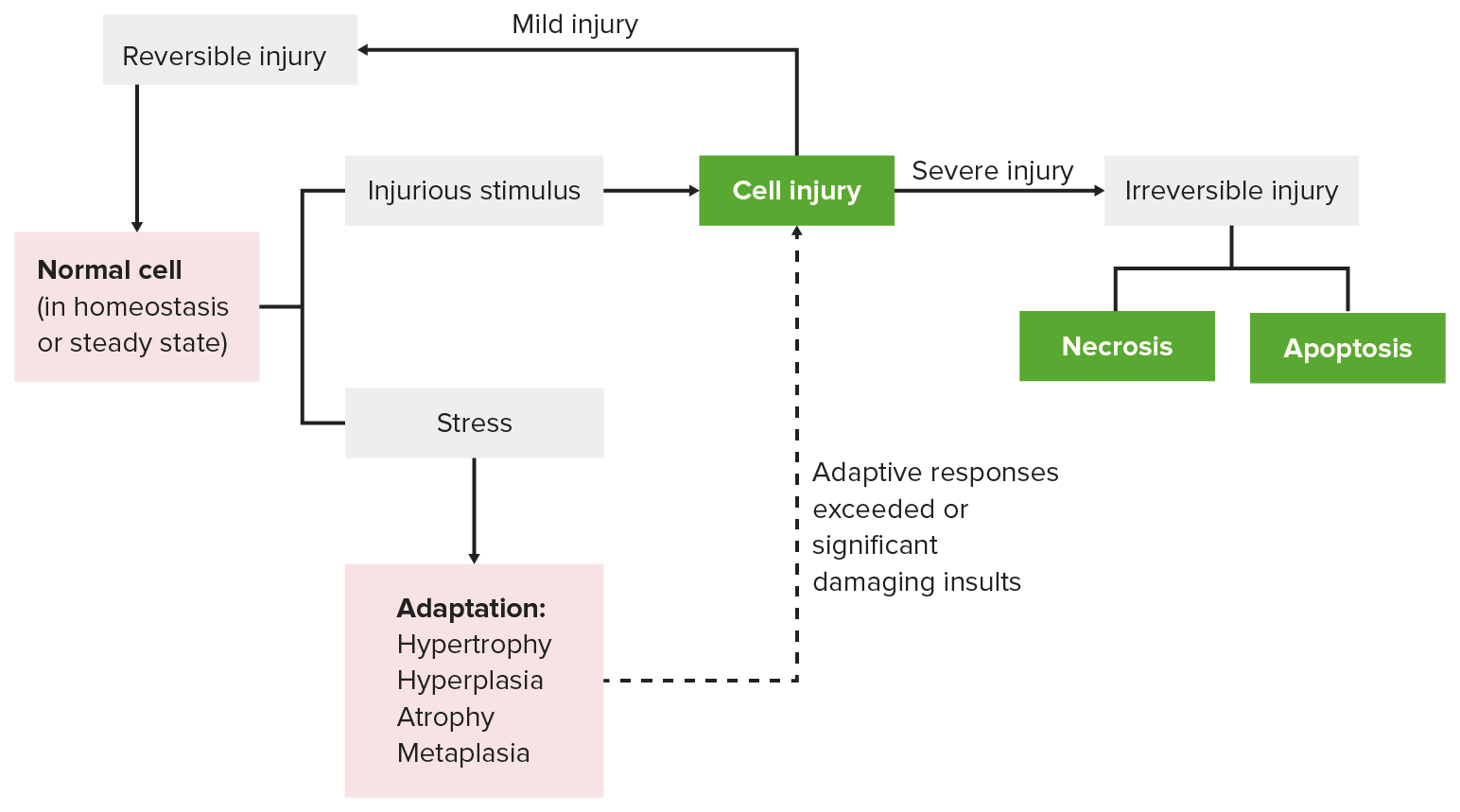
I. Definition & The Infarction Link

In the study of hemodynamic disorders, it is critical to distinguish between the mechanism of injury and the final resulting lesion. While an infarction is the ultimate pathological result (the localized area of ischemic tissue necrosis), an embolism is the primary mechanical cause that initiated the cascade.

Embolism Defined

  • Embolism: The pathophysiological process of partial or complete obstruction of a part of the cardiovascular system by any foreign mass carried within the bloodstream.
  • Embolus: The transported intravascular mass itself. It is a detached material that has broken free from its original site of origin and is now traveling freely through the circulatory system.
  • Physical States: An embolus is not exclusively a blood clot. It can be a detached intravascular solid (clots, tumor clumps), a liquid (fat droplets, amniotic fluid), or a gaseous mass (air bubbles, nitrogen).

The Key Pathophysiological Sequence:

  1. Formation: An embolus forms or is introduced into the vasculature (e.g., a deep vein thrombosis breaks loose, or a fractured bone releases marrow fat).
  2. Transit: The embolus travels seamlessly through the larger upstream vessels of the bloodstream.
  3. Impaction: It eventually reaches a downstream vessel where the lumen diameter narrows to the point that it is physically too small for the mass to pass.
  4. Obstruction & Ischemia: The mass tightly wedges into the vessel, instantly halting anterograde arterial blood flow, leading directly to localized tissue hypoxia and ischemia.
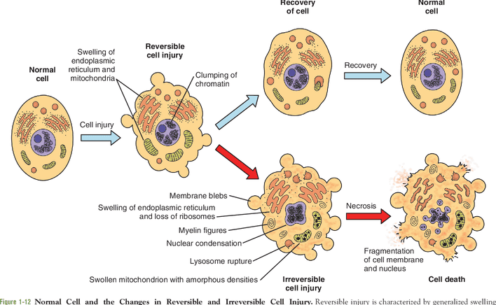
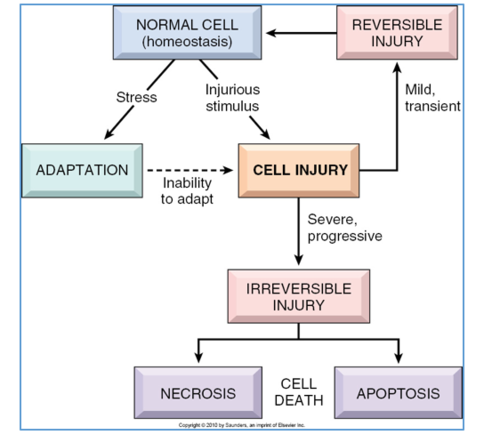
  5. Infarction: If the occlusion is severe, sudden, or prolonged—and if collateral circulation is inadequate—the ischemic cells pass the point of no return, culminating in irreversible coagulative necrosis (infarction).

💡 The Golden Rule of Embolism vs. Thrombosis

A Thrombus forms locally and stays exactly where it was created, growing firmly attached to the endothelial wall of the vessel. An Embolus travels. It is a rogue, detached piece of material that breaks off, hops onto the cardiovascular "highway," and violently crashes when the road narrows. All thromboemboli started as thrombi, but not all thrombi become emboli!


II. Classification of Emboli

Emboli are highly heterogeneous. Pathologists classify them based on the matter they are composed of, whether they carry an infectious payload, their specific anatomical source, and their eventual destination.

Depending on the Matter (State)
  • Solid: Detached blood clots (Thromboemboli - which astonishingly make up 90-99% of all clinical emboli), fragmented atheromatous plaques (cholesterol crystals), aggressive tumor cell clumps, necrotic tissue fragments, parasites, or bacterial vegetations.
  • Liquid: Globules of marrow fat, amniotic fluid during childbirth, or inadvertently injected foreign fluids.
  • Gaseous: Atmospheric air bubbles (introduced via IV lines or trauma) or other gases like Nitrogen precipitating out of the blood in decompression sickness.
Depending on Infection
  • Bland: Completely sterile, containing no viable bacteria. They cause pure ischemic damage.
  • Septic: Heavily infected with pyogenic bacteria. When a septic embolus lodges in distant tissue, it doesn't just cause ischemia; it actively seeds the dying tissue with bacteria, rapidly forming a highly destructive metastatic abscess.
Depending on the Source
  • Cardiac emboli: Arising from the chambers of the heart (primarily the left atrium or left ventricle).
  • Arterial emboli: Originating from systemic arteries (e.g., an ulcerated plaque in the internal carotid artery firing emboli into the cerebral circulation).
  • Venous emboli: Originating from systemic veins (e.g., deep leg veins) and traveling to the right heart.
  • Lymphatic emboli: Usually composed of metastatic tumor cells traveling through the lymphatic vessels to regional lymph nodes.

The 8 Major Clinical Types of Embolism:

In clinical practice, these are the primary culprits causing morbidity and mortality:

  1. Pulmonary embolism (PE)
  2. Systemic (Arterial) embolism
  3. Fat embolism
  4. Air embolism
  5. Decompression sickness (Nitrogen gas embolism)
  6. Amniotic fluid embolism
  7. Atheroembolism (Cholesterol embolism)
  8. Tumour embolism

III. Thromboembolism (90%+ of all Emboli)

A detached thrombus, or a fragment of a thrombus that breaks off, constitutes the overwhelming majority of emboli. Because the cardiovascular system is a closed loop split into two distinct halves (pulmonary and systemic), the origin of the thrombus absolutely dictates its deadly destination.

A. Arterial (Systemic) Thromboembolism

  • Origin: Around 80% of systemic arterial emboli arise from intracardiac mural thrombi. These predominantly occur in a diseased left heart (e.g., mural thrombi forming on the scarred wall of the left ventricle following a massive myocardial infarction, stagnant blood in a fibrillating left atrium, or vegetative endocarditis on the mitral and aortic valves). The remaining 20% derive from aortic aneurysms, carotid atherosclerosis, or paradoxical emboli.
  • Destination: Because it starts in the arterial system, it is propelled OUT to the systemic capillary beds of the body. They invariably cause localized infarction at the sites of lodgement.

Specific Effects & Locations of Arterial Emboli:

The ultimate clinical consequence depends heavily upon the size of the embolus, the specific site of lodgment, and the adequacy of local collateral circulation.

  • Lower Limbs (70-75%): The most common destination. Results in Acute Limb Ischemia. Clinicians look for the classic "6 Ps": Pain, Pallor (paleness), Pulselessness, Paresthesia (tingling/numbness), Paralysis, and Poikilothermia (the limb feels ice cold). If collateral circulation is inadequate and surgery is delayed, the infarction progresses to irreversible Gangrene.
  • The Brain (10%): Lodging in the cerebral circulation (often the Middle Cerebral Artery), causing a massive ischemic stroke and potentially sudden death.
  • Internal Viscera (10%): Splenic infarctions, renal infarctions (causing flank pain and hematuria), or severe acute mesenteric ischemia in the intestines (a highly lethal condition causing excruciating abdominal pain out of proportion to the physical exam).
  • Myocardial Infarction: An extremely rare event where an embolus is swept directly into the coronary arteries.

B. Venous Thromboembolism (VTE)

  • Origin: The vast majority (over 95%) of venous emboli originate from Deep Vein Thrombosis (DVT) within the large, deep veins of the lower extremities (specifically the popliteal, femoral, and iliac veins). Less common sources include severe varicosities, pelvic venous plexuses (especially post-surgery or post-partum), or the intracranial cavernous sinus.
  • Destination: Because it originates in the systemic veins, the clot flows with venous return, gradually moving into larger and larger vessels (up the Inferior Vena Cava). It dumps directly into the right atrium, passes into the right ventricle, and is violently pumped out into the pulmonary arterial tree.
  • The Ultimate Consequence: Rapid, acute obstruction of the pulmonary arterial circulation, causing Pulmonary Embolism (PE).
Mnemonic

Arterial vs. Venous Emboli Destination

Remember the simple directional plumbing of the heart to never confuse the two on an exam:

  • Venous emboli go to the Ventilators: (Lungs = Pulmonary Embolism).
  • Arterial emboli go to the Appendages and Organs: (Brain, Kidneys, Legs = Stroke / Acute Limb Ischemia).

IV. Pulmonary Embolism (PE)

Pulmonary embolism is the most common, preventable, and highly fatal form of venous thromboembolism. It involves the sudden, acute occlusion of the pulmonary arterial tree by a traveling blood clot.

Etiology & Risk Factors (Virchow's Triad)

The formation of the initial DVT is driven by Virchow's Triad: endothelial injury, hypercoagulability, and venous stasis.

  • Highly prevalent in hospitalized, post-operative, or bed-ridden patients due to severe venous stasis in the legs.
  • Ambulatory patients with underlying genetic hypercoagulable states (e.g., Factor V Leiden mutation).
  • Specific Hormonal Triggers: Late pregnancy, the puerperal (post-partum) state, and the use of oral contraceptive pills. Estrogen fundamentally alters the liver's production of clotting factors, tipping the blood into a pro-thrombotic state.

Pathogenesis (The Hemodynamic Journey):

  1. Detachment of a fragile, propagating thrombus from the deep leg veins.
  2. The thrombo-embolus flows effortlessly through the widening venous drainage into the Inferior Vena Cava (IVC).
  3. Drains seamlessly through the Right Atrium and into the Right Ventricle.
  4. Pumped out forcefully into the pulmonary artery, where the vascular tree begins to branch and rapidly narrow.

Outcome A (Saddle Embolus): If the thrombus is massive (often shaped like a long snake reflecting the leg vein it came from), it gets impacted directly at the main bifurcation (the "crotch") of the pulmonary artery. It straddles both the left and right main pulmonary arteries like a saddle on a horse.

Outcome B (Multiple Small Emboli): Multiple smaller emboli, or a large one that fragments through the mechanical churning of the right ventricle, will bypass the main bifurcation and impact in a number of smaller, peripheral vessels, heavily favoring the highly-perfused lower lobes of the lungs.

Consequences of Pulmonary Embolism:

The severity of the resulting syndrome depends heavily upon the size of the occluded vessel, the sheer number of emboli showering the lungs, and the pre-existing baseline cardiovascular health of the patient.

1. Sudden Death

A massive pulmonary embolism (like a Saddle Embolus) results in virtually instantaneous death, often without the patient even having time to complain of chest pain or dyspnea!
Pathophysiology: It physically blocks 100% of blood from leaving the right heart. Consequently, the left heart receives absolutely no blood. Systemic cardiac output instantly drops to zero, leading to cardiovascular collapse and Pulseless Electrical Activity (PEA). If death is slightly delayed, the clinical features mimic a massive myocardial infarction (severe chest pain, sweating, profound cardiogenic shock).

2. Acute Cor Pulmonale

Occurs when numerous small emboli suddenly obstruct over 60% of the total pulmonary circulation. The right ventricle, which is normally a thin-walled, low-pressure pump, suddenly faces an impenetrable wall of resistance. It must pump incredibly hard, resulting in rapid, acute right heart failure (severe acute dilatation of the right ventricle). This pushes the intraventricular septum leftward, further crushing left ventricular filling.

3. Pulmonary Infarction

Occurs upon obstruction of relatively small-sized, peripheral pulmonary arterial branches. Interestingly, because the lung has a dual blood supply (Pulmonary arteries + Bronchial arteries), true infarction only occurs if the patient's underlying cardiovascular status is already compromised (e.g., existing heart failure). Clinical features include severe pleuritic chest pain (pain worsens upon taking a deep breath), dyspnea, and hemoptysis (coughing up blood) as the necrotic lung tissue bleeds into the alveoli.

4. Pulmonary Hemorrhage

Obstruction of terminal branches (end-arteries). The dual bronchial blood supply manages to keep the tissue alive (no infarction), but the ischemic endothelial damage causes the capillaries to leak massively, leading to central pulmonary hemorrhage. Features include hemoptysis and dyspnea, but less pleuritic chest pain due to the central location away from the sensitive pleura.

5. Resolution (The Best Case)

The vast majority (60-80%) of very small, clinically silent pulmonary emboli are effectively dissolved and cleared by the body's natural fibrinolytic defense system (plasmin aggressively dissolving the fibrin clot network within days to weeks).

6. Chronic Cor Pulmonale / Pulmonary Hypertension

These are the devastating long-term sequelae of multiple, recurrent small thromboemboli that undergo organization (fibrotic scarring) rather than fibrinolytic resolution. Over years, these organized scars permanently narrow and stiffen the pulmonary vascular bed, driving pulmonary pressures steadily upward until the right heart ultimately fails.


V. Contrasting Pulmonary Thrombosis vs. Thromboembolism

Although extremely rare, localized thrombosis can occur directly within the pulmonary arteries (usually secondary to existing pulmonary atherosclerosis, severe pulmonary hypertension, or local trauma). It is a diagnostic necessity for a pathologist to differentiate a primary local thrombus from a traveling embolus during an autopsy.

Diagnostic Feature Pulmonary Thrombosis (Primary) Pulmonary Thromboembolism (Secondary)
Pathogenesis Locally formed at the exact site of occlusion. Travelled from a distant source (usually deep leg veins).
Anatomical Location Typically found in small arteries and distal branches where flow is naturally sluggish. Found abruptly lodged in major arteries and main bifurcations (can be massive in size).
Wall Attachment Firmly, biologically adherent to the vessel wall. Difficult to scrape off. Loosely attached, wedged tightly, or lying completely free within the lumen.
Gross Appearance Distinct architecture: the head is pale (platelet-rich), the tail is dark red (RBC trapped). No distinct distinction in head/tail; usually has a smooth-surfaced, dry, dull, cast-like appearance matching the leg vein.
Microscopic Architecture Platelets and fibrin are laid down in beautifully distinct, alternating microscopic layers. Lines of Zahn are clearly and prominently seen. Haphazardly mixed with blood clot due to churning in the heart. Lines of Zahn are generally rare, disrupted, or entirely absent.

VI. Detection and Diagnosis of Emboli and Infarction

Because emboli (especially a massive PE) can lead to rapid right ventricular strain, profound cardiovascular collapse, and sudden death within minutes, immediate and highly accurate diagnosis is a life-saving critical priority.

Clinical Signs & Symptoms (High Suspicion):

  • Tachypnea: An abnormally fast breathing rate (>20 breaths/min), seen in an overwhelming 96% of cases.
    Pathophysiology: The embolus creates a massive Ventilation-Perfusion (V/Q) mismatch. It creates "alveolar dead space"—areas of the lung where oxygen enters the alveoli, but absolutely no blood is flowing past to pick it up. The brain detects low oxygen and forces the body to hyperventilate to try and compensate.
  • Tachycardia: A rapid, pounding heart rate (>100 beats/min) as the heart struggles to maintain cardiac output against the blockage.
  • DVT Signs: Unilateral leg swelling, throbbing pain, warmth, and erythema (tenderness usually localized deep in the calf muscle).
  • Cyanosis: A disturbing bluish discoloration of the lips and nail beds due to profound, systemic hypoxemia.

Advanced Imaging Tests (The Gold Standards):

  • CT Pulmonary Angiography (CTPA): The absolute most preferred and highly accurate diagnostic test! It utilizes rapid 3D CT imaging combined with an intravenous iodine contrast dye to physically visualize the pulmonary tree. An embolus appears as a dark, definitive "filling defect" cutting off the bright white dye.
  • Ventilation-Perfusion (V/Q) Scan: A highly specialized nuclear medicine scan used to map air flow (ventilation) against blood flow (perfusion) in the lungs. Particularly vital if a CTPA cannot be performed (e.g., in a pregnant patient to avoid high-dose chest radiation, or in a patient with severe kidney failure where CT iodine contrast is highly nephrotoxic). A PE shows normal ventilation but totally absent perfusion.
  • Doppler Ultrasound: Used to scan the deep veins of the legs for a DVT. Finding the primary source strongly suggests the presence of a PE in a symptomatic patient without needing to radiate the chest.
  • Echocardiogram: An ultrasound of the heart that cannot usually see the lung arteries directly, but can definitively identify right heart strain (the right ventricle ballooning outward = acute cor pulmonale) or, occasionally, actually visualize a massive clot actively "in transit" whipping around inside the right atrium or ventricle.

Laboratory Blood Tests:

  • D-Dimer Test: Measures the presence of D-dimer, a highly specific protein fragment released exclusively when the body's plasmin is actively breaking down a cross-linked fibrin clot.
    Clinical Utility & Caveat: A perfectly normal/negative D-dimer safely and definitively rules OUT a clot (it has an exceptionally high negative predictive value). However, a high/positive level is extremely non-specific! D-dimer elevates in pregnancy, cancer, severe inflammation, and post-surgery. Therefore, a high D-dimer demands a CTPA to actually confirm the diagnosis.
  • Arterial Blood Gas (ABG): Measures exact oxygen and carbon dioxide levels in the deep arterial blood. In a classic PE, the ABG reveals severe hypoxemia (low oxygen) and a profound respiratory alkalosis (low CO2) because the patient is hyperventilating and blowing off all their acid.

❓ Applied Clinical Question: The Post-Op Patient

Case: A 55-year-old female is recovering in the hospital 4 days after undergoing major orthopedic surgery on her right knee. She suddenly calls frantically for the nurse, complaining of sharp, stabbing chest pain specifically when she breathes in (pleuritic pain), and she begins coughing up bright red blood (hemoptysis). Her respiratory rate is 28, and her right calf is visibly swollen, red, and warm to the touch.

What is the most likely diagnosis, what was the exact path the object took to get there, and what is the gold-standard imaging test you should order immediately?

Answer: She is suffering from an acute Pulmonary Embolism that has progressed to a pulmonary infarction (which is the direct cause of the hemoptysis and pleuritic pain).
The Path: A Deep Vein Thrombosis (DVT) formed in the stagnant veins of her immobilized right leg -> it broke free -> traveled up the Iliac vein -> into the Inferior Vena Cava (IVC) -> Right Atrium -> Right Ventricle -> Pulmonary Artery -> and permanently lodged in a small peripheral lung branch.
The Test: The physician must immediately order a CT Pulmonary Angiography (CTPA) to visualize the clot and initiate immediate systemic anticoagulation therapy (e.g., Heparin).


VII. Septic Embolism & Systemic Embolism

While we explored systemic arterial emboli previously, the fundamental nature of the material traveling through the arteries can drastically, and dangerously, alter the clinical outcome.

Systemic Arterial Embolism (A Brief Review):

These originate predominantly from sterile thrombi residing in a diseased left heart. Because they travel out through the aorta, they invariably cause pure, sterile ischemic infarction at the highly perfused sites of lodgement (legs, brain, viscera).

Septic Emboli: The Infected Missiles

  • These are highly dangerous emboli containing dense, active colonies of viable bacteria.
  • Origin: They originate classically from Vegetative Mural Endocarditis (massive, friable clumps of highly active bacteria and fibrin aggressively growing on and destroying the inner heart valves, notably the mitral or aortic valves, frequently caused by Staphylococcus aureus or Streptococcus viridans).
  • Pathology: When these infected fragments break off and lodge in distant, healthy tissues, they do not merely cause ischemic necrosis; they actively seed the dying tissue with millions of bacteria. This dual-action injury rapidly liquifies the dead tissue, forming a highly destructive metastatic abscess filled with purulent exudate (pus) wherever the embolus lands (e.g., brain abscesses, renal abscesses).
Diagnostic Hallmark

Clinical Sign of Septic Emboli: Janeway Lesions

If a patient is suffering from Acute Bacterial Endocarditis, tiny fragments of the infected valve vegetations can shoot down the arteries of the arm and lodge tightly in the tiny capillary beds of the hands and feet.

This creates Janeway Lesions: Small, completely painless, non-tender subcutaneous maculopapular (flat to slightly raised) hemorrhagic lesions typically found on the thick pulp of the fingers or the palms.

Pathophysiological Distinction: Because Janeway lesions are essentially painless, physical micro-abscesses resulting from traveling bacteria, they are clinically distinguished from Osler's Nodes. Osler's Nodes are intensely painful, raised, immune-complex deposits found on the finger pads in subacute endocarditis! (Memory Trick: Osler's nodes equal Ouch!).


VIII. Emboli Dependent Upon the Flow of Blood

Usually, emboli predictably follow the standard, directional plumbing of the cardiovascular system. However, two highly specialized types completely and astonishingly defy normal circulatory logic.

A. Paradoxical Embolus: The "Crossed" Clot

  • Definition: An embolus which is carried from the venous side of the circulation directly to the arterial side (or vice versa), entirely bypassing the massive capillary filter network of the lungs! This is termed a paradoxical or "crossed" embolus.
  • Mechanism: It absolutely requires the presence of an abnormal, congenital arteriovenous communication (a right-to-left intracardiac shunt). The most common structural defects allowing this are a Patent Foramen Ovale (PFO), an Atrial Septal Defect (ASD), or a Ventricular Septal Defect (VSD).
  • Clinical Scenario: A patient has a massive, silent DVT in their leg. They go to the bathroom and bear down aggressively (the Valsalva maneuver). This heavy straining temporarily raises right atrial pressure above left atrial pressure. In that split second, the right-to-left shunt opens. The leg clot arrives, bypasses the right ventricle, slips through the PFO directly into the left atrium, drops into the left ventricle, and shoots straight up the carotid artery into the brain, causing a massive, unexplainable ischemic stroke in an otherwise healthy young person!

B. Retrograde Embolus: Flowing Backwards

  • Definition: An embolus which miraculously travels in the exact opposite direction of the normal forward flow of blood.
  • Classic Example: Widespread metastatic tumor deposits found inexplicably in the high thoracic and cervical spine originating from an early-stage Prostate Carcinoma.
  • The Mechanism: The prostatic venous plexus normally drains cleanly into the internal iliac vein. However, it also heavily interconnects with the vertebral venous plexus (Batson's Plexus) that runs the entire length of the spine.
  • These complex intraspinal/vertebral veins uniquely operate under incredibly low pressure and, crucially, they possess absolutely NO one-way valves.
  • During conditions of high, sustained intra-pelvic or intra-abdominal pressure (such as severe, chronic coughing, straining, or heavy lifting), blood is aggressively forced backward (retrograde movement) from the prostate gland directly up into the spinal column. Insidious tumor cells ride this backward vascular wave, effortlessly leading to severe, destructive bone metastasis far from the primary tumor!

IX. Fat and Tumour Embolism

Not all occlusions are driven by coagulated blood. Lipids and rapidly dividing neoplastic cells frequently hijack the vasculature.

Fat Embolism

Defined as the lethal obstruction of arterioles and capillaries by circulating fat globules. If the obstruction is caused by actual, intact microscopic fragments of adipose (fat) tissue, it is specifically termed a fat-tissue embolism.

  • Traumatic Causes: The most common, defining cause is severe physical, crushing trauma to the skeletal bones (e.g., severe long bone fractures like a shattered femur or crushed pelvis in a high-speed car accident). The violent fracture forcefully releases large volumes of semi-liquid, yellow bone marrow fat directly into the freshly ruptured, low-pressure venous sinusoids of the bone.
  • Non-Traumatic Causes: Severe fatty liver disease, Diabetes Mellitus (DM), aggressive liposuction procedures, or severe acute pancreatitis.

💡 High-Yield Clinical Triad: Fat Embolism Syndrome (FES)

Fat embolism is uniquely destructive due to both a mechanical and a highly toxic biochemical pathway. If a patient severely fractures their femur and 24 to 72 hours later develops a highly specific triad of symptoms, they have FES:

  1. Hypoxemia (Acute Respiratory Distress): The fat droplets physically clog the massive pulmonary capillary bed, leading to severe shortness of breath. Furthermore, the biochemical theory states that pulmonary lipase enzymes aggressively break down the fat droplets into highly toxic Free Fatty Acids, which chemically burn and destroy the delicate alveolar pneumocytes, causing Acute Respiratory Distress Syndrome (ARDS).
  2. Neurologic Abnormalities: The tiny fat micro-globules squeeze through the pulmonary filter and shower the brain, causing sudden confusion, agitation, delirium, or deep coma.
  3. Petechial Rash: The hallmark sign. A fine, pinpoint, non-blanching red rash appears specifically on the chest, neck, conjunctiva, and armpits (axillae). This is caused by fat micro-emboli rupturing the dermal capillaries, combined with a sudden massive drop in blood platelets as they aggregate around the fat droplets.

Tumour Embolism

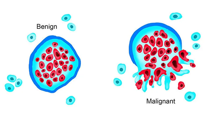
Malignant, highly aggressive tumor cells utilize the bloodstream for distant colonization. They secrete enzymes that degrade the extracellular matrix, allowing them to actively invade local, thin-walled blood vessels (especially veins). Clumps of these cells break off to form circulating tumor emboli, lodging in distant capillary beds and establishing secondary metastatic tumor deposits.

Notable Examples:

  • Clear cell carcinoma of the kidney: A notorious cancer that actively invades the renal vein, forms an organized embolic mass, and literally grows backward like a solid snake entirely up the Inferior Vena Cava (IVC), sometimes reaching all the way into the Right Atrium!
  • Carcinoma of the lung: Frequently sending embolic metastases to the brain and adrenal glands.
  • Malignant Melanoma: Extremely aggressive, utilizing both lymphatic and hematogenous embolic spread widely throughout the body.

X. Amniotic Fluid Embolism (AFE)

A rare (approx. 1 in 40,000 deliveries), highly unpredictable, catastrophic, and exceptionally lethal obstetric complication of pregnancy and childbirth. Mortality rates historically exceed 60-80% if untreated immediately.

  • Definition: AFE occurs when amniotic fluid—which is heavily contaminated with fetal squamous skin cells, lanugo (fetal hair), vernix, meconium, and dense cellular debris—aggressively and inappropriately enters the maternal pulmonary circulation. This typically happens through a tear in the placental membranes or deeply ruptured uterine veins during intense labor, complicated delivery, or immediate post-partum trauma.

Pathophysiology (Far beyond a simple physical blockage!)

The profound lethality of AFE is not merely due to fetal debris physically plugging the maternal lung vessels. It triggers a massive, systemic, two-phased immune and hemodynamic collapse:

  1. Phase 1: Severe Pulmonary Vasospasm & Anaphylaxis. The maternal immune system recognizes the fetal debris as a massive foreign antigen attack. It triggers a profound, systemic inflammatory response (similar to severe anaphylactic shock). The pulmonary arteries violently spasm shut, leading to immediate, severe hypoxemia, acute right heart failure, and rapid cardiogenic shock.
  2. Phase 2: Disseminated Intravascular Coagulation (DIC). The amniotic fluid is incredibly, dangerously rich in Tissue Factor (thromboplastin). When massive amounts of Tissue Factor enter the mother's central blood supply, it completely overwhelms the coagulation cascade. It aggressively forces the mother's body to form millions of microscopic blood clots everywhere, instantly consuming and depleting all of her platelets and clotting factors.

The Devastating Result: Because all clotting factors are utterly exhausted by the microscopic clots, the mother immediately transitions into uncontrollable, profuse, widespread bleeding from every orifice and surgical site (DIC). She suffers simultaneous hypoxic respiratory failure, cardiogenic shock, and exsanguination.


XI. Air and Gas Embolism

It is a common misconception that only solid objects cause blockages. Air, nitrogen, and other gases can produce highly resilient physical bubbles within the circulation. Due to surface tension, these gas bubbles act exactly like solid, impenetrable physical clots, relentlessly obstructing blood vessels and causing severe downstream tissue hypoxia. There are two main forms: Air Embolism and Decompression Sickness.

A. Air Embolism

Occurs when ambient atmospheric room air is accidentally, or traumatically, introduced into either the venous or arterial circulation.

1. Venous Air Embolism:

Occurs when air is inadvertently sucked into the low-pressure systemic venous system. It can occur under the following specific circumstances:

  • Operations on the head & neck / Severe Trauma: Accidental opening of a major vein (like the large internal jugular vein). Because venous pressure in the neck is actually sub-atmospheric (lower than the outside air pressure) when a patient is sitting upright, air is rapidly and forcefully drawn into the open vessel like a vacuum.
  • Obstetrical operations & trauma: During normal vaginal delivery, caesarean sections, or aggressive abortions, massive, fatal air embolism may occasionally result from pressurized air entering the huge, opened-up uterine venous sinuses and deep endometrial veins.
  • Intravenous (IV) Infusion / Iatrogenic: Can occur if improper, un-purged IV lines are used, or if positive pressure is incorrectly employed to push blood or fluid bags that run completely empty.
  • Angiography / Catheterization: During a venogram or central line placement, air may be accidentally entrapped in the massive catheter lumen and injected directly into the deep central venous system.

Factors determining the extreme severity of Venous Air Embolism:

  1. Amount of air introduced: Highly variable, but usually 100 to 150 ml of rapid air entry is considered definitively fatal in a healthy adult. (In severely ill or compromised patients, as little as 40 ml can be highly lethal). The air enters the Right Ventricle and mixes with the blood to create a dense, frothy, incompressible foam that acts as an "air lock," physically preventing the ventricle from pumping any blood forward into the lungs.
  2. Rapidity of entry: A sudden massive bolus is far more lethal than a slow, steady leak that the body can gradually absorb.
  3. General cardiovascular condition of the patient.
  4. Position of the patient: If the patient's head is elevated higher than their trunk (upright/sitting position), air bubbles—being lighter than blood—will ascend rapidly directly up the superior vena cava and reach the cerebral venous sinuses, causing massive neurological damage!

2. Arterial Air Embolism:

Occurs when air directly enters the pulmonary vein (via chest trauma or lung surgery) or its tributaries. It travels instantly to the left heart and is pumped directly out to the delicate systemic organs. It is extraordinarily highly fatal even in miniscule amounts if the tiny bubbles reach the extremely sensitive coronary arteries (causing instant myocardial infarction) or cerebral arteries (causing severe embolic stroke).

Physiology Expansion

Durant's Maneuver (The Life-Saving Position)

If you are a nurse or physician and you suspect a massive venous air embolism (e.g., a massive central line becomes completely disconnected, and you hear a distinct "hissing" sound), why do doctors instantly instruct you to place the patient in the Left Lateral Decubitus position with the head tilted severely down (Trendelenburg)?

The Physical Rationale: You urgently want the massive, lethal air bubble to rise and trap itself safely against the non-obstructing lateral wall and apex of the Right Ventricle! If the patient sits up or lies flat, the lighter air bubble will float directly up into the pulmonary outflow tract, causing an impenetrable "air lock." This totally stops all forward blood flow to the lungs, dropping cardiac output to zero, causing instant death. By tilting them left and down, you trap the air away from the exit valve, allowing blood to flow underneath it until the air can be slowly absorbed or surgically aspirated via a catheter.


XII. Decompression Sickness (Nitrogen Gas Embolism)

This is a highly specialized, physics-driven form of gas embolism known historically by various names: Caisson's Disease, Divers' Palsy, The Bends, or Aeroembolism. It relies entirely on the complex physics of dissolved atmospheric gases reacting under extreme environmental pressure.

The Physics (Henry's Law)

Henry's Law dictates that the physical solubility of a gas in a liquid is directly proportional to the immense pressure of that gas resting above the liquid. Nitrogen (N2) is a largely inert gas that makes up roughly 78% of normal breathing air.

When a deep-sea diver descends into the depths (or an industrial worker in a highly pressurized caisson/underwater diving-bell performs heavy labor), the immense, crushing physical pressure of the surrounding water forces massive, unnatural amounts of Nitrogen gas to dissolve completely and invisibly into the diver's blood and deep tissue fluids.

Pathogenesis of the Embolism

  • The highly dissolved Nitrogen is entirely harmless as long as the diver remains at depth and the pressure remains constantly high.
  • The Danger (Ascending too rapidly): If the individual decompresses too suddenly (e.g., panicking and swimming from high-pressure depths to the normal-pressure surface in seconds, or an astronaut flying in an unpressurized cabin rapidly from normal ground level to low-pressure extreme altitudes), the dissolved nitrogen gases physically cannot remain in solution. They instantly precipitate out, expanding violently as thousands of minute gas bubbles directly within the blood and solid tissues. (Think exactly of rapidly unscrewing the cap on a highly shaken, pressurized bottle of soda!).
  • These minute, expanding bubbles quickly coalesce (join together) in the venous system to form massive, highly obstructive Nitrogen emboli.

Effects & Severity Factors

The overall severity of the resulting tissue damage depends strictly on: 1) The extreme depth/altitude reached, 2) The total duration of high-pressure exposure, 3) The exact rate of ascent, and 4) The general physiological condition of the individual.

  • Obesity Risk Factor: The physiological changes are vastly more serious and lethal in obese persons. Why? Because Nitrogen gas is highly lipophilic (it is up to five times more soluble in fat tissue than in normal body fluids). Obese divers absorb vastly higher, dangerous reserves of Nitrogen into their adipose tissue!
  • Sudden decompression from high pressure to normal levels (divers) causes a much more massive gradient shift, and thus is significantly more pronounced and dangerous than moving from normal to low pressure (pilots).

Clinical Effects of Decompression Sickness

1. Acute Form (Immediate Presentation)

Occurs due to the sudden, acute obstruction of thousands of small blood vessels simultaneously.

  • 'The Bends': The hallmark symptom. The patient literally doubles over in agony in bed due to excruciating, severe, sharp pain localized deep in the major joints (shoulders, knees), ligaments, and skeletal muscles, caused by gas bubbles aggressively tearing apart the tissue planes and blocking local ischemia.
  • 'The Chokes': Massive accumulation of countless tiny nitrogen bubbles in the pulmonary microvasculature, resulting in severe acute respiratory distress, heavy coughing, and suffocating chest pain.
  • Cerebral Effects: Nitrogen bubbles expanding directly within the brain or spinal cord may manifest rapidly as severe vertigo, vision loss, instant coma, and sometimes sudden death.

2. Chronic Form (Delayed Presentation)

Occurs in professional divers who suffer repeated, minor, poorly-treated decompression events over years.

  • Avascular Necrosis of Bones (Caisson Disease of Bone): Chronic, tiny nitrogen emboli relentlessly occlude and destroy the delicate blood supply to the bones. Classic, highly tested locations include the heavy destruction of the head of the femur, the tibia, and the humerus, leading to early, crippling osteoarthritis.
  • Neurological Symptoms: Severe, permanent ischemic damage to the spinal cord tracts includes intractable paraesthesia (numbness/tingling) and irreversible paraplegia (paralysis of the lower body).
  • Lung Involvement: Chronic micro-infarctions can present as pulmonary hemorrhage, severe edema, emphysema, and lung atelectasis (collapse).
  • Skin Manifestations (The Creeps): Cutaneous itching (pruritus), patchy red erythema, cyanosis (mottling), and pitting edema as bubbles block dermal lymphatics.

❓ Final Module Review Clinical Question

Case: A 35-year-old male, slightly overweight, presents to the ER via ambulance with excruciating, crippling hip and knee pain, extreme shortness of breath accompanied by a dry cough ("the chokes"), and distinct patchy erythema mottling his skin. He reports that he is a professional deep-sea pipeline welder and his oxygen equipment malfunctioned earlier today, forcing him to drop his gear and swim to the surface as fast as humanly possible.

Based entirely on the strict pathogenesis of his condition, what specific gas is currently obstructing his microvasculature, and what is the absolute definitive, life-saving medical treatment?

Answer: The specific gas obstructing his vessels is Nitrogen (N2), which rapidly bubbled out of solution due to his extremely rapid ascent causing a sudden drop in ambient pressure (Henry's Law). He is suffering from severe, acute Decompression Sickness (The Bends). The definitive, non-negotiable medical treatment is immediate transport to and placement inside a Hyperbaric Oxygen Chamber. The chamber safely and heavily re-pressurizes his entire body to forcefully crush the nitrogen bubbles back into a dissolved liquid solution within his blood, and then painstakingly slowly decompresses him over hours so his lungs can safely exhale the nitrogen normally.


XIII. References & Recommended Reading

  • Kumar, V., Abbas, A. K., & Aster, J. C. (2020). Robbins and Cotran Pathologic Basis of Disease (10th ed.). Elsevier. (Chapters on Hemodynamic Disorders, Thromboembolism, and Shock).
  • Loscalzo, J., Fauci, A., Kasper, D., Hauser, S., Longo, D., & Jameson, J. L. (2022). Harrison's Principles of Internal Medicine (21st ed.). McGraw Hill. (Sections on Pulmonary Embolism and Deep Vein Thrombosis).
  • Hall, J. E., & Hall, M. E. (2020). Guyton and Hall Textbook of Medical Physiology (14th ed.). Elsevier. (Aviation, High Altitude, and Space Physiology; Deep-Sea Diving and Other Hyperbaric Conditions).
  • Kearon, C., et al. (2016). Antithrombotic Therapy for VTE Disease: CHEST Guideline and Expert Panel Report. Chest, 149(2), 315-352.

Quick Quiz

Embolism Quiz

Pathology - mobile-friendly and focused practice.

Privacy: Your details are used only for quiz tracking and certificates.

Embolism in Pathology Read More »

Infarction

Infarctions in Pathology

Pathology Master Guide: Infarction

Module Learning Objectives

By the conclusion of this exhaustive guide, you will be deeply conversant with:

  • The precise definition and core mechanisms of infarction and ischemic necrosis.
  • The comprehensive Etiology of Hypoperfusion utilizing the TEVACTR mnemonic.
  • The morphological classification of infarcts (Red vs. White vs. Septic) and the anatomical reasons behind their coloration.
  • The four critical factors influencing the development and severity of an infarct.
  • The chronological pathogenesis and microscopic sequence of infarction healing.
  • Extensive, detailed breakdowns of Specific Organ Infarctions (Lung, Kidney, Spleen, Liver, Brain, and Heart).

I. Definition & Introduction

Infarction is a fundamental, life-threatening concept in general and systemic pathology. It represents the ultimate, irreversible consequence of severe, unresolved vascular compromise.

What is an Infarct?

  • Definition: An infarction is defined as the formation of a localized area of ischemic necrosis (cell death) within a tissue or organ.
  • Cause: It results most often from a sudden, catastrophic reduction or complete interruption of arterial blood supply, or occasionally from the sudden obstruction of its venous drainage.
  • The Core Mechanism: The underlying driver is Hypoperfusion (decreased blood flow) leading to severe oxygen deprivation (hypoxia) and the subsequent failure of aerobic cellular metabolism. Without oxygen, ATP production halts, ion pumps fail, and the cell is rapidly destroyed.
Pathophysiology Expansion: Ischemia vs. Hypoxia
While hypoxia is simply low oxygen, ischemia (loss of blood flow) is far more damaging. Ischemia not only deprives the tissue of oxygen, but it also deprives it of vital metabolic substrates (like glucose) and fails to wash away highly toxic metabolic waste products (like lactic acid), leading to much faster and more severe cellular necrosis.

II. Etiology: Causes of Hypoperfusion

What actually physically blocks the blood vessels to cause an infarct? The mechanical and systemic causes of hypoperfusion can be grouped using a classic, highly effective mnemonic.

Mnemonic: TEVACTR

Mechanical Causes of Vascular Occlusion

  • T - Thrombi: A blood clot forming locally within the intact vascular system. Example: A coronary thrombosis forming over a ruptured plaque, leading to a heart attack.
  • E - Emboli: An unattached, free-floating mass (can be a blood clot, fat globule, air bubble, or amniotic fluid) that travels through the bloodstream and wedges into a smaller downstream arterial capillary. Example: A Deep Vein Thrombosis (DVT) breaking off and causing a Pulmonary Embolism (PE).
  • V - Vasospasm: A sudden, intense, localized arterial spasm (severe vasoconstriction) leading to tissue ischemia. Example: Prinzmetal angina or cocaine-induced coronary vasospasm.
  • A - Atheroma (Expansion): The gradual, insidious accumulation of lipid/cholesterol material in the inner layer (intima) of an artery wall, which can chronically expand or suddenly rupture.
  • C - Compression (Extrinsic): A vessel is squeezed shut from the outside. Example: A growing malignant tumor compressing a local artery, or a loop of bowel trapped and strangulated in an inguinal hernia.
  • T - Twisting of a vessel (Torsion): The physical twisting of an organ cuts off its tortuous blood vessels. Example: Testicular torsion or bowel volvulus.
  • R - Rupture (Traumatic): Traumatic tearing of a vessel. Example: A ruptured aortic aneurysm, hemorrhagic stroke, or compartment syndrome where pooled blood edema causes secondary vascular compromise.

Non-Occlusive Causes:

Infarction doesn't always require a physical, localized blockage. Non-occlusive circulatory insufficiency can cause massive infarcts if the global blood flow or systemic oxygenation drops too low to sustain vulnerable tissues. Examples include prolonged global hypotension (hypovolemic, cardiogenic, or septic shock) or severe hypoxic encephalopathy (e.g., drowning or cardiac arrest leading to global brain damage).


III. Types of Infarction (Morphological Classification)

Pathologists classify infarcts based primarily on their gross color and the presence or absence of bacterial infection. The color of an infarct tells you a massive amount about the tissue's underlying vascular anatomy.

A. Red (Hemorrhagic) Infarcts:

These infarcts are engorged with blood and appear dark red or purple. They occur under very specific, well-defined anatomical conditions:

  • Venous Occlusions: Such as in ovarian torsion or testicular torsion. Blood can continue to pump in through thick-walled, high-pressure arteries, but it cannot escape through the collapsed, thin-walled, low-pressure veins. Blood backs up, choking the tissue and causing massive hemorrhagic necrosis.
  • Loose, Spongy Tissues: Such as the lung. The spongy, honeycomb structure of the lung alveoli allows leaked blood to easily collect and pool in massive quantities within the infarcted zone.
  • Tissues with Dual Circulation: Such as the lung (supplied by pulmonary & bronchial arteries) and the small intestine (supplied by multiple anastomosing mesenteric arcades). If one main artery is blocked, the other still pumps blood into the dead area, but the flow isn't strong enough to rescue the tissue—it merely causes massive hemorrhage into the necrotic zone.
  • Previously Congested Tissues: Tissues that were already severely swollen with sluggish venous outflow (e.g., chronic passive congestion of the liver) before the acute infarct occurred.
  • Reperfusion Injury: When blood flow is suddenly restored to an area of pale infarction (e.g., dissolving a clot with tPA or performing angioplasty), blood rushes into the dead tissue and leaks through the damaged capillaries, turning a white infarct red.

B. White (Anemic/Pale) Infarcts:

These infarcts appear distinctly pale, white, and bloodless.

  • They occur strictly with Arterial Occlusions in Solid Organs that have End-Arterial Circulation.
  • Pathophysiology: Because the organ is solid and highly dense (not spongy), there is simply no room for blood to seep in. Because it has "end-arterial" supply (meaning there is no dual circulation or collateral backup), once that single artery is blocked, absolutely no alternative blood can reach the area. The tissue dies and is completely drained of blood.
  • Classic Examples: Heart (Myocardial Infarction), Spleen, and Kidney.

C. Septic Infarcts:

  • Occur when the occluding embolus contains live bacteria. Example: A piece of a highly infected heart valve (vegetation) from infectious endocarditis breaking off and lodging in the brain, kidney, or spleen.
  • The bacteria rapidly multiply in the nutrient-rich dead tissue, transforming the infarct into a walled-off abscess filled with pus and acute inflammatory cells.
  • Infarcts that are completely free of bacterial contamination are termed Bland infarcts.

💡 Chronology of an Infarct: Does the color change?

Yes! Most red/hemorrhagic infarcts will actually become pale later on. This happens because macrophages enter the dead tissue over the following weeks and aggressively lyse, digest, and clear away the dead red blood cells.

The Exception: Pulmonary (lung) infarcts never become pale because the amount of bleeding into the spongy tissue is simply too extensive for macrophages to fully clear!


IV. Factors Influencing the Development of an Infarct

Why does a blocked blood vessel cause a massive, fatal infarct in one patient, but only a tiny, harmless scar in another? The clinical outcome of a vascular occlusion is heavily influenced by four key variables:

  1. Nature of the Vascular Supply (The Most Important Factor!):
    • The availability of an alternative (collateral) blood supply completely determines whether an occlusion will cause severe damage.
    • Organs with dual blood supply (e.g., the Lungs have pulmonary AND bronchial arteries; the Liver has the portal vein AND hepatic artery) are highly resistant to infarction.
    • Organs with single end-arteries (e.g., Kidney, Spleen) will infarct immediately and severely if that single vessel is blocked.
  2. Rate of Development of Occlusion:
    • Slowly developing occlusions are much less likely to cause an infarction because they provide time for the body to grow new blood vessels (angiogenesis) to bypass the blockage.
    • Example: There are three major coronary arteries in the heart. If one slowly occludes over 10 years, alternative perfusion pathways (collateral circulation) develop. This can sufficiently prevent infarction even when the major artery eventually closes completely (Stable Angina). Sudden occlusions (like a ruptured plaque) do not allow time for collaterals to form, resulting in massive necrosis (Acute MI).
  3. Vulnerability of the Tissue to Hypoxia:
    • Different cells have vastly different metabolisms and tolerances to oxygen deprivation.
    • Neurons (Brain): Extremely sensitive. They undergo irreversible damage and death when deprived of blood supply for only 3 to 4 minutes.
    • Myocardial Cells (Heart): Hardier than neurons, but will inevitably die after 20 to 30 minutes of total ischemia.
    • Fibroblasts (Connective Tissue): Highly resilient. They can remain viable even after many hours of complete ischemia!
  4. Oxygen Content of the Blood:
    • The partial pressure of oxygen in the blood at the exact time of the blockage determines the outcome.
    • A partial flow obstruction in a normal, healthy person might have zero clinical effect. However, that exact same partial obstruction in a patient who is heavily anemic, cyanotic, or has severe heart failure (low baseline oxygen tension) will readily tip the scales and lead to complete tissue infarction.

❓ Applied Clinical Pathology Question

Case: A 60-year-old male arrives at the morgue after a sudden death. Autopsy reveals a completely occluded left renal artery and a completely occluded left pulmonary artery. The pathologist notes a firm, pale, wedge-shaped lesion in the kidney, but a dark red, spongy, wedge-shaped lesion in the lung. Explain the difference in colors.

Answer: The kidney suffers a White (Pale) Infarct because it is a dense, solid organ with a single end-arterial supply; no collateral blood can enter the dead zone. The lung suffers a Red (Hemorrhagic) Infarct because it is a loose, spongy tissue with a dual blood supply. Even though the pulmonary artery is blocked, the bronchial artery continues to pump blood under high pressure into the dead spongy tissue, causing massive hemorrhage into the infarcted area.


V. Pathogenesis: The Sequence of Infarction

When a blood vessel is completely occluded, the tissue undergoes a highly predictable, step-by-step sequence of pathological events leading to permanent scarring.

  1. Localized Hyperemia: The immediate surrounding area becomes engorged with blood as collateral vessels maximally dilate attempting to compensate and rescue the tissue.
  2. Edema and Hemorrhage: The dying capillary walls become highly leaky, allowing fluid and red blood cells to seep freely into the surrounding tissue interstitium.
  3. Cellular Changes (The Ischemic Cascade): The ischemic cells undergo severe hypoxia, failing to produce ATP. This leads to the failure of the Na+/K+ pump, massive cellular swelling, calcium influx, and eventual irreversible coagulative necrosis.
  4. Progressive Proteolysis & Lysis of RBCs: The dead tissue and leaked red blood cells are chemically broken down by endogenous enzymes.
  5. Acute Inflammatory Reaction & Hyperemia: The body's immune system recognizes the dead tissue as "foreign" and mounts an intense acute inflammatory response (dominated by neutrophils) at the margins of the infarct to begin cleaning up.
  6. Blood Pigments Liberated: As the leaked red blood cells are destroyed (hemolysis) by macrophages, they release their iron content, which is converted into Hemosiderin (leaving a distinct brown/rust-colored pigment in the tissue).
  7. Progressive Ingrowth of Granulation Tissue: Fibroblasts and new, fragile, leaky blood vessels grow into the dead area to replace the necrotic tissue with a permanent, non-functional fibrous scar.

VI. General Pathologic Changes of Infarcts

Gross Appearance (Macroscopic):

  • Shape: Infarcts of solid organs are characteristically wedge-shaped.
  • Orientation: The apex (the pointed tip of the wedge) points directly toward the occluded artery. The wide base rests heavily on the outer surface (capsule, pleura, or epicardium) of the organ.
  • Color Evolution: As extensively discussed, arterial occlusions in solid organs are pale, while venous obstruction or dual-supply spongy organs cause hemorrhagic (red) infarcts. Most red infarcts (except in the lungs) pale over time as macrophages clear the blood.

Microscopic Appearance:

  • The Pathognomonic Change: The defining cellular change in almost all infarcts is Coagulative (Ischemic) Necrosis.
  • What does it look like? The basic architectural outline of the tissue is preserved for several days, but the cells are dead. You will see "ghosts" of cells—they retain their basic cellular shape and membranes, but completely lack intact nuclei and functional cytoplasmic content. They stain deeply, homogeneously pink (eosinophilic).
  • The Cerebral Exception: Infarcts in the brain do not undergo coagulative necrosis. They characteristically undergo Liquefactive Necrosis (the dead brain tissue completely digests itself and turns to liquid mush).

The Sequence at the Periphery of an Infarct:

At the margin of an infarct, a predictable inflammatory reaction is noted.

  • Initially (1-3 Days): Neutrophils predominate (acute inflammation) to break down the dead cells.
  • Later (3-7 Days): Macrophages arrive to heavily phagocytize the debris, and Fibroblasts begin to appear.
  • 1-2 Weeks: The edges are replaced by highly vascularized pink granulation tissue.
  • Eventually (Months): The necrotic area is entirely replaced by a firm, white, fibrous scar tissue (collagen). This scar may undergo dystrophic calcification (calcium depositing blindly into dead/dying tissue).
  • The Brain Exception: In cerebral infarcts, liquefactive necrosis is followed by Gliosis (not fibrosis). The dead fluid-filled space is surrounded by proliferating astrocytes, and the lipid debris from dead myelin is eaten by microglial cells, which become massively distended with fat (known clinically as Gitter cells).

VII. Summary Table: Infarcts of Different Organs

Location Gross Appearance Clinical Outcome / Notes
1. Myocardial Infarction Pale / White Frequently lethal. Major cause of arrhythmias and cardiogenic shock.
2. Pulmonary Infarction Hemorrhagic (Red) Less commonly fatal, but causes severe pleuritic chest pain and hemoptysis (coughing up blood).
3. Cerebral Infarction Hemorrhagic & Pale Fatal if massive. Results in permanent focal neurologic deficits (stroke) and liquefactive cysts.
4. Intestinal Infarction Hemorrhagic (Red) Frequently lethal. Causes severe abdominal pain out of proportion to physical exam, leading to bowel gangrene.
5. Renal (Kidney) Infarction Pale / White Not lethal unless massive & bilateral. Causes sharp flank pain and hematuria.
6. Splenic Infarction Pale / White Not lethal. Causes severe Left Upper Quadrant (LUQ) pain radiating to the shoulder (Kehr's sign).
7. Liver Infarction Pale (True Infarct) Not lethal. Extremely rare due to massive dual blood supply.
8. Lower Extremity Infarct Pale initially, turning black Not acutely lethal, but leads directly to dry gangrene requiring surgical amputation.

VIII. Specific Organ Infarctions

A. Lung Infarction
  • Etiology: Caused almost exclusively by thromboembolism of the pulmonary arteries (usually originating from Deep Vein Thrombosis - DVT in the legs).
  • Precondition: Because the lungs have a robust dual blood supply, a pulmonary embolus will only cause a *true* infarction in patients who already have inadequate overall circulation (e.g., those with severe Chronic Lung Diseases or Congestive Heart Failure). In healthy people, it just causes transient ischemia without cell death.
  • Gross Appearance: Wedge-shaped, base firmly on the pleura, predominantly in the lower lobes. It is profoundly hemorrhagic (dark purple). A cut surface will reveal the blocked thromboembolus near the apex of the infarcted area. Old, healed pulmonary infarcts appear as retracted fibrous scars.
  • Microscopic: Coagulative necrosis of the delicate alveolar walls. Initially shows infiltration by neutrophils and intense alveolar capillary congestion, eventually replaced by massive amounts of hemosiderin-laden phagocytes and granulation tissue.
B. Kidney (Renal) Infarction
  • Etiology: Renal infarcts are very common. They are almost exclusively caused by Thromboemboli originating from the left side of the heart (e.g., mural thrombi in the left atrium during atrial fibrillation, pieces of a myocardial infarction clot, or vegetative endocarditis on the mitral/aortic valves). Less commonly caused by renal artery atherosclerosis or sickle cell anemia.
  • Gross Appearance: Often multiple and bilateral. Characteristically pale and wedge-shaped with the wide base resting just under the renal capsule and the apex pointing towards the medulla.
  • High-Yield Note (Capsular Sparing): A narrow rim of perfectly preserved, living renal tissue is spared immediately beneath the capsule. Why? Because it receives a secondary, alternative collateral blood supply from the capsular and perforating arteries!
  • Chronology: First 2-3 days = Red and congested. By the 4th day = The center turns pale yellow. At 1 week = Typically anemic (pale) and depressed below the surface as the scar contracts.
  • Microscopic: Classic coagulative necrosis. "Ghosts" of renal tubules and glomeruli without intact nuclei. Margin shows acute inflammation transitioning to macrophages.
C. Spleen Infarction
  • Etiology: A very common site for infarcts. Results from occlusion of the splenic artery or its branches, most commonly by thromboemboli arising from the heart. Less frequently caused by obstruction of the microcirculation (e.g., severe sickle cell anemia, myeloproliferative diseases, Hodgkin's disease).
  • Gross & Microscopic: Grossly, they are often multiple, intensely pale (anemic), and wedge-shaped, with the base at the periphery and the apex pointing toward the hilum. Microscopically identical to the kidney: extensive coagulative necrosis followed by a shrunken, retracted fibrous scar.
D. Liver Infarction & The Infarct of Zahn
  • Etiology: True ischemic infarction of the liver is extremely uncommon because of its immensely rich dual blood supply (Portal Vein provides 75% flow, Hepatic Artery provides 25% flow). True infarcts only occur with catastrophic obstruction of the hepatic artery and appear grossly pale or hemorrhagic.
  • The Infarct of Zahn (Crucial Distinction): This occurs with obstruction of the Portal Vein (secondary to hepatic cirrhosis, or IV invasion by a primary carcinoma of the liver or pancreas).
  • Why is it special? It generally does NOT produce ischemic necrosis! Instead, the reduced portal blood flow causes severe *atrophy* of the hepatocytes and massive *dilatation* of the sinusoids. Grossly, it produces a sharply defined red-blue area. Because there is no actual cell death, it is formally termed a "non-ischemic infarct."

IX. Cerebral Infarction (Stroke)

Cerebral infarctions represent a massive medical burden, resulting in severe neurological deficits or death.

Etiology (Causes):

  • Local Vascular Occlusion: Arterial occlusion (thrombi forming locally over a ruptured carotid/cerebral plaque, or emboli flying up from the heart) or Venous occlusion.
  • Non-Occlusive Causes: Compression of the cerebral arteries from the outside (which occurs during catastrophic brain herniation due to swelling) or from profound hypoxic encephalopathy (global drop in oxygen).
  • Venous Occlusion (Infrequent): Occurs rarely due to the good collateral communication of cerebral venous drainage. However, in cancer patients, pregnant women, or hypercoagulable states, Superior Sagittal Sinus thrombosis may occur, leading to devastating bilateral, parasagittal, multiple hemorrhagic infarcts.

Clinical Presentation:

Signs and symptoms depend entirely upon the specific region of the brain infarcted (e.g., blocking the Middle Cerebral Artery causes contralateral face/arm paralysis). The focal neurologic deficit is termed a stroke. Significant atherosclerotic cerebrovascular disease that causes temporary blockage without permanent necrosis may produce Transient Ischemic Attacks (TIAs).

Pathologic Changes of the Brain (Detailed Sequence):

  • Gross Appearance: Can be anemic or hemorrhagic.
    • Early (0-12 hours): No macroscopic change.
    • 12-24 Hours: The affected area becomes soft and swollen, causing a blurring of the junction between grey and white matter.
    • 2-3 Days: The infarct undergoes massive softening and degeneration (encephalomalacia). Recent infarcts are slightly elevated over the surface due to severe edema.
    • Months Later: Central liquefaction occurs with a peripheral firm glial reaction and thickened leptomeninges, forming a permanent cystic infarct. Old infarcts are fluid-filled, shrunken, and depressed under the surface of the brain.
    • Note: Small cavitary infarcts deep in the brain are called lacunar infarcts, commonly found as a complication of severe systemic hypertension.
  • Microscopic Sequence (High-Yield):
    • Initially (12-24 hrs): The hallmark is eosinophilic neuronal necrosis (neurons rapidly shrink, lose their Nissl substance, and turn bright pink/red, known universally as "red neurons"). Lipid vacuolization is produced by the breakdown of myelin.
    • Days 1-3: Infiltration by neutrophils.
    • Days 3-5: Progressive invasion by macrophages, along with astrocytic and vascular proliferation.
    • Late Stage (Weeks): Macrophages aggressively clear away the necrotic debris by phagocytosis (becoming massive, lipid-laden "Gitter cells"), followed by reactive astrocytosis at the edges. Hemorrhagic infarcts will contain phagocytes loaded with hemosiderin.
    • Months 3-4: An old cystic infarct is fully formed, showing a fluid-filled cyst transversed by small blood vessels and walled off by peripheral fibrillary gliosis.

X. Myocardial Infarction (Heart Attack)

Myocardial Infarction (MI) is the most important and deadly consequence of Coronary Artery Disease (CAD). The patient may die within the first few hours of the onset (due to fatal arrhythmias like Ventricular Fibrillation), while survivors suffer from the long-term effects of impaired cardiac pump function (Heart Failure). It occurs at all ages, but is exponentially more common in the elderly.

Etiopathogenesis (How it happens):

  • Predisposing Factors: Hyperlipidaemia (high cholesterol), Hypertension, Diabetes Mellitus (DM), and Cigarette smoking.
  • The Core Mechanism: A critical imbalance between Myocardial Oxygen Demand (increased by exercise, emotion) and Diminished Coronary Blood Flow (decreased by CAD or shock). Note: Severe hypertrophy of the heart without a simultaneous increase in blood flow (e.g., from severe hypertension or aortic stenosis) can also cause profound ischemia.
  • The Role of Platelets: The process usually begins with the sudden rupture of a previously stable atherosclerotic plaque. This rupture exposes highly thrombogenic sub-endothelial collagen to the blood. Platelets instantly bind to the collagen, undergo aggregation, activation, and the release reaction. This rapid build-up of a platelet mass gives rise to emboli or initiates a massive, acute occlusive thrombosis, entirely blocking blood flow and oxygen to the heart muscle.

Complicated Plaques:

  • Superimposed coronary thrombosis: Seen in about half of the cases of acute MI. (This is exactly why infusing fibrinolysins/clot-busters or placing a stent in the first few hours restores blood flow and saves the heart muscle!).
  • Intramural hemorrhage: Bleeding *into* the core of the plaque itself causes it to rapidly balloon outward, physically occluding the vessel. Found in about 1/3 of cases.
  • Non-Atherosclerotic Causes: Coronary vasospasm (Prinzmetal angina), coronary ostial stenosis, embolism, thrombotic diseases, and severe trauma/outside compression.

Anatomy of the Infarction (Where does it happen?):

The area of infarcted heart muscle is strictly dictated by which specific coronary arterial trunk is obstructed. The Left Ventricle (LV) is massively affected, while the Right Ventricle (RV) and Left Atrium (LA) are relatively protected due to thinner walls (less oxygen demand) and direct diffusion of oxygen from the blood pools inside the chambers.

LAD (40-50%)

Left Anterior Descending Artery

  • The most common site of infarction (often called the "Widowmaker").
  • Infarcts the anterior wall of the left ventricle, the apex of the heart, and the anterior two-thirds of the interventricular septum.
RCA (30-40%)

Right Coronary Artery

  • The next most frequent site.
  • Infarcts the posterior/inferior wall of the left ventricle, the right ventricle, and the posterior one-third of the interventricular septum.
LCX (15-20%)

Left Circumflex Artery

  • The least frequently involved major artery.
  • Infarcts the lateral wall of the left ventricle.

Transmural vs. Subendocardial Infarcts:

Feature Transmural Infarct (STEMI) Subendocardial Infarct (NSTEMI)
Definition Full-thickness, solid necrosis of the entire heart wall. Inner one-third to one-half of the wall (the zone furthest from blood supply), often patchy.
Frequency Most frequent (approx 95% of major cases). Less frequent.
Distribution Specific, localized area matching a single coronary supply (e.g., LAD territory). Often Circumferential (affects the inner ring of the whole ventricle).
Pathogenesis > 75% coronary stenosis (usually complete acute thrombotic blockage). Global, transient hypoperfusion of the myocardium (e.g., profound shock or severe anemia).
Coronary Thrombosis Very Common. Rare.
Epicarditis Common (inflammation reaches the outer surface of the heart, causing a friction rub). None.

Microscopic changes (The Timeline): The sequential cellular changes are a classic board testing point.
0-4 hours: No visible change.
4-24 hours: Coagulative necrosis, wavy fibers, and contraction bands (dark mottling).
1-3 days: Massive acute neutrophil infiltration.
3-7 days: Macrophage infiltration begins cleaning up dead cells (tissue is soft yellow and prone to rupture).
7-14 days: Granulation tissue with plump fibroblasts and new vessels.
> 2 months: Dense, white, acellular fibrous collagen scar.

❓ Applied Clinical Question: The Widowmaker

Case: A 65-year-old male dies of a massive heart attack. Autopsy reveals a full-thickness area of pale, firm scar tissue encompassing the entire anterior wall of the left ventricle and the front portion of the septum. Which specific artery was occluded to cause this, and what type of infarct is this (Transmural or Subendocardial)?

Answer: The Left Anterior Descending (LAD) artery was completely occluded. Because the scar involves the full-thickness of the ventricular wall from the inside to the outside, it is definitively a Transmural infarct.


XI. Recommended References

  • Robbins & Cotran Pathologic Basis of Disease (Kumar, Abbas, Aster) - The undisputed gold standard for general and systemic pathology.
  • Rubin's Pathology: Clinicopathologic Foundations of Medicine (Rubin, Reisner) - Excellent for gross and microscopic morphological descriptions.
  • Rapid Review Pathology (Edward F. Goljan) - Highly recommended for synthesizing high-yield clinical correlations and pathogenesis timelines.

Quick Quiz

Infarction Quiz

Pathology - mobile-friendly and focused practice.

Privacy: Your details are used only for quiz tracking and certificates.

Infarctions in Pathology Read More »

Neoplasia & Oncology

Neoplasia & Oncology

Neoplasia & Oncology

Neoplasia & Oncology

Exam Focus & Objectives

Neoplasia is one of the most heavily tested topics in medical exams. To master this, you must shift your thinking from normal physiology to pathological, rogue biology. By the end of this exhaustive guide, you will master:

  • The precise nomenclature (naming rules) of tumors, including the famous "exceptions" that appear on every exam.
  • The distinct morphological and behavioral differences between Benign vs. Malignant tumors.
  • The intricate genetic mechanisms (Oncogenes vs. Tumor Suppressor Genes) and viral etiologies of cancer.
  • The systemic effects of cancer, especially the high-yield Paraneoplastic Syndromes.
  • The crucial difference between Grading and Staging.

1. Definitions & Anatomy of a Tumor

Before we classify tumors, we must define exactly what we are dealing with. The terminology is precise and highly testable.

  • Cancer: A genetic disorder caused by DNA mutations. It is not a single disease, but a collection of disorders driven by corrupted genetic code.
  • Neoplasia: Literally translates to "new growth." A neoplasm is an abnormal mass of tissue whose growth exceeds and is uncoordinated with that of normal tissues, and persists even after the stimuli that evoked the change is removed.
  • Tumor: Literally means "Swelling." While originally a sign of inflammation, in modern medicine, "tumor" is used interchangeably with "neoplasm." Tumors can be Benign (innocent, localized) or Malignant (cancerous, spreading).
  • Oncology: The clinical and scientific study of tumors (from the Greek oncos, meaning tumor).

The Two Basic Components of ALL Tumors

Whether a tumor is benign or malignant, it is constructed of two main parts. Think of a tumor like a rogue city:

  1. The Parenchyma: These are the transformed neoplastic cells. These are the actual mutated "bad guys." The parenchyma determines the biological behavior of the tumor and is what we use to name the tumor.
  2. The Stroma: This is the supporting, host-derived, non-neoplastic tissue. It is made up of connective tissue, blood vessels, and host-derived inflammatory cells.
The Rogue Army Analogy: The parenchyma is the rogue army; the stroma is the supply lines (blood vessels) and infrastructure (connective tissue) the army forces the host to build for them so they can survive. Without a blood supply (stromal angiogenesis), a tumor cannot physically grow larger than 1-2 millimeters!

2. Nomenclature: How We Name Tumors

Tumor nomenclature is based entirely on the parenchyma (the cell of origin). This is a heavily tested area where suffixes give away the diagnosis.

A. Benign Tumors

General Rule: Benign tumors are designated by attaching the suffix "-oma" to the cell type from which the tumor arises.

  • Fibroblast + oma = Fibroma (Benign tumor of fibrous tissue).
  • Chondrocyte (cartilage) + oma = Chondroma.
  • Lipocyte (fat) + oma = Lipoma (e.g., benign lipoma of the small intestine or under the skin).
  • Osteocyte (bone) + oma = Osteoma.
  • Smooth Muscle + oma = Leiomyoma (e.g., uterine fibroids are actually benign leiomyomas!).

Special Benign Epithelial Tumors:

  • Papillomas: Benign epithelial neoplasms growing on any surface that produce microscopic or macroscopic finger-like fronds (e.g., Squamous cell papilloma).
  • Polyp: A mass that projects above a mucosal surface (like in the gut/colon) to form a macroscopically visible structure. (Note: A polyp is a descriptive macroscopic term. It can technically be benign or malignant, but most are benign adenomatous polyps).
  • Cystadenomas: Hollow, cystic masses that typically arise in the ovary.
  • Adenoma: A benign epithelial tumor that either arises from glands or forms a glandular pattern.

B. Malignant Tumors (CANCERS)

Malignant tumors are named based on their embryological origin (Mesenchymal vs. Epithelial).

SARCOMAS

Mesenchymal / Connective Tissue Origin

Malignant tumors arising in solid mesenchymal tissues (bone, cartilage, fat, muscle, blood vessels).

  • Fibrosarcoma
  • Liposarcoma
  • Chondrosarcoma
  • Osteogenic sarcoma (Osteosarcoma)
  • Angiosarcoma (blood vessels)
  • Leiomyosarcoma (smooth muscle)
  • Rhabdomyosarcoma (skeletal/striated muscle)

Liquid Mesenchymal: Malignancies arising from blood-forming cells are called Leukemias or Lymphomas.

CARCINOMAS

Epithelial Origin

Malignant neoplasms of epithelial cell origin (regardless of which of the 3 germ layers the epithelium came from). Carcinomas are the most common cancers in adults.

  • Adenocarcinoma: Carcinomas that grow in a glandular pattern (e.g., Colon adenocarcinoma, Prostate adenocarcinoma).
  • Squamous cell carcinoma: Carcinomas that produce squamous cells (often arising in the skin, cervix, or lung). Microscopically, these often show "pink keratin pearls" or "intercellular bridges".

CRITICAL EXAM TRAPS: The Malignant "-omas"

The suffix "-oma" usually means benign. However, examiners LOVE to test the famous exceptions that sound benign but are absolutely, lethally MALIGNANT. Memorize these:

  • Melanoma: Malignant tumor of melanocytes (skin).
  • Lymphoma: Malignant tumor of lymphoid tissue.
  • Mesothelioma: Malignant tumor of the mesothelium (pleura of lung, strongly linked to asbestos exposure).
  • Seminoma: Malignant tumor of testicular germ cells.

C. Mixed Tumors & Teratomas

  • Mixed Tumors: Arise from a single clone of cells capable of differentiating into more than one cell type (e.g., Pleomorphic adenoma of the salivary gland, containing both epithelial tissue and cartilage-like stroma).
  • Teratoma: A special type of mixed tumor containing recognizable mature or immature cells/tissues derived from more than one germ cell layer (endoderm, mesoderm, ectoderm), and sometimes all three!
    • Origin: They originate from totipotential germ cells (cells with the capacity to turn into ANY tissue in the body). These normally reside in the ovary and testis, or abnormally in midline embryonic rests.
    • Pathology: Because germ cells can differentiate into anything, a teratoma might contain hair, bone, epithelium, muscle, fat, and teeth all thrown together in a disorganized "helter-skelter" fashion! (e.g., Ovarian cystic teratoma / dermoid cyst).

D. The "Fake Tumors" (Non-Neoplastic Lesions)

These two are often tested to confuse you. They sound like tumors, but they are congenital anomalies or disorganized normal tissue.

Hamartoma

A mass of disorganized tissue indigenous (native) to that particular site. It is the right tissue, just messy.

Example: A disorganized mass of normal lung cartilage and respiratory epithelium in the lung, or bile ducts inside the liver.

Note: Newer evidence shows some have clonal mutations and are now considered benign neoplasms, but classically they are disorganized native tissue.

Choristoma

A congenital anomaly consisting of a heterotopic (out of place) nest of normal cells. It is perfectly normal tissue, completely lost.

Example: A perfectly normal, tiny piece of pancreatic tissue found living inside the wall of the stomach. It functions normally, it is just in the wrong zip code.

Mnemonic to remember the difference:
Hamartoma = Here (Right tissue, wrong organization).
Choristoma = Completely out of place (Normal tissue, wrong location).


3. Characteristics of Benign vs. Malignant Neoplasms

There are four fundamental features used by pathologists to distinguish a benign tumor from a malignant cancer. Metastasis is the absolute most reliable discriminator.

1. Differentiation and Anaplasia

Differentiation refers to the extent to which neoplastic cells resemble their normal parenchymal cells of origin, both morphologically (how they look) and functionally (what they do).

  • Benign Tumors: Usually well-differentiated. A benign lipoma looks exactly like normal fat cells under a microscope. Mitoses (cell divisions) are rare and look normal.
  • Malignant Tumors: Range from well-differentiated to entirely undifferentiated.

Anaplasia literally means "backward formation" and refers to a complete lack of differentiation. Anaplastic cells look nothing like their tissue of origin. Anaplasia is a hallmark of malignancy.

Microscopic Features of Anaplasia (Highly Testable)

If a pathologist sees these features on a slide, they are looking at aggressive cancer:

  • Pleomorphism: Extreme variation in the size and shape of the cells and their nuclei. (They don't look uniform like healthy cells).
  • Nuclear Abnormalities:
    • Extreme hyperchromatism (darkly staining, ink-black nuclei because of massive amounts of mutated, condensed DNA).
    • Variation in nuclear size/shape. Prominent single or multiple nucleoli.
    • Abnormal Nuclear-to-Cytoplasmic (N:C) ratio. (Normal is 1:4 or 1:6; cancer is often 1:1, meaning the massive, mutated nucleus takes up the entire cell!).
  • Atypical Mitoses: You see cells dividing rapidly, but the mitotic spindles are bizarre, tripolar, or multipolar (looks like a Mercedes-Benz sign under the microscope), not normal bipolar spindles.
  • Tumor Giant Cells: Massive cells with single huge polymorphic nuclei or multiple nuclei (not to be confused with foreign body giant cells).

2. Dysplasia & Carcinoma in Situ (The Pre-Cancer Spectrum)

Dysplasia means "disorderly proliferation." It is encountered primarily in epithelia (e.g., the cervix or respiratory tract).

  • Dysplastic epithelium shows a loss in the uniformity of individual cells and a loss in their architectural orientation.
  • It is a precursor to cancer, but it is not yet cancer because it has not broken through the basement membrane.
  • Carcinoma in situ (CIS): When dysplastic changes are so severe that they involve the entire thickness of the epithelium, it is called CIS. It is the absolute final pre-invasive stage of cancer. Once it breaches the basement membrane into the stroma, it officially becomes invasive carcinoma.

3. Local Invasion (Encapsulation vs. Infiltration)

  • Benign Tumors: Grow as cohesive, expansile masses that remain localized. Because they grow slowly, they compress the surrounding normal tissue, causing the host fibroblasts to deposit a fibrous capsule. This capsule makes the tumor discrete, moveable, and easily excisable by a surgeon (surgical enucleation).
    Exception Exam Trap: Not all benign tumors have capsules! Hemangiomas (benign blood vessel tumors) are not encapsulated and can be messy to remove.
  • Malignant Tumors (Cancer): Growth is accompanied by progressive infiltration, invasion, and destruction of surrounding tissues. They do not have well-defined capsules. They send out "crab-like" penetrating roots into normal tissue. (Note: Invasiveness is the feature that most reliably distinguishes local cancers from benign tumors).

4. Metastasis

Metastasis unequivocally marks a tumor as malignant. By definition, benign neoplasms DO NOT metastasize.

Metastasis is the spread of a tumor to sites that are physically discontinuous with the primary tumor. The invasiveness of cancers allows them to penetrate blood vessels, lymphatics, and body cavities to spread.


4. Dissemination Pathways (How Cancer Spreads)

Cancers spread via three main routes. Examiners love matching the cancer type to the route of spread:

1. Seeding Body Cavities

Occurs when neoplasms invade a natural body cavity (like the peritoneum or pleura).

Classic Example: Ovarian cancer frequently penetrates the surface of the ovary and coats the entire peritoneal cavity with cancerous "seeds." This often leads to massive abdominal fluid accumulation (ascites).

2. Lymphatic Spread

The tumor invades lymphatic vessels and travels to regional lymph nodes.

This is the most typical pathway for CARCINOMAS (epithelial cancers like breast cancer).

Sentinel Lymph Node: The very first regional lymph node that receives lymph flow from a primary tumor. Surgeons inject blue dye or radiolabeled tracers into the tumor to find this exact node. If a biopsy of the sentinel node is negative for cancer, it means the cancer likely hasn't spread further down the chain, sparing the patient from massive, debilitating lymph node removal surgeries.

3. Hematogenous Spread

The tumor invades veins and travels through the bloodstream. (Arteries are harder to penetrate due to their thick muscular walls).

This is the favored pathway for SARCOMAS (connective tissue cancers).

Because all venous blood eventually drains through the liver (portal system) and the lungs (caval system), the LIVER and LUNGS are the most common secondary sites for metastatic tumors.

Exam Exception: Renal Cell Carcinoma and Hepatocellular Carcinoma are carcinomas, but they famously prefer to spread via the blood (hematogenous) by invading the renal vein and portal vein, respectively!

Note: There are numerous interconnections between the lymphatic and vascular systems, so all forms of cancer may eventually disseminate through either or both systems.


5. Rate of Growth

In general, rapid growth signifies malignancy, but many malignant tumors grow slowly, so growth rate alone is not a perfect discriminator.

Tumor growth rate is determined by three factors:

  1. Doubling time of the tumor cells.
  2. The Growth Fraction: The fraction of tumor cells that are actively in the replicative pool (actively dividing in the cell cycle).
  3. Cell Loss: The rate at which cells are shed, die by apoptosis, or are lost due to a lack of blood supply in the growing lesion.

Clinical Correlate: Why does Chemotherapy cause hair loss?

Traditional chemotherapy drugs do not "know" which cell is cancer. They simply target and kill any cell that is actively dividing (cells in the Growth Fraction). Cancers usually have a high growth fraction, so they take heavy damage. However, your hair follicles, GI tract lining, and bone marrow also have naturally high growth fractions to keep your body renewed. The chemotherapy destroys these healthy dividing cells too, resulting in alopecia (hair loss), severe nausea, and anemia/immunosuppression.


6. Etiology: Risk Factors and Pre-disposing Conditions

A. Environmental Risk Factors

  • Diet: High fat, low fiber linked to colorectal cancer.
  • Smoking: Heavily linked to lung squamous cell carcinoma, mouth, throat, and notably bladder cancers (carcinogens are excreted in urine).
  • Alcohol consumption: Liver, mouth, esophagus cancers.
  • Reproductive history: Nulliparity (no pregnancies) increases risk of breast/endometrial cancer due to a lifetime of prolonged, uninterrupted estrogen cycles.
  • Infectious agents: Viruses (HPV, Hepatitis) and Bacteria (H. pylori).
  • Age: Most cancers occur between ages 55-75 years. This is simply because it takes decades for a cell to accumulate enough random somatic mutations to become cancerous.

B. Acquired Predisposing Conditions (Pre-Malignant Lesions)

Certain chronic irritations cause tissues to change (metaplasia) and eventually become disorderly (dysplasia). These are high-risk states for cancer:

Condition (The Precursor) Associated Cancer Risk
Squamous metaplasia and dysplasia of bronchial mucosa (seen in habitual smokers). Lung cancer (Squamous cell carcinoma).
Endometrial hyperplasia and dysplasia (seen in women with unopposed estrogenic stimulation, e.g., PCOS or obesity). Endometrial carcinoma.
Leukoplakia (thick, un-scrapeable white patches) of oral cavity, vulva, and penis. Squamous cell carcinoma.
Villous adenoma of the colon. High risk for progression to Colorectal carcinoma.
Barrett's Esophagus (acid reflux changing lower esophagus to intestinal columnar epithelium). Esophageal Adenocarcinoma.

7. The Genetics of Cancer (Carcinogenesis)

Cancer is fundamentally a genetic disease. No single mutation is sufficient to transform a normal cell into a cancer cell. Carcinogenesis is a multistep process resulting from the accumulation of multiple genetic alterations. Genetic evolution shaped by Darwinian selection explains why cancers become more aggressive and resistant to therapy over time (the cells that survive chemo mutate and reproduce).

The Four Main Classes of Cancer Genes

The Gas Pedal

1. Oncogenes

Mutated versions of normal growth genes (proto-oncogenes). When mutated, they are permanently turned "ON," inducing a transformed phenotype by promoting unchecked cell growth.

Analogy: A brick stuck on the gas pedal of a car.

The Brakes

2. Tumor Suppressor Genes (TSGs)

Genes that normally prevent uncontrolled growth. When these are mutated or lost, the cell loses its brakes, allowing the transformed phenotype to develop.

Analogy: The brakes of the car are completely cut.

The Self-Destruct

3. Genes that regulate Apoptosis

These genes normally program severely damaged cells to die (suicide). Mutations here enhance cell survival, making the cancer cell immortal.

The Logistics

4. Tumor/Host Interaction Genes

Genes that help the tumor evade the immune system or recruit blood vessels (angiogenesis to feed the growing tumor).

Inherited Predisposition to Cancer (The Genetic Syndromes)

This table is heavily tested. Memorize the gene associated with the disease!

Inherited Syndrome Mutated Gene(s) Type / Mechanism
Autosomal Dominant Cancer Syndromes
Retinoblastoma (Eye cancer in children) RB Tumor Suppressor
Li-Fraumeni syndrome (Patient gets multiple cancers at young ages: sarcomas, breast, brain, leukemias) TP53 Tumor Suppressor (p53 is known as "The Guardian of the Genome")
Melanoma CDKN2A Tumor Suppressor
Familial Adenomatous Polyposis (FAP) / Colon cancer (100% chance of colon cancer by age 40) APC Tumor Suppressor
Neurofibromatosis 1 and 2 NF1, NF2 Tumor Suppressor
Breast and Ovarian tumors BRCA1, BRCA2 DNA Repair / Tumor Suppressor
Multiple Endocrine Neoplasia (MEN) 1 and 2 MEN1, RET Tumor Suppressor (MEN1) / Oncogene (RET)
Hereditary Nonpolyposis Colon Cancer (HNPCC / Lynch Syndrome) MSH2, MLH1, MSH6 DNA Mismatch Repair defect
Nevoid basal cell carcinoma syndrome (Gorlin syndrome) PTCH1 Tumor Suppressor
Autosomal Recessive Syndromes of Defective DNA Repair
Xeroderma pigmentosum (Extreme sensitivity to UV light / massive risk of skin cancers) Diverse genes Defective Nucleotide Excision Repair (Cannot fix UV damage)
Ataxia-telangiectasia ATM Defective DNA repair
Bloom syndrome BLM Defective DNA repair
Fanconi anemia Diverse genes Defective repair of DNA cross-links

8. Etiology: Carcinogenic Agents

Carcinogens inflict the genetic damage that lies at the heart of carcinogenesis. There are 3 main classes:

  1. Chemicals: E.g., Tobacco smoke, asbestos, aflatoxin (from moldy grains, causes liver cancer).
  2. Radiant energy: UV radiation from the sun (causes pyrimidine dimers in DNA), Ionizing radiation (X-rays, nuclear disasters).
  3. Microbial products: Viruses and bacteria.

Viral and Microbial Oncogenesis (High Yield)

ONCOGENIC RNA VIRUSES:

  • Human T-cell Leukemia Virus type 1 (HTLV-1): Causes adult T-cell leukemia/lymphoma. Endemic in Japan, Caribbean, South America, and Africa. Transmitted via sexual intercourse, blood, breast milk. Leukemia occurs in 3-5% of infected individuals.
    • Mechanism: The viral genome encodes a protein called Tax, which stimulates T-cell proliferation, enhances cell survival, and interferes with cell cycle controls.

ONCOGENIC DNA VIRUSES:

Human Papillomavirus (HPV) - Classic Board Topic

  • Low-risk (HPV 1, 2, 4, 7, 6, 11): Cause benign squamous papillomas (warts) and genital warts. Very low malignant potential.
  • High-risk (HPV 16 & 18): Cause several cancers, particularly Squamous Cell Carcinoma of the cervix and anogenital region.
    The Lethal Mechanism: The virus produces viral proteins E6 and E7.
    -> E6 binds and destroys human tumor suppressor p53.
    -> E7 binds and destroys human tumor suppressor RB.
    By destroying both the "brakes" and the "guardian" of the cell, cancer flourishes.
  • Epstein-Barr Virus (EBV): Strongly associated with Burkitt lymphoma (a B-cell lymphoma endemic in Africa, often presenting as a jaw mass), Hodgkin lymphoma, and Nasopharyngeal carcinoma.
  • Hepatitis B (HBV) and Hepatitis C (HCV) viruses: Chronic infection leads to cirrhosis and is strongly associated with Hepatocellular carcinoma (Liver cancer).
  • Kaposi Sarcoma Herpesvirus (Human Herpesvirus-8 [HHV-8]): Causes Kaposi Sarcoma, a vascular tumor heavily seen as dark skin lesions in immunocompromised HIV/AIDS patients.
  • Merkel cell polyoma virus: Causes Merkel cell carcinoma (a rare, aggressive skin cancer).

ONCOGENIC BACTERIA:

  • Helicobacter pylori (H. pylori): A stomach bacteria implicated in the genesis of both Gastric adenocarcinomas and Gastric lymphomas (MALTomas).

9. Clinical Aspects of Neoplasia

Both malignant and benign tumors cause problems for patients because of:

  • Location and impingement: A tiny 1cm benign meningioma growing in the brain can kill a patient by physically compressing vital respiratory centers.
  • Functional activity: Tumors of endocrine glands may overproduce hormones (e.g., a benign beta-cell adenoma of the pancreas producing massive insulin, causing fatal hypoglycemia).
  • Bleeding and infections: When a tumor expands, it often outgrows its blood supply, necrotizes, and ulcerates through adjacent surfaces (like the bowel wall), causing massive bleeding or peritonitis. Symptoms from rupture or infarction.
  • Cachexia: Severe wasting, weight loss, and muscle atrophy seen in terminal cancer patients, caused by inflammatory cytokines (like TNF-alpha) released by the tumor and host macrophages.

Paraneoplastic Syndromes

Symptom complexes that occur in patients with cancer that cannot be readily explained by local/distant spread of the tumor or by the elaboration of hormones indigenous to the tissue of origin. (Basically, the tumor mutates and starts acting like an endocrine gland it has no business being).

Clinical Syndrome Major Forms of Neoplasia (Classic exam associations) Causal Mechanism / Hormone Secreted
Cushing syndrome (Weight gain, central obesity, moon face, striae) Small cell carcinoma of lung Ectopic production of ACTH
SIADH (Syndrome of Inappropriate ADH - severe water retention, hyponatremia/low sodium) Small cell carcinoma of lung Ectopic Anti-diuretic hormone (ADH)
Hypercalcemia (High blood calcium: "Stones, bones, groans, psychiatric overtones") Squamous cell carcinoma of lung, Breast, Renal CA PTHrP (Parathyroid hormone-related protein) mimics normal PTH.
Polycythemia (Too many red blood cells) Renal cell carcinoma, Hepatocellular carcinoma Ectopic Erythropoietin (EPO)
Myasthenia (Muscle weakness) Bronchogenic carcinoma, Thymoma Immunologic cross-reactivity (antibodies against tumor attack muscles)
Acanthosis nigricans (Dark, velvety skin folds on neck/axilla) Gastric carcinoma, Lung carcinoma Secretion of epidermal growth factor
Hypertrophic osteoarthropathy / Clubbing of fingers Bronchogenic carcinoma Unknown mechanism
Trousseau phenomenon (Migratory venous thrombosis/recurrent blood clots) Pancreatic carcinoma, Bronchogenic CA Tumor products (mucins) that activate clotting cascade

10. Grading, Staging, and Laboratory Diagnosis

Grading vs. Staging (Know the Difference!)

This is a fundamental concept in oncology. Between the two, STAGING is always the most important prognostic indicator (it tells you how likely the patient is to survive).

  • GRADING (Microscopic): Based on the pathologist looking under a microscope at the degree of differentiation of the tumor cells, the number of mitoses, and the architectural features.
    -> Grade 1 = Well differentiated / low grade / less aggressive.
    -> Grade 4 = Anaplastic / high grade / highly aggressive.
  • STAGING (Macroscopic/Clinical): Based on the physical footprint of the cancer in the patient's body. It looks at the size of the primary lesion, extent of spread to lymph nodes, and presence of blood-borne metastases.
    Uses the TNM System:
    • T = Tumor: Primary Tumor size and depth of invasion (T1-T4).
    • N = Nodes: Regional Lymph Node involvement (N0 = no nodes, N1-N3 = increasing node spread).
    • M = Metastasis: Distant blood-borne metastases (M0 = no spread, M1 = spread to distant organs). Note: Any M1 makes it automatically Stage IV cancer, generally incurable.

Laboratory Diagnosis of Cancer

  • Morphological Methods: Looking at tissue. Rule: The laboratory evaluation is only as good as the specimen submitted. The specimen must be adequate, representative, and properly preserved (e.g., in formalin).
  • Sampling Methods:
    • FNA (Fine Needle Aspiration): Sucking out single cells with a tiny needle (e.g., used for thyroid or breast nodules).
    • Cytology (Pap Smear): Scraping cells from a surface (e.g., cervix) to look for dysplasia.
    • Excision Biopsy: Cutting out the whole lesion.
    • Frozen Sections: Rapid freezing and slicing of tissue while the patient is still anesthetized on the operating table. The pathologist tells the surgeon immediately if the margins are clear of cancer, deciding if the surgeon needs to cut out more tissue right then and there.
  • Immunohistochemistry (IHC): Using tagged antibodies to identify specific protein markers on cancer cells (e.g., determining if a breast cancer is Estrogen Receptor positive, which dictates if hormonal therapy will work).
  • Flow Cytometry: Using lasers to analyze cells suspended in fluid (crucial for diagnosing specific types of liquid cancers like leukemias and lymphomas).
  • Tumor Markers: Biochemical indicators found in blood/urine (e.g., PSA for prostate, AFP for liver, CEA for colon). Clinical Note: They lack sensitivity/specificity for definitive initial diagnosis (benign conditions can raise them), but they are excellent for monitoring therapy response or detecting recurrence after surgery.
  • Molecular Diagnosis: DNA sequencing and PCR to detect specific mutations (like BRCA or BCR-ABL) to guide modern targeted therapies.

Quick Quiz

Teaching Methodology Day 1 Quiz

Teaching Methodology - mobile-friendly and focused practice.

Privacy: Your details are used only for quiz tracking and certificates.

Neoplasia & Oncology Read More »

Neoplasia & Oncology

Neoplasia & Oncology

Neoplasia & Oncology

Exam Focus & Objectives

Neoplasia is one of the most heavily tested topics in medical exams. To master this, you must shift your thinking from normal physiology to pathological, rogue biology. By the end of this exhaustive guide, you will master:

  • The precise nomenclature (naming rules) of tumors, including the famous "exceptions" that appear on every exam.
  • The distinct morphological and behavioral differences between Benign vs. Malignant tumors.
  • The intricate genetic mechanisms (Oncogenes vs. Tumor Suppressor Genes) and viral etiologies of cancer.
  • The systemic effects of cancer, especially the high-yield Paraneoplastic Syndromes.
  • The crucial difference between Grading and Staging.

1. Core Definitions & Anatomy of a Tumor

Before we classify tumors, we must define exactly what we are dealing with. The terminology is precise and highly testable.

  • Cancer: A genetic disorder caused by DNA mutations. It is not a single disease, but a collection of disorders driven by corrupted genetic code.
  • Neoplasia: Literally translates to "new growth." A neoplasm is an abnormal mass of tissue whose growth exceeds and is uncoordinated with that of normal tissues, and persists even after the stimuli that evoked the change is removed.
  • Tumor: Literally means "Swelling." While originally a sign of inflammation, in modern medicine, "tumor" is used interchangeably with "neoplasm." Tumors can be Benign (innocent, localized) or Malignant (cancerous, spreading).
  • Oncology: The clinical and scientific study of tumors (from the Greek oncos, meaning tumor).

The Two Basic Components of ALL Tumors

Whether a tumor is benign or malignant, it is constructed of two main parts. Think of a tumor like a rogue city:

  1. The Parenchyma: These are the transformed neoplastic cells. These are the actual mutated "bad guys." The parenchyma determines the biological behavior of the tumor and is what we use to name the tumor.
  2. The Stroma: This is the supporting, host-derived, non-neoplastic tissue. It is made up of connective tissue, blood vessels, and host-derived inflammatory cells.
The Rogue Army Analogy: The parenchyma is the rogue army; the stroma is the supply lines (blood vessels) and infrastructure (connective tissue) the army forces the host to build for them so they can survive. Without a blood supply (stromal angiogenesis), a tumor cannot physically grow larger than 1-2 millimeters!

2. Nomenclature: How We Name Tumors

Tumor nomenclature is based entirely on the parenchyma (the cell of origin). This is a heavily tested area where suffixes give away the diagnosis.

A. Benign Tumors

General Rule: Benign tumors are designated by attaching the suffix "-oma" to the cell type from which the tumor arises.

  • Fibroblast + oma = Fibroma (Benign tumor of fibrous tissue).
  • Chondrocyte (cartilage) + oma = Chondroma.
  • Lipocyte (fat) + oma = Lipoma (e.g., benign lipoma of the small intestine or under the skin).
  • Osteocyte (bone) + oma = Osteoma.
  • Smooth Muscle + oma = Leiomyoma (e.g., uterine fibroids are actually benign leiomyomas!).

Special Benign Epithelial Tumors:

  • Papillomas: Benign epithelial neoplasms growing on any surface that produce microscopic or macroscopic finger-like fronds (e.g., Squamous cell papilloma).
  • Polyp: A mass that projects above a mucosal surface (like in the gut/colon) to form a macroscopically visible structure. (Note: A polyp is a descriptive macroscopic term. It can technically be benign or malignant, but most are benign adenomatous polyps).
  • Cystadenomas: Hollow, cystic masses that typically arise in the ovary.
  • Adenoma: A benign epithelial tumor that either arises from glands or forms a glandular pattern.

B. Malignant Tumors (CANCERS)

Malignant tumors are named based on their embryological origin (Mesenchymal vs. Epithelial).

SARCOMAS

Mesenchymal / Connective Tissue Origin

Malignant tumors arising in solid mesenchymal tissues (bone, cartilage, fat, muscle, blood vessels).

  • Fibrosarcoma
  • Liposarcoma
  • Chondrosarcoma
  • Osteogenic sarcoma (Osteosarcoma)
  • Angiosarcoma (blood vessels)
  • Leiomyosarcoma (smooth muscle)
  • Rhabdomyosarcoma (skeletal/striated muscle)

Liquid Mesenchymal: Malignancies arising from blood-forming cells are called Leukemias or Lymphomas.

CARCINOMAS

Epithelial Origin

Malignant neoplasms of epithelial cell origin (regardless of which of the 3 germ layers the epithelium came from). Carcinomas are the most common cancers in adults.

  • Adenocarcinoma: Carcinomas that grow in a glandular pattern (e.g., Colon adenocarcinoma, Prostate adenocarcinoma).
  • Squamous cell carcinoma: Carcinomas that produce squamous cells (often arising in the skin, cervix, or lung). Microscopically, these often show "pink keratin pearls" or "intercellular bridges".

CRITICAL EXAM TRAPS: The Malignant "-omas"

The suffix "-oma" usually means benign. However, examiners LOVE to test the famous exceptions that sound benign but are absolutely, lethally MALIGNANT. Memorize these:

  • Melanoma: Malignant tumor of melanocytes (skin).
  • Lymphoma: Malignant tumor of lymphoid tissue.
  • Mesothelioma: Malignant tumor of the mesothelium (pleura of lung, strongly linked to asbestos exposure).
  • Seminoma: Malignant tumor of testicular germ cells.

C. Mixed Tumors & Teratomas

  • Mixed Tumors: Arise from a single clone of cells capable of differentiating into more than one cell type (e.g., Pleomorphic adenoma of the salivary gland, containing both epithelial tissue and cartilage-like stroma).
  • Teratoma: A special type of mixed tumor containing recognizable mature or immature cells/tissues derived from more than one germ cell layer (endoderm, mesoderm, ectoderm), and sometimes all three!
    • Origin: They originate from totipotential germ cells (cells with the capacity to turn into ANY tissue in the body). These normally reside in the ovary and testis, or abnormally in midline embryonic rests.
    • Pathology: Because germ cells can differentiate into anything, a teratoma might contain hair, bone, epithelium, muscle, fat, and teeth all thrown together in a disorganized "helter-skelter" fashion! (e.g., Ovarian cystic teratoma / dermoid cyst).

D. The "Fake Tumors" (Non-Neoplastic Lesions)

These two are often tested to confuse you. They sound like tumors, but they are congenital anomalies or disorganized normal tissue.

Hamartoma

A mass of disorganized tissue indigenous (native) to that particular site. It is the right tissue, just messy.

Example: A disorganized mass of normal lung cartilage and respiratory epithelium in the lung, or bile ducts inside the liver.

Note: Newer evidence shows some have clonal mutations and are now considered benign neoplasms, but classically they are disorganized native tissue.

Choristoma

A congenital anomaly consisting of a heterotopic (out of place) nest of normal cells. It is perfectly normal tissue, completely lost.

Example: A perfectly normal, tiny piece of pancreatic tissue found living inside the wall of the stomach. It functions normally, it is just in the wrong zip code.

Mnemonic to remember the difference:
Hamartoma = Here (Right tissue, wrong organization).
Choristoma = Completely out of place (Normal tissue, wrong location).


3. Characteristics of Benign vs. Malignant Neoplasms

There are four fundamental features used by pathologists to distinguish a benign tumor from a malignant cancer. Metastasis is the absolute most reliable discriminator.

1. Differentiation and Anaplasia

Differentiation refers to the extent to which neoplastic cells resemble their normal parenchymal cells of origin, both morphologically (how they look) and functionally (what they do).

  • Benign Tumors: Usually well-differentiated. A benign lipoma looks exactly like normal fat cells under a microscope. Mitoses (cell divisions) are rare and look normal.
  • Malignant Tumors: Range from well-differentiated to entirely undifferentiated.

Anaplasia literally means "backward formation" and refers to a complete lack of differentiation. Anaplastic cells look nothing like their tissue of origin. Anaplasia is a hallmark of malignancy.

Microscopic Features of Anaplasia (Highly Testable)

If a pathologist sees these features on a slide, they are looking at aggressive cancer:

  • Pleomorphism: Extreme variation in the size and shape of the cells and their nuclei. (They don't look uniform like healthy cells).
  • Nuclear Abnormalities:
    • Extreme hyperchromatism (darkly staining, ink-black nuclei because of massive amounts of mutated, condensed DNA).
    • Variation in nuclear size/shape. Prominent single or multiple nucleoli.
    • Abnormal Nuclear-to-Cytoplasmic (N:C) ratio. (Normal is 1:4 or 1:6; cancer is often 1:1, meaning the massive, mutated nucleus takes up the entire cell!).
  • Atypical Mitoses: You see cells dividing rapidly, but the mitotic spindles are bizarre, tripolar, or multipolar (looks like a Mercedes-Benz sign under the microscope), not normal bipolar spindles.
  • Tumor Giant Cells: Massive cells with single huge polymorphic nuclei or multiple nuclei (not to be confused with foreign body giant cells).

2. Dysplasia & Carcinoma in Situ (The Pre-Cancer Spectrum)

Dysplasia means "disorderly proliferation." It is encountered primarily in epithelia (e.g., the cervix or respiratory tract).

  • Dysplastic epithelium shows a loss in the uniformity of individual cells and a loss in their architectural orientation.
  • It is a precursor to cancer, but it is not yet cancer because it has not broken through the basement membrane.
  • Carcinoma in situ (CIS): When dysplastic changes are so severe that they involve the entire thickness of the epithelium, it is called CIS. It is the absolute final pre-invasive stage of cancer. Once it breaches the basement membrane into the stroma, it officially becomes invasive carcinoma.

3. Local Invasion (Encapsulation vs. Infiltration)

  • Benign Tumors: Grow as cohesive, expansile masses that remain localized. Because they grow slowly, they compress the surrounding normal tissue, causing the host fibroblasts to deposit a fibrous capsule. This capsule makes the tumor discrete, moveable, and easily excisable by a surgeon (surgical enucleation).
    Exception Exam Trap: Not all benign tumors have capsules! Hemangiomas (benign blood vessel tumors) are not encapsulated and can be messy to remove.
  • Malignant Tumors (Cancer): Growth is accompanied by progressive infiltration, invasion, and destruction of surrounding tissues. They do not have well-defined capsules. They send out "crab-like" penetrating roots into normal tissue. (Note: Invasiveness is the feature that most reliably distinguishes local cancers from benign tumors).

4. Metastasis

Metastasis unequivocally marks a tumor as malignant. By definition, benign neoplasms DO NOT metastasize.

Metastasis is the spread of a tumor to sites that are physically discontinuous with the primary tumor. The invasiveness of cancers allows them to penetrate blood vessels, lymphatics, and body cavities to spread.


4. Dissemination Pathways (How Cancer Spreads)

Cancers spread via three main routes. Examiners love matching the cancer type to the route of spread:

1. Seeding Body Cavities

Occurs when neoplasms invade a natural body cavity (like the peritoneum or pleura).

Classic Example: Ovarian cancer frequently penetrates the surface of the ovary and coats the entire peritoneal cavity with cancerous "seeds." This often leads to massive abdominal fluid accumulation (ascites).

2. Lymphatic Spread

The tumor invades lymphatic vessels and travels to regional lymph nodes.

This is the most typical pathway for CARCINOMAS (epithelial cancers like breast cancer).

Sentinel Lymph Node: The very first regional lymph node that receives lymph flow from a primary tumor. Surgeons inject blue dye or radiolabeled tracers into the tumor to find this exact node. If a biopsy of the sentinel node is negative for cancer, it means the cancer likely hasn't spread further down the chain, sparing the patient from massive, debilitating lymph node removal surgeries.

3. Hematogenous Spread

The tumor invades veins and travels through the bloodstream. (Arteries are harder to penetrate due to their thick muscular walls).

This is the favored pathway for SARCOMAS (connective tissue cancers).

Because all venous blood eventually drains through the liver (portal system) and the lungs (caval system), the LIVER and LUNGS are the most common secondary sites for metastatic tumors.

Exam Exception: Renal Cell Carcinoma and Hepatocellular Carcinoma are carcinomas, but they famously prefer to spread via the blood (hematogenous) by invading the renal vein and portal vein, respectively!

Note: There are numerous interconnections between the lymphatic and vascular systems, so all forms of cancer may eventually disseminate through either or both systems.


5. Rate of Growth

In general, rapid growth signifies malignancy, but many malignant tumors grow slowly, so growth rate alone is not a perfect discriminator.

Tumor growth rate is determined by three factors:

  1. Doubling time of the tumor cells.
  2. The Growth Fraction: The fraction of tumor cells that are actively in the replicative pool (actively dividing in the cell cycle).
  3. Cell Loss: The rate at which cells are shed, die by apoptosis, or are lost due to a lack of blood supply in the growing lesion.

Clinical Correlate: Why does Chemotherapy cause hair loss?

Traditional chemotherapy drugs do not "know" which cell is cancer. They simply target and kill any cell that is actively dividing (cells in the Growth Fraction). Cancers usually have a high growth fraction, so they take heavy damage. However, your hair follicles, GI tract lining, and bone marrow also have naturally high growth fractions to keep your body renewed. The chemotherapy destroys these healthy dividing cells too, resulting in alopecia (hair loss), severe nausea, and anemia/immunosuppression.


6. Etiology: Risk Factors and Pre-disposing Conditions

A. Environmental Risk Factors

  • Diet: High fat, low fiber linked to colorectal cancer.
  • Smoking: Heavily linked to lung squamous cell carcinoma, mouth, throat, and notably bladder cancers (carcinogens are excreted in urine).
  • Alcohol consumption: Liver, mouth, esophagus cancers.
  • Reproductive history: Nulliparity (no pregnancies) increases risk of breast/endometrial cancer due to a lifetime of prolonged, uninterrupted estrogen cycles.
  • Infectious agents: Viruses (HPV, Hepatitis) and Bacteria (H. pylori).
  • Age: Most cancers occur between ages 55-75 years. This is simply because it takes decades for a cell to accumulate enough random somatic mutations to become cancerous.

B. Acquired Predisposing Conditions (Pre-Malignant Lesions)

Certain chronic irritations cause tissues to change (metaplasia) and eventually become disorderly (dysplasia). These are high-risk states for cancer:

Condition (The Precursor) Associated Cancer Risk
Squamous metaplasia and dysplasia of bronchial mucosa (seen in habitual smokers). Lung cancer (Squamous cell carcinoma).
Endometrial hyperplasia and dysplasia (seen in women with unopposed estrogenic stimulation, e.g., PCOS or obesity). Endometrial carcinoma.
Leukoplakia (thick, un-scrapeable white patches) of oral cavity, vulva, and penis. Squamous cell carcinoma.
Villous adenoma of the colon. High risk for progression to Colorectal carcinoma.
Barrett's Esophagus (acid reflux changing lower esophagus to intestinal columnar epithelium). Esophageal Adenocarcinoma.

7. The Genetics of Cancer (Carcinogenesis)

Cancer is fundamentally a genetic disease. No single mutation is sufficient to transform a normal cell into a cancer cell. Carcinogenesis is a multistep process resulting from the accumulation of multiple genetic alterations. Genetic evolution shaped by Darwinian selection explains why cancers become more aggressive and resistant to therapy over time (the cells that survive chemo mutate and reproduce).

The Four Main Classes of Cancer Genes

The Gas Pedal

1. Oncogenes

Mutated versions of normal growth genes (proto-oncogenes). When mutated, they are permanently turned "ON," inducing a transformed phenotype by promoting unchecked cell growth.

Analogy: A brick stuck on the gas pedal of a car.

The Brakes

2. Tumor Suppressor Genes (TSGs)

Genes that normally prevent uncontrolled growth. When these are mutated or lost, the cell loses its brakes, allowing the transformed phenotype to develop.

Analogy: The brakes of the car are completely cut.

The Self-Destruct

3. Genes that regulate Apoptosis

These genes normally program severely damaged cells to die (suicide). Mutations here enhance cell survival, making the cancer cell immortal.

The Logistics

4. Tumor/Host Interaction Genes

Genes that help the tumor evade the immune system or recruit blood vessels (angiogenesis to feed the growing tumor).

Inherited Predisposition to Cancer (The Genetic Syndromes)

This table is heavily tested. Memorize the gene associated with the disease!

Inherited Syndrome Mutated Gene(s) Type / Mechanism
Autosomal Dominant Cancer Syndromes
Retinoblastoma (Eye cancer in children) RB Tumor Suppressor
Li-Fraumeni syndrome (Patient gets multiple cancers at young ages: sarcomas, breast, brain, leukemias) TP53 Tumor Suppressor (p53 is known as "The Guardian of the Genome")
Melanoma CDKN2A Tumor Suppressor
Familial Adenomatous Polyposis (FAP) / Colon cancer (100% chance of colon cancer by age 40) APC Tumor Suppressor
Neurofibromatosis 1 and 2 NF1, NF2 Tumor Suppressor
Breast and Ovarian tumors BRCA1, BRCA2 DNA Repair / Tumor Suppressor
Multiple Endocrine Neoplasia (MEN) 1 and 2 MEN1, RET Tumor Suppressor (MEN1) / Oncogene (RET)
Hereditary Nonpolyposis Colon Cancer (HNPCC / Lynch Syndrome) MSH2, MLH1, MSH6 DNA Mismatch Repair defect
Nevoid basal cell carcinoma syndrome (Gorlin syndrome) PTCH1 Tumor Suppressor
Autosomal Recessive Syndromes of Defective DNA Repair
Xeroderma pigmentosum (Extreme sensitivity to UV light / massive risk of skin cancers) Diverse genes Defective Nucleotide Excision Repair (Cannot fix UV damage)
Ataxia-telangiectasia ATM Defective DNA repair
Bloom syndrome BLM Defective DNA repair
Fanconi anemia Diverse genes Defective repair of DNA cross-links

8. Etiology: Carcinogenic Agents

Carcinogens inflict the genetic damage that lies at the heart of carcinogenesis. There are 3 main classes:

  1. Chemicals: E.g., Tobacco smoke, asbestos, aflatoxin (from moldy grains, causes liver cancer).
  2. Radiant energy: UV radiation from the sun (causes pyrimidine dimers in DNA), Ionizing radiation (X-rays, nuclear disasters).
  3. Microbial products: Viruses and bacteria.

Viral and Microbial Oncogenesis (High Yield)

ONCOGENIC RNA VIRUSES:

  • Human T-cell Leukemia Virus type 1 (HTLV-1): Causes adult T-cell leukemia/lymphoma. Endemic in Japan, Caribbean, South America, and Africa. Transmitted via sexual intercourse, blood, breast milk. Leukemia occurs in 3-5% of infected individuals.
    • Mechanism: The viral genome encodes a protein called Tax, which stimulates T-cell proliferation, enhances cell survival, and interferes with cell cycle controls.

ONCOGENIC DNA VIRUSES:

Human Papillomavirus (HPV) - Classic Board Topic

  • Low-risk (HPV 1, 2, 4, 7, 6, 11): Cause benign squamous papillomas (warts) and genital warts. Very low malignant potential.
  • High-risk (HPV 16 & 18): Cause several cancers, particularly Squamous Cell Carcinoma of the cervix and anogenital region.
    The Lethal Mechanism: The virus produces viral proteins E6 and E7.
    -> E6 binds and destroys human tumor suppressor p53.
    -> E7 binds and destroys human tumor suppressor RB.
    By destroying both the "brakes" and the "guardian" of the cell, cancer flourishes.
  • Epstein-Barr Virus (EBV): Strongly associated with Burkitt lymphoma (a B-cell lymphoma endemic in Africa, often presenting as a jaw mass), Hodgkin lymphoma, and Nasopharyngeal carcinoma.
  • Hepatitis B (HBV) and Hepatitis C (HCV) viruses: Chronic infection leads to cirrhosis and is strongly associated with Hepatocellular carcinoma (Liver cancer).
  • Kaposi Sarcoma Herpesvirus (Human Herpesvirus-8 [HHV-8]): Causes Kaposi Sarcoma, a vascular tumor heavily seen as dark skin lesions in immunocompromised HIV/AIDS patients.
  • Merkel cell polyoma virus: Causes Merkel cell carcinoma (a rare, aggressive skin cancer).

ONCOGENIC BACTERIA:

  • Helicobacter pylori (H. pylori): A stomach bacteria implicated in the genesis of both Gastric adenocarcinomas and Gastric lymphomas (MALTomas).

9. Clinical Aspects of Neoplasia

Both malignant and benign tumors cause problems for patients because of:

  • Location and impingement: A tiny 1cm benign meningioma growing in the brain can kill a patient by physically compressing vital respiratory centers.
  • Functional activity: Tumors of endocrine glands may overproduce hormones (e.g., a benign beta-cell adenoma of the pancreas producing massive insulin, causing fatal hypoglycemia).
  • Bleeding and infections: When a tumor expands, it often outgrows its blood supply, necrotizes, and ulcerates through adjacent surfaces (like the bowel wall), causing massive bleeding or peritonitis. Symptoms from rupture or infarction.
  • Cachexia: Severe wasting, weight loss, and muscle atrophy seen in terminal cancer patients, caused by inflammatory cytokines (like TNF-alpha) released by the tumor and host macrophages.

Paraneoplastic Syndromes

Symptom complexes that occur in patients with cancer that cannot be readily explained by local/distant spread of the tumor or by the elaboration of hormones indigenous to the tissue of origin. (Basically, the tumor mutates and starts acting like an endocrine gland it has no business being).

Clinical Syndrome Major Forms of Neoplasia (Classic exam associations) Causal Mechanism / Hormone Secreted
Cushing syndrome (Weight gain, central obesity, moon face, striae) Small cell carcinoma of lung Ectopic production of ACTH
SIADH (Syndrome of Inappropriate ADH - severe water retention, hyponatremia/low sodium) Small cell carcinoma of lung Ectopic Anti-diuretic hormone (ADH)
Hypercalcemia (High blood calcium: "Stones, bones, groans, psychiatric overtones") Squamous cell carcinoma of lung, Breast, Renal CA PTHrP (Parathyroid hormone-related protein) mimics normal PTH.
Polycythemia (Too many red blood cells) Renal cell carcinoma, Hepatocellular carcinoma Ectopic Erythropoietin (EPO)
Myasthenia (Muscle weakness) Bronchogenic carcinoma, Thymoma Immunologic cross-reactivity (antibodies against tumor attack muscles)
Acanthosis nigricans (Dark, velvety skin folds on neck/axilla) Gastric carcinoma, Lung carcinoma Secretion of epidermal growth factor
Hypertrophic osteoarthropathy / Clubbing of fingers Bronchogenic carcinoma Unknown mechanism
Trousseau phenomenon (Migratory venous thrombosis/recurrent blood clots) Pancreatic carcinoma, Bronchogenic CA Tumor products (mucins) that activate clotting cascade

10. Grading, Staging, and Laboratory Diagnosis

Grading vs. Staging (Know the Difference!)

This is a fundamental concept in oncology. Between the two, STAGING is always the most important prognostic indicator (it tells you how likely the patient is to survive).

  • GRADING (Microscopic): Based on the pathologist looking under a microscope at the degree of differentiation of the tumor cells, the number of mitoses, and the architectural features.
    -> Grade 1 = Well differentiated / low grade / less aggressive.
    -> Grade 4 = Anaplastic / high grade / highly aggressive.
  • STAGING (Macroscopic/Clinical): Based on the physical footprint of the cancer in the patient's body. It looks at the size of the primary lesion, extent of spread to lymph nodes, and presence of blood-borne metastases.
    Uses the TNM System:
    • T = Tumor: Primary Tumor size and depth of invasion (T1-T4).
    • N = Nodes: Regional Lymph Node involvement (N0 = no nodes, N1-N3 = increasing node spread).
    • M = Metastasis: Distant blood-borne metastases (M0 = no spread, M1 = spread to distant organs). Note: Any M1 makes it automatically Stage IV cancer, generally incurable.

Laboratory Diagnosis of Cancer

  • Morphological Methods: Looking at tissue. Rule: The laboratory evaluation is only as good as the specimen submitted. The specimen must be adequate, representative, and properly preserved (e.g., in formalin).
  • Sampling Methods:
    • FNA (Fine Needle Aspiration): Sucking out single cells with a tiny needle (e.g., used for thyroid or breast nodules).
    • Cytology (Pap Smear): Scraping cells from a surface (e.g., cervix) to look for dysplasia.
    • Excision Biopsy: Cutting out the whole lesion.
    • Frozen Sections: Rapid freezing and slicing of tissue while the patient is still anesthetized on the operating table. The pathologist tells the surgeon immediately if the margins are clear of cancer, deciding if the surgeon needs to cut out more tissue right then and there.
  • Immunohistochemistry (IHC): Using tagged antibodies to identify specific protein markers on cancer cells (e.g., determining if a breast cancer is Estrogen Receptor positive, which dictates if hormonal therapy will work).
  • Flow Cytometry: Using lasers to analyze cells suspended in fluid (crucial for diagnosing specific types of liquid cancers like leukemias and lymphomas).
  • Tumor Markers: Biochemical indicators found in blood/urine (e.g., PSA for prostate, AFP for liver, CEA for colon). Clinical Note: They lack sensitivity/specificity for definitive initial diagnosis (benign conditions can raise them), but they are excellent for monitoring therapy response or detecting recurrence after surgery.
  • Molecular Diagnosis: DNA sequencing and PCR to detect specific mutations (like BRCA or BCR-ABL) to guide modern targeted therapies.

Quick Quiz

Neoplasia & Oncology Quiz

Pathology - mobile-friendly and focused practice.

Privacy: Your details are used only for quiz tracking and certificates.

Neoplasia & Oncology Read More »

Genetic Disorders

Genetic Disorders

Learning Objectives

Genetics can feel overwhelming because it deals with the invisible instruction manuals of our bodies. We will break this down step-by-step so that by the end of this guide, you will be able to:

  • Understand the general classification of genetic disorders and how they arise.
  • Master the principal aspects of Mendelian disorders (the rules of inheritance).
  • Confidently identify examples of different Mendelian disorders, as well as recognize the physical signs (phenotype) and genetic blueprints (genotype) needed to make a diagnosis.

Impact of Genetic Disorders

Genetic disorders are far more common than is widely appreciated by the general public. They are not rare anomalies; they are a fundamental part of human medicine.

  • Lifetime Prevalence: The estimated lifetime prevalence of genetic diseases is 670 per 1000 individuals. This means that over the course of a lifetime, more than half the population will experience a disease that has a genetic component.
  • Early Gestation: It is estimated that 50% of spontaneous abortions (miscarriages) during the early months of pregnancy occur because the embryo has a demonstrable chromosomal abnormality incompatible with life.
  • Newborns and Youth: About 1% of all newborn infants possess a gross (large-scale, easily visible) chromosomal abnormality. Furthermore, approximately 5% of individuals under age 25 develop a serious disease with a significant genetic component.

Genetics versus Genomics

While these terms sound similar, they represent different scales of study:

  • Genetics: The study of single genes or a few specific genes and their phenotypic effects (the physical traits they produce). Example: Studying the single mutated gene that causes Cystic Fibrosis.
  • Genomics: The comprehensive study of all the genes in the entire genome and how they interact with each other. Example: DNA microarray analysis of tumors is an excellent example of genomics in current clinical use. It looks at thousands of genes at once to understand a cancer's behavior.
Analogy to make it stick: Genetics is like studying a single instrument in an orchestra to see if it is out of tune. Genomics is listening to the entire symphony to understand how all the instruments interact to create the overall sound. The most important contribution of genomics to human health will be identifying multifactorial diseases (like heart disease or diabetes) that arise from interactions among multiple genes and environmental factors.

The Spectrum of Human Diseases

Every human disease falls somewhere on a spectrum based on what causes it:

  • Environmentally determined: Caused purely by outside factors (e.g., getting a sunburn or breaking a bone in a car accident).
  • Genetically determined: Caused purely by DNA (e.g., Sickle cell anemia).
  • Environmentally AND Genetically determined: A mixture of both. Someone might have a genetic predisposition to lung cancer, but smoking (environment) triggers it.

Some Definitions

Before looking at the specific diseases, we must clearly define the terminology used in medical genetics.

  • Genetic Disorders: A heterogeneous (diverse) group of disorders caused by abnormalities in genes or whole chromosomes.
  • Hereditary Disorders: These are derived from one’s parents and are transmitted through the germ line (sperm and egg cells) across generations. Therefore, these conditions are familial (they run in families).
  • Congenital: Simply means "born with." It is crucial to note that not all congenital diseases are genetic (e.g., a baby born with syphilis acquired it from the mother during birth, which is environmental), and not all genetic diseases are congenital (e.g., Huntington's disease is genetic but symptoms don't appear until age 40).
  • Mutations: A permanent change in the DNA sequence.

Germ Cells versus Somatic Cells

Where a mutation happens dictates whether it can be passed on to children:

  • Germ Cell Mutations: Mutations that affect sperm or egg cells. These are transmitted to the progeny (offspring) and give rise to inherited diseases.
  • Somatic Cell Mutations: Mutations that arise in the regular cells of the body (like skin, liver, or lung cells) after birth. Understandably, these do not cause hereditary diseases because they are not in the sperm or egg. However, they are immensely important in the genesis of cancers and some congenital malformations.

Classification of Mutations

Mutations are classified by the "size" of the DNA mistake:

  • Genome mutations: The largest errors. The loss or gain of whole chromosomes. This gives rise to monosomy (missing a chromosome) or trisomy (having an extra one, like Trisomy 21 causing Down Syndrome).
  • Chromosome mutations: The rearrangement of genetic material. A whole chunk of a chromosome might break off and attach somewhere else. These give rise to visible structural changes in the chromosome under a microscope. Most of these are highly destructive and incompatible with survival.
  • Gene mutations: The smallest, but most common, errors. These may result in partial or complete deletion of a specific gene, or more often, affect just a single base (a single "letter" in the DNA code).

General Classification of Genetic Disorders

Genetic disorders are grouped into three massive categories:

  1. Disorders related to mutant genes of large effect (Mendelian disorders).
  2. Diseases with multifactorial inheritance.
  3. Chromosomal disorders.

Mendelian Disorders (Mutant Genes of Large Effect)

Named after Gregor Mendel (the father of genetics), these disorders are the result of expressed mutations in single genes that have a very large, obvious effect on the body. An estimated 80% to 85% of these mutations are familial (inherited from parents).

Most of these diseases are recessive, meaning a person needs two bad copies of the gene to show symptoms. Because of this, many people carry these mutations without having any serious phenotypic effect themselves.

The Laws Governing Mendelian Inheritance

To understand how these traits are passed down, we rely on Mendel's fundamental laws, which were later proven by the discovery of meiosis (the cell division process that creates sperm and eggs).

  • Law of Segregation (The "First Law"): States that when any individual produces gametes (sperm/eggs), the two copies of a gene separate, so that each gamete receives only one copy. A gamete will receive one allele or the other. In meiosis, the paternal and maternal chromosomes get physically separated, segregating the characters into two different gametes.
  • Law of Independent Assortment (The "Second Law"): Also known as the "Inheritance Law," it states that alleles of different genes assort independently of one another during gamete formation. Mendel concluded that different traits are inherited independently of each other. Example: The gene for hair color is passed down completely independently from the gene for blood type. There is no relation between them.

Important Concept: Codominance and Partial Expression

Although gene expression is often described as strictly "dominant" or "recessive," genetics is not always black and white.

  • Codominance: In some cases, both of the alleles of a gene pair may be fully expressed in the heterozygote. A perfect example is blood group antigens (If you inherit an 'A' allele from mom and a 'B' allele from dad, you have AB blood—both are fully expressed) and Histocompatibility antigens (immune system markers).
  • Partial Expression (Sickle Cell Anemia): Sickle cell anemia is caused by the substitution of normal hemoglobin (HbA) with mutant hemoglobin S (HbS).
    • If a person is homozygous (has two mutant HbS genes), all their hemoglobin is abnormal. With normal saturation of oxygen, the disorder is fully expressed, causing severe anemia and pain crises.
    • If a person is heterozygous (has one normal HbA and one mutant HbS gene), they have the "Sickle Cell Trait." Only a proportion of their hemoglobin is HbS. They are largely healthy, and possibly hemolysis (red blood cell destruction) occurs only when there is exposure to severely lowered oxygen tension (like climbing a high mountain).

Transmission Patterns of Single-Gene Disorders

Mendelian disorders follow three main transmission patterns: Autosomal Dominant, Autosomal Recessive, and X-Linked.

A. Autosomal Dominant Disorders

These disorders occur when you only need one mutant copy of a gene to show the disease. The abnormal gene is located on one of a pair of autosomes (the non-sex chromosomes, pairs 1-22).

  • They are manifested in the heterozygous state.
  • At least one parent of an index case (the patient) is usually affected.
  • Both males and females are affected equally, and both can transmit the condition.
  • New Mutations: Some patients do not have affected parents. Such patients owe their disorder to brand new (de novo) mutations in either the egg or sperm from which they were derived.
  • Delayed Onset: In many autosomal dominant conditions, the age at onset is delayed. Symptoms and signs do not appear until adulthood (a prime example is Huntington disease, which often strikes in a person's 40s).

Modifying Factors in Autosomal Dominant Diseases:

The clinical features can be altered by two major phenomena:

  1. Reduced Penetrance: Think of this as an "on/off" switch that fails. Some individuals inherit the mutant gene but are phenotypically completely normal. The gene is there, but it fails to penetrate and cause the disease.
  2. Variable Expressivity: Think of this as a "volume dial." The trait is observed in all individuals carrying the mutant gene, but it is expressed very differently among individuals. One person might have a severe form, while their sibling has a very mild form.

Examples of Autosomal Dominant Disorders:

  • Brachydactyly: Characterized by unusually short fingers and toes due to abnormal bone growth.
  • Huntington’s chorea: A devastating neurodegenerative disease causing uncontrollable movements and cognitive decline in adulthood.
  • Marfan’s syndrome: A connective tissue disorder resulting in a tall stature, long limbs, and dangerous cardiovascular issues.
  • Familial polyposis: A condition where hundreds of polyps form in the colon, inevitably leading to colon cancer if untreated.
  • Multiple neurofibromatosis: Causes tumors to grow on nerves throughout the body.

B. Autosomal Recessive Disorders

These disorders result only when both alleles at a given gene locus are mutants (homozygous state). If you have one good copy, it produces enough protein to keep you healthy.

  • The trait does not usually affect the parents (they are just healthy carriers). However, siblings may show the disease.
  • The condition appears in one-quarter (25%) of the brothers and sisters of an affected individual.
  • Parents of the affected individual are often consanguineous (blood relatives, like first cousins). This increases the chance that both parents carry the exact same rare mutant recessive gene.
  • The expression of the defect tends to be much more uniform than in autosomal dominant disorders (less variable expressivity).
  • Complete penetrance is common (if you have two bad copies, you *will* get the disease).
  • Onset is frequently very early in life (often seen in infants or toddlers).

Examples of Autosomal Recessive Disorders:

  • Cystic fibrosis: Causes thick, sticky mucus to build up in the lungs and digestive tract. Often leads to physical signs like clubbed fingers (swollen, rounded fingertips due to chronic low oxygen).
  • Phenylketonuria (PKU): An inability to break down the amino acid phenylalanine. This builds up in the brain and causes severe mental retardation. Elaboration: This is why newborns routinely receive a "heel prick" blood test shortly after birth; catching PKU early allows it to be treated completely with a strict diet.
  • Galactosemia: An inability to process galactose (a sugar found in milk), also tested for via the newborn heel prick.
  • Wilson disease: A failure of copper metabolism. Copper accumulates in the liver (causing a bumpy, cirrhotic liver) and in the eyes (creating a visible brown/golden ring around the cornea called a Kayser-Fleischer ring).
  • Sickle cell anemia: Causes red blood cells to deform into a sickle shape, blocking blood vessels.
  • Spinal muscular atrophy: Causes severe muscle wasting and weakness in infants.

C. X-Linked Disorders

These are mutations on the sex chromosomes. Females are XX, males are XY.

  • Almost all of these disorders are X-linked recessive.
  • The Y Chromosome: Several genes are encoded in the male-specific region of the Y chromosome; all these are related to spermatogenesis. Males with Y-chromosome mutations are usually infertile.

Transmission Rules:

  • An affected male does not transmit the disorder to his sons (because he gives his sons his Y chromosome, not his X).
  • However, an affected male transmits the mutant X to all his daughters, making them all carriers.
  • Sons of heterozygous carrier women have a 1:2 (50%) chance of inheriting the mutant gene and getting the disease.
  • Female Protection: The heterozygous female usually does not express the full phenotypic change because she has a paired, normal allele on her other X chromosome to compensate. Males have no backup, which is why X-linked disorders predominantly affect males.

Examples of X-Linked Disorders:

  • Hemophilia A and B: Severe bleeding disorders where the blood fails to clot.
  • Glucose-6-phosphate dehydrogenase (G6PD) deficiency: Causes red blood cells to break down in response to certain medications, infections, or foods (like fava beans).
  • Diabetes insipidus: Causes extreme thirst and heavy urination (kidneys cannot conserve water).
  • Fragile X syndrome: A major cause of inherited intellectual disability.

Biochemical and Molecular Basis of Single-Gene Disorders

To truly understand Mendelian disorders, we must look at what the mutated gene actually failed to build. Genes are instructions for proteins. The defects fall into four main categories:

1. Receptors & Transport

Defects in Receptors and Transport Systems

Example: Familial Hypercholesterolemia. This is possibly the most frequent Mendelian disorder. It is the direct consequence of a mutation in the gene encoding the receptor for low-density lipoprotein (LDL). Without this receptor, the liver cannot remove cholesterol from the blood, leading to massive, early-onset cardiovascular disease.

2. Structural Proteins

Disorders associated with Defects in Structural Proteins

Example: Marfan Syndrome. An autosomal dominant disorder of the connective tissue. It results from an inherited defect in an extracellular glycoprotein called fibrillin-1. Without strong fibrillin, the body's scaffolding is weak. It is manifested principally by changes in the skeleton (long, thin fingers and tall stature), eyes (lens dislocation), and the cardiovascular system (deadly aortic aneurysms). 70% to 85% of cases are familial.

3. Enzymes

Enzyme Defects and their consequences

Enzymes act as biological scissors. If an enzyme is broken, waste products build up in the cells.

Examples: Gaucher disease and Niemann-Pick disease (both involve toxic accumulation of fatty substances in organs like the spleen and brain).

4. Cell Growth

Defects in Proteins that Regulate Cell Growth

Example: Neurofibromatosis (Types 1 and 2). These comprise two autosomal dominant disorders where cells grow without proper braking.

  • Type 1 (previously called von Recklinghausen disease): Characterized by multiple neurofibromas (bumpy tumors growing on nerves under the skin), numerous pigmented skin lesions (flat brown spots called café-au-lait macules), and pigmented iris hamartomas inside the eye (called Lisch nodules).
  • Type 2 (acoustic neurofibromatosis): Tumors grow specifically on the acoustic nerve, leading to deafness.
5. Drug Reactions

Genetically determined adverse reactions to drugs

An example includes G6PD deficiency reacting poorly to anti-malarial drugs, causing hemolysis.


Disorders with Multifactorial Inheritance

These disorders do not follow simple Mendelian rules. Instead, they result from the combined actions of environmental influences AND two or more mutant genes having additive effects. No single gene is fully responsible.

  • Interestingly, a massive number of normal phenotypic characteristics are governed by multifactorial inheritance, such as hair color, eye color, skin color, height, and intelligence. They exist on a spectrum because many genes are adding up together.
  • The risk of expressing a multifactorial disorder is conditioned strictly by the number of mutant genes inherited. The more "bad" genes you inherit, the closer you get to the threshold of disease.
  • The rate of recurrence of the disorder for all first-degree relatives of an affected individual is 2% to 7%. This means if a couple has a child with a multifactorial heart defect, the chance their next child has it is about 2-7% (much lower than the 25% or 50% seen in Mendelian disorders).

Examples of Multifactorial Disorders:

These are the most common diseases seen in modern hospitals:

  • Cleft lip or cleft palate: A birth defect where the lip or roof of the mouth does not form properly.
  • Congenital heart disease: Structural heart defects present at birth.
  • Coronary heart disease: Plaque buildup in the heart arteries (driven by genes regulating cholesterol + diet/smoking).
  • Hypertension: High blood pressure.
  • Gout: Painful joint inflammation due to uric acid buildup.
  • Diabetes mellitus: Particularly Type 2, driven heavily by genetic predisposition interacting with dietary and lifestyle environments.
  • Pyloric stenosis: A narrowing of the opening from the stomach to the intestines in infants.

Quick Quiz

Genetic Disorders Quiz

Pathology - mobile-friendly and focused practice.

Privacy: Your details are used only for quiz tracking and certificates.

Genetic Disorders Read More »

Chronic Inflammation

Chronic Inflammation

Chronic Inflammation

Learning Objectives

By the end of this detailed study guide, you should be able to clearly understand and explain:

  • The precise definition of chronic inflammation and identify the key cellular mediators involved.
  • The specific mechanisms and different types of granulomatous inflammation (foreign body vs. immune).
  • The various examples of giant cells, distinguishing between those that occur naturally (physiological) and those that indicate disease (pathological).

Introduction to Chronic Inflammation


Defining Chronic Inflammation

Chronic inflammation is defined as a physiological response of prolonged duration—lasting for weeks or months (and sometimes even years). Unlike acute inflammation, which is a rapid, short-lived response characterized by fluid leakage and neutrophil infiltration, chronic inflammation is entirely different in its nature.

The defining characteristic of chronic inflammation is that three distinct processes occur simultaneously at the site of the lesion:

  1. Active Inflammation: The immune system continues to fight.
  2. Tissue Injury: The ongoing battle causes collateral damage to the host's own tissues.
  3. Attempts at Repair: The body simultaneously tries to heal the damage, usually through scarring (fibrosis) and the creation of new blood vessels.

Onset and Progression

Chronic inflammation does not always start the same way. It can initiate in two main ways:

  • Following Acute Inflammation: If an initial acute inflammatory response fails to clear the offending agent (like a persistent bacterial infection), the body transitions the response into a chronic state.
  • Insidious (Sneaky) Onset: It may begin as a low-grade, smoldering response from the very beginning. In these cases, there are absolutely no manifestations of a preceding acute reaction. The patient might not even realize it is happening until significant tissue damage has already occurred (e.g., in rheumatoid arthritis or atherosclerosis).

Causes of Chronic Inflammation

The immune system generally wants to clear a threat quickly and return to normal. Chronic inflammation only occurs when an offending agent is highly stubborn or when the immune system becomes misdirected.

A. Persistent Infections

Certain microorganisms are extremely difficult for the immune system to eradicate. They possess unique defense mechanisms that allow them to survive inside the body for long periods. Examples include:

  • Mycobacteria: The bacteria responsible for Tuberculosis and Leprosy. They have waxy cell walls that resist being digested by immune cells.
  • Treponema pallidum: The spiral-shaped bacterium that causes Syphilis.
  • Certain Viruses, Fungi, and Parasites: These pathogens can hide within host cells or evade immune detection, leading to a constant, unresolved immune battle.

B. Immune-Mediated Inflammatory Diseases (Hypersensitivity)

Sometimes, the immune system inappropriately attacks the body's own tissues or harmless environmental substances. Because the eliciting antigens (the targets of the attack) cannot be eliminated—since they are part of the body itself or constantly present in the environment—these disorders tend to be chronic and highly intractable (hard to treat).

  • Autoimmune diseases: Conditions like Rheumatoid Arthritis or Systemic Lupus Erythematosus.
  • Allergic diseases: Chronic asthma is a prime example of continuous inflammation driven by environmental antigens.

C. Prolonged Exposure to Potentially Toxic Agents

Continuous exposure to toxic substances over a long period forces the body into a state of chronic inflammation. These agents can be from outside the body (exogenous) or inside the body (endogenous).

  • Exogenous Materials (Outside the body): An example is inhaled particulate silica. When inhaled, silica dust cannot be degraded by the lungs' immune cells. The constant presence of these sharp, indestructible particles induces a chronic inflammatory response known as silicosis.
  • Endogenous Agents (Inside the body): An example is chronically elevated plasma lipid components (high blood cholesterol). The persistent presence of toxic oxidized lipids in the blood vessel walls triggers a relentless inflammatory response that contributes heavily to the development of atherosclerosis (hardening of the arteries).

Morphologic Features (What it looks like under a microscope)

If a pathologist examines a tissue sample experiencing chronic inflammation under a microscope, they will observe three hallmark features that clearly distinguish it from acute inflammation.

  1. Infiltration with Mononuclear Cells: Acute inflammation is dominated by polymorphonuclear cells (neutrophils). In contrast, chronic inflammation is dominated by mononuclear cells—cells with a single, round, or un-lobed nucleus. These specifically include macrophages, lymphocytes, and plasma cells.
  2. Tissue Destruction: There is visible damage and destruction of the normal tissue architecture. This destruction is induced directly by the persistent offending agent (like a toxin or virus) or indirectly as collateral damage caused by the highly potent chemicals released by the accumulating inflammatory cells.
  3. Attempts at Healing: Because the tissue is being destroyed, the body desperately tries to patch the holes. This connective tissue replacement of damaged tissue is accomplished by two simultaneous processes:
    • Angiogenesis: The sprouting and creation of new, fragile blood vessels to bring nutrients to the healing area.
    • Fibrosis: The massive deposition of fibrous connective tissue (collagen) by fibroblasts, resulting in heavy scarring.

Cells and Mediators of Chronic Inflammation

The chronic inflammatory response is orchestrated by a specific cast of cellular characters. The major players include Macrophages, Lymphocytes, Plasma cells, Eosinophils, Neutrophils (in specific ongoing scenarios), and Mast cells.

A. Macrophages: The Dominant Cells

Macrophages are the undisputed heavyweights and dominant cells of chronic inflammation. They are large, highly capable tissue cells derived from circulating blood white blood cells called monocytes. After monocytes emigrate from the bloodstream into the tissues, they transform into macrophages.

The Mononuclear Phagocyte System

Macrophages are normally diffusely scattered in most connective tissues throughout the body to act as local guards. Together, these cells comprise the mononuclear phagocyte system (older name: reticuloendothelial system). Depending on the organ they reside in, they are given special names:

  • Kupffer cells: Found in the Liver.
  • Sinus histiocytes: Found in the Spleen and Lymph nodes.
  • Microglial cells: Found in the Central Nervous System (Brain and Spinal Cord).
  • Alveolar macrophages: Found in the Lungs.

In all tissues, these cells act as essential filters (eating particulate matter, microbes, and dead/senescent cells) and as sentinels to alert the specific components of the adaptive immune system (T and B lymphocytes) to injurious stimuli.

Lifecycle and Activation of Macrophages

  • Migration: The half-life of circulating blood monocytes is very short, about 1 day. Under the influence of chemical signals (adhesion molecules and chemotactic factors), they begin to migrate to a site of injury within 24 to 48 hours after the onset of acute inflammation.
  • Transformation: When monocytes reach the extravascular tissue, they undergo a transformation into larger macrophages. These new tissue macrophages have much longer half-lives and a significantly greater capacity for phagocytosis (eating debris) than their blood monocyte precursors.
  • Activation: To fight tough infections, macrophages must become "activated." This results in an increased cell size, an increased content of deadly lysosomal enzymes, a more active metabolism, and a much greater ability to kill ingested organisms.
  • Epithelioid Cells: By light microscopy, these activated macrophages appear large, flat, and pink (when stained with H&E). Because this appearance makes them look very similar to squamous epithelial cells (skin-like cells), these highly activated macrophages are sometimes called epithelioid cells.

Macrophage Activation Signals and Secreted Products

Macrophages do not activate themselves; they require specific activation signals, which include:

  • Bacterial endotoxins and other microbial products.
  • Cytokines secreted by sensitized T lymphocytes, in particular the highly potent cytokine IFN-γ (Interferon-gamma).
  • Various mediators produced during the acute inflammation phase.
  • Extracellular Matrix (ECM) proteins such as fibronectin.

Once activated, macrophages become secretory factories. They secrete a wide variety of biologically active products. If left unchecked, these very products cause the severe tissue injury and fibrosis characteristic of chronic inflammation. These products include:

  • Acid and neutral proteases: Enzymes that literally digest and break down proteins and tissue matrix.
  • Plasminogen activator and other enzymes: These greatly amplify the generation of further proinflammatory substances.
  • ROS (Reactive Oxygen Species) and NO (Nitric Oxide): Highly toxic free radicals meant to destroy bacteria, but which also heavily damage host cells.
  • Arachidonic Acid (AA) metabolites: Known as eicosanoids (prostaglandins and leukotrienes) that sustain inflammation.
  • Cytokines: Specifically IL-1 (Interleukin-1) and TNF (Tumor Necrosis Factor), which recruit even more immune cells.
  • Growth factors: These chemicals influence the proliferation of smooth muscle cells and fibroblasts, directly driving the overproduction of ECM (scar tissue).

The Fate of Macrophages

After the initiating stimulus is finally eliminated and the inflammatory reaction abates, macrophages eventually die off or wander away into the lymphatic vessels to be cleared. However, in chronic inflammatory sites, macrophage accumulation persists, and the macrophages can even proliferate directly at the site. This steady accumulation is maintained by a continuous release of lymphocyte-derived chemokines that recruit and immobilize them.

If the battle is incredibly difficult, IFN-γ can induce several individual macrophages to fuse together into massive, multi-nucleated super-cells called Giant Cells.

B. Eosinophils

Eosinophils are a specialized type of white blood cell characteristically found in inflammatory sites surrounding parasitic infections (like worms) or as part of immune reactions mediated by the IgE antibody (which is heavily associated with allergies and asthma).

  • Their recruitment is driven by specific adhesion molecules and targeted chemokines, most notably eotaxin, which is derived from leukocytes or epithelial cells.
  • Eosinophil granules are packed with a substance called Major Basic Protein. This is a highly charged cationic protein that is highly toxic to invading parasites, but tragically, it also causes severe necrosis (death) of the host's own epithelial cells.

C. Mast Cells

Mast cells act as sentinel (guard) cells widely distributed throughout the connective tissues of the body. They participate in both acute and chronic inflammatory responses.

  • In atopic (allergy-prone) individuals, mast cells become "armed" with IgE antibodies that are specific to certain environmental antigens (like pollen or peanut protein).
  • When these specific antigens are subsequently encountered, they bind to the IgE, triggering the mast cells to aggressively release histamines and Arachidonic Acid (AA) metabolites. These chemicals elicit massive vascular changes (vasodilation and leakiness).
  • Because of this mechanism, IgE-armed mast cells are the central players in allergic reactions, up to and including fatal anaphylactic shock.
  • Additionally, mast cells can elaborate cytokines such as TNF and chemokines, playing a beneficial role in fighting off some infections.

Granulomatous Inflammation

Granulomatous inflammation is a very specific, unique morphological pattern of chronic inflammation. It is a protective response essentially acting as a cellular quarantine.

Definition: It is characterized by the collection of highly activated macrophages that assume an epithelioid appearance, often surrounded by a collar of T lymphocytes, and sometimes featuring central tissue necrosis.

Purpose: Granuloma formation is a desperate cellular attempt to wall off and contain an offending agent that is extremely difficult or impossible to completely eradicate.

Types of Granulomas

Granulomas are broadly divided into two main categories based on what incited their creation:

  • Foreign Body Granulomas:
    • These are incited by completely inert (non-living, non-reactive) foreign bodies.
    • They induce inflammation in the absence of a T-cell mediated immune response (because the material does not present proteins for the T-cells to react to).
    • Causes include splinters, talc powder, large surgical sutures, or other indigestible fibers that are simply too large for a single macrophage to engulf via phagocytosis. The body walls them off instead.
  • Immune Granulomas:
    • These are caused by a variety of biological agents (like specific bacteria or fungi).
    • These agents are fully capable of inducing a persistent, ongoing T-cell mediated immune response. The T-cells continuously release cytokines (like IFN-γ) that keep the macrophages gathered and highly activated.

Morphology: Components of a Granuloma

A fully formed granuloma is a microscopic structure consisting of several distinct layers and components:

  • Epithelioid cells: The core is packed with activated macrophages that have changed shape to look like epithelial cells.
  • Multinucleated giant cells: Frequently, epithelioid cells fuse to form massive giant cells (such as Langhans’ giant cells).
  • Lymphocytes: A surrounding ring or collar of T-cells that constantly secrete cytokines to maintain the structure.
  • Fibroblasts: Cells on the outermost rim laying down collagen to physically wall off the structure.
  • Caseous necrosis: In certain diseases (like Tuberculosis), the very center of the granuloma dies and turns into a cheese-like, amorphous mass known as caseous necrosis.

Causes and Examples of Granulomatous Inflammation

Many distinct agents can trigger this intense form of inflammation.

General Causes

  • Bacterial: Tuberculosis (TB), Leprosy, Syphilis, Cat-scratch disease.
  • Parasitic: Schistosomiasis, Leishmaniasis.
  • Fungi: Histoplasmosis, Cryptococcosis, Coccidioides immitis.
  • Inorganic Metals or Dusts: Silicosis (silica dust), Berylliosis (beryllium exposure).
  • Foreign Body:
    • Endogenous (From inside): Keratin, Uric acid crystals (causing Gout), necrotic bone fragments.
    • Exogenous (From outside): Surgical sutures, splinters of wood.
  • Drugs: Allopurinol, Sulphonamides.
  • Unknown Etiology: Diseases where the body forms granulomas, but the exact trigger remains a medical mystery, such as Sarcoidosis and Crohn's disease.

Specific Disease Reactions (Detailed Breakdown)

Disease Cause Tissue Reaction / Morphological Description
Tuberculosis Mycobacterium tuberculosis Characterized by a Caseating granuloma (referred to as a tubercle). It features a focus of activated macrophages (epithelioid cells) rimmed by fibroblasts, lymphocytes, and histiocytes. Occasional Langhans giant cells are present. The defining feature is central necrosis containing amorphous granular debris. Acid-fast bacilli may be found.
Leprosy Mycobacterium leprae Acid-fast bacilli are heavily present inside macrophages. Forms noncaseating granulomas (meaning the center does not undergo the cheese-like death seen in TB).
Syphilis Treponema pallidum Forms a specific lesion known as a Gumma. This is a microscopic to grossly visible lesion enclosing a wall of histiocytes and a plasma cell infiltrate. The central cells are necrotic but uniquely occur without the loss of cellular outlines.
Cat-scratch disease Gram-negative bacillus Forms a rounded or stellate (star-shaped) granuloma. It contains central granular debris and recognizable neutrophils. Giant cells are uncommon in this specific disease.
Sarcoidosis Unknown etiology Characterized heavily by Noncaseating granulomas filled with abundant activated macrophages. No central dead zone is present.
Crohn disease Immune reaction against intestinal bacteria, possibly self-antigens Occasional noncaseating granulomas found deeply embedded in the wall of the intestine, accompanied by a dense chronic inflammatory infiltrate.

Types of Giant Cells

Giant cells are massive, multi-nucleated cells formed by the fusion of many individual cells. They are categorized based on whether they are part of normal, healthy bodily function (physiological) or the result of a disease process (pathological).

Physiological Giant Cells

Normal Function

These cells naturally possess multiple nuclei to perform massive tasks for the body.

  • Osteoclasts: Large cells responsible for the resorption (breaking down) of bone tissue during normal bone remodeling and growth.
  • Megakaryocytes: Massive bone marrow cells responsible for the continuous production of blood platelets.
  • Striated muscle cells: Skeletal muscle fibers are naturally formed by the fusion of many individual myoblasts, resulting in long, multi-nucleated fibers.
  • Syncytiotrophoblast: The outer layer of the placenta that actively invades the uterine wall during pregnancy, forming a continuous multi-nucleated layer without cell boundaries.
Pathological Giant Cells

Disease States

These are formed aberrantly due to chronic inflammation or cancer.

  • Langhans’ giant cell: Characteristically seen in Tuberculosis (TB). The multiple nuclei are distinctively arranged in a horseshoe or circular pattern at the periphery of the cell membrane.
  • Foreign body giant cell: Formed to digest foreign material (like surgical sutures). The nuclei are haphazardly clustered together in the center of the cell, without any distinct pattern.
  • Touton giant cell: Characterized by a ring of nuclei surrounded by foamy, lipid-filled cytoplasm; commonly seen in lesions with high lipid content (xanthomas).
  • Tumor giant cell: Highly irregular, monstrous cells with bizarre, massive nuclei found in highly malignant cancers.
  • Warthin-Finkeldey giant cells: Specifically found in the hyperplastic lymph nodes of individuals infected with Measles and HIV.
  • Aschoff body (Anitschkow cells): A specific type of enlarged, altered macrophage found within the heart muscle in patients suffering from Rheumatic fever.
  • Reed-Sternberg cell: A massive, bi-nucleated or multi-nucleated malignant cell that famously resembles an "owl's face." It is the hallmark diagnostic cell of Hodgkin lymphoma.

Quick Quiz

Chronic Inflammation Quiz

Pathology - mobile-friendly and focused practice.

Privacy: Your details are used only for quiz tracking and certificates.

Chronic Inflammation Read More »

cell injury death

Cell Injury & Death

Cell Injury, Death, and Adaptation

Learning Objectives

By the end of this comprehensive guide, you will be deeply conversant with:

  • The different cellular responses to injury and the specific agents that cause them.
  • The various types and stages of cell injury (Reversible vs. Irreversible).
  • The underlying biochemical mechanisms and distinct morphological changes observed in cell injury.
  • The defining characteristics of Cell Death (Necrosis vs. Apoptosis) and other emerging pathways.
  • The different forms of cellular adaptations (Hypertrophy, Hyperplasia, Atrophy, Metaplasia) and cellular depositions, along with their clinicopathological relevance.

Cellular Responses to Stress

Cells are active, dynamic participants in their environment. They do not merely exist; they constantly adjust their internal structure and function to accommodate changing physiological demands and extracellular stresses. Under normal conditions, cells maintain a steady, balanced state called homeostasis, where the intracellular environment is kept within a highly regulated, narrow range of physiologic parameters.

However, when a cell encounters stress or a pathologic stimulus, it follows a specific progression of responses:

  1. Adaptation: The cell's first response is to adapt. This allows the cell to achieve a "new steady state," preserving its viability and function in a hostile environment (e.g., a muscle cell growing larger to handle heavy lifting).
  2. Reversible Cell Injury: If the adaptive capability is exceeded, or if the external stress is inherently harmful but mild/transient, the cell sustains injury. However, up to a certain point, this injury is reversible, and the cell can return to a stable baseline if the stress is removed.
  3. Irreversible Cell Injury and Cell Death: If the injurious stimulus is severe, persistent, or rapid in onset, the cell passes a "point of no return." It suffers irreversible injury and ultimately dies via Necrosis or Apoptosis.
Clinical Example

The Myocardium (Heart Muscle)

  • Adaptation: In a patient with chronic high blood pressure, the heart must work harder to pump blood. In response to this increased hemodynamic load, the heart muscle cells enlarge (Hypertrophy). This adaptation helps short-term but increases the oxygen/metabolic demand of the heart.
  • Reversible Injury: If a coronary artery narrows and the blood supply becomes inadequate (ischemia), the muscle first suffers reversible injury. The cells may swell and stop contracting efficiently, but they are not dead yet.
  • Irreversible Injury: Unless the blood supply is rapidly restored (e.g., via a stent or clot-busting drug), the cells cross the threshold into irreversible injury, leading to cell death (Myocardial Infarction / Heart Attack).

Causes of Cell Injury

The agents that can injure cells range from the very large (physical trauma) to the submicroscopic (genetic mutations). They are generally grouped into seven categories:

1. Oxygen Deprivation (Hypoxia & Ischemia)

Hypoxia is a deficiency of oxygen, which fundamentally injures cells by reducing aerobic oxidative respiration (halting ATP production). It is an extremely important and common cause of cell injury and death.

  • Ischemia (Arterial obstruction): The most common cause of hypoxia. Ischemia is a loss of blood supply. Elaboration: Ischemia is actually worse than pure hypoxia because it not only deprives the tissue of oxygen but also deprives it of metabolic substrates (glucose) and fails to remove toxic metabolic waste products (like lactic acid).
  • Inadequate oxygenation of the blood: Due to cardiorespiratory failure (e.g., severe pneumonia or heart failure).
  • Decreased oxygen-carrying capacity: Seen in severe anemia, severe blood loss, or Carbon Monoxide (CO) poisoning (where CO irreversibly binds hemoglobin, blocking oxygen).

2. Physical Agents

Physical forces can physically tear, burn, or irradiate cells. Examples include:

  • Mechanical trauma (crush injuries, lacerations).
  • Extremes of temperature (burns causing protein coagulation, deep cold causing ice crystal formation).
  • Sudden changes in atmospheric pressure (decompression sickness).
  • Radiation (ionizing radiation directly breaks DNA and creates free radicals).
  • Electric shock (causes cardiac arrhythmias and severe tissue burning).

3. Chemical Agents and Drugs

The list of chemicals that may produce cell injury defies compilation; almost any substance can be toxic in the wrong amount.

  • Harmless substances in excess: Simple chemicals like glucose or salt in hypertonic concentrations can cause cell injury directly by deranging electrolyte and fluid balance, drawing water out of cells and killing them. Even pure oxygen at high concentrations is toxic (causing oxidative stress).
  • Poisons: Strong acids and alkalis directly destroy cell membranes.
  • Toxins and Drugs: Trace amounts of poisons (cyanide, arsenic), environmental pollutants (asbestos, carbon monoxide), social drugs (ethanol/alcohol, cigarette smoke), and even therapeutic drugs (chemotherapy) can injure cells.

4. Infectious Agents

Biological pathogens attack cells either by directly invading them or by releasing toxins. These range from submicroscopic viruses (which hijack the cell's DNA/RNA machinery) to bacteria, fungi, protozoa, and metazoa (tapeworms several feet in length).

5. Immunologic Reactions

While the immune system defends against pathogens, friendly fire can cause massive tissue damage.

  • Autoimmune diseases: Injurious reactions to endogenous self-antigens (the body attacks its own cells).
  • Allergic reactions: Exaggerated immune responses to external agents (viruses, environmental substances, allergens) are important causes of tissue injury.

6. Genetic Abnormalities

Errors in the DNA code can lead to cell death by preventing the creation of essential proteins.

  • Deficient protein function: Such as enzyme defects leading to inborn errors of metabolism.
  • Accumulation of damage: Misfolded proteins or damaged DNA trigger cell death (apoptosis) if they are beyond repair.
  • Chromosomal abnormalities: Examples include congenital malformations associated with Down Syndrome (Trisomy 21).

7. Nutritional Imbalances

A major global cause of cell injury.

  • Deficiencies: Protein-calorie deficiencies cause an appalling number of deaths, chiefly among low-income populations. Specific vitamin deficiencies (e.g., Scurvy from Vitamin C deficiency) are not uncommon even in developed countries.
  • Excesses: Nutritional excesses, such as obesity and high-fat diets, are significant factors in cellular injury leading to atherosclerosis, diabetes, and cancer.

Factors Affecting Cell Response to Injury

Why does one cell survive a stress while another dies? The outcome depends on two sets of variables:

Variables of the Injurious Agent

  • Nature of the injury: (Is it a mild toxin or a severe physical crush?)
  • Duration of injury: (Did the ischemia last for 5 minutes or 50 minutes?)
  • Severity: (A low dose vs. a massive overdose of a drug).

Variables of the Cell

  • Type: Skeletal muscle cells can withstand hypoxia for 2-3 hours without dying. A brain neuron will die in 3-5 minutes of hypoxia.
  • State: A well-nourished cell full of glycogen has a better chance of surviving ischemia than a starved cell.
  • Adaptability: Some cells are inherently better at changing their metabolic pathways to survive.

Note: Any injurious stimulus may simultaneously trigger multiple, interconnected mechanisms that damage cells.


Mechanisms of Cell Injury

At the biochemical level, cellular injury usually targets four critical cellular components: Mitochondria, Cell Membranes, DNA, and Calcium homeostasis.

1. Mitochondrial Damage

Mitochondria are the "powerhouses" of the cell, supplying life-sustaining energy by producing ATP. They are critical players in all pathways leading to cell injury and death. Three major consequences arise when they are damaged:

  • ATP Depletion: Decreased ATP synthesis is heavily associated with hypoxic and chemical injury.
  • The Domino Effect of ATP Depletion: Without ATP, the sodium-potassium (Na+/K+) membrane pump fails. Sodium rushes into the cell, dragging water with it, causing the cell and endoplasmic reticulum (ER) to swell. Anaerobic glycolysis ramps up to compensate, producing lactic acid which drops the cellular pH. The acidic pH causes ribosomes to detach from the rough ER, resulting in a severe reduction in protein synthesis.
  • Incomplete Oxidative Phosphorylation: Damaged mitochondria fail to process oxygen correctly, leading to the creation of toxic Reactive Oxygen Species (ROS).
  • Leakage of Pro-Apoptotic Proteins: Damage to the mitochondrial membrane allows proteins (like Cytochrome c) to leak into the cytoplasm, signaling the cell to commit suicide (Apoptosis).

2. Membrane Damage

Early loss of selective membrane permeability is a consistent feature of most forms of cell injury (except apoptosis, where the membrane initially stays intact). Damage can occur to the outer plasma membrane, the mitochondrial membrane, or the lysosomal membrane.

  • Causes: ATP depletion reduces phospholipid synthesis. Increased calcium activates phospholipases (which destroy the membrane) and proteases (which damage the cellular cytoskeleton).
  • Result: Loss of the plasma membrane allows cellular contents to leak out and extracellular fluids to rush in. Loss of lysosomal membranes unleashes highly acidic, destructive enzymes into the cytoplasm, digesting the cell from the inside out.

3. Damage to DNA and Proteins

Damage to nuclear DNA activates specific sensors (like the p53 tumor suppressor protein). If the DNA damage is too severe to be repaired, p53 triggers apoptosis to prevent the cell from becoming cancerous.

  • Causes of DNA Damage: Exposure to radiation, chemotherapeutic drugs, ROS, or spontaneous aging (e.g., deamination of cytosine to uracil).
  • Protein Damage: Accumulation of misfolded proteins causes "ER stress," which also initiates apoptotic death pathways.

4. Influx of Intracellular Calcium (Loss of Calcium Homeostasis)

Normally, cytosolic calcium is kept extremely low. Calcium ions normally serve as tightly controlled second messengers. However, injurious agents (like ischemia or toxins) cause calcium to rush in from the extracellular fluid and leak out of the smooth ER and mitochondria.

Excess calcium is highly toxic because it inappropriately activates cellular enzymes:

  • Phospholipases: Break down cell membranes.
  • Proteases: Break down membrane and cytoskeletal proteins.
  • Endonucleases: Fragment DNA and chromatin.
  • ATPases: Accelerate the depletion of whatever ATP is left.

5. Accumulation of Oxygen-Derived Free Radicals (Oxidative Stress)

Free radicals are chemical species with a single unpaired electron in their outer orbit. This makes them highly unstable and aggressively reactive. They smash into adjacent molecules (lipids, proteins, DNA) to steal electrons, causing a chain reaction of damage. Cell injury by Reactive Oxygen Species (ROS) is a critical mechanism in chemical/radiation injury, ischemia-reperfusion injury, aging, and microbial killing.

The Three Main Free Radicals (ROS)

  1. Superoxide anion (O2•⁻): Contains one extra electron. Generated by incomplete reduction of oxygen during oxidative phosphorylation or by phagocytes.
  2. Hydrogen peroxide (H2O2): Contains two extra electrons. Converted from superoxide by Superoxide Dismutase (SOD).
  3. Hydroxyl radical (•OH): Contains three extra electrons. The most fiercely reactive ROS. Generated from water by radiation, or from H2O2 via the Fenton reaction.

Pathologic Effects of Free Radicals:

  • Lipid Peroxidation: Radicals attack the double bonds of polyunsaturated lipids in cell membranes, destroying membrane integrity.
  • Protein Modification: They promote protein cross-linking, breakdown, and misfolding.
  • DNA Damage: They cause single- and double-strand breaks in DNA, leading to mutations and cell death.

The Progression of Cell Injury

All stresses and noxious influences exert their effects first at the molecular or biochemical level. There is a distinct time lag between the stress occurring and the morphologic (structural) changes becoming visible.

  • Biochemical alterations (loss of ATP, enzyme activation) happen almost instantly.
  • Ultrastructural changes (swelling of mitochondria, visible only under an electron microscope) happen minutes to hours later.
  • Light microscopic changes (visible to a pathologist on a slide) take considerably longer (hours to days).
  • Gross morphologic changes (visible to the naked eye, like a pale, dead piece of heart tissue) take the longest.

Reversible Cell Injury

Reversible injury characterizes the early stages or mild forms of injury. The functional and structural alterations are correctable if the damaging stimulus is removed. Two main morphological features are consistently seen:

1. Cellular Swelling

Cellular swelling is the very first manifestation of almost all forms of injury to cells. It is a direct result of the failure of energy-dependent ion pumps (the ATP-dependent Na+/K+ plasma membrane pump) due to ATP depletion from hypoxia or toxins. Sodium accumulates inside the cell, creating an osmotic pull that brings water rushing in.

  • Gross Appearance: The affected organ (like the kidney, liver, or heart) becomes enlarged, pale, and heavy. The cut surface bulges outward and is slightly opaque.
  • Microscopic Terminology: Pathologists historically refer to this using several terms:
    • Cloudy swelling: Describing the gross/microscopic haziness.
    • Hydropic change: Reflecting the accumulation of water.
    • Vacuolar degeneration: Because the swollen, pinched-off segments of the ER appear as clear vacuoles inside the cytoplasm.
  • Early Alterations include: Generalized swelling of the cell and organelles, blebbing of the plasma membrane, detachment of ribosomes from the ER, and early clumping of nuclear chromatin.

2. Fatty Change (Steatosis)

Fatty change describes the abnormal accumulation of triglycerides within parenchymal cells. It is most often seen in the liver because the liver is the major organ involved in fat metabolism, but it also occurs in the heart, muscle, and kidney.

  • Mechanism: Toxic injury disrupts normal metabolic pathways (such as the inability to package triglycerides into apoproteins to export them), leading to a rapid accumulation of lipid-filled vacuoles in the cytoplasm.
  • Causes: Toxins, protein malnutrition, obesity, diabetes mellitus, anoxia, and importantly, Alcohol abuse.

3. Intracellular vs Extracellular Depositions: Hyaline Change

The word "Hyaline" means glassy. It is a descriptive histologic term, not a specific substance. It refers to any alteration that yields a glassy, homogeneous, pink (eosinophilic) appearance in H&E stained tissue sections.

  • Intracellular Hyaline: Mainly seen in epithelial cells.
    • Hyaline droplets: Seen in proximal tubular epithelial cells of the kidney due to excessive protein reabsorption.
    • Mallory’s hyaline: Aggregates of intermediate filaments in liver cells, a hallmark of alcoholic liver disease.
    • Viral inclusions: Nuclear or cytoplasmic hyaline masses seen in viral infections (e.g., Cytomegalovirus).
    • Russell bodies: Excessive immunoglobulins accumulating in the rough ER of plasma cells, forming pink, glowing globules.
  • Extracellular Hyaline: Seen in connective tissues.
    • Hyaline degeneration in old leiomyomas (fibroids) of the uterus.
    • Hyalinized old scars consisting of dense fibro-collagenous tissue.
    • Hyaline arteriolosclerosis: Thickening of small renal blood vessels due to hypertension and diabetes mellitus.
    • Hyalinized glomeruli in chronic kidney disease.

Irreversible Cell Injury and Cell Death

When the injury is too severe or prolonged, the cell passes a point of no return. There are two principal types of cell death: Necrosis and Apoptosis. They differ fundamentally in their mechanisms, morphology, and roles in physiology and disease.

A. NECROSIS

Necrosis is strictly a pathologic process. It is "cell murder." It is the culmination of irreversible cell injury.

Mechanism: Severe injury (ischemia, microbes, burns, chemicals) causes the cell membrane to fail. Intracellular proteins denature, and cellular contents leak out. This leakage invariably triggers a robust inflammatory response from the host to clean up the dead cells. Leakage of specific cellular enzymes into the blood is the basis for clinical blood tests (e.g., elevated Troponin indicates necrotic heart muscle).

Morphological Changes in Necrosis

Nuclear Changes (The hallmark of cell death):

  • Pyknosis The nucleus dramatically shrinks and condenses into a solid, dark, basophilic (blue/purple) mass.
  • Karyorrhexis The pyknotic nucleus undergoes fragmentation, breaking apart into destructive pieces.
  • Karyolysis The chromatin totally breaks up and dissolves (fades away) due to DNAse enzymes, leaving an empty, "ghost" cell.

Cytoplasmic Changes:

  • Increased eosinophilia: The cytoplasm turns intensely pink/red because denatured proteins bind eosin dye strongly, and the blue-staining RNA has been destroyed.
  • Myelin figures: Whorled, clumped phospholipid masses derived from damaged cell membranes.
  • Vacuolation: The cytoplasm appears bubbly and moth-eaten as organelles are digested.

Patterns of Tissue Necrosis

When masses of cells die, the gross and microscopic appearance takes on specific patterns depending on the cause and location:

Coagulative Necrosis

The most common form. The architecture of the dead tissue is preserved for several days. The injury denatures not only structural proteins but also the enzymes that would normally digest the cell, so the cell maintains its shape as a firm, pale "ghost." This is characteristic of infarcts (areas of ischemic necrosis) in all solid organs except the brain.

Liquefactive Necrosis

Characterized by the rapid digestion of dead cells, transforming the tissue into a viscous liquid mass. Seen in focal bacterial/fungal infections because microbes strongly stimulate white blood cells. Leukocytes release digestive enzymes, liquefying the tissue into creamy yellow pus. Curiously, hypoxic death in the Central Nervous System (brain) always manifests as liquefactive necrosis.

Gangrenous Necrosis

Not a specific pattern, but a clinical term. Usually applied to a limb (e.g., lower leg or toes) that lost its blood supply and underwent coagulative necrosis across multiple tissue planes (Dry Gangrene). If a bacterial infection is superimposed, bacteria and inflammatory cells liquefy the dead tissue, turning it into Wet Gangrene.

Caseous Necrosis

The term means "cheeselike," referring to the friable, white, crumbly appearance of the dead tissue. It is most often encountered in foci of tuberculous (TB) infection. Microscopically, it appears as a structureless collection of lysed cells and granular debris enclosed within a distinct inflammatory border called a granuloma.

Fat Necrosis

Refers to focal areas of fat destruction resulting from the release of activated pancreatic lipases (seen in severe emergency acute pancreatitis). The enzymes liquefy fat cell membranes in the peritoneum, splitting triglycerides into fatty acids. These fatty acids rapidly combine with calcium to produce grossly visible, chalky-white areas—a process known as fat saponification.

Fibrinoid Necrosis

A special form of vascular damage seen in immune reactions. It occurs when complexes of antigens and antibodies deposit in the walls of arteries (vasculitis syndromes). These immune complexes, mixed with leaked plasma proteins, produce a bright pink, amorphous appearance on an H&E stain, resembling fibrin.


B. APOPTOSIS

Apoptosis is highly regulated, programmed cell death. It is "cell suicide." The cell activates enzymes that degrade its own nuclear DNA and nuclear/cytoplasmic proteins.

Key characteristic: The apoptotic cell breaks up into plasma membrane-bound fragments called apoptotic bodies. Because the membrane remains intact, cellular contents do not leak out. Therefore, apoptosis does not elicit an inflammatory reaction. The cell is quietly devoured by macrophages.

Causes of Apoptosis

  • Physiologic (Normal): Required for normal embryogenesis (e.g., deleting webbing between fingers), hormone-dependent involution (menstruation), and eliminating cells that have outlived their usefulness.
  • Pathologic (Disease): Eliminates cells injured beyond repair to prevent collateral damage. Causes include:
    • DNA Damage: From radiation or chemotherapy. If the cell cannot fix the DNA, it kills itself to prevent cancer.
    • Accumulation of Misfolded Proteins: Leads to Endoplasmic Reticulum (ER) stress, triggering apoptosis.
    • Infections: Especially viruses. Cytotoxic T-Lymphocytes (CTLs) recognize viral proteins on infected cells and forcibly induce apoptosis to eliminate the reservoir of infection.

Morphology of Apoptosis

  • Cell Shrinkage: The cell becomes smaller, cytoplasm becomes dense, and organelles pack tightly together.
  • Chromatin Condensation: The most characteristic feature. Chromatin forms dense masses against the nuclear membrane, and the nucleus breaks into fragments.
  • Cytoplasmic Blebs & Apoptotic Bodies: The membrane bubbles outward (blebbing) and pinches off, forming membrane-bound packets containing cytoplasm and organelles.
  • Phagocytosis: Macrophages recognize receptors on the apoptotic bodies, ingest them rapidly, and degrade them without any surrounding inflammation.

Mechanism of Apoptosis (The Caspase Cascade)

Apoptosis is governed by a balance of death and survival signals. The ultimate goal is the activation of Caspases (enzymes that act as cellular executioners). The process involves an Initiation Phase (caspases become active) and an Execution Phase (caspases tear the cell apart).

There are two distinct initiation pathways that converge on execution:

  1. The Mitochondrial (Intrinsic) Pathway:
    • This is the major pathway in most physiologic and pathologic situations.
    • It is controlled by the permeability of the mitochondrial outer membrane, which is governed by the BCL2 family of proteins (20+ members).
    • Anti-apoptotic (The Protectors): BCL2, BCL-XL, MCL1. They reside in the mitochondrial membrane and keep it sealed, preventing death.
    • Pro-apoptotic (The Killers): BAX and BAK. When activated, they oligomerize (clump together) to punch channels in the mitochondrial membrane.
    • Sensors (The Initiators / BH3-only proteins): BAD, BIM, BID, Puma, Noxa. When the cell senses stress (DNA damage, loss of growth factors), these sensors are activated. They neutralize the protectors and activate the killers (BAX/BAK).
    • Once BAX/BAK punch holes in the mitochondria, Cytochrome C leaks into the cytoplasm. This initiates the caspase cascade by activating the initiator Caspase-9.
  2. The Death Receptor (Extrinsic) Pathway:
    • Initiated by the engagement of "death receptors" on the plasma membrane. These are members of the Tumor Necrosis Factor (TNF) family, characterized by a cytoplasmic "death domain."
    • The best-known receptors are TNFR1 and Fas (CD95).
    • Mechanism: A T-lymphocyte expressing Fas Ligand (FasL) binds to the Fas receptor on a target cell. This causes several Fas molecules to group together inside the cell, forming a binding site for an adaptor protein called FADD.
    • FADD pulls together inactive pro-caspases, forcing them to cleave each other and generate the active initiator Caspase-8 (or 10).
    • Note: Viruses can produce a protein called FLIP, which blocks FADD binding, preventing Caspase-8 activation and allowing the virus to keep the host cell alive.
    • Cross-talk: Caspase-8 can also cleave a protein called BID, linking the extrinsic pathway into the intrinsic mitochondrial pathway for an amplified death signal.
  3. The Execution Phase:
    • Both pathways converge to activate the executioner caspases, notably Caspase-3 and Caspase-6. These enzymes act like molecular scissors. They cleave inhibitors of DNAse (allowing DNA degradation to begin), and they break down the structural cytoskeleton, leading to the physical fragmentation of the cell.

C. Other Mechanisms of Cell Death

  • Necroptosis: A hybrid. Morphologically, it looks exactly like messy necrosis (cell swelling, lysis, inflammation). Mechanistically, it is strictly controlled by a signal transduction pathway like apoptosis. It is often called "programmed necrosis."
  • Pyroptosis: Programmed cell death accompanied by the massive release of the fever-inducing cytokine IL-1 (causing a fiery inflammatory response).
  • Ferroptosis: Triggered when excessive intracellular iron or reactive oxygen species overwhelm the cell's glutathione-dependent antioxidant defenses, leading to unchecked, fatal membrane lipid peroxidation.

Cellular Adaptations to Stress

If a stress is not immediately lethal, cells adapt. Adaptations are reversible changes in the number, size, phenotype, metabolic activity, or functions of cells. They can be Physiologic (normal responses to hormones, like uterus growth in pregnancy) or Pathologic (responses to disease/stress to escape injury).

1. Hypertrophy

Definition: An increase in the size of existing cells, resulting in an increase in the size of the organ. There are no new cells. The bigger cells just contain more structural proteins and organelles to handle increased demand.

  • Where it happens: Primarily in cells that cannot divide (Permanent cells like cardiac and skeletal muscle).
  • Physiological Examples: Massive growth of the uterus during pregnancy (driven by estrogen). Bulging skeletal muscles in weightlifters due to increased demand.
  • Pathological Examples: Cardiac muscle hypertrophy due to chronic hemodynamic overload (e.g., chronic hypertension or aortic valve disease). Smooth muscle hypertrophy in the stomach/intestines proximal to a stricture or obstruction (e.g., pyloric stenosis).
  • Compensatory hypertrophy: If one kidney is removed, the remaining kidney undergoes massive hypertrophy (and some hyperplasia) to filter the body's blood alone.
2. Hyperplasia

Definition: An increase in the number of cells in an organ or tissue. It frequently occurs concurrently with hypertrophy.

  • Where it happens: Only in cell populations capable of dividing (Labile cells like skin/mucosa, and Stable cells like liver/kidney/glands). It does not occur in permanent cells (neurons, cardiac muscle).
  • Physiological Examples: Hormonal: Breast glandular proliferation during puberty and lactation. Endometrial proliferation during the normal menstrual cycle. Compensatory: Liver regeneration. If a surgeon removes 70% of a healthy liver, the remaining cells rapidly undergo hyperplasia to restore the liver to its original size.
  • Pathological Examples: (Usually driven by excessive hormone or growth factor stimulation). Endometrial hyperplasia: Excess estrogen causes abnormal thickening of the uterine lining, leading to heavy bleeding (and a risk of cancer). Benign Prostatic Hyperplasia (BPH) in older men, driven by androgens. Viral infections: Human Papillomavirus (HPV) forces skin cells to divide rapidly, causing skin warts and mucosal lesions.
3. Atrophy

Definition: Shrinkage in the size of the cell by the loss of cell substance, resulting in the reduction of organ size. It represents a retreat to a smaller size at which survival is still possible.

  • Mechanisms: A combination of decreased protein synthesis (due to reduced metabolic demand) and increased protein degradation (primarily via the ubiquitin-proteasome pathway). It is often accompanied by increased autophagy ("self-eating").
  • Physiological Examples: Involution of the postpartum uterus; shrinkage of the thyroglossal duct in embryogenesis; atrophy of gonads after menopause.
  • Pathological Examples: Disuse atrophy: Muscle wasting in a limb immobilized by a plaster cast. Denervation atrophy: Muscle wasting following nerve damage (e.g., polio or spinal cord injury). Ischemic atrophy: Brain shrinkage in late adulthood due to atherosclerotic narrowing of cerebral arteries. Nutritional atrophy: Marasmus/starvation. Cachexia in late-stage cancer. Endocrine atrophy: Loss of pituitary hormones causes adrenal and thyroid atrophy. Pressure atrophy: A slowly expanding benign tumor or aneurysm exerts physical pressure on surrounding healthy tissue, compromising its blood supply.
4. Metaplasia

Definition: A reversible cellular adaptation in which one fully mature (adult) cell type—either epithelial or mesenchymal—is replaced by another mature cell type. It occurs via the genetic reprogramming of local stem cells.

  • Squamous Metaplasia: The most common form. Smokers: In the respiratory tract, delicate ciliated columnar epithelium is repeatedly irritated by smoke and reprogrammed to produce rugged stratified squamous epithelium (losing mucus secretion and ciliary clearance). Vitamin A deficiency: Induces squamous metaplasia in the respiratory, urinary, and salivary tracts. Chronic irritation: Gallstones or kidney stones can induce squamous metaplasia in excretory ducts.
  • Columnar Metaplasia: Barrett's Esophagus: In chronic acid reflux, the normal squamous epithelium of the lower esophagus is continuously burned by stomach acid. It undergoes metaplasia to become intestinal-like columnar epithelium (a precursor to esophageal cancer).
  • Mesenchymal Metaplasia: Bone or cartilage forming in soft tissues where it doesn't belong (e.g., bone formation in injured muscle or aging arterial walls).

Quick Quiz

Cell Injury & Death Quiz

Pathology - mobile-friendly and focused practice.

Privacy: Your details are used only for quiz tracking and certificates.

Cell Injury & Death Read More »

Routes of Administration

Routes of Administration

Routes of Administration


Fundamental Definitions and Concepts


What is a Drug?

In the strictest scientific sense, a drug is defined as any chemical agent or substance which affects, alters, or modifies any biological process within a living organism. It is important to realize that the body does not distinguish between a "therapeutic medication," an "environmental toxin," or a "recreational substance"—to the body's cells, they are all simply foreign chemicals (xenobiotics) that bind to biological targets and induce a change.

What is Pharmacology?

Pharmacology is the comprehensive scientific study of exactly how these drugs affect biological systems. It investigates the entire lifecycle of a drug interaction: from how the drug is manufactured and sourced, to how it travels through the bloodstream, how it binds to microscopic cellular receptors, and ultimately, how the body destroys and removes it.

The Five Major Branches of Pharmacology

To fully understand drug action, pharmacology is systematically divided into distinct domains:

1

Pharmacokinetics

"What the body does to the drug." This encompasses the four pillars of drug biodisposition: Absorption (getting in), Distribution (moving around), Metabolism/Biotransformation (being broken down by enzymes), and Excretion (leaving the body).

2

Pharmacodynamics

"What the drug does to the body." This looks at the microscopic level: drug receptors (the protein locks that drugs fit into), the physiological effects of the drug, cellular responses, and potential toxicity or adverse effects.

3

Pharmacotherapeutics

The clinical study of the strictly medical use of drugs to prevent, diagnose, or treat diseases.

4

Pharmacognosy

The highly specialized study of identifying, extracting, and isolating crude materials from natural sources to be used as drugs.

5

Toxicology

The study of the poisonous, adverse, and toxic effects of chemicals on living systems.


Sources of Drugs and Forms of Medication


Sources of Drugs (Pharmacognosy)

Historically, all drugs came from nature. Today, we source drugs from five primary categories:

  • Plants: The oldest source of medicine. Examples include Morphine (from the opium poppy), Digoxin (from the foxglove plant for heart failure), and Quinine (from the cinchona tree bark for malaria).
  • Animals: Historically, many hormones were extracted from slaughtered livestock. Examples include Insulin (previously extracted from pig and cow pancreases) and Heparin (a blood thinner extracted from pig intestines).
  • Minerals: Earthly elements used directly for health. Examples include Iron (for anemia), Lithium (for bipolar disorder), and Magnesium (as an antacid or laxative).
  • Synthetic: Today, the vast majority of drugs are entirely synthesized in chemistry laboratories. This allows for massive scaling, precise purity, and the structural modification of natural drugs to reduce side effects.
  • Microbes: Many life-saving drugs are produced by harnessing bacteria and fungi. The most famous example is Penicillin (produced by the Penicillium fungus).

Forms of Medication

Medications are practically never pure, raw chemicals. They are carefully formulated into specific "preparations" or "dosage forms." The form of the medication strictly dictates its route of administration. The composition of the medicine is intricately designed by pharmaceutical scientists to enhance its absorption, dictate its metabolism rate, and ensure patient compliance.

Common forms include:

  • Tablet: A solid dosage form made by highly compressing powdered drug and inactive binders into a hard pill.
  • Capsule: A drug enclosed within a gelatin shell. They dissolve quickly in the stomach, releasing the powder or liquid inside.
  • Elixir: A clear, sweetened, hydro-alcoholic liquid intended for oral use, perfect for drugs that do not dissolve easily in pure water.
  • Enteric-coated: A specially designed tablet with an acid-resistant shell. It passes through the highly acidic stomach unharmed and only dissolves when it reaches the alkaline environment of the small intestine. This protects sensitive drugs from destruction and protects the stomach from irritating drugs.
  • Suppository: A solid, bullet-shaped mass that is inserted into a body cavity (rectum or vagina) which is explicitly designed to melt at exact human body temperature (37°C) to release the drug.
  • Suspension: A liquid preparation containing undissolved solid drug particles. Because the drug settles at the bottom, these must be shaken vigorously before administration.
  • Transdermal patch: An adhesive patch placed on the skin that delivers a specific, slow, and continuous dose of medication through the skin and directly into the bloodstream.

Routes of Drug Administration

A route of administration is the specific anatomical path by which a drug, fluid, poison, or other substance is brought into contact with the body.

Routes of administration are broadly classified into three main channels based on whether they act locally or systematically, and whether they involve the digestive tract:

  • Enteral: Through the gastrointestinal tract (Oral, Sublingual, Buccal, Rectal).
  • Parenteral: Bypassing the gastrointestinal tract, usually via injection (IV, IM, SC, etc.).
  • Topical: Applied to a specific surface for a localized effect (Skin, eyes, ears, lungs).

Enteral Routes of Administration

The term Enteral comes from the Greek word enteron, meaning intestine. It refers to anything involving the alimentary tract, from the mouth down to the rectum.

A. Oral Route or Per Os (P.O.)

The oral route involves swallowing a drug. It is the most common, oldest, and generally most universally accepted route of administration. It utilizes the body's natural machinery used for digesting food, absorbing nutrients, and eliminating wastes.

Advantages of the Oral Route:

  • It is safe: Because absorption is relatively slow, there is a window of opportunity to induce vomiting or pump the stomach in case of an accidental overdose.
  • It is convenient: Patients can take it themselves anywhere.
  • It is cheap: Tablets and capsules do not require sterile manufacturing conditions like injectable fluids do.
  • No skilled personnel required: The patient does not need a nurse or doctor to administer the dose.

Disadvantages of the Oral Route:

  • Unpalatable drugs: Bitter or foul-tasting drugs can cause severe irritation to the intestinal tract, resulting in nausea, vomiting, and diarrhea.
  • Destruction by enzymes and acid: Some drugs are completely annihilated by stomach acid (HCl) or digestive enzymes before they can be absorbed. For example, Insulin is a protein; if swallowed, the stomach digests it just like a piece of meat, destroying its therapeutic value.
  • Not suitable for emergencies: It takes time for a pill to reach the stomach, dissolve, and be absorbed into the blood. When quick, life-saving action is desired, this route is too slow.
  • Not suitable for unconscious patients: An unconscious or actively vomiting patient cannot safely swallow a pill due to the high risk of choking or aspirating the drug into the lungs.
  • Requires patient cooperation: Uncooperative patients (e.g., small children, psychiatric patients, or animals) may refuse to swallow or secretly spit the pill out.
  • Slow, unpredictable, and irregular absorption: The presence of food (which delays gastric emptying), the varied stages of digestion, and the fluctuating acidity/alkalinity of digestive juices create massive variability in how much drug actually gets absorbed.
Crucial Concept

The First-Pass Effect

The oral route is highly not recommended for drugs undergoing an extensive First-Pass Effect.

What is it? When a drug is absorbed through the stomach and small intestine, it does NOT go straight to the heart to be pumped to the rest of the body. Instead, the blood from the gut is funneled directly into the Hepatic Portal Vein, which leads straight into the liver.

The liver acts as a chemical checkpoint. It is packed with drug-metabolizing enzymes. Many drugs are heavily metabolized (destroyed or altered) by the liver to a great extent before they ever reach the systemic circulation to be distributed to their site of action. If a drug has a 90% first-pass effect, swallowing 100mg means only 10mg will actually reach the rest of the body.

B. Sublingual Route

Derived from Latin (sub = under, lingua = tongue), this route involves placing the drug strictly underneath the tongue.

The mucosa (inner lining) under the tongue is extremely thin and supported by a massive, rich network of small blood vessels (capillaries). Drugs placed here dissolve in saliva and diffuse directly across the thin membrane into these veins.

Sublingual Classic Example: Nitroglycerine

Nitroglycerin is a highly lipid-soluble drug used to treat severe angina (crushing chest pain caused by the heart muscle not getting enough oxygenated blood). If given orally, the liver destroys nearly 100% of it via the first-pass effect. When placed sublingually, it bypasses the liver entirely, jumping directly into the systemic venous circulation. It reaches the heart in seconds, dilating blood vessels and saving the patient's life instantly.

Advantages:

  • Rapid absorption: Due to the rich blood supply and thin membrane.
  • Low enzyme activity: Saliva does not have the harsh drug-destroying enzymes that the stomach does.
  • NO first-pass effect: The veins under the tongue drain into the superior vena cava, bypassing the liver entirely.
  • Quick termination: If the patient experiences a bad side effect, they can simply spit the remaining tablet out to immediately stop absorption.

Disadvantages:

  • Discomfort: Holding a tablet under the tongue and avoiding swallowing saliva is uncomfortable.
  • Possibility of swallowing: If accidentally swallowed, the drug will be subjected to the first-pass effect and rendered useless.
  • Unpalatable & bitter drugs: It is highly unpleasant to hold a bad-tasting drug in the mouth.
  • Irritation: Can cause ulcers or irritation of the delicate oral mucosa.
  • Volume limitations: Only very small quantities of a drug can be administered this way.

C. Buccal Cavity Route

Similar to sublingual, but the dosage form is placed snugly between the gums and the inner lining of the cheek (the buccal pouch).

Advantages:

  • Ease of administration and termination: Can be easily placed and easily removed.
  • Avoidance of hepatic first-pass metabolism: Like the sublingual route, it drains directly into systemic veins.
  • Salivary secretion: Ensures adequate dissolution of the drug.
  • Bypasses stomach acid: Highly suitable for drugs prone to acidic degradation.
  • Minimal diffusion hindrance: A lack of heavy mucus secretion from goblet cells in the cheek means the drug diffuses easily without a mucus barrier building up beneath it.
  • Can be used in unconscious patients: Can be slipped into the cheek pouch of an unresponsive patient safely (if formulated correctly to avoid choking).
  • Controlled release: Initial mucoadhesion (sticking to the cheek) time can be engineered into the tablet to provide a steady, slow release of the drug over hours.

Limitations:

  • Not suitable for drugs requiring high, bulky doses.
  • High possibility that the patient forgets the tablet is there and accidentally swallows it.
  • Eating, drinking, and talking may be severely restricted while the tablet is in place.
  • Restricted for drugs that are severe irritants, have a terribly bitter taste/odor, or are unstable at salivary pH.
  • Limited surface area available for drug absorption compared to the massive surface area of the small intestine.
  • Lower permeability: The buccal membrane is thicker and slightly less permeable than the incredibly thin sublingual membrane.

D. Rectal Administration

In this route, the drug is administered deep into the rectum. The drug may be given rectally for a localized effect (like treating hemorrhoids) or for a full systemic effect when the patient cannot take medications orally.

Different Forms of Rectal Administration:

  • Suppositories: Small, solid, cone-shaped medicated masses. They are inserted into the rectum where they melt cleanly at body temperature. Example: Ergotamine suppositories for severe migraine headaches when the patient is too nauseous to swallow pills.
  • Enemas: The procedure of introducing large volumes of liquid (solutions or suspensions) directly into the rectum and colon via the anus.
    • Evacuant Enema: Used as a bowel stimulant to treat severe constipation (e.g., soft soap enema or MgSO4 enema). The volume may reach up to 2 liters. Note: They should be warmed to body temperature before administration to prevent thermal shock to the bowel.
    • Retention Enema: Volume does not exceed 100 ml, and no warming is strictly needed. Designed to be held in the rectum to be absorbed.
      • Local effect: e.g., A Barium enema used as a contrast substance to allow doctors to take highly detailed radiological imaging (X-rays) of the lower bowel.
      • Systemic effect: The administration of substances into the bloodstream. Done when mouth delivery is impossible (e.g., antiemetics to stop vomiting, or nutrient enemas containing carbohydrates, vitamins, and minerals for starving patients who cannot eat).

Advantages of Rectal Administration:

  • Incredibly useful for delivering drugs during active, severe vomiting or when the patient is totally unable to swallow (dysphagia or unconsciousness).
  • Suitable for drugs that are highly irritant to the stomach lining, which would otherwise cause severe ulcers (e.g., Aminophylline, Indomethacin).
  • Of particular, exceptional value in pediatric medicine, especially for small, uncooperative children who refuse to swallow bitter pills or syrups.
  • Partial avoidance of First-Pass Effect: The venous drainage of the rectum is split. The lower and middle rectal veins drain straight into the systemic circulation (bypassing the liver), while only the superior rectal vein drains into the portal system. Thus, it experiences little to no first-pass effect compared to oral ingestion.
  • Higher blood concentrations can often be rapidly achieved compared to oral dosing.

Disadvantages of Rectal Administration:

  • Inconvenient and Embarrassing: Most patients (and caregivers) find this route culturally or personally objectionable and deeply embarrassing.
  • Absorption is slow, erratic, and irregular: The rectum does not have the microvilli of the small intestine, making absorption highly unpredictable, especially if the rectum is full of fecal matter.
  • Irritation: Repeated administration can easily cause severe inflammation, proctitis, or irritation of the delicate rectal mucosa.

Parenteral Routes of Administration

The term parenteral is literally translated from the Greek words: para (meaning outside or alongside) and enteron (meaning the intestine). Therefore, parenteral administration means any delivery method that bypasses the intestinal tract.

Practically, parenteral administration involves injection or infusion by means of a hollow needle or catheter inserted directly through the skin barrier into the body tissues or blood vessels.

Parenteral forms deserve extremely special clinical attention due to:

  • Their structural and manufacturing complexity (they must be absolutely 100% sterile and free of pyrogens).
  • Their widespread use in modern medicine.
  • Their massive potential for profound therapeutic benefit (saving lives instantly) coupled with severe danger (if the wrong dose is injected, it cannot be easily removed).

General Advantages of Parenteral Administration:

  • The drug is never destroyed by destructive gastric acid or digestive enzymes.
  • A much higher, more accurate concentration of the drug in the blood is almost always achieved because hepatic metabolism via the First-Pass Effect is completely, 100% avoided.
  • Absorption into the bloodstream is usually complete, highly measurable, and highly predictable.
  • In emergency medicine, this method is unparalleled. If a patient is unconscious, seizing, uncooperative, or violently vomiting, parenteral therapy is absolutely necessary to save their life.

General Disadvantages of the Parenteral Route:

  • It is highly expensive because all parenteral preparations require rigorous sterilization, specialized glass ampoules, and single-use syringes.
  • Pain, fear, and psychological distress almost always accompany or follow the injection.
  • It strictly requires the services of a professionally skilled personnel (nurses, doctors, paramedics) because it is technically difficult, dangerous, and physically awkward for a patient to safely perform a deep injection on themselves (with some exceptions like insulin pens).

Specific Parenteral Routes:


A. Subcutaneous (S.C.)

The drug is dissolved in a small volume of vehicle (liquid) and injected deep beneath the epidermis and dermis, directly into the fatty subcutaneous tissue.

  • Because fat tissue has a relatively poor blood supply compared to muscle, absorption is slow and highly uniform.
  • Because absorption is slow, the duration of drug action is heavily prolonged. This makes it incredibly useful when continuous, steady presence of the drug in tissues is needed over a long period.
  • Depot Preparations: The usefulness is astronomically enhanced by "depot" preparations. These are chemically modified drugs that dissolve incredibly slowly in the fat, releasing the active drug over hours, days, or even months (e.g., long-acting basal insulins).
  • Implants: An extreme form of SC delivery. A small incision is made in the skin, and a solid, sterile pellet or porous capsule is surgically slipped into the loose tissues and stitched up. It releases drugs for years (e.g., hormonal contraceptive implants like Nexplanon).
Caution: Substances causing chemical irritation to tissues must never be injected S.C., otherwise they will cause agonizing pain, sloughing, and severe necrosis (deadening/rotting of the tissues) at the injection site.

B. Intramuscular (I.M.)

The injection is made deep, straight down (usually at a 90-degree angle) directly into the belly of skeletal muscle tissue. The best and safest sites are the large, thick muscles: the deltoid muscle in the shoulder, or the gluteus muscles in the buttocks.

Advantages:
  • Absorption is reasonably uniform.
  • Rapid onset of action: Muscle tissue is highly vascularized (rich in blood vessels), meaning the drug is swept into the bloodstream much faster than a subcutaneous injection.
  • Mild irritants can be given: Muscle tissue is much less sensitive to pain and chemical irritation than subcutaneous fat.
  • Absorption is complete, predictable, and fully avoids gastric factors and the first-pass effect.
  • The speed of absorption depends on the liquid vehicle: aqueous (water-based) solutions absorb very quickly, while oily preparations absorb slowly and act as a depot.
Disadvantages:
  • Volume limits: Only up to about 10mL of drug can be forced into a muscle before it becomes dangerous and tearing occurs.
  • Local pain, soreness, and potentially a sterile abscess can form.
  • Risk of infection if the skin isn't cleaned properly.
  • Nerve Damage: If injected in the wrong quadrant of the gluteus, the needle can strike and permanently sever or chemically burn the massive sciatic nerve, causing permanent leg paralysis.

C. Intravenous (I.V.)

The drug solution is injected directly through the wall of a vein into the lumen, where it instantly mixes and is diluted in the returning venous blood. The drug is carried straight to the Right side of the Heart, pumped to the lungs, and then circulated to all body tissues.

Advantages:
  • 100% Bioavailability: Since it goes directly into the blood, the desired therapeutic concentration is achieved immediately, within seconds. This rapid onset is not possible by any other procedure.
  • This is the only route for giving massive volumes of therapeutic fluids (e.g., 1-2 Liters of saline for dehydration, or whole Blood Transfusions).
  • Certain drugs that are highly irritant can only be given IV. Why? Because the rapid flow of blood inside the vein dilutes the irritant instantly, protecting the vessel wall.
Disadvantages:
  • No turning back: Once the drug is pushed into the vein, nothing can be done to physically retrieve it or prevent its action. An overdose here is a catastrophic emergency.
  • Requires immense technical skill to find a vein, insert the needle correctly, and minimize the risk of the needle slipping out of the vein (extravasation). If an irritant drug leaks into the surrounding S.C. tissues, it causes severe necrosis.
  • Air Embolism: If the syringe contains a large air bubble, injecting it into the vein can cause the air to travel to the heart or lungs, blocking blood flow and causing sudden death.
  • Local vein complications: Irritation, cellulitis, and Thrombophlebitis (inflammation and blood clotting of the vein).
  • Generally considered the "less safe" route simply due to the severity and speed of potential adverse reactions.

D. Intradermal (I.D.)

A very shallow injection where the drug is placed exactly into the papillary layer of the dermis (the thick layer of skin just beneath the very outer epidermis). It produces a small "bleb" or blister-like bump on the skin.

  • It is highly painful because the dermis is packed with sensory pain nerves.
  • Main uses:
    • Inoculations: Administration of specific vaccines that require powerful local immune responses (e.g., the BCG vaccination for active immunization against Tuberculosis, or the historical smallpox vaccine).
    • Sensitivity/Allergy Testing: Injecting minute amounts of a substance (like Penicillin, Anti-Tetanus Serum - ATS, or environmental allergens) to visually watch for a localized allergic skin reaction before giving a full systemic dose.

E. Intra-articular (Intra-synovial)

The needle is advanced directly into the joint cavity (the space between two bones filled with synovial fluid). This localizes the drug's intense action precisely at the site of administration without affecting the rest of the body.

  • Example: Injecting strong corticosteroids (like Hydrocortisone acetate) directly into a swollen knee joint for the treatment of severe Rheumatoid Arthritis.
  • Because joints are incredibly sensitive, a local anesthetic is almost always added to the syringe to minimize the agonizing pain of the fluid expansion.
  • Strict asepsis (absolute sterility) must be maintained. Introducing even a single skin bacteria into a joint cavity can cause a devastating, cartilage-destroying joint infection.

F. Intra-cardiac

The needle is plunged through the chest wall, between the ribs, and directly into the muscular wall or chamber of the heart.

  • Used almost exclusively in dramatic cardiac arrest scenarios where intra-cardiac injection of Adrenaline (Epinephrine) is made for emergency resuscitation to restart a stopped heart.
  • Note: Very few modern case reports support this "Pulp Fiction" style injection in closed-chest CPR due to the risk of lacerating coronary arteries. It is largely reserved for use during an emergent open thoracotomy (chest is already cracked open).

G. Intra-arterial

The drug is injected directly into a high-pressure artery (which carries blood away from the heart to a specific organ).

  • It is used to violently localize a drug's effects in one particular tissue, organ, or limb, intentionally starving the rest of the body of the drug.
  • Examples: Potent, highly toxic anticancer drugs (chemotherapy) are shot directly into the artery feeding a tumor, destroying the tumor while sparing the patient systemic toxicity. Also used for injecting radio-opaque contrast dyes to diagnose peripheral vascular blockages via X-ray.
  • Requires a highly competent, specialized physician.
  • There is absolutely zero fear of the first-pass effect, as arterial blood goes straight to the organ tissues.

Inhalation and Topical Routes


A. Inhalation (Pulmonary Absorption)

Gaseous and highly volatile liquid drugs are inhaled deeply into the lungs. The lungs possess a massive surface area of pulmonary endothelium (millions of microscopic alveoli) surrounded by a dense web of capillaries.

  • Because the blood-air barrier is incredibly thin, drugs are absorbed immediately and reach the systemic circulation and brain rapidly (e.g., general anesthetics like Isoflurane).
  • Localized Inhalation: Drugs like Bronchodilators (e.g., Albuterol/Salbutamol for asthma) are given via metered-dose inhalers in aerosolized form. Modern inhalers allow the supply of accurately metered, microgram doses of drugs straight to the smooth muscle of the airways, minimizing systemic side effects like heart palpitations.

B. Topical Routes of Administration

Topical administration is the direct physical application of a drug strictly to the surface of the skin or a specific mucous membrane.

1. Skin (Epidermal / Transdermal)

Normally, drugs applied to healthy, unbroken skin are very poorly absorbed because the outer epidermis (stratum corneum) is a tough, dead, waterproof shield. However, the living layer beneath it (the dermis) is highly permeable to solutes.

  • Local Action: Drugs are applied as creams, thick ointments, pastes, or poultices for local conditions (rashes, eczema).
  • Enhanced Absorption: Systemic absorption happens rapidly and dangerously through abraded, burned, or denuded skin where the barrier is gone. Severe inflammation, which brings massive cutaneous blood flow to the skin, also radically promotes absorption.
  • Inunction: The physical act of vigorously rubbing a drug suspended in a highly oily/lipid vehicle deep into the skin to force absorption.
  • Transdermal Patches: A specialized adhesive patch that deliberately drives drug absorption entirely through the intact skin for a systemic action.
    • Provides beautifully stable, flat-line blood levels of the drug for days.
    • Completely bypasses hepatic first-pass metabolism.
    • Limitation: The drug must be incredibly potent (active at microgram levels) and highly lipophilic (fat-soluble) to penetrate the skin. If a drug requires a large dose, the patch would have to be absurdly, impractically large. Examples include Nicotine patches, Fentanyl pain patches, and Scopolamine motion-sickness patches.

2. Mucous Membranes

Mucous membranes line all the wet, internal pathways of the body exposed to the outside. Drugs are applied here primarily for their local action.

  • Mouth and Pharynx:
    • Bitters: Foul tasting liquids applied to the tongue strictly for their neurological reflex action to stimulate saliva and gastric acid to improve sluggish digestion.
    • Boroglycerine and Gentian Violet: Thick paints applied as astringents and antiseptics for localized mouth ulcers or oral thrush (fungal infections) directly on the buccal mucosa.
  • Stomach & Intestine: While swallowing is usually "enteral," taking a liquid Antacid to chemically neutralize secreted stomach HCl, or an Emetic to locally irritate the stomach to induce violent vomiting after poisoning, are considered local topical actions within the gut tube.
  • Respiratory Tract: For severe sinus infections or colds, Tincture of Benzoin in steam inhalations acts locally to soothe raw airways and give relief from chest congestion. Phenylephrine nasal drops physically shrink swollen local blood vessels to clear a blocked nose.
  • Vagina: Drugs formulated as a solid pessary, cream, or dissolving tablet are inserted to treat aggressive local vaginal infections (like yeast infections or bacterial vaginosis). While some systemic absorption can occur due to the rich blood supply, this route is clinically restricted to local treatment.
  • Conjunctivae (The Eyes): The delicate, wet membrane lining the eyelids and covering the eyeball.
    • Mydriatics: Eye drops forced to locally dilate the pupil (used by eye doctors to see into the back of the eye).
    • Miotics: Drops used to aggressively constrict the pupil (often to treat Glaucoma).
    • Local anesthetics, antiseptic drops, and antibiotic ointments are applied here strictly for superficial eye infections or surgeries.

Summary: Advantages & Disadvantages of Topical Routes

  • Advantages: Provides spectacular local therapeutic effects directly where the problem is. Because it is poorly absorbed into the deeper layers and systemic blood, there is a massively lower risk of severe systemic side effects. The Transdermal sub-route offers the holy grail of steady-state drug levels without pills or needles.
  • Disadvantages: Highly limited to localized problems (with the exception of specialized patches). Messy, can stain clothing (ointments/pastes), and is heavily dependent on the physical condition of the skin barrier.

Quick Quiz

Routes of Drug Admin Quiz

Pharmacology - mobile-friendly and focused practice.

Privacy: Your details are used only for quiz tracking and certificates.

Routes of Administration Read More »

Introduction to Basic Pharmacology

Introduction to Basic Pharmacology

Introduction to Basic Pharmacology and Pharmacology Practicals (Instrumentation)

Learning Outcomes of the Lecture

By the end of this comprehensive guide, students should be fully equipped to:

  • Define pharmacology and clearly outline its major branches and sub-disciplines.
  • Distinguish definitively between the concepts of pharmacodynamics and pharmacokinetics.
  • Explain the critical importance of pharmacology practicals and laboratory experiments in medical and scientific training.
  • Identify and understand the major instruments and equipment used in modern and historical pharmacology laboratories.
  • Describe the specific functions of organ bath systems, transducers, and recording devices in depth.
  • Recognize and apply ethical considerations (such as the 3Rs) in experimental pharmacology.

Introduction to Pharmacology


Pharmacology is broadly defined as the rigorous scientific study of drugs and their interactions with living systems. Derived from the Greek words pharmakon (drug or poison) and logos (study), it is a vast field that examines every aspect of how drugs produce their physiological effects, how the human (or animal) body processes these foreign substances, and how these chemicals can be utilized therapeutically to treat disease, or experimentally to understand biological processes.

Definition: A Drug

A drug, in the context of pharmacology, can be defined as any chemical substance (natural, synthetic, or endogenous) that modifies physiological or biochemical functions when administered to a living organism. This includes everything from life-saving antibiotics to everyday pain relievers, as well as substances of abuse and environmental toxins.

The Multidisciplinary Nature of Pharmacology

Pharmacology does not exist in isolation. It acts as a bridge between the physical sciences and the biological sciences. It integrates core knowledge from several crucial disciplines, including:

  • Physiology: Understanding normal body functions is essential before one can understand how a drug alters those functions.
  • Biochemistry: Provides the foundation for understanding the chemical basis of drug action at the enzymatic and metabolic levels.
  • Molecular Biology: Helps in understanding how drugs interact with genetic material, intracellular signaling, and protein synthesis.
  • Medicinal Chemistry: Focuses on the structural design, synthesis, and optimization of pharmaceutical drugs.
  • Toxicology: The study of the adverse or poisonous effects of chemicals, closely tied to drug safety.
  • Clinical Medicine: The ultimate application of pharmacological knowledge to diagnose, prevent, and treat illnesses in human patients.

Major Branches of Pharmacology

To fully grasp pharmacology, the field is traditionally divided into several distinct, yet deeply interconnected, branches.

1. Pharmacodynamics

Pharmacodynamics essentially studies what the drug does to the body. It delves into the specific biochemical and physiological effects of drugs and their mechanisms of action.

Key aspects of pharmacodynamics include:

  • Mechanism of Drug Action: Exactly how a drug produces its effect at the cellular level.
  • Drug–Receptor Interactions: How drugs bind to specific protein targets (receptors) to initiate or block a biological response.
  • Cellular Targets: Drugs typically exert their effects by interacting with four main regulatory proteins:
    • Ion Channels: Drugs can act as openers (increasing ion influx) or blockers (preventing ion passage).
    • Enzymes: Drugs often act as inhibitors, preventing the enzyme from converting a substrate into a product (e.g., aspirin inhibiting COX enzymes).
    • Transporters: Drugs can act as transport inhibitors, preventing the movement of molecules across cell membranes.
    • Receptors: Drugs can be Agonists (activating the receptor for signal transduction) or Antagonists (blocking the receptor and preventing activation).
  • Dose–Response Relationships: The mathematical and graphical relationship between the amount of drug given (dose) and the magnitude of the effect produced. As the log of the drug concentration increases, the effect typically increases until a maximum plateau is reached.
  • Therapeutic and Toxic Effects: Determining the primary intended effects versus unintended side effects.
Example of Pharmacodynamics

How β-blockers reduce heart rate: A beta-blocker (like Atenolol) acts as an antagonist. It specifically targets and blocks β1-adrenergic receptors located in the heart muscle. By blocking these receptors, it prevents adrenaline from binding, which structurally and functionally reduces the heart rate and blood pressure (this is what the drug does to the body).

2. Pharmacokinetics

Pharmacokinetics studies what the body does to the drug. It traces the journey of a drug molecule from the moment it enters the body until it is completely removed.

It involves four major, continuous processes, universally remembered by the acronym ADME:

A - Absorption

The movement of a drug from its site of administration (e.g., gut, muscle, skin) into the systemic blood circulation. Factors like route of administration, lipid solubility, and pH heavily influence this.

D - Distribution

The reversible transfer of a drug from one location to another within the body, typically from the bloodstream into tissues, organs, and intracellular spaces. It is affected by blood flow, tissue binding, and membrane permeability (e.g., the blood-brain barrier).

M - Metabolism (Biotransformation)

The chemical modification or breakdown of drugs, primarily occurring in the liver. The body attempts to make the drug more water-soluble so it can be easily excreted.

E - Excretion

The irreversible elimination of the drug and its metabolites from the body. The kidneys (via urine) are the primary route, but drugs can also be excreted through bile, feces, sweat, saliva, tears, and lungs (exhaled air).

Example of Pharmacokinetics

First-pass metabolism of drugs like propranolol: When propranolol is taken orally, it is absorbed by the digestive tract and carried directly to the liver via the hepatic portal vein. The liver highly metabolizes (destroys) a large portion of the drug before it ever reaches the systemic circulation. This "first-pass effect" drastically reduces the bioavailability of the drug, which is an example of what the body does to the drug.

3. Therapeutics (Clinical Pharmacology/Pharmacotherapeutics)

This branch focuses strictly on the clinical use of drugs to prevent, diagnose, or treat diseases. It is the practical application of pharmacology in a healthcare setting, emphasizing evidence-based medicine, rational prescribing, and patient care.

  • Antihypertensive therapy: Using drugs to lower high blood pressure and prevent cardiovascular events.
  • Antidiabetic therapy: Managing blood sugar levels using insulin or oral hypoglycemic agents.
  • Antimicrobial therapy: Utilizing antibiotics, antivirals, or antifungals to eradicate infections while minimizing harm to the host.

4. Toxicology

Toxicology is the study of the harmful, adverse, or toxic effects of drugs, chemicals, and environmental poisons on living systems. Paracelsus famously stated, "The dose makes the poison," highlighting that any drug can be toxic if taken in excess.

It includes the study of:

  • Acute toxicity: Harmful effects occurring rapidly after a single or short-term exposure.
  • Chronic toxicity: Harmful effects resulting from prolonged, long-term repeated exposure.
  • Organ-specific toxicity: Such as hepatotoxicity (liver damage), nephrotoxicity (kidney damage), or cardiotoxicity (heart damage).
  • Poison management: The clinical strategies to treat overdoses, including the administration of specific antidotes.

5. Experimental Pharmacology

This branch studies drug effects under strictly controlled laboratory conditions using various experimental models. It forms the crucial foundation for the entire pharmaceutical industry's drug discovery pipeline and preclinical testing phases (before a drug is ever tested in humans).

Models include:

  • Isolated tissues: Organs or tissues removed from an animal and kept alive in nutrient solutions (e.g., isolated heart, intestine).
  • Laboratory animals: Whole living organisms (in vivo studies), usually rodents like mice, rats, or guinea pigs, to observe systemic effects.
  • Cellular models: Cultured human or animal cells grown in petri dishes (in vitro studies).
  • Molecular assays: Biochemical tests to observe drug-target interactions at the genetic or protein level.

The Importance of Pharmacology Practicals

Theoretical knowledge alone is insufficient for scientific mastery. Pharmacology practicals (laboratory sessions) are a cornerstone of medical and scientific curricula. They serve to bridge the gap between textbook theories and real-world biological phenomena.

Practicals help students and researchers to:

  • Understand drug actions experimentally: Seeing a physical tissue respond to a drug solidifies abstract concepts.
  • Learn fundamental research techniques: Mastering the use of delicate instruments, precise pipetting, and tissue handling.
  • Develop skills in experimental design: Learning how to formulate hypotheses, set up controls, and execute a valid scientific test.
  • Interpret dose-response relationships: Practically gathering data points to plot logarithmic curves and calculate metrics like ED50 (Effective Dose 50%).
  • Understand biological variability: Recognizing that living tissues do not behave like perfect mathematical machines; responses vary between individual animals and tissues.
  • Practice scientific data recording and analysis: Learning the rigor of maintaining lab notebooks, statistically analyzing data, and drawing objective conclusions.

In modern pharmacology laboratories, experiments may involve:

  • Isolated tissue preparations: (Ex vivo) Testing drugs on organs kept alive outside the body.
  • Animal experiments: (In vivo studies) Measuring parameters like blood pressure, behavior, or toxicology in a whole living animal.
  • Computer simulation experiments: (In silico) Using advanced software to simulate biological responses without using living tissues.
  • Drug bioassays: Determining the concentration or potency of a substance by measuring its biological response relative to a standard.
  • Pharmacokinetic studies: Tracking drug absorption and elimination rates by taking serial blood or urine samples over time.

Introduction to Pharmacology Laboratory Instrumentation

Instrumentation is the lifeblood of experimental pharmacology. High-quality, properly calibrated instruments are absolutely essential for the accurate measurement, recording, and analysis of drug effects.


1. The Organ Bath System

The organ bath is a classic and foundational apparatus used to study the physiological effects of drugs on isolated tissues. By removing a tissue and placing it in a controlled environment, researchers can study local drug effects without interference from systemic reflexes or central nervous system control.

Typical tissues studied include:

  • Ileum (part of the small intestine, commonly from a guinea pig or rat).
  • Uterus (to study drugs that induce or inhibit labor contractions).
  • Trachea (windpipe tissue to study bronchodilators used in asthma).
  • Aorta (blood vessel tissue to study vasoconstriction and vasodilation).
  • Heart muscle (atria or ventricles to study drugs affecting heart rate and contractility).

Components of a Student Organ Bath Assembly:

  • Tissue Chamber (Organ Tube): A specialized inner glass tube where the isolated tissue is suspended. It contains a physiological salt solution (PSS) that mimics the body's natural fluids (e.g., Tyrode's or Krebs solution) to keep the tissue alive.
  • Outer Water Bath: A larger chamber filled with water that surrounds the inner tissue chamber.
  • Temperature Control (Thermostat & Heater): Maintains the water (and thereby the inner solution) at exact body temperature (~37°C for mammals). A stirrer ensures uniform temperature distribution.
  • Aeration System (O2/CO2): Tissues require oxygen to survive. An aeration tube delivers gas (often "carbogen" - 95% Oxygen and 5% Carbon dioxide) directly into the physiological solution. The bubbling also helps mix the drug.
  • Tissue Holder and Hooks: The bottom of the tissue is tied to a fixed hook (aeration tube base), while the top is tied via a fine thread to a transducer or writing lever.
  • Transducer / Recording System: Detects the mechanical movement or tension of the tissue and converts it into a readable format.
Function & Example Experiment

The organ bath allows for precise measurement of muscle contraction, muscle relaxation, drug potency, and the generation of dose-response curves.

Example: Effect of Acetylcholine on Guinea Pig Ileum.
A piece of guinea pig intestine is suspended in the bath. When Acetylcholine (a neurotransmitter) is added via a micropipette into the physiological solution, it binds to muscarinic receptors on the smooth muscle of the ileum, causing a rapid, measurable contraction. By adding increasing doses, a student can plot a dose-response curve.

2. Physiological Recording Systems

These systems are responsible for capturing the physical biological response (like a muscle twitch) and recording it for analysis.

  • a) Kymograph (Classical Instrument)

    The kymograph is a historically significant, mechanical instrument. It essentially records tissue contraction on a rotating drum wrapped with smoked paper.

    • Principle: The physical, mechanical movement from a contracting tissue pulls a thread connected to a magnifying lever (e.g., a simple or frontal writing lever). The tip of the lever lightly touches a rotating drum covered in a layer of black soot (smoked paper). As the tissue contracts, the lever moves up and scratches away the soot, leaving a white line tracing the contraction wave.
    • Historical Use: While largely replaced by digital systems today, it was historically the backbone of isolated tissue studies, muscle contraction experiments, and early physiology research.
  • b) Polygraph / Physiograph

    These are the transitional electronic recording systems. Instead of a mechanical lever scratching paper, they use electronic sensors to record multiple physiological parameters simultaneously onto a scrolling chart paper or basic digital screen. They can concurrently record: Blood pressure, Heart rate, Muscle contraction, and Respiration depth/rate.

  • c) Data Acquisition Systems (Modern Standard)

    Modern laboratories have almost exclusively transitioned to highly sophisticated computer-based systems. Leading examples include systems manufactured by ADInstruments (PowerLab) and Harvard Apparatus.

    • Components: Transducers (to capture the biological signal), Amplifiers (to boost the microscopic electrical signals), Data recording modules (hardware converting analog to digital), and Computer software (such as LabChart, which displays, stores, and analyzes data).
    • Advantages: These modern systems allow for absolute real-time data recording, intricate digital analysis (calculating area under the curve, exact frequencies), and immediate graph generation for publication.

3. Transducers

A transducer is a critical intermediary device. Its primary function is to convert biological signals (mechanical force, pressure, displacement) into electrical signals that a computer or physiograph can understand and record.

There are two major types used in tissue baths:

Isometric Transducers
  • Definition: "Iso" = same, "metric" = length. These measure the force or tension generated by a muscle without allowing the muscle to change its length.
  • Application: Used heavily in smooth muscle contraction studies and vascular tissue (blood vessel) experiments where the tension developed against a fixed resistance is the critical metric.
Isotonic Transducers
  • Definition: "Iso" = same, "tonic" = tension. These measure the physical change in tissue length (shortening) during contraction while keeping the load/tension constant.
  • Application: Used when studying the actual physical shortening of a tissue, such as a piece of gut pulling a lever upward.

4. Perfusion Pumps

Perfusion pumps are automated mechanical devices designed to ensure a steady, constant flow of physiological solutions or drugs to a tissue or animal over extended periods.

  • Applications: Crucial in organ perfusion experiments (e.g., keeping an entire isolated heart continuously supplied with nutrients via the Langendorff setup) and continuous drug delivery studies.

Types include:

  • Peristaltic pumps: Use rotating rollers to squeeze fluid through flexible tubing. Excellent because the fluid never touches the pump machinery, ensuring sterility.
  • Syringe pumps: Slowly and mechanically depress the plunger of a loaded syringe to deliver highly precise, minute volumes of drugs (micro-infusions).

5. Analytical Instruments in Pharmacology Labs

Beyond tissue responses, modern pharmacology practicals frequently involve biochemical and analytical chemistry to determine drug concentration analysis within biological fluids.

  • Spectrophotometers:
    • Function: Used to highly accurately measure drug concentration by evaluating how much light a specific solution absorbs (based on the Beer-Lambert law).
    • Example Type: UV-Visible Spectrophotometer (utilizes ultraviolet and visible light spectrums).
    • Applications: Conducting drug assays, studying enzyme kinetics, and performing metabolic breakdown studies.
  • Centrifuges:
    • Function: Utilize rapid spinning (centrifugal force) to separate components of biological samples based on density.
    • Applications: Separating clear blood plasma from heavy red blood cells, or preparing tissue homogenates (blended tissues) for molecular analysis.
  • Micropipettes:
    • Function: Essential hand-held tools used for the extremely accurate measurement and transfer of very small liquid volumes, usually measured in microliters (µL). They are indispensable for adding exact drug doses to an organ bath.

5. Laboratory Safety and Ethical Considerations


Safety Equipment in Pharmacology Labs

Pharmacology labs deal with potent chemicals, biologically active drugs, and animal tissues. Safety is paramount to protect the researcher and the environment. Standard safety equipment includes:

  • Fume hoods: Ventilated enclosures used to safely handle volatile toxic chemicals, preventing inhalation of hazardous vapors.
  • Personal Protective Equipment (PPE): Specifically, nitrile gloves to prevent skin absorption of drugs, and heavy cotton lab coats to protect clothing and skin from spills. Safety goggles protect the eyes.
  • Biohazard containers: Specially marked, puncture-proof bins (often red or yellow) for the safe disposal of biological tissues, blood-contaminated items, and sharp objects (needles/scalpels).
  • Animal handling equipment: Specialized cages, thick gloves, and restraints to safely handle live animals without causing stress to the animal or injury to the handler.
  • Emergency wash stations: Eye-wash basins and full-body safety showers to immediately dilute and flush away accidental chemical splashes.

These elements are strictly essential for the safe handling of drugs, hazardous chemicals, and biological samples.

Ethical Considerations in Pharmacology Practicals

The use of live animals in science is a serious ethical issue. Modern pharmacology is strictly governed by ethical boards and humane principles. Any animal experiment must follow the internationally recognized framework known as The 3Rs Principle:

The 3Rs Principle

  • Replacement: The absolute first step is to question if an animal is needed at all. Researchers must use alternative methods where possible, such as cell cultures (in vitro) or computer models.
  • Reduction: If animals must be used, the experiment must be statistically designed to minimize the number of animals required to obtain valid, scientifically significant data.
  • Refinement: Experimental procedures must be optimized to minimize animal suffering, pain, and distress. This includes proper housing, adequate anesthesia, and humane endpoints.

The Rise of Computer Simulations

In many modern educational institutions, to adhere to the principle of Replacement, computer simulations are increasingly used to entirely replace animal experiments for undergraduate teaching.

A prime example of this is ExPharm (and similar pharmacology simulation software). These programs allow students to administer "virtual drugs" to simulated tissues (like a virtual rat intestine or dog blood pressure model) on a screen. They generate realistic physiological graphs and data, allowing students to learn dose-response concepts and practical analysis without sacrificing a single animal life.

Quick Quiz

Intro to Pharma practical Quiz

Pharmacology - mobile-friendly and focused practice.

Privacy: Your details are used only for quiz tracking and certificates.

Introduction to Basic Pharmacology Read More »

intracellular accumulation

Intracellular Accumulation & Acute Inflammation

Intracellular Accumulation & Acute Inflammation


Intracellular Accumulations

Intracellular accumulations are the buildup of substances—such as lipids, proteins, glycogen, or pigments—within cells due to metabolic derangements, genetic defects, or environmental factors.

These accumulations occur in the cytoplasm or nucleus, ranging from harmless to severely toxic, causing reversible or irreversible cell injury. Key mechanisms include increased production, defective metabolism/transport, or lack of enzymes to break down substances.

General Principles

Cells often act as reservoirs for metabolic products or exogenous substances. These accumulations represent a sign of metabolic derangement.

Subcellular Localization

  • Cytoplasm: Most common (e.g., Fatty change, Glycogen).
  • Organelles: Specifically within Lysosomes (e.g., Pompe disease) or the Endoplasmic Reticulum (e.g., Protein folding defects).
  • Nucleus: Rare, but seen in certain viral infections or lead poisoning.

The Four Pathological Mechanisms

  1. Abnormal Metabolism: A normal endogenous substance (like water, lipids, or proteins) is produced at a normal or increased rate, but the metabolic rate is inadequate for its removal (e.g., Steatosis).
  2. Defect in Protein Folding/Transport: Genetic mutations or acquired defects cause proteins to misfold. These "garbage" proteins build up because they cannot be exported or degraded (e.g., α1-antitrypsin deficiency).
  3. Enzymatic Deficiency: An inherited lack of a vital enzyme (usually lysosomal) means a specific substrate cannot be broken down, leading to massive buildup—known as Storage Diseases.
  4. Inability to Degrade Exogenous Material: The cell encounters a substance (like carbon or silica) for which it has no natural enzymes to digest.

Examples of Abnormal Accumulations


1. Fatty Change (Steatosis)

The abnormal accumulation of triglycerides within parenchymal (functional) cells.

  • Organ Involvement: Primarily the Liver (yellow, greasy, enlarged). It is also significant in the Heart (where it can cause "Tiger effect" banding) and the Kidneys.
  • Etiology (The "Why"):
    • Toxins: Most notably Alcohol, which is a mitochondrial toxin that impairs fat oxidation.
    • Protein Malnutrition: Lack of "apoproteins" needed to carry fat out of the liver.
    • Anoxia: Lack of oxygen prevents the oxidation (burning) of fatty acids.
    • Diabetes Mellitus & Obesity: Causes an oversupply of free fatty acids to the liver.

2. Cholesterol and Cholesteryl Esters

  • Pathology: Unlike triglycerides, cholesterol is usually stored in macrophages or smooth muscle cells.
  • Atherosclerosis: The most critical clinical result. Phagocytic cells in the large arteries become overloaded with lipid, forming "Foam Cells." These accumulate in the intimal layer of arteries, leading to yellow fatty streaks and eventually plaques.

3. Proteins

  • Morphology: Appear as rounded, eosinophilic (bright pink) droplets, vacuoles, or aggregates.
  • Clinical Examples:
    • Nephrotic Syndrome: Excess protein leaks into the kidney tubules; the cells reabsorb it, creating pink protein droplets.
    • Russell Bodies: Found in plasma cells (overproduction of immunoglobulins).
    • Misfolded Proteins: Build up in the brain (Amyloid plaques in Alzheimer's).

4. Glycogen

  • Association: Highly associated with Glucose metabolism disorders.
  • Diabetes Mellitus: Glycogen is found in the epithelial cells of the distal segments of the renal tubules and the liver.
  • Glycogen Storage Diseases (GSD): Genetic defects where glycogen cannot be converted back to glucose, leading to massive cell death and organ failure.

5. Pigments: The "Colored" Pathologies

  • Exogenous (Environmental):
    • Carbon (Anthracosis): The most ubiquitous pigment. Inhaled carbon is phagocytosed by alveolar macrophages. These macrophages travel through the lymphatics to the tracheobronchial lymph nodes. In coal miners, this leads to "Black Lung" disease (Coal Workers' Pneumoconiosis).
  • Endogenous (Produced by the body):
    • Lipofuscin: A "wear-and-tear" pigment. It is a sign of free radical injury and lipid peroxidation. It does not harm the cell but is a tell-tale marker of aging.
    • Melanin: An insoluble brown-black pigment produced by melanocytes in the epidermis to protect against UV radiation.
    • Hemosiderin: A hemoglobin-derived, golden-yellow to brown, granular pigment. It represents local or systemic Iron excess.
    • Staining Tip: On a standard H&E slide, it looks like brown granules. To prove it is iron, pathologists use the Prussian Blue Histochemical Stain (the iron turns bright blue).

Pathologic Calcification

Pathologic calcification is the abnormal deposition of calcium salts (phosphates, carbonates) in soft tissues, commonly due to injury or metabolic dysfunction.

Calcification is a permanent marker of past or present tissue injury. It occurs in two main forms: dystrophic (normal serum calcium, damaged tissue) and metastatic (high serum calcium, normal tissue).

I. Dystrophic Calcification (Local Injury)

Occurs in dead or dying tissues (necrosis) despite normal serum calcium levels, often seen in atherosclerosis, damaged heart valves, or tuberculous lymph nodes.

  • Requirement: Occurs in non-viable (dead) or dying tissues.
  • Calcium Levels: Serum calcium levels are Normal; there is no systemic mineral imbalance.
  • Pathogenesis: Necrotic cells have damaged membranes; calcium binds to the phospholipids in these membranes, initiating the "crystallization" of calcium phosphate.
  • Clinical Examples:
    • Atherosclerosis: The core of an old plaque is often "bone-hard" due to calcification.
    • Tuberculosis (TB): Areas of Caseous Necrosis often calcify, making them visible on X-rays (Ghon complex).
    • Aging/Damaged Heart Valves: Leads to stenosis (narrowing of the valve).

II. Metastatic Calcification (Systemic Imbalance)

Occurs in normal tissues due to hypercalcemia (high calcium levels in the blood), often caused by renal failure, hyperparathyroidism, or Vitamin D intoxication.

  • Requirement: Occurs in normal, healthy tissues.
  • Calcium Levels: Always associated with Hypercalcemia (Elevated blood calcium).
The Four Major Causes of Hypercalcemia
  1. Hyperparathyroidism: Either a primary tumor of the parathyroid gland or "ectopic" secretion of PTH-related protein by cancers (like lung or breast cancer).
  2. Rapid Bone Destruction:
    • Multiple Myeloma: A cancer of plasma cells that "eats" bone.
    • Paget's Disease: Disorganized bone remodeling.
    • Immobilization: Long-term bedrest leads to bone resorption.
  3. Vitamin D Disorders: Intoxication (overdose) or Sarcoidosis (where lung macrophages inappropriately activate Vitamin D).
  4. Renal Failure: Leads to phosphate retention, which triggers a secondary rise in PTH, pulling calcium out of the bones and into the tissues.

Morphology and Distribution

  • Gross (Macroscopic): Calcium deposits are white, chalky granules. When a pathologist cuts through the tissue, it feels "gritty" (like cutting through sand or eggshells).
  • Microscopic (Histology):
    • Stains Basophilic (deep blue/purple) with H&E.
    • Can be found inside cells (mitochondria) or outside cells in the matrix.
    • Psammoma Bodies: In some tumors (like thyroid cancer), the calcification forms beautiful, laminated, sand-like concentric circles.
  • Preferred "Metastatic" Targets: High-calcium levels prefer tissues that have an alkaline (basic) internal environment, which promotes salt precipitation. This includes:
    • Gastric Mucosa (stomach lining).
    • Kidneys (can lead to "nephrocalcinosis" or kidney stones).
    • Lungs (alveolar walls).
    • Systemic Arteries and Pulmonary Veins.

Acute Inflammation

Inflammation is the response of vascularized tissues that delivers leukocytes and host defense molecules from the circulation to the sites of infection and cell damage. Its primary objective is to eliminate the offending agent.

It is a protective response. Without it, infections remain unchecked, wounds fail to heal, and injured tissues become permanent festering sores.

  • Dual Purpose:
    • Destruction of the initial cause of injury (e.g., microbes, toxins).
    • Management of the consequences of injury (e.g., necrotic cells and debris).
  • The Mediators of Defense:
    • Phagocytic Leukocytes: Cells that eat and digest foreign matter.
    • Antibodies: Proteins that identify and neutralize targets.
    • Complement Proteins: A system of plasma proteins that punch holes in bacterial membranes.

The Sequence of an Inflammatory Reaction

An inflammatory response follows a specific, step-by-step biological "protocol":

  1. Recognition: Receptors on host cells identify the noxious agent (the initiating stimulus).
  2. Recruitment: Leukocytes and plasma proteins move from the blood into the extravascular tissues.
  3. Removal: Phagocytic cells ingest and destroy microbes and dead cells.
  4. Regulation: The body activates control mechanisms to terminate the response once the threat is gone.
  5. Repair: A series of events (regeneration or scarring) heals the damaged tissue.

Comparison: Acute vs. Chronic Inflammation

Feature Acute Inflammation Chronic Inflammation
Onset Fast: Seconds, minutes, or hours. Slow: Days to weeks.
Duration Short: Minutes to a few days. Long: Weeks, months, or years.
Cellular Infiltrate Mainly Neutrophils. Monocytes, Macrophages, and Lymphocytes.
Tissue Injury Mild and self-limited. Severe and progressive.
Fibrosis (Scarring) Absent or minimal. Prominent and permanent.
Signs Prominent: Redness, heat, swelling, pain. Subtle: Less obvious local signs.

Diseases Caused by Inflammatory Reactions

When inflammation is misdirected or overactive, it causes specific clinical disorders:

1. Acute Disorders

(Neutrophil/Antibody-Driven)

  • Acute Respiratory Distress Syndrome (ARDS): Neutrophils damage the alveolar-capillary membrane in the lungs.
  • Asthma: Driven by Eosinophils and IgE antibodies, causing bronchial constriction.
  • Glomerulonephritis: Antibodies and Complement proteins attack the kidney's filtration units.
  • Septic Shock: An explosion of Cytokines leads to systemic vasodilation and organ failure.
2. Chronic Disorders

(Macrophage/Lymphocyte-Driven)

  • Arthritis: Lymphocytes and macrophages destroy joint cartilage.
  • Atherosclerosis: Macrophages and lymphocytes drive the formation of plaques in arteries.
  • Pulmonary Fibrosis: Macrophages and Fibroblasts replace lung tissue with thick scar tissue.

The 5 Cardinal Signs of Inflammation

  1. Rubor (Redness): Caused by Hyperemia (increased blood flow).
  2. Calor (Warmth): Caused by heat from the increased blood flow.
  3. Dolor (Pain): Caused by the release of chemical mediators (prostaglandins) and pressure on nerve endings.
  4. Tumor (Swelling): Caused by Edema (fluid accumulation).
  5. Functio Laesa (Loss of Function): Resulting from the combination of pain and swelling.

Component 1: Vascular Changes (The Fluid Response)

Acute inflammation has three major vascular components:

  1. Alteration in Vascular Caliber: Vasodilation increases blood flow to the area.
  2. Structural Changes: The microvasculature becomes "leaky," allowing plasma proteins and leukocytes to leave the blood.
  3. Leukocyte Emigration: Cells accumulate at the focus of injury to eliminate the agent.

Changes in Flow and Caliber

  • Vasodilation: This is the earliest manifestation. It is induced by mediators like Histamine acting on vascular smooth muscle.
  • Increased Permeability: Protein-rich fluid pours into the extravascular tissues.
  • Stasis: As fluid leaves the vessels, blood flow slows. Red blood cells become concentrated and "packed," leading to engorgement of small vessels.

Understanding the Fluid (Edema)

  • Exudation: The escape of fluid, proteins, and blood cells into the interstitial tissue.
  • Exudate: A fluid with high protein concentration, cellular debris, and high specific gravity (>1.020). Indicates an increase in vascular permeability.
  • Transudate: A fluid with low protein concentration, little cellular material, and low specific gravity (<1.012). It is an ultrafiltrate caused by osmotic/hydrostatic imbalance, not increased permeability.
  • Pus (Purulent Exudate): An inflammatory exudate rich in neutrophils, dead cell debris, and microbes.

Component 2: The Lymphatic Response

  • Drainage: Lymphatics act as a "filter" for extravascular fluids. In inflammation, lymph flow increases to drain the accumulating edema.
  • Lymphangitis: Secondary inflammation of the lymphatic vessels (often seen as red streaks).
  • Lymphadenitis: Inflammation of the draining lymph nodes (causing them to become swollen and painful).

Component 3: Leukocyte Recruitment (The Cellular Response)

Vascular endothelium in its normal state does not bind circulating cells. In inflammation, the endothelium is activated.

Step 1: In the Lumen (Margination, Rolling, and Adhesion)

  • Margination: As blood flow slows (stasis), leukocytes leave the center of the vessel and move toward the endothelial wall.
  • Rolling: Leukocytes "tumble" and bind transiently to the endothelium. This is mediated by the Selectin family of adhesion molecules.
  • Adhesion: Leukocytes stop rolling and stick firmly to the vessel wall. This is mediated by Integrins.

Step 2: Migration Across the Endothelium

  • Also known as Diapedesis or Transmigration. Leukocytes "squeeze" through the junctions between endothelial cells to enter the tissue.

Step 3: Chemotaxis

  • Leukocytes follow a chemical "scent" toward the injury site.
  • Chemotactic Stimuli: These include bacterial products, complement components (C5a), and cytokines (Chemokines).

Inflammatory Mediators

Mediators are substances that initiate or regulate inflammatory reactions. They are either cell-derived or plasma protein-derived.

  1. Vasoactive Amines: Histamine and Serotonin. These are stored in mast cells and platelets and cause immediate vasodilation and increased permeability.
  2. Lipid Products: Prostaglandins (cause pain and fever) and Leukotrienes (increase permeability and chemotaxis).
  3. Cytokines: Small proteins (like TNF and IL-1) that mediate the recruitment and activation of leukocytes.
  4. Complement Activation Products: Proteins (C3a, C5a) that increase vascular permeability and "coat" microbes for easier digestion (opsonization).

Morphologic Patterns & Systemic Effects of Acute Inflammation

This is the exhaustive, high-detail master set for the Morphologic Patterns and Systemic Effects of Acute Inflammation. Regardless of the specific pattern, every acute inflammatory reaction is defined by two fundamental microscopic features:

  1. Dilation of Small Blood Vessels: Resulting in increased blood volume at the site.
  2. Accumulation of Leukocytes and Fluid: The migration of cells and protein-rich fluid into the extravascular tissue (Interstitium).

Specific Morphologic Patterns


1. Serous Inflammation

  • Defining Feature: The exudation of cell-poor fluid into spaces created by cell injury or into body cavities (Peritoneum, Pleura, Pericardium).
  • Fluid Composition: The fluid does not contain microbes or large numbers of leukocytes.
  • Sources of Fluid:
    • Plasma: Leaking from blood vessels due to increased permeability.
    • Mesothelial Cells: Secretions from the cells lining the body cavities.
  • Clinical Terminology: The accumulation of this fluid in body cavities is termed an Effusion.
  • Classic Example: A skin blister resulting from a burn or viral infection.

2. Fibrinous Inflammation

  • Mechanism: When vascular permeability increases significantly, large molecules like Fibrinogen escape the blood. Once in the extravascular space, fibrinogen is converted into Fibrin, which is deposited.
  • Stimulus: Occurs when vascular leaks are large or when there is a local procoagulant stimulus (e.g., cancer cells or certain bacteria).
  • Location: Characteristically found in the linings of body cavities: Meninges (brain), Pericardium (heart), and Pleura (lungs).
  • Histology: Fibrin appears as an eosinophilic (bright pink) meshwork of threads or an amorphous (shapeless) coagulum.
  • Outcome: If the fibrin is not removed (dissolved by fibrinolysis), it leads to the ingrowth of fibroblasts and blood vessels, resulting in scarring (Adhesions).

3. Purulent (Suppurative) Inflammation & Abscess

  • Defining Feature: The production of Pus.
  • Pus Composition: A thick exudate containing Neutrophils, liquefied debris of necrotic cells, and edema fluid.
  • Clinical Example: Acute Appendicitis is a common example of acute suppurative inflammation.
  • Abscesses: These are localized collections of pus caused by suppuration buried deep within a tissue, an organ, or a confined space. They often require surgical drainage because they are "walled off" from the blood supply.

4. Ulcers

  • Definition: A local defect or excavation of the surface of an organ or tissue.
  • Mechanism: Produced by the sloughing (shedding) of inflamed, necrotic tissue.
  • Requirement: Ulceration occurs only when tissue necrosis and inflammation exist on or near a surface.
  • Common Sites:
    • Mucosa: Mouth, stomach, intestines, or genitourinary tract.
    • Skin/Subcutaneous Tissue: Particularly in the lower extremities of patients with vascular insufficiency (e.g., Diabetes, Sickle Cell Anemia, or Peripheral Vascular Disease).

Systemic Effects of Inflammation

Inflammation is not just local; it triggers the Acute-Phase Response throughout the body.

1. Fever

  • Elevation: Temperature rises by 1–4° Celsius.
  • Mediators: Induced specifically by IL-1 and TNF. These cytokines trigger the production of prostaglandins in the hypothalamus, resetting the body's "thermostat."

2. Acute-Phase Proteins

Plasma proteins synthesized in the liver increase rapidly during inflammation:

  • C-reactive protein (CRP) & Fibrinogen: Synthesis is stimulated by the cytokine IL-6.
  • Serum Amyloid A (SAA): Synthesis is stimulated by IL-1 or TNF.
  • Note: Elevated fibrinogen causes red blood cells to stack (Rouleaux), increasing the Erythrocyte Sedimentation Rate (ESR), a common clinical test for inflammation.

3. Leukocytosis

  • Definition: An increase in the white blood cell count in the blood.
  • Trigger: Induced by bacterial infections.
  • Leukemoid Reaction: When the count reaches extreme levels (15,000–20,000 cells/ml), mimicking leukemia.
  • Mediators: Driven by TNF and IL-1, which accelerate the release of cells from the bone marrow.

4. Other Clinical Manifestations

  • Circulatory: Increased pulse and blood pressure.
  • Thermoregulation: Decreased sweating, Rigors (shivering), and Chills (seeking warmth).
  • Constitutional: Anorexia (loss of appetite), Somnolence (excessive sleepiness), and Malaise (general feeling of being unwell).

Septic Shock: High Cytokine Levels

In severe infections (Sepsis), massive amounts of cytokines enter the blood, leading to a clinical triad known as Septic Shock:

  1. Disseminated Intravascular Coagulation (DIC): Widespread blood clotting that consumes all clotting factors, leading to hemorrhage.
  2. Hypotensive Shock: Extreme drop in blood pressure due to systemic vasodilation.
  3. Metabolic Disturbances: Including insulin resistance and Hyperglycemia (high blood sugar).

Outcomes of Acute Inflammation

Every acute inflammatory event ends in one of three ways:

  1. Complete Resolution: The injury is short-lived, there is little tissue destruction, and the tissue returns to its normal state.
  2. Healing by Connective Tissue Replacement: Occurs after substantial tissue destruction or in tissues that cannot regenerate. This results in Scarring or Fibrosis.
  3. Progression to Chronic Inflammation: Occurs when the offending agent is not removed or there is interference with the normal healing process.

Quick Quiz

Accumulation & Acute Inflammation Quiz

Pathology - mobile-friendly and focused practice.

Privacy: Your details are used only for quiz tracking and certificates.

Intracellular Accumulation & Acute Inflammation Read More »

Want notes in PDF? Join our classes!!

Send us a message on WhatsApp
0726113908

Scroll to Top
Enable Notifications OK No thanks