
❤️ Heart & Circulation
CN-1102: Cardiovascular System
🫀 1. INTRODUCTION TO THE CARDIOVASCULAR SYSTEM
The cardiovascular system is a closed circuit that transports blood throughout the body, delivering oxygen and nutrients while removing waste products. For Certificate Nurses, understanding this system is critical because cardiac emergencies are common and require immediate recognition!
🫀 Heart (pump)
🩸 Blood vessels (pipes)
💉 Blood (transport medium)
🏛️ 2. HEART STRUCTURE & CHAMBERS
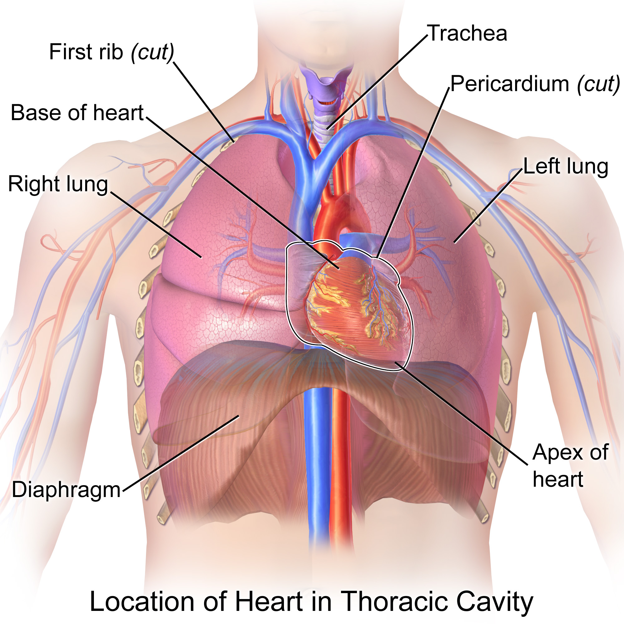
LOCATION & SIZE
📏 Size: About your clenched fist
⚖️ Weight: ~300g in adult
🔊 Apex beat: 5th intercostal space, midclavicular line (LEFT side)
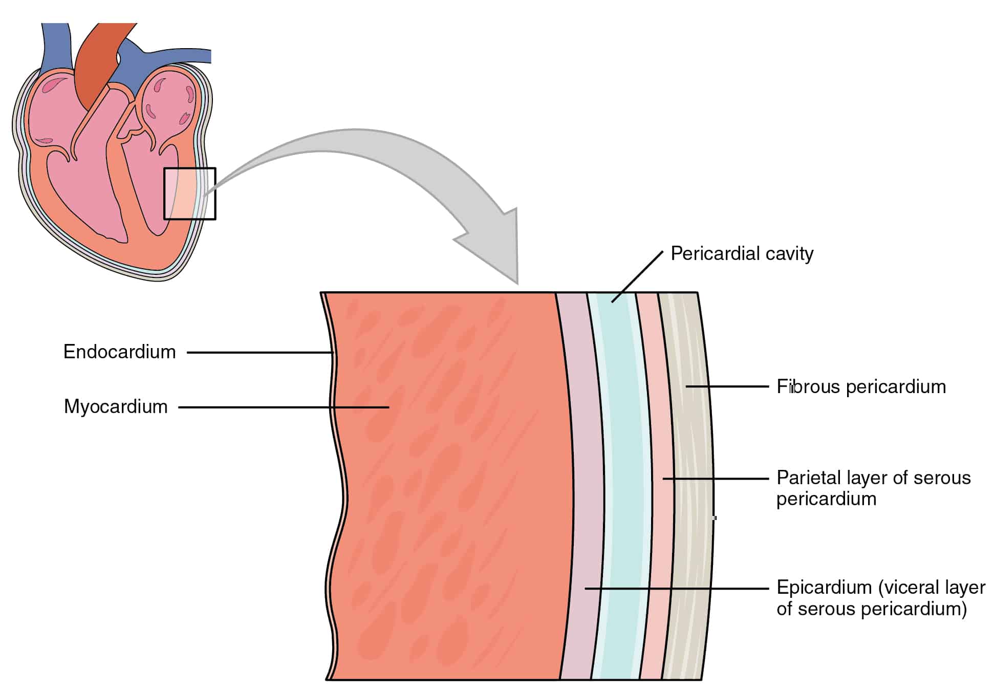
3 LAYERS OF HEART WALL (MUST KNOW!)
| Layer | Description | Function |
|---|---|---|
| Epicardium | Outer layer (visceral pericardium) | Protection, reduces friction |
| Myocardium | Middle layer (CARDIAC MUSCLE) | Contraction & pumping |
| Endocardium | Inner layer (smooth membrane) | Lines chambers & valves |
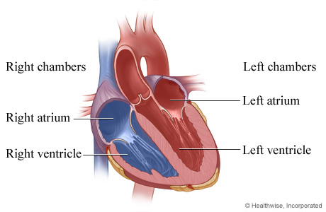
4 CHAMBERS (2 Atria, 2 Ventricles)
➡️ Right Atrium → Right Ventricle → LUNGS
LEFT SIDE (Oxygen-rich blood)
⬅️ Left Atrium ← Left Ventricle ← LUNGS
SEPTUM divides right & left sides
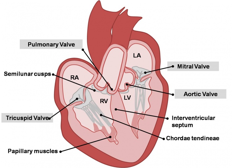
🚪 3. HEART VALVES (ONE-WAY DOORS!)
ATRIOVENTRICULAR (AV) VALVES
- Tricuspid Valve: Between Right Atrium & Right Ventricle (3 cusps)
- Mitral (Bicuspid) Valve: Between Left Atrium & Left Ventricle (2 cusps)
SEMILUNAR VALVES
- Pulmonary Valve: Between Right Ventricle & Pulmonary Artery
- Aortic Valve: Between Left Ventricle & Aorta
T = Tricuspid, P = Pulmonary, M = Mitral, A = Aortic
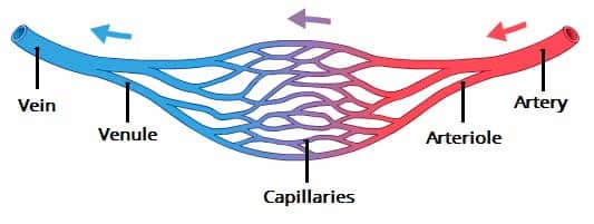
🩸 4. BLOOD VESSELS
| Type | Function | Wall Thickness | Key Features |
|---|---|---|---|
| ARTERIES | Carry blood AWAY from heart | THICK, muscular | High pressure, pulsate |
| VEINS | Carry blood TO the heart | THIN | Low pressure, have valves |
| CAPILLARIES | Exchange O₂, CO₂, nutrients | ONE cell thick | Microscopic, network |
🫄 Pulmonary Artery carries DEOXYGENATED blood to lungs
🫃 Pulmonary Vein carries OXYGENATED blood to heart
🔄 5. PULMONARY & SYSTEMIC CIRCULATION

PULMONARY CIRCULATION (RIGHT SIDE)
Purpose: Oxygenate blood & remove CO₂
SYSTEMIC CIRCULATION (LEFT SIDE)
Purpose: Deliver O₂ & nutrients to tissues
Right → Lungs (Respiration)
Left → Body (Life support)
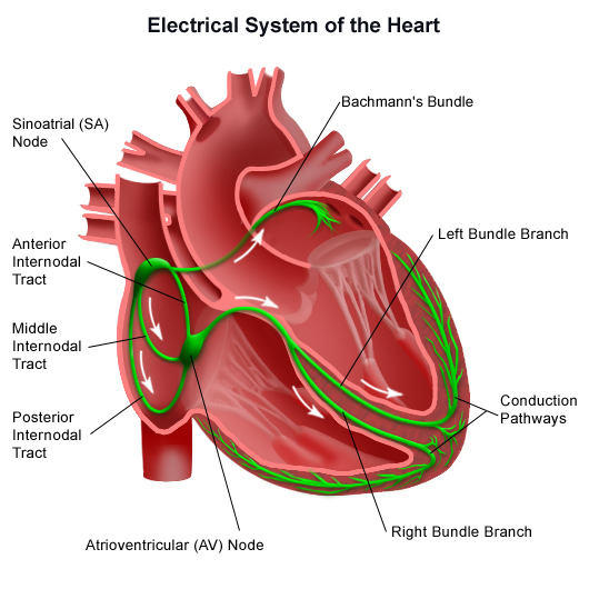
⚡ 6. CARDIAC CONDUCTION SYSTEM
| Structure | Location | Rate | Function |
|---|---|---|---|
| SA Node PACEMAKER! | Right atrium wall | 60-100 bpm | Initiates heartbeat |
| AV Node | Between atria & ventricles | 40-60 bpm | Delays impulse |
| Bundle of His | Interventricular septum | - | Conducts impulse |
| Purkinje Fibers | Ventricle walls | 20-40 bpm | Spread impulse to ventricles |
"Silly Ants Visit Bumpy Places"
📊 7. KEY CARDIAC VALUES FOR NURSES
| Measurement | Normal Range | Clinical Significance |
|---|---|---|
| Heart Rate | 60-100 bpm | Bradycardia <60, Tachycardia>100 |
| Blood Pressure | 120/80 mmHg | HTN >140/90, Hypotension <90 /60 |
| Cardiac Output | 4-8 L/min | HR × Stroke Volume |
| Stroke Volume | 70 mL/beat | Amount pumped per contraction |
👩⚕️ 8. CLINICAL SIGNIFICANCE FOR UGANDAN NURSES
COMMON CARDIAC CONDITIONS IN UGANDA
- Hypertension: Leading cause of heart disease
- Rheumatic Heart Disease: Common in children
- Heart Failure: Often from untreated HTN
- Anemia: Increases cardiac workload
NURSE'S RESPONSIBILITIES
✅ Administer cardiac medications safely
✅ Observe for side effects & report
✅ Patient education on diet, exercise, medication compliance
✅ Recognize emergency signs (chest pain, dyspnea, syncope)
📝 LIKELY EXAM QUESTIONS FOR DAY 8
1. FILL-IN-THE-BLANK (2 marks)
The heart is located in the mediastinum, which is the space between the lungs.
2. MULTIPLE CHOICE (2 marks)
Which structure is known as the natural pacemaker of the heart?
A) AV Node
B) Bundle of His
C) SA Node ⭐CORRECT
D) Purkinje Fibers
3. MULTIPLE CHOICE (3 marks)
Which side of the heart handles pulmonary circulation?
A) Left side
B) Right side ⭐CORRECT
C) Both sides equally
D) Neither side
4. SHORT ANSWER (5 marks)
Explain the difference between arteries and veins, and give one example of each.
• Arteries carry blood AWAY from heart, thick walls, high pressure (e.g., Aorta)
• Veins carry blood TO the heart, thin walls, have valves (e.g., Vena Cava)
• Mention pulmonary artery/vein as exceptions
5. LIST QUESTION (10 marks)
List the path of blood through the heart starting from the vena cava.
Vena Cava → Right Atrium → Tricuspid Valve → Right Ventricle → Pulmonary Valve → Pulmonary Artery → LUNGS → Pulmonary Vein → Left Atrium → Mitral Valve → Left Ventricle → Aortic Valve → AORTA → Body
6. PRACTICAL SCENARIO (10 marks)
You are a certificate nurse in a rural health centre. A patient presents with a pulse rate of 45 bpm, complaining of dizziness and fatigue. What actions do you take?
• Recognize bradycardia (<60 bpm) is abnormal
• Assess for other symptoms (BP, consciousness, chest pain)
• Keep patient at rest, monitor closely
• Inform doctor immediately
• Prepare for possible emergency interventions
• Document all findings accurately