
Muscles of the Upper Limbs and The Brachial Plexus
The upper limb is a area designed for both power (like lifting heavy weights) and precision (like performing microsurgery or playing the violin). By the end of this guide, you will master:
- The complex "wiring" of the upper limb via the Brachial Plexus.
- The origins, insertions, and innervations of the Chest, Shoulder, Arm, Forearm, and Hand muscles.
- Real-world biomechanical examples of how these muscles function in daily life.
- The devastating clinical consequences (Pathology and Palsies) when these nerves or muscles are injured.
Part 1: The Brachial Plexus
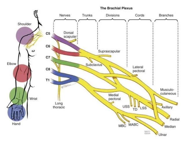
Before we can study the muscles, we must understand how the brain communicates with them. The brachial plexus is a highly complex, interwoven network of nerves formed by the anterior rami (branches) of the lower four cervical nerves (C5, C6, C7, C8) and the first thoracic nerve (T1). It is exclusively responsible for the motor and sensory innervation of the entire upper limb.
To remember the anatomical progression of the plexus from the spine down to the arm, use the famous mnemonic:
"Real Texans Drink Cold Beer"
(Roots → Trunks → Divisions → Cords → Branches)
1. Roots (C5, C6, C7, C8, T1)
The five roots are the anterior primary rami of the spinal nerves. They emerge from the spinal cord and pass through a tight space in the neck between the anterior and middle scalene muscles. (Clinical tip: Tight neck muscles can compress these roots, causing numbness down the arm—a condition called Thoracic Outlet Syndrome).
- Key Branches from Roots:
- Dorsal Scapular Nerve (C5): Innervates the Rhomboids and Levator Scapulae (pulls the shoulder blades together).
- Long Thoracic Nerve (C5, C6, C7): (Mnemonic: "C5, 6, 7 keep your wings from heaven"). Innervates the Serratus Anterior.
2. Trunks (Superior, Middle, Inferior)
As the roots travel laterally over the first rib, they merge to form three distinct trunks:
- Upper (Superior) Trunk: Formed by the union of C5 and C6 roots.
- Middle Trunk: A direct, straight continuation of the C7 root.
- Lower (Inferior) Trunk: Formed by the union of C8 and T1 roots.
- Key Branches from Trunks:
- Suprascapular Nerve (C5, C6): Originates strictly from the Upper Trunk. It dives deep to innervate the Supraspinatus and Infraspinatus (key rotator cuff muscles).
- Nerve to Subclavius (C5, C6): Innervates the subclavius muscle.
3. Divisions (Anterior and Posterior)
As the trunks pass under the clavicle (collarbone), each of the three trunks splits into an Anterior and a Posterior division. This is a crucial evolutionary separation!
- The 3 Posterior Divisions: Combine to supply all the future extensors of the upper limb (triceps, wrist extensors).
- The 3 Anterior Divisions: Combine to supply all the future flexors of the upper limb (biceps, wrist flexors).
4. Cords (Lateral, Posterior, Medial)
The six divisions regroup in the axilla (armpit) to form three cords. They are named strictly for their physical position relative to the massive Axillary Artery.
- Lateral Cord (C5-C7): Formed by the anterior divisions of the upper and middle trunks. Gives off the Lateral Pectoral Nerve.
- Posterior Cord (C5-T1): Formed by the posterior divisions of all three trunks. Gives off the Upper & Lower Subscapular Nerves and the Thoracodorsal Nerve (which powers the mighty Latissimus Dorsi).
- Medial Cord (C8-T1): Formed by the anterior division of the lower trunk. Gives off the Medial Pectoral Nerve and Medial Cutaneous Nerves of the arm and forearm.
5. Branches (The 5 Major Terminal Nerves)
The cords finally split into the five major nerves that travel down the arm.
(Mnemonic: MARMU)
The "Popeye" Nerve.
- Motor: Powers the entire anterior arm compartment (Biceps Brachii, Brachialis, Coracobrachialis). Responsible for elbow flexion.
- Sensory: Pierces the muscle to become the lateral cutaneous nerve, supplying the skin of the lateral forearm.
The "Shoulder Pad" Nerve.
- Motor: Wraps around the surgical neck of the humerus to power the Deltoid and Teres Minor.
- Sensory: Skin over the lower deltoid (the "regimental badge area").
The "Great Extensor" Nerve.
- Motor: Powers EVERY single muscle in the posterior compartments of the arm and forearm (Triceps and all wrist/finger extensors).
- Sensory: Posterior skin of arm/forearm, and the dorsal aspect of the lateral 2.5 digits (the anatomical snuffbox area).
The "Laborer's Nerve".
- Motor: Powers most anterior forearm muscles (flexors/pronators), and the critical thenar (thumb) muscles for grasping.
- Sensory: Skin of the lateral palm and palmar aspect of the lateral 3.5 digits.
The "Musician's Fine-Tuning" Nerve.
- Motor: Powers only 1.5 muscles in the forearm (Flexor Carpi Ulnaris, medial half of FDP), but powers almost ALL the intrinsic fine-motor muscles of the hand (interossei/lumbricals).
- Sensory: Skin of the medial 1.5 digits (the pinky and half the ring finger).
Brachial Plexus Summary Table
| Level | Components | Key Nerve Branches | Clinical Notes |
|---|---|---|---|
| ROOTS | Anterior Rami of C5, C6, C7, C8, T1 | Dorsal Scapular N (C5): Rhomboids, Levator Scapulae Long Thoracic N (C5-C7): Serratus Anterior |
Emerge between Scalenes. Injury to Long Thoracic N. → Winged Scapula. |
| TRUNKS | Upper: C5 + C6 Middle: C7 Lower: C8 + T1 |
Suprascapular N (C5, C6): Supraspinatus, Infraspinatus N. to Subclavius (C5, C6): Subclavius |
Pass over 1st rib. Erb-Duchenne palsy is an upper trunk injury. |
| DIVISIONS | Each trunk divides into an Anterior & Posterior Division | No direct named branches. | Posterior divisions strictly supply extensors; Anterior supply flexors. |
| CORDS | Lateral: Ant. divisions of Upper & Middle Posterior: Post. divisions of all 3 Medial: Ant. division of Lower |
Lateral Pectoral N. Upper & Lower Subscapular N., Thoracodorsal N. Medial Pectoral N., Medial Cutaneous Nerves |
Named for physical position wrapping around the axillary artery. |
| BRANCHES | Terminal Nerves | Musculocutaneous N. Axillary N. Radial N. Median N. Ulnar N. |
Major nerves of the upper limb. Injuries lead to highly distinct motor & sensory deficits. |
Brachial Plexus Injuries and Clinical Correlates
Nerve injuries are devastating and present with highly specific, testable postures:
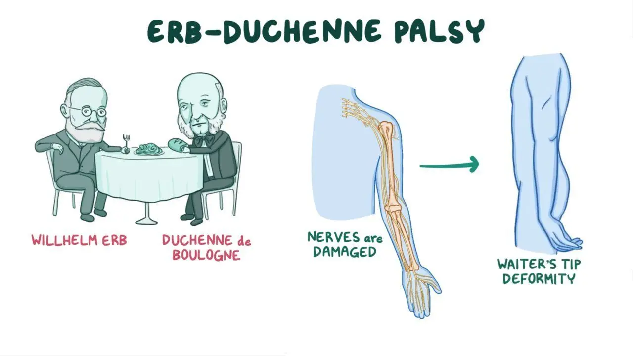
- Upper Plexus Injury (Erb-Duchenne Palsy): Affects C5-C6 roots. Usually caused by traumatic tearing, such as shoulder dystocia during a difficult childbirth or a motorcycle accident where the head and shoulder are violently pushed apart.
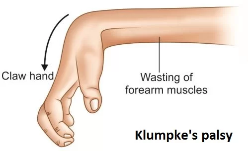
Result: The classic "Waiter's Tip" position. The arm hangs limply adducted (deltoid paralyzed), medially rotated (external rotators paralyzed), and the elbow is extended with the wrist flexed. - Lower Plexus Injury (Klumpke's Palsy): Affects C8-T1 roots. Caused by excessive, violent abduction of the arm (e.g., grabbing a tree branch to stop a fall from a height, or pulling a baby out by its arm).
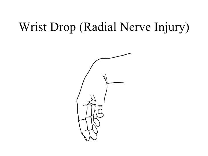
Result: Affects intrinsic hand muscles, leading to a total "Claw Hand" of all digits. - Radial Nerve Injury (Wrist Drop): Commonly caused by mid-shaft humeral fractures (the nerve spirals tightly around the bone) or compression in the axilla ("Saturday night palsy" from passing out with an arm over a chair, or improperly fitted crutches).
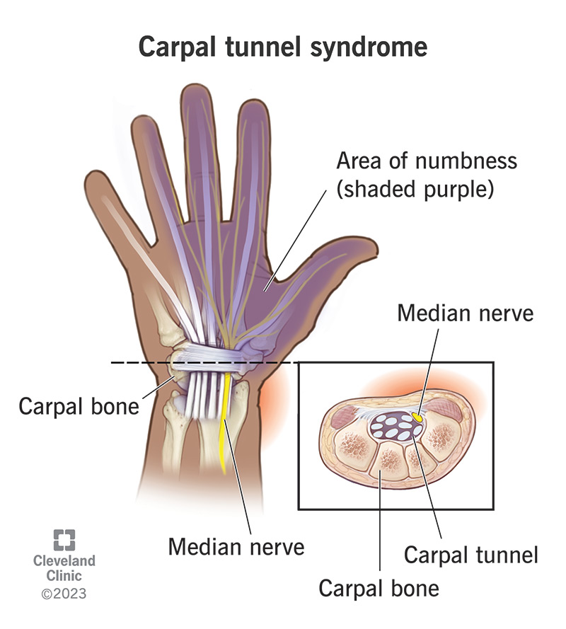
Result: Absolute inability to extend the wrist and fingers. The hand hangs completely limp (Wrist Drop). - Median Nerve Injury (Carpal Tunnel Syndrome): Compression of the median nerve as it passes under the flexor retinaculum at the wrist (often due to repetitive typing or vibration).
Result: Causes numbness and "pins and needles" in the lateral 3.5 digits, and severe weakness/wasting of the fleshy base of the thumb (thenar atrophy), producing an "Ape Hand" deformity. - Ulnar Nerve Injury ("Claw Hand"): Injury usually occurs where it wraps around the medial epicondyle of the elbow (hitting your "funny bone") or at the wrist.
Result: Loss of intrinsic hand muscles leads to specific "clawing" (hyperextension of MCP and flexion of IP joints) of the 4th and 5th digits, and severe sensory loss over the medial pinky side of the hand.
Part 2: Muscles of the Chest (Pectoral Region)
The muscles of the pectoral region serve a dual purpose: the superficial layers anchor the heavy upper limb to the axial skeleton of the thorax, while the deeper layers are the biological bellows responsible for the mechanics of breathing.
1. Superficial Muscles of the Pectoral Region
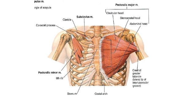
- a. Pectoralis Major: A massive, fan-shaped muscle covering the upper chest. It has two parts: a clavicular head and a larger sternocostal head.
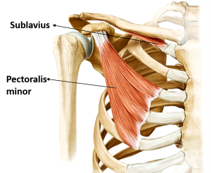
Biomechanical Example: It is the ultimate hugging muscle. It is a powerful adductor and medial rotator of the arm. The clavicular head flexes the arm (e.g., throwing a bowling ball underhand), while the sternocostal head extends it from a flexed position (e.g., the downward pull in a butterfly swimming stroke or performing a push-up). - b. Pectoralis Minor: A thin, triangular muscle lying completely hidden deep to the Pectoralis Major.
Biomechanical Example: It depresses the shoulder and protracts the scapula (pulls it forward and downward). Think of the motion of reaching down to pick up a heavy suitcase. - c. Subclavius: A tiny, cylindrical muscle located directly inferior to the clavicle.
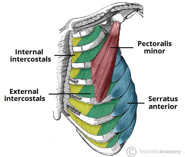
Function: It actively anchors and depresses the clavicle. More importantly, it acts as a soft biological cushion to protect the underlying subclavian vessels and brachial plexus if the collarbone fractures. - d. Serratus Anterior: Visually distinct, saw-toothed slips of muscle on the lateral thoracic wall.
Biomechanical Example: Known as the "Boxer's Muscle". It is the prime mover for aggressively protracting the scapula (the motion of throwing a punch or pushing a heavy stalled car). It is also essential for upwardly rotating the scapula, which is required to lift your arm entirely above your head. Paralysis (Long Thoracic N. injury) leads to the pathognomonic "winged scapula".
2. Deep Muscles of the Thorax (Associated with Respiration)
- a. Intercostal Muscles (External, Internal, Innermost): Three distinct layers of muscles woven tightly in the intercostal spaces between the ribs.
Function: The External Intercostals lift and elevate the ribs out and up, expanding chest volume for forced inspiration (taking a deep breath before diving into water). The Internal and Innermost Intercostals violently depress the ribs for forced expiration (blowing out all the candles on a birthday cake). - b. Transversus Thoracis: A thin, star-like muscle on the inner anterior thoracic wall (behind the sternum) that weakly depresses the ribs.
Summary Table of Chest Muscles
| Muscle | Origin | Insertion | Innervation | Main Actions |
|---|---|---|---|---|
| SUPERFICIAL PECTORAL MUSCLES | ||||
| Pectoralis Major | Clavicle, Sternum, Costal Cartilages 1-6 | Intertubercular groove of humerus (lateral lip) | Lat & Med Pectoral N. | Adducts & medially rotates arm; flexes & extends arm. |
| Pectoralis Minor | Ribs 3-5 | Coracoid process of scapula | Medial Pectoral N. | Depresses shoulder; protracts the scapula. |
| Subclavius | 1st rib | Inferior surface of clavicle | N. to Subclavius | Depresses & anchors the clavicle tightly. |
| Serratus Anterior | Ribs 1-9 | Medial border of scapula (anterior surface) | Long Thoracic N. | Protracts & upwardly rotates scapula (prevents winging). |
| DEEP THORACIC (RESPIRATORY) MUSCLES | ||||
| External Intercostals | Rib above | Rib below | Intercostal Nerves | Elevate ribs (forced, deep inspiration). |
| Internal Intercostals | Rib above | Rib below | Intercostal Nerves | Depress ribs (forced, active expiration). |
Part 3: Muscles of the Upper Limbs
The muscles of the upper limb enable a remarkable range of movements. The shoulder provides maximum 360-degree mobility, the elbow acts as a powerful hinge lever, the forearm provides rotational positioning, and the hand yields unparalleled dexterity. We divide them regionally.
1. Muscles of the Shoulder (Scapulohumeral Region)
These muscles act primarily on the incredibly shallow, highly mobile glenohumeral (shoulder) joint, providing both movement and vital dynamic stability.
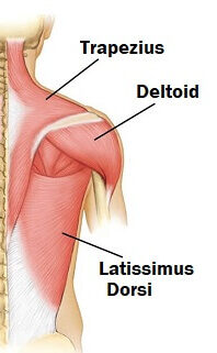
- a. Deltoid: The massive, thick, triangular muscle forming the rounded, armored contour of the shoulder.
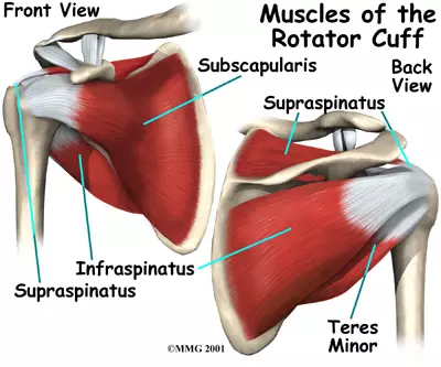
Biomechanical Example: Its three parts (anterior, middle, posterior) allow it to perform almost every action. The entire muscle is the absolute prime mover of arm abduction (lifting the arm out to the side like a bird flapping its wings) only after the first 15 degrees. The anterior fibers flex and medially rotate (reaching forward to shake a hand), while the posterior fibers extend and laterally rotate (pulling open a heavy vault door). - b. Rotator Cuff Muscles (SITS): A critical group of four muscles that tightly surround the shoulder joint like a biological cuff. Their tendons physically blend directly into the joint capsule, dynamically sucking the humerus into the shallow socket to prevent dislocation during movement. Remembered by the mnemonic SITS.
- Supraspinatus: Action: Initiates the very first 15 degrees of arm abduction (before the deltoid takes over). Clinical: It is the most commonly torn rotator cuff muscle because its tendon gets aggressively pinched (impingement syndrome) under the acromion bone during repetitive overhead lifting.
- Infraspinatus: Action: A powerful lateral rotator of the arm (e.g., the motion of winding up to pitch a baseball).
- Teres Minor: Action: Synergist to the infraspinatus; also laterally rotates the arm.
- Subscapularis: Action: The only rotator cuff muscle on the front of the scapula. It is a powerful medial rotator of the arm (e.g., the motion of tucking your shirt into the back of your pants).
- c. Teres Major: A thick, rounded muscle inferior to Teres Minor.
Biomechanical Example: Often colloquially called "Lat's Little Helper." It is NOT part of the rotator cuff. Its main actions are to powerfully extend, adduct, and medially rotate the arm. Think of the aggressive, downward pulling motion of chopping wood with an axe or performing a pull-up.
Summary Table of Shoulder Muscles
| Muscle | Origin | Insertion | Innervation | Main Actions |
|---|---|---|---|---|
| Deltoid | Clavicle, acromion, spine of scapula | Deltoid tuberosity of humerus | Axillary N. (C5, C6) | Abducts arm (>15°); flexes & medially rotates; extends & laterally rotates. |
| Supraspinatus | Supraspinous fossa of scapula | Greater tubercle of humerus (superior facet) | Suprascapular N. (C5, C6) | Initiates arm abduction (first 15°); stabilizes joint. |
| Infraspinatus | Infraspinous fossa of scapula | Greater tubercle of humerus (middle facet) | Suprascapular N. (C5, C6) | Laterally rotates arm; stabilizes joint. |
| Teres Minor | Lateral border of scapula | Greater tubercle of humerus (inferior facet) | Axillary N. (C5, C6) | Laterally rotates arm; stabilizes joint. |
| Subscapularis | Subscapular fossa (anterior scapula) | Lesser tubercle of humerus | Upper & Lower Subscapular N. | Medially rotates arm; provides massive anterior stability. |
| Teres Major | Inferior angle of scapula | Intertubercular groove of humerus (medial lip) | Lower Subscapular N. | Extends, adducts, medially rotates arm (woodchopper). |
2. Muscles of the Arm (The Brachium)
The true "arm" (the segment between the shoulder and elbow) is cleanly divided into an anterior (flexor) and posterior (extensor) compartment by tough, fibrous intermuscular septa.
Anterior (Flexor) Compartment
Innervation: Musculocutaneous Nerve (C5, C6, C7)
Arterial Supply: Brachial artery
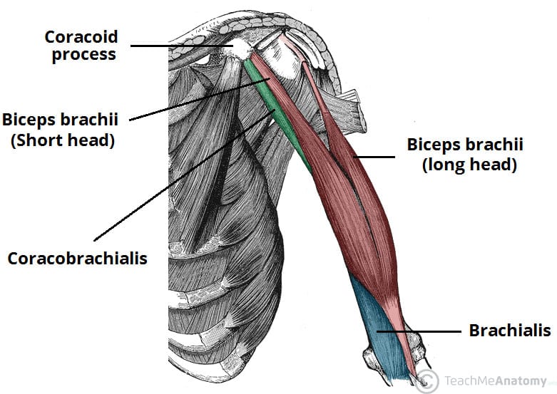
- Biceps Brachii: A prominent two-headed muscle (Long and Short head) that astonishingly doesn't attach to the humerus at all!
Biomechanical Example: Because it attaches to the radius, it is not just a flexor, but the most powerful supinator of the forearm. Think of aggressively driving a corkscrew into a wine bottle (supination) and then pulling the cork out (flexion). - Brachialis: Lies deep to the biceps.
Biomechanical Example: This is the absolute "workhorse" and primary flexor of the elbow. Because it attaches to the ulna (which doesn't rotate), it simply flexes the elbow powerfully regardless of hand position (e.g., doing heavy hammer curls or lifting a heavy mug of beer). - Coracobrachialis: The smallest of the three. It assists in weak flexion and adduction of the arm tightly to the body at the shoulder.
Posterior (Extensor) Compartment
Innervation: Radial Nerve (C6, C7, C8, T1)
Arterial Supply: Deep brachial artery (Profunda brachii)
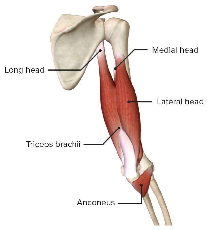
- Triceps Brachii: The massive, sole muscle occupying the entire posterior arm, possessing three heads (long, lateral, medial).
Biomechanical Example: It is the powerful, absolute extensor of the forearm at the elbow. Think of the violent snapping motion of throwing a dart, hammering a nail, or doing a triceps push-down. The long head (crossing the shoulder joint) also assists in extending and pulling the arm backward. - Anconeus: A tiny, triangular muscle at the posterior elbow. It assists the triceps in locking out forearm extension and prevents the joint capsule from being pinched during movement.
Summary Table of Arm Muscles
| Muscle | Origin | Insertion | Innervation | Main Actions |
|---|---|---|---|---|
| ANTERIOR COMPARTMENT | ||||
| Biceps Brachii | Long head: Supraglenoid tubercle Short head: Coracoid process |
Radial tuberosity & bicipital aponeurosis | Musculocutaneous N. | Powerfully supinates forearm; flexes forearm. |
| Brachialis | Anterior, distal half of humerus | Coronoid process & tuberosity of ulna | Musculocutaneous N. | Primary, pure flexor of forearm (the workhorse). |
| Coracobrachialis | Coracoid process of scapula | Middle medial surface of humerus | Musculocutaneous N. | Flexes and adducts arm at the shoulder. |
| POSTERIOR COMPARTMENT | ||||
| Triceps Brachii | Long: Infraglenoid tubercle Lat/Med: Posterior surface of humerus |
Olecranon process of ulna | Radial N. | Powerful, primary extensor of forearm. |
| Anconeus | Lateral epicondyle of humerus | Lateral surface of olecranon & superior ulna | Radial N. | Assists triceps in extension; stabilizes elbow capsule. |
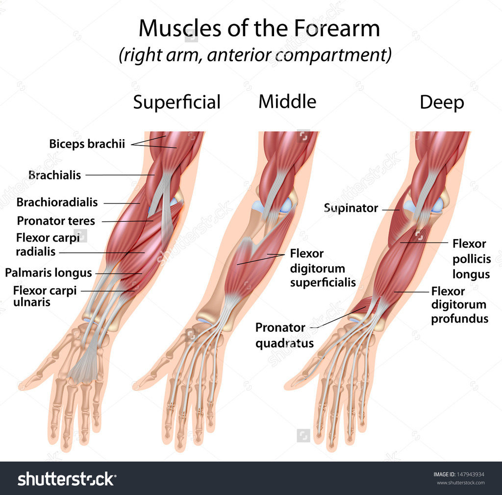
3. Muscles of the Forearm (The Antebrachium)
The numerous muscles of the forearm are complexly arranged in stacked layers. They are cleanly divided into an anterior (flexor/pronator) and posterior (extensor/supinator) compartment. Their long tendons pass through the wrist to control the hand like strings on a puppet.
Anterior (Flexor-Pronator) Compartment
Innervation: Mostly Median Nerve. Exception: Flexor Carpi Ulnaris & the medial half of FDP are powered by the Ulnar Nerve.
Main Actions: Flexion of the wrist and fingers; pronation (turning palm down) of the forearm.
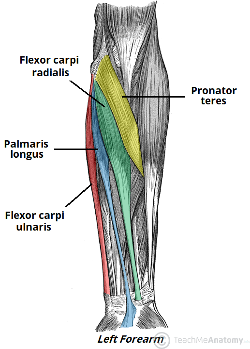
- Superficial Layer (4 muscles originating from the Medial Epicondyle):
- Pronator Teres: Pronates (turns palm down) and weakly flexes the forearm.
- Flexor Carpi Radialis (FCR): Flexes and abducts the wrist.
- Palmaris Longus: Flexes the wrist. (Interesting fact: It is entirely absent in about 15% of the human population! When present, its long, useless tendon is frequently harvested by surgeons for tendon grafts).
- Flexor Carpi Ulnaris (FCU): Flexes and heavily adducts the wrist (ulnar deviation).
- Intermediate Layer (1 muscle):
- Flexor Digitorum Superficialis (FDS): Its tendons split in half. It specifically flexes the middle phalanges (PIP joints) of digits 2-5 (e.g., the specific curling finger motion used when playing a piano).
- Deep Layer (3 muscles):
- Flexor Digitorum Profundus (FDP): The deepest flexor. It passes entirely through the split tendons of the FDS to flex the absolute distal tips (DIP joints) of digits 2-5. Think of the aggressive, crushing fingertip grip needed by professional rock climbers.
- Flexor Pollicis Longus (FPL): Flexes the distal phalanx of the thumb (the primary muscle used when rapidly texting on a smartphone).
- Pronator Quadratus: A square muscle at the wrist. The primary, deep pronator of the forearm.
Posterior (Extensor-Supinator) Compartment
Innervation: Radial Nerve and its deep branch (Posterior Interosseous Nerve).
Main Actions: Extension of the wrist and fingers; supination (turning palm up) of the forearm.
- Superficial Layer: Includes the wrist extensors (ECRL, ECRB, ECU), finger extensors (Extensor Digitorum, Extensor Digiti Minimi), and the highly unique Brachioradialis.
Biomechanical Example: The Brachioradialis is the "drinking muscle". It sits in the extensor compartment and is innervated by the radial nerve, but it actually flexes the elbow when the arm is in a mid-pronated position (like bringing a glass of water to your mouth). - Deep Layer: Includes the Supinator muscle, and the three "outcropping" muscles of the thumb: Abductor Pollicis Longus (APL), Extensor Pollicis Brevis (EPB), and Extensor Pollicis Longus (EPL). These three tendons form the triangular depression on the back of the hand known as the Anatomical Snuffbox. Also includes the Extensor Indicis, which allows for independent extension of the index finger (pointing at something).
Summary Table of Forearm Muscles
| Muscle | Origin | Insertion | Innervation | Main Actions |
|---|---|---|---|---|
| ANTERIOR COMPARTMENT | ||||
| Pronator Teres | Medial epicondyle, coronoid process | Lateral surface of radius | Median N. | Pronates & flexes forearm. |
| Flexor Carpi Radialis | Medial epicondyle | Base of 2nd & 3rd metacarpals | Median N. | Flexes & abducts wrist (radial deviation). |
| Palmaris Longus | Medial epicondyle | Palmar aponeurosis | Median N. | Tenses palmar fascia; flexes wrist. |
| Flexor Carpi Ulnaris | Medial epicondyle, olecranon | Pisiform, hook of hamate, 5th metacarpal | Ulnar N. | Flexes & adducts wrist (ulnar deviation). |
| Flexor Digitorum Superficialis | Medial epicondyle, coronoid, anterior radius | Middle phalanges of digits 2-5 (splits) | Median N. | Flexes PIP joints and wrist. |
| Flexor Digitorum Profundus | Anterior ulna, interosseous membrane | Distal phalanges of digits 2-5 | Median N. (lat half), Ulnar N. (med half) | Flexes DIP joints (crushing grip). |
| Flexor Pollicis Longus | Anterior radius, interosseous membrane | Distal phalanx of thumb | Median N. (AIN) | Flexes distal thumb joint (texting). |
| Pronator Quadratus | Distal, anterior ulna | Distal, anterior radius | Median N. (AIN) | Primary pronator of forearm. |
| POSTERIOR COMPARTMENT | ||||
| Brachioradialis | Lateral supracondylar ridge of humerus | Styloid process of radius | Radial N. | Flexes forearm in mid-pronated position. |
| Extensor Carpi Radialis Longus | Lateral supracondylar ridge of humerus | Base of 2nd metacarpal | Radial N. | Extends & abducts wrist. |
| Extensor Carpi Ulnaris | Lateral epicondyle, posterior ulna | Base of 5th metacarpal | Radial N. (PIN) | Extends & adducts wrist. |
| Supinator | Lateral epicondyle, ulna crest | Proximal third of radius | Radial N. (Deep br.) | Primary supinator of forearm (driving a screw). |
4. Muscles of the Hand (The Intrinsic Controllers)
While the heavy lifting is done by the forearm muscles acting via long tendons, the intrinsic muscles of the hand (contained entirely within the hand itself) are exclusively responsible for fine motor control, delicate pinching, and exquisite dexterity.
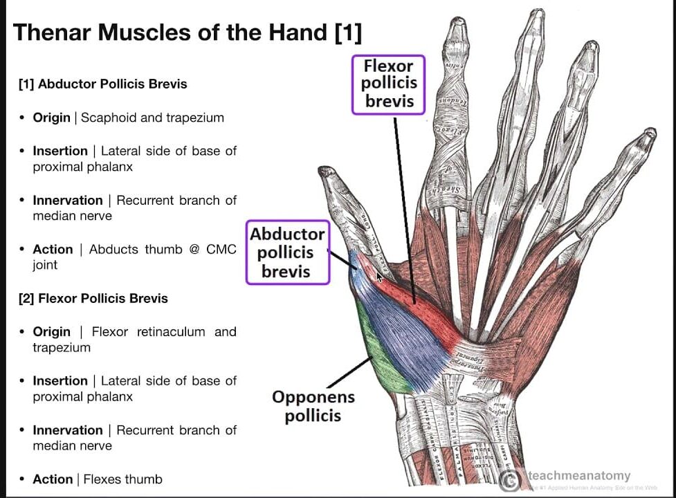
a. Thenar Muscles (Ball of the Thumb)
This fleshy group acts strictly on the thumb (pollux). All are innervated by the Recurrent Branch of the Median Nerve, EXCEPT for the massive Adductor Pollicis.
- Abductor Pollicis Brevis (APB): Pulls the thumb away from the palm.
- Flexor Pollicis Brevis (FPB): Flexes the proximal joint of the thumb.
- Opponens Pollicis (OP): Swings the thumb across the palm to touch the other fingers (the evolutionary hallmark of human dexterity).
- Adductor Pollicis: The heavy muscle in the web space. It violently adducts the thumb (innervated by the Ulnar Nerve). Think of clamping a piece of paper tightly between your thumb and index finger.
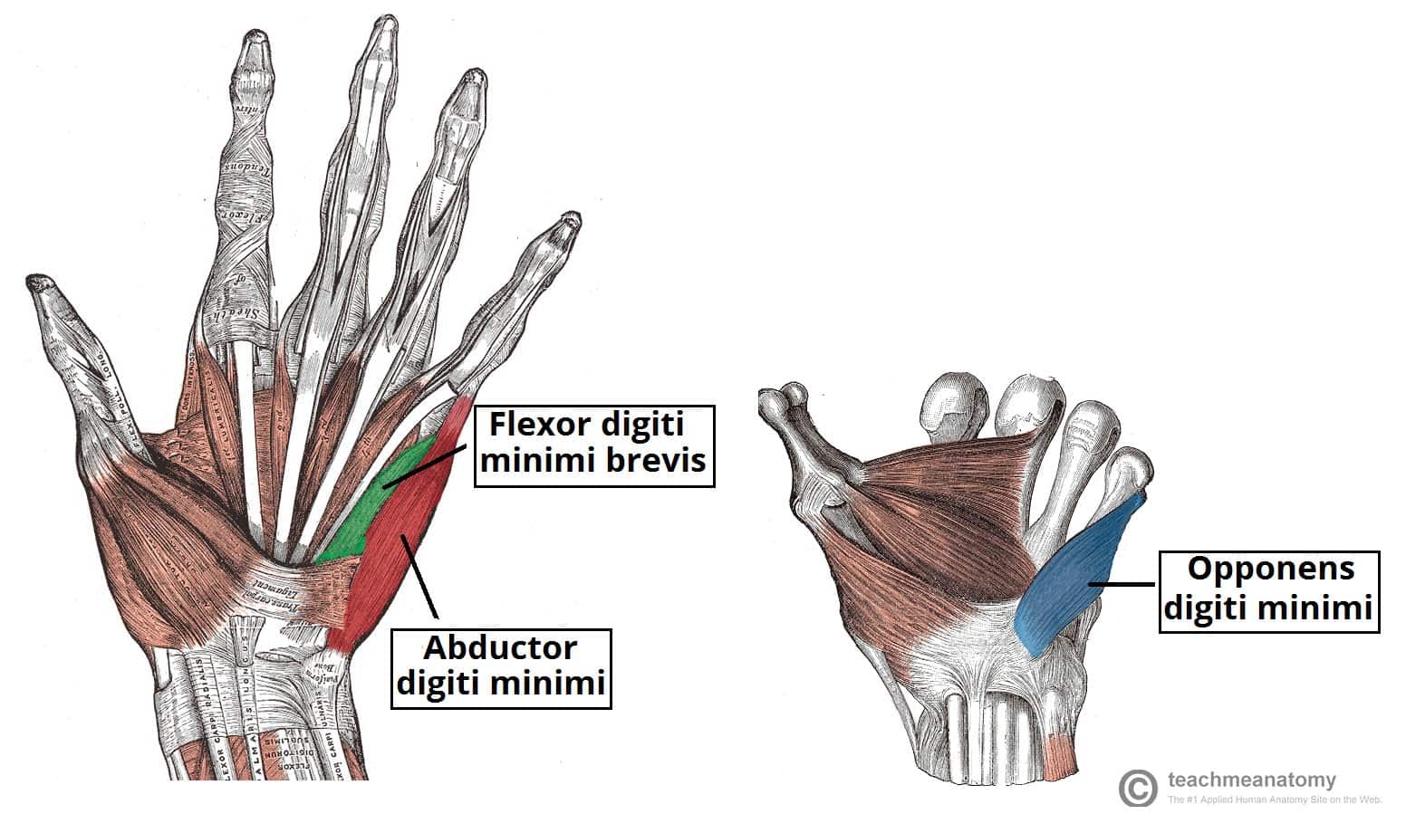
b. Hypothenar Muscles (Ball of the Little Finger)
This group acts on the little finger (digiti minimi). All are innervated by the Deep Branch of the Ulnar Nerve.
- Abductor Digiti Minimi (ADM): Pulls the pinky away.
- Flexor Digiti Minimi Brevis (FDMB): Flexes the pinky.
- Opponens Digiti Minimi (ODM): Opposes the little finger (rotates it to meet the thumb, forming a "cup" in your palm to hold water).
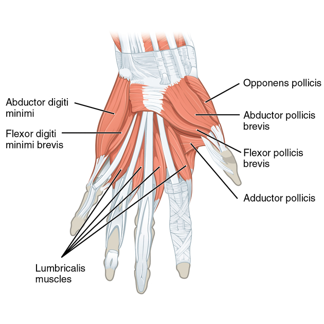
c. Deep Intrinsic Muscles
- Lumbricals (4 muscles): Tiny, worm-shaped muscles that magically originate entirely from the moving tendons of the FDP, rather than bone!
Action: They flex the MCP joints and extend the IP joints. (This creates the "bye-bye" hand wave or the "tabletop" position). Lateral two = Median N., Medial two = Ulnar N. - Interossei (7 muscles): Muscles tightly packed between the metacarpal bones. All innervated by the Ulnar N.
Mnemonic Action: The 4 Dorsal Interossei Abduct the fingers (DAB - spreading fingers wide). The 3 Palmar Interossei Adduct the fingers (PAD - squeezing fingers together).
Summary Table of Hand Muscles
| Group | Muscle | Origin | Insertion | Innervation | Action |
|---|---|---|---|---|---|
| Thenar | Abductor Pollicis Brevis | Flexor retinaculum, scaphoid, trapezium | Proximal phalanx of thumb | Median N. (Recurrent br.) | Abducts thumb |
| Flexor Pollicis Brevis | Flexor retinaculum, trapezium | Proximal phalanx of thumb | Median N. (Recurrent br.) | Flexes thumb | |
| Opponens Pollicis | Flexor retinaculum, trapezium | 1st metacarpal (radial side) | Median N. (Recurrent br.) | Opposes thumb | |
| Adductor Pollicis | Capitate, 2nd & 3rd metacarpals | Proximal phalanx of thumb | Ulnar N. (Deep br.) | Adducts thumb (pinching paper) | |
| Hypothenar | Abductor Digiti Minimi | Pisiform bone | Proximal phalanx of digit 5 | Ulnar N. (Deep br.) | Abducts little finger |
| Flexor Digiti Minimi Brevis | Hook of hamate, flexor retinaculum | Proximal phalanx of digit 5 | Ulnar N. (Deep br.) | Flexes little finger | |
| Opponens Digiti Minimi | Hook of hamate, flexor retinaculum | 5th metacarpal (ulnar border) | Ulnar N. (Deep br.) | Opposes little finger (cupping palm) | |
| Deep Intrinsic | Lumbricals (4) | Tendons of Flexor Digitorum Profundus | Extensor expansions of digits 2-5 | Lat 2: Median N. Med 2: Ulnar N. |
Flex MCP joints, Extend IP joints (tabletop) |
| Dorsal Interossei (4) | Adjacent sides of two metacarpals (bipennate) | Proximal phalanges & extensor expansions | Ulnar N. (Deep br.) | Abduct fingers (DAB) | |
| Palmar Interossei (3) | Palmar surfaces of 2nd, 4th, 5th metacarpals | Proximal phalanges & extensor expansions | Ulnar N. (Deep br.) | Adduct fingers (PAD) |
References
The anatomical and biomechanical descriptions provided in this guide have been synthesized in accordance with standard international anatomical literature and surgical texts, including:
- Moore, K. L., Dalley, A. F., & Agur, A. M. R. (2018). Clinically Oriented Anatomy (8th ed.). Wolters Kluwer.
- Standring, J. (Ed.). (2015). Gray's Anatomy: The Anatomical Basis of Clinical Practice (41st ed.). Elsevier.
- Netter, F. H. (2018). Atlas of Human Anatomy (7th ed.). Elsevier.
- Drake, R., Vogl, A. W., & Mitchell, A. W. M. (2019). Gray's Anatomy for Students (4th ed.). Elsevier.
- Snell, R. S. (2011). Clinical Anatomy by Regions (9th ed.). Lippincott Williams & Wilkins.