
Nervous Tissue: The Master Communication Network
Nervous tissue can be thought of as the ultimate high-speed, fiber-optic telecommunications network of the human body. By the end of this exhaustively detailed guide, you will be deeply conversant with:
- The overarching anatomy and primary functions of nervous tissue.
- The intricate structural and functional classification of Neurons.
- The specialized roles of the six distinct types of Neuroglia (Glial Cells) in both the CNS and PNS.
- The exact step-by-step electrophysiology behind Resting Membrane Potentials, Graded Potentials, and Action Potentials.
- The mechanism of Synaptic Transmission and chemical signaling.
1. Introduction to Nervous Tissue
Nervous tissue is the master controller and communication system of the body. It forms the brain, spinal cord, and peripheral nerves. Its primary function is to regulate, coordinate, and integrate all body functions by rapidly transmitting electrical signals. Without nervous tissue, there is no consciousness, no movement, no sensation, and no homeostasis.
General Characteristics
- Primary Function: To receive stimuli (changes in the internal or external environment), transmit electrical impulses, and process information to precisely control the body's responses.
- Location: Makes up the Central Nervous System (CNS)—the brain and spinal cord—and the Peripheral Nervous System (PNS)—the cranial, spinal, and peripheral nerves.
The Two Main Cell Types
The nervous system is comprised of two principal types of cells that work in absolute, inseparable concert:
- Neurons (Nerve Cells): These are the primary functional, signaling cells that are specialized to transmit electrical signals (nerve impulses). They send and receive messages using chemical signals called neurotransmitters across junctions known as synapses.
- Neuroglia (Glial Cells): These are the non-excitable, supporting cells of the nervous system. They provide physical scaffolding, metabolic support, electrical insulation (myelin), and immune defense for the neurons. Examples include Astrocytes, Oligodendrocytes, and Schwann Cells.
2. The Neuron (Nerve Cell)
Neurons are the excitable cells strictly responsible for transmitting electrical signals. They are highly specialized and possess three extreme characteristics:
- Extreme Longevity: Given good nutrition, they can function optimally for a lifetime (over 100 years).
- Amitotic: With very few exceptions (like olfactory epithelium and some hippocampal regions), mature neurons lose their ability to divide. If they are destroyed, they cannot be replaced.
- High Metabolic Rate: They require a continuous, uninterrupted supply of abundant oxygen and glucose to survive. They cannot survive for more than a few minutes without oxygen.
Key Properties of Neurons
- Excitability (Irritability): The ability to intensely respond to a stimulus (mechanical, chemical, or electrical) by generating a massive electrical change across its cell membrane (membrane potential).
- Conductivity: The ability to propagate these electrical signals (nerve impulses or action potentials) rapidly along the cell membrane to distant locations.
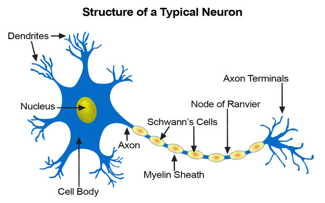
Structural Components of a Typical Neuron
A typical neuron is structurally divided into three functional zones: a receptive region, a conducting region, and a secretory region.
- Cell Body (Soma / Perikaryon):
- The neuron's main nutritional and metabolic center.
- It contains the nucleus, a nucleolus, and most of the standard cellular organelles.
- It features highly prominent Nissl bodies (a specialized, extremely dense form of rough endoplasmic reticulum), reflecting the neuron's phenomenally high rate of protein synthesis required to constantly build neurotransmitters and membrane proteins.
- Dendrites:
- Numerous, short, highly branched, tree-like processes.
- They act as the main receptive (input) regions. They provide an enormous surface area to receive incoming signals from other neurons and convey those signals (as graded potentials) towards the cell body.
- Axon:
- A single, long process (can be over a meter long in human legs!) that acts as the conducting region.
- It originates from a cone-shaped area of the cell body called the Axon Hillock (the "trigger zone" where action potentials are actually generated).
- Its job is generating and transmitting nerve impulses (action potentials) away from the cell body.
- It terminates in thousands of tiny branches called Axon Terminals (Synaptic Boutons), which represent the secretory region where neurotransmitters are released.
The Myelin Sheath
Many axons, especially long ones, are covered by a fatty, whitish, insulating layer called the myelin sheath. This sheath is formed by specific glial cells (Schwann cells in the PNS and Oligodendrocytes in the CNS) wrapping themselves tightly around the axon like a jelly roll.
- Function: It electrically insulates the axon and dramatically speeds up nerve impulse transmission.
- Nodes of Ranvier: The myelin sheath is not continuous. It has microscopic gaps between the myelin segments called Nodes of Ranvier.
- Saltatory Conduction: Because myelin blocks ion flow, the action potential cannot travel continuously down the axon. Instead, the electrical signal is forced to "jump" rapidly from node to node. This leaping process is called saltatory conduction, and it is up to 30 times faster than continuous conduction in unmyelinated fibers.
Clinical Scenario: Demyelinating Diseases
Multiple Sclerosis (MS): An autoimmune disease where the body's immune system attacks and destroys the myelin sheaths specifically in the Central Nervous System (CNS). Without myelin, the electrical currents short-circuit or slow down drastically. Patients experience visual disturbances, muscle weakness, speech problems, and eventual paralysis.
Guillain-Barré Syndrome: A similar autoimmune demyelinating condition, but it strictly attacks the Schwann cells in the Peripheral Nervous System (PNS), leading to rapid-onset muscle weakness spreading from the legs upward.
3. Classification of Neurons
Neurons are immensely diverse, but we classify them based on what they do (function) and what they look like (structure).
A. Functional Classification of Neurons
This classification focuses on the direction the nerve impulse travels relative to the Central Nervous System.
- Sensory (Afferent) Neurons: Transmit impulses from sensory receptors in the skin, internal organs, and special sense organs towards the CNS. They act as the body's surveillance cameras. (Almost all are unipolar).
- Motor (Efferent) Neurons: Transmit impulses away from the CNS to effector organs (muscles and glands). They carry the "commands" that tell the body to move or secrete. (Almost all are multipolar).
- Interneurons (Association Neurons): Lie completely within the CNS, sandwiched between sensory and motor neurons. They integrate, process, and interpret incoming information to decide on the appropriate motor response. They make up over 99% of all neurons in the body!
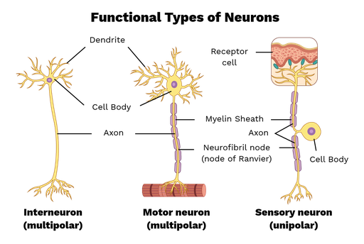
B. Structural Classification of Neurons
This classification focuses on the number of processes (extensions) extending directly from the cell body.
Possess three or more processes (one axon, and many dendrites).
- Prevalence: The most common type in humans (over 99% of neurons). The major neuron type in the CNS.
- Examples: Purkinje cells of the cerebellum, Pyramidal cells of the motor cortex, and all skeletal muscle motor neurons.
Possess exactly two processes (one axon, one single dendrite) extending from opposite sides of the cell body.
- Prevalence: Exceedingly rare.
- Examples: Exclusively found in special sense organs where they act as receptor cells (e.g., the retina of the eye, and the olfactory mucosa in the nose).
Possess a single, short process extending from the cell body that divides T-like into two branches (a peripheral process and a central process).
- Prevalence: Found primarily in the PNS.
- Examples: They function almost exclusively as Sensory neurons. Their cell bodies are famously clustered together in the Dorsal Root Ganglia just outside the spinal cord.
4. Neuroglia (Glial Cells)
Neuroglia are non-excitable cells that intimately surround, support, insulate, and protect neurons. Though they do not transmit electrical signals, without them, neurons cannot function. They are far more numerous than neurons (outnumbering them 10 to 1) and, crucially, they retain the ability to divide throughout life.
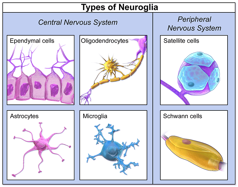
There are six types of neuroglia: four found exclusively in the CNS and two found exclusively in the PNS.
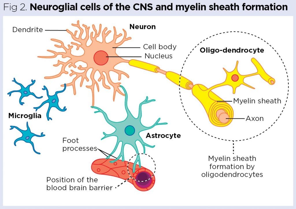
A. Neuroglia of the Central Nervous System (CNS)
- Astrocytes (Star Cells):
- The most abundant, versatile, and highly branched glial cells.
- Functions: They act as braces, physically anchoring neurons to their nutrient supply lines (blood capillaries). They strictly control the chemical environment around neurons, "mopping up" leaked potassium ions and recycling neurotransmitters. Most importantly, their "feet" wrap around brain capillaries to form the Blood-Brain Barrier (BBB), preventing toxins in the blood from entering brain tissue.
- Microglial Cells:
- Small, ovoid cells with thorny processes.
- Functions: They are the resident macrophages (immune cells) of the CNS. Since regular immune cells cannot cross the blood-brain barrier, microglia are responsible for monitoring neuron health and migrating toward injured cells to phagocytize (eat) microorganisms, dead tissue, and cellular debris.
- Ependymal Cells:
- Range in shape from squamous to columnar, and many are ciliated.
- Functions: They line the central cavities (ventricles) of the brain and the central canal of the spinal cord. They interact with capillary tangles to produce Cerebrospinal Fluid (CSF). The beating of their cilia constantly circulates the CSF, cushioning the brain.
- Oligodendrocytes:
- Have fewer processes than astrocytes.
- Functions: They line up along the thicker nerve fibers in the CNS and wrap their broad, flat processes tightly around them to produce insulating Myelin Sheaths. Unlike Schwann cells, a single oligodendrocyte can branch out and myelinate up to 60 different adjacent axons simultaneously!
B. Neuroglia of the Peripheral Nervous System (PNS)
- Satellite Cells:
- Surround the cell bodies of neurons located in PNS ganglia.
- Functions: They are thought to have many of the same functions in the PNS as astrocytes do in the CNS, providing structural support and heavily regulating the chemical environment.
- Schwann Cells (Neurolemmocytes):
- Surround all nerve fibers in the PNS.
- Functions: They form the Myelin sheaths around the thicker nerve fibers. Unlike oligodendrocytes, one Schwann cell can only myelinate one tiny segment of one single axon. They are absolutely crucial for the regeneration of damaged peripheral nerve fibers, forming a "regeneration tube" to guide a severed nerve back to its target.
Clinical Correlate: Brain Tumors (Gliomas)
Because mature neurons cannot divide (amitotic), they very rarely form tumors. Almost all primary brain tumors in adults are Gliomas—tumors that arise from the runaway, uncontrolled division of Glial cells (like Astrocytomas or Ependymomas). Because glial cells form a massive supporting web, these tumors are often highly invasive and difficult to remove surgically.
5. Nerve Impulse (Action Potential) Generation and Transmission
The entire ability of the nervous system to communicate relies on the generation and propagation of electrical signals. This involves moving charged particles (ions, specifically Sodium Na⁺ and Potassium K⁺) back and forth across the cell membrane.
Step-by-Step Electrophysiology
- Resting Membrane Potential:
- A neuron at rest is "polarized." It has a voltage difference across its membrane of about -70mV (the inside of the cell is 70 millivolts more negative than the outside).
- This extreme tension is maintained by the Sodium-Potassium (Na⁺/K⁺) Pump (which constantly burns ATP to pump 3 Na⁺ ions out and 2 K⁺ ions in) and thousands of ion leak channels.
- Graded Potentials:
- These are short-lived, localized, minor changes in membrane potential occurring on the dendrites or cell body.
- They "grade" the signal. If a graded potential is strong enough to travel to the axon hillock and reach the magical Threshold Potential (~ -55mV), it triggers an unstoppable, full-blown action potential.
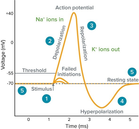
- Action Potential (Nerve Impulse):
This is a brief, rapid, "all-or-none" electrical impulse that travels down the axon without losing strength. It has distinct phases:
- Depolarization Phase: Hitting the -55mV threshold violently snaps open voltage-gated Sodium channels. Positive Na⁺ ions rush into the cell. The voltage rockets from -70mV up to +30mV.
- Repolarization Phase: At +30mV, the Na⁺ channels slam shut. Simultaneously, slow voltage-gated Potassium channels open. Positive K⁺ ions rush out of the cell, restoring the internal negativity.
- Hyperpolarization: The K⁺ channels stay open a fraction too long, causing a brief "overshoot" where the cell drops to about -90mV before the Na⁺/K⁺ pump restores the resting state of -70mV.
- Refractory Periods:
- Absolute Refractory Period: During depolarization, the neuron cannot respond to another stimulus, no matter how strong. This ensures the action potential only travels in one direction (forward).
- Relative Refractory Period: During hyperpolarization, an exceptionally strong stimulus *could* trigger another action potential.
6. Synapses and Chemical Transmission
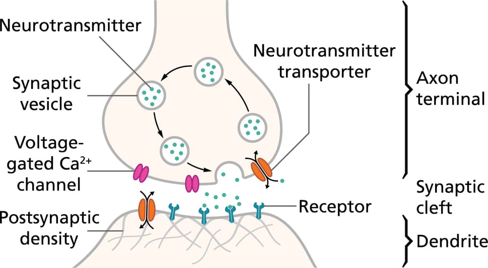
Electrical action potentials cannot jump across the empty space between two neurons. The junction where information is transferred from one neuron to the next (or from a neuron to a muscle) is called a Synapse.
- The electrical action potential travels down the axon and arrives at the Axon Terminal (Presynaptic neuron).
- The arrival of the electrical charge forces voltage-gated Calcium (Ca²⁺) channels to open. Ca²⁺ rushes into the axon terminal.
- The massive influx of calcium acts as an explosive trigger. It causes synaptic vesicles (tiny bubbles filled with chemical neurotransmitters) to fuse with the membrane via SNARE proteins and empty their contents via exocytosis.
- The neurotransmitters diffuse across the tiny fluid-filled gap (Synaptic Cleft).
- The neurotransmitters bind to highly specific protein receptors on the Postsynaptic membrane (the next neuron or muscle cell).
- This binding opens ion channels on the new cell, creating a graded potential (either an Excitatory Postsynaptic Potential / EPSP, or an Inhibitory Postsynaptic Potential / IPSP), continuing or stopping the message!
Examples of Critical Neurotransmitters:
| Neurotransmitter | Primary Action / Location | Clinical Significance |
|---|---|---|
| Acetylcholine (ACh) | Excitatory at skeletal muscles; regulates parasympathetic nervous system (Rest & Digest). | Deficiency in the brain is heavily linked to Alzheimer's Disease. Blocked by Botox and Curare. |
| Dopamine | Feel-good reward pathways; highly involved in coordinating smooth motor movement. | Deficiency in the substantia nigra causes Parkinson's Disease. Excess is linked to Schizophrenia. |
| Serotonin (5-HT) | Inhibitory; regulates mood, sleep, appetite, and nausea. | Low levels are the primary cause of clinical Depression. Treated with SSRIs (Prozac). |
| GABA | The principal INHIBITORY neurotransmitter in the brain. It calms everything down. | Enhanced by alcohol, Valium, and sedatives. Lack of GABA can lead to seizures and severe anxiety. |
References
- Marieb, E. N., & Hoehn, K. (2018). Human Anatomy & Physiology (11th ed.). Pearson.
- Tortora, G. J., & Derrickson, B. (2017). Principles of Anatomy and Physiology (15th ed.). Wiley.
- Guyton, A. C., & Hall, J. E. (2020). Textbook of Medical Physiology (14th ed.). Elsevier.
- Netter, F. H. (2018). Atlas of Human Anatomy (7th ed.). Elsevier.
- Mescher, A. L. (2018). Junqueira's Basic Histology: Text and Atlas (15th ed.). McGraw-Hill Education.