
Anatomy of the Eye, Orbit, and Extraocular Muscles
By the end of this exhaustive, highly detailed master guide, you will be deeply conversant with:
- The complex embryological development of the eye and its associated congenital anomalies.
- The intricate bony architecture of the orbit, including all foramina, fissures, and their neurovascular contents.
- The extraocular muscles, their precise actions, laws of innervation, and clinical paralysis patterns.
- The macroscopic and microscopic structure of the eyeball, the visual pathway, and physiological reflexes.
- A comprehensive breakdown of clinical ophthalmic pathologies and emergencies.
1. Embryology of the Eye
The development of the eye is an incredibly complex, highly orchestrated process that requires flawless interactions between the neural ectoderm, surface ectoderm, and mesenchyme. The master control gene for this entire process is the PAX6 gene, often referred to as the "master architect" of eye development.
1. Early Development (Optic Vesicles)
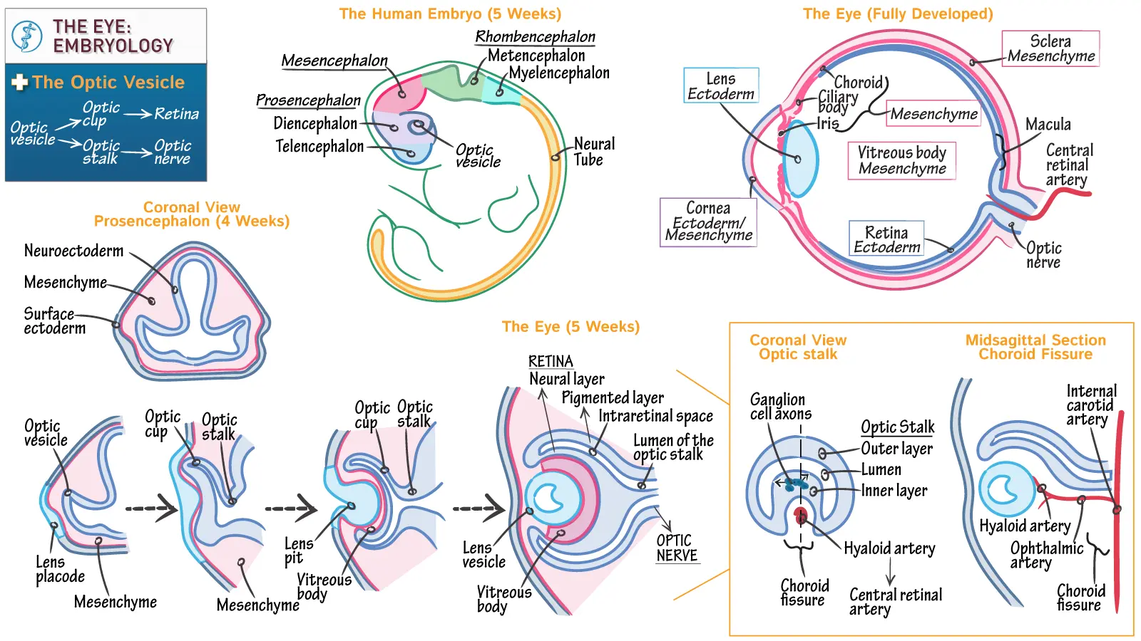
- Around day 22 of embryonic development, the eye begins its journey as a pair of shallow indentations called optic grooves located on the sides of the forebrain (diencephalon).
- With the closure of the neural tube, these grooves rapidly evaginate (push outward) to form optic vesicles, which are distinct outpocketings of the forebrain.
- These optic vesicles then grow laterally, moving outward until they make direct physical contact with the overlying surface ectoderm. This physical contact is crucial for the next stage of cellular communication (induction).
2. Lens Formation
- The optic vesicle releases chemical signals that induce the overlying surface ectoderm to thicken and invaginate, forming a specialized region called the lens placode.
- The lens placode then invaginates further inward to form the hollow lens vesicle.
- By the 5th week of intrauterine life, the lens vesicle completely loses contact with the surface ectoderm and drops down to lie within the mouth of the newly forming optic cup.
- Germ Layer Origin: The lens is formed entirely from the surface ectoderm.
3. Optic Cup Formation
- As the lens vesicle forms, the optic vesicle simultaneously invaginates back into itself to form a double-walled, goblet-like structure called the optic cup.
- This complex invagination also creates a groove called the choroid fissure (or optic fissure) running along the inferior surface of the optic cup and optic stalk.
- The choroid fissure is vital because it serves as a protected pathway for the hyaloid artery (which later becomes the central artery of the retina) to reach the inner chamber of the developing eye and supply the lens.
- During the 7th week, the lips of the choroid fissure must perfectly align and fuse. Failure of this fusion results in a structural defect called a coloboma.
- The anterior opening of the optic cup, formed by the successful fusion of the choroid fissure lips, eventually becomes the future pupil.
Derivatives of the Optic Cup Layers
| Optic Cup Layer | Derived Retinal Layers (Posterior 4/5, Pars Optica Retinae) | Derived Iris & Ciliary Body Layers (Anterior 1/5) |
|---|---|---|
| Outer Pigmented Layer | Pigment epithelium of the retina. | Outer layer of the iris (pigmented epithelium) and pigmented epithelium of the ciliary body. |
| Inner (Neural) Layer |
1. Rods and cones (photoreceptors) 2. External limiting lamina 3. Outer nuclear layer (rod & cone cell bodies with nuclei) 4. Outer plexiform layer 5. Inner nuclear layer (bipolar, horizontal, amacrine cells) 6. Inner plexiform layer 7. Ganglion cell layer 8. Fibrous layer (axons of ganglion cells) 9. Nerve fiber layer (axons forming optic nerve) 10. Inner limiting lamina | Inner layer of the iris (pigmented epithelium) and non-pigmented epithelium of the ciliary body (which forms the ciliary processes and contributes to aqueous humor production). |
2. Congenital Eye Abnormalities
Any disruption in the delicate sequence of embryological events can lead to a range of severe visual impairments. Understanding these provides deep insight into developmental biology.
- Cause: Failure of the choroid fissure to close during the 7th week of development.
- Presentation: A persistent cleft, most commonly in the iris (coloboma iridis), resulting in a distinctive keyhole-shaped pupil. However, the cleft can extend much deeper into the ciliary body, retina, choroid, or even the optic nerve.
- Association: Often associated with other systemic defects. Optic nerve colobomas are highly linked to PAX2 gene mutations and can be part of Renal Coloboma Syndrome (a condition involving both severe eye and kidney defects).
- Cause: Failure of the embryonic membrane (which temporarily covers the pupil during development) to resorb during the final formation of the anterior chamber.
- Presentation: Visible as fine, web-like strands of tissue stretching across the pupil. While often benign and asymptomatic, a very dense membrane can significantly impair vision and require surgical removal.
- Cause: The normally transparent lens becomes opaque (cloudy) during intrauterine life.
- Etiology: Can be genetically determined or caused by severe intrauterine infections (the "TORCH" infections). A classic example is Rubella (German measles) infection in the mother between the 4th and 7th weeks of pregnancy. Clinical Note: Infection after the 7th week might spare the lens but frequently causes profound deafness due to severe cochlear abnormalities.
- Normal Degeneration: The distal portion of the hyaloid artery (which supplied the developing lens) normally degenerates entirely before birth, with its proximal part remaining to form the central artery of the retina.
- Anomaly: Persistence of the distal portion can lead to a fibrous cord (Mittendorf dot) or a prominent cyst floating in the vitreous humor, potentially obstructing the visual axis.
- Microphthalmia: The eye is abnormally small, sometimes only 2/3 of its normal volume. Usually associated with severe intrauterine infections like Cytomegalovirus (CMV) and Toxoplasmosis.
- Anophthalmia: Complete, absolute absence of the eye. This is an extreme developmental failure often accompanied by severe cranial and brain abnormalities.
- Aphakia: Complete absence of the lens.
- Aniridia: Complete absence of the iris.
- Genetic Link: Both are exceptionally rare and caused by profound disturbances in tissue induction. Mutations in the PAX6 gene are heavily associated with aniridia, and this mutation can simultaneously contribute to anophthalmia and microphthalmia.
8. Cyclopia & Synophthalmia
These represent a devastating spectrum of midline defects occurring during early gestation.
- Cyclopia: Development of a single, central eye.
- Synophthalmia: The fusion of two eyes (partial or complete) into the center of the face.
- Spectrum & Timing: Represents a massive loss of midline tissue during early gestation (specifically days 19-21 or later, deeply affecting facial development).
- Association: Invariably linked to severe, fatal cranial defects like Holoprosencephaly (where the embryonic forebrain fails to divide into two separate merged cerebral hemispheres).
- Etiology: Factors devastating the midline include severe maternal alcohol exposure, critical mutations in the Sonic Hedgehog (SHH) signaling pathway, and profound abnormalities in cholesterol metabolism (which physically disrupt SHH signaling).
3. The Bony Orbit
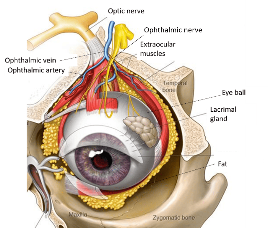
The orbit is a deep, pyramidal-shaped bony cavity designed to house and aggressively protect the eyeball and its associated muscles, nerves, and fat.
1. Bones Forming the Orbit
Each bony orbit is formed by a complex jigsaw puzzle of exactly seven bones. (Mnemonic to remember: "Many Friendly Zebras Enjoy Lazy Summer Picnics")
- Maxilla
- Frontal
- Zygomatic
- Ethmoid
- Lacrimal
- Sphenoid
- Palatine
2. Boundaries of the Orbit
- Apex: The deepest point, containing the optic foramen (located in the lesser wing of the sphenoid bone).
- Base (Orbital Rim): The strong outer edge that you can feel on your face.
- Superiorly: Frontal bone.
- Medially: Frontal process of the maxilla.
- Inferiorly: Zygomatic process of the maxilla and the zygomatic bone.
- Laterally: Zygomatic bone, frontal process of the zygomatic bone, and zygomatic process of the frontal bone.
- Roof (Superior Wall): Mainly the orbital part of the frontal bone. Posteriorly, it is completed by the lesser wing of the sphenoid bone.
- Medial Wall: The thinnest, most fragile wall (the ethmoid portion is called the lamina papyracea or "paper sheet"). Composed of four bones: frontal process of maxilla, lacrimal bone, orbital plate of the ethmoid bone, and a small part of the sphenoid bone (body). The medial walls of the two orbits run parallel to each other.
- Floor (Inferior Wall): Primarily the orbital surface of the maxilla. Anterolaterally, the zygomatic bone. Posteriorly, the orbital process of the palatine bone. (Clinical Note: This is a common site for "Blowout fractures").
- Lateral Wall: The thickest wall. Anteriorly, the zygomatic bone. Posteriorly, the greater wing of the sphenoid bone.
3. Orbital Fissures, Foramina, and their Contents
These openings serve as crucial passageways for nerves, vessels, and other structures entering or leaving the eye socket.
| Orbital Opening | Boundaries / Location | Contents (Nerves & Vessels) |
|---|---|---|
| Optic Canal (Foramen) | Lies within the lesser wing of the sphenoid bone, between its two roots. | Optic Nerve (CN II) and the Ophthalmic Artery (a major branch of the internal carotid artery). |
| Superior Orbital Fissure | Located between the greater and lesser wings of the sphenoid bone. Connects the orbit deeply with the middle cranial fossa. | Cranial Nerves: Oculomotor (CN III), Trochlear (CN IV), Ophthalmic division of Trigeminal (CN V1 - specific branches include Lacrimal, Frontal, and Nasociliary nerves), and Abducens (CN VI). Vessels: Superior Ophthalmic Vein. Other: Sympathetic fibers traveling to the ciliary ganglion. |
| Inferior Orbital Fissure | Located between the lateral wall (greater wing of sphenoid/zygomatic bone) and the floor (maxilla/orbital process of palatine bone). Connects the orbit with the pterygopalatine and infratemporal fossae. | Nerves: Zygomatic nerve (branch of CN V2), Infraorbital nerve (another branch of CN V2), Orbital branches of the pterygopalatine ganglion. Vessels: Inferior Ophthalmic Vein (which drains backward into the pterygoid plexus), Infraorbital Artery and Vein. |
| Supraorbital Foramen (or Notch) | Located on the superior orbital margin (frontal bone). | Supraorbital Nerve (the terminal branch of the frontal nerve, which is a branch of V1) and the Supraorbital Artery. |
| Infraorbital Foramen | Located on the anterior surface of the maxilla, directly below the inferior orbital rim. | Infraorbital Nerve (the continuation of V2 after it passes through the infraorbital canal) and the Infraorbital Artery and Vein. |
| Anterior Ethmoidal Foramen | Located in the medial wall of the orbit, exactly between the frontal bone and the ethmoid bone. | Anterior Ethmoidal Nerve (a branch of the nasociliary nerve, from V1) and the Anterior Ethmoidal Artery and Vein. |
| Posterior Ethmoidal Foramen | Located in the medial wall of the orbit, posterior to the anterior ethmoidal foramen, between the frontal and ethmoid bones. | Posterior Ethmoidal Nerve (branch of nasociliary nerve, from V1) and the Posterior Ethmoidal Artery and Vein. |
| Nasolacrimal Canal | Formed by the lacrimal bone and maxilla. It drains tears away from the lacrimal sac straight down into the inferior meatus of the nasal cavity. | Contains the Nasolacrimal duct. (This is why your nose runs when you cry!) |
4. Extrinsic (Extraocular) Muscles of the Eye
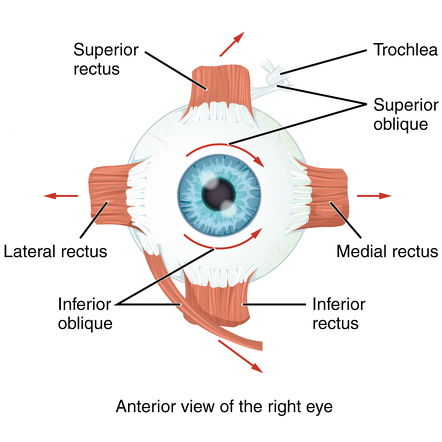
These specialized muscles precisely control the movement of the eyeball. They act in perfect coordination and are primarily innervated by Cranial Nerves III, IV, and VI. (Mnemonic: LR6-SO4-Rest3 -> Lateral Rectus=CN VI, Superior Oblique=CN IV, Rest=CN III).
1. Origin and Insertion
- Common Origin: All extrinsic muscles (with the strict exception of the inferior oblique) arise from a tough, common tendinous ring called the Annulus of Zinn, which firmly surrounds the optic canal and part of the superior orbital fissure at the apex.
- Inferior Oblique Origin: This unique muscle originates anteriorly from the orbital surface of the maxilla, right near the inferior orbital rim.
- Insertions: They all insert onto the tough sclera of the eyeball.
- The Recti muscles insert anterior to the equator (the middle line) of the eyeball, pulling the eye toward them.
- The Oblique muscles insert posterior to the equator, acting from behind to rotate the eye.
2. Muscle Actions and Innervation
The 'primary action' is the most effective movement produced when the eye starts in the primary (straight-ahead) position.
| Muscle | Innervation | Primary Action (from primary gaze) | Secondary Action(s) |
|---|---|---|---|
| Superior Rectus | Oculomotor Nerve (CN III) | Elevation (moves eye upward) | Adduction, Intorsion (medial rotation) |
| Inferior Rectus | Oculomotor Nerve (CN III) | Depression (moves eye downward) | Adduction, Extorsion (lateral rotation) |
| Medial Rectus | Oculomotor Nerve (CN III) | Adduction (moves eye medially/inward toward nose) | - |
| Lateral Rectus | Abducens Nerve (CN VI) | Abduction (moves eye laterally/outward toward ear) | - |
| Superior Oblique | Trochlear Nerve (CN IV) | Intorsion (medial rotation, especially when the eye is adducted) | Depression (when eye is adducted), Abduction. Passes through the cartilaginous trochlea pulley! |
| Inferior Oblique | Oculomotor Nerve (CN III) | Extorsion (lateral rotation, especially when the eye is adducted) | Elevation (when eye is adducted), Abduction |
| Levator Palpebrae Superioris | Oculomotor Nerve (CN III) (and sympathetic fibers for Müller's muscle) | Elevates the upper eyelid (keeps the eye open) | - |
Key Considerations for Muscle Actions
- Recti Muscles: All recti muscles physically pull the eye towards their origin at the apex of the orbit. Because they originate medially to the sagittal axis of the eyeball, all recti (except the pure lateral rectus) have a natural adduction component.
- Oblique Muscles: Because they insert posterior to the equator, they pull from the back.
- The Superior Oblique passes through a cartilaginous pulley (the trochlea) before inserting. When it pulls, it depresses and intorts the eye (most effective when the eye is adducted).
- The Inferior Oblique elevates and extorts when the eye is adducted.
3. Laws of Ocular Innervation
- Hering's Law of Equal Innervation: States that synergistic muscles (muscles that work together in both eyes to produce a specific gaze direction) receive exactly equal and simultaneous innervation. Example: When you look to the right, your brain fires an identical, equal electrical signal to both the right lateral rectus and the left medial rectus.
- Sherrington's Law of Reciprocal Innervation: States that when an agonist muscle actively contracts, its opposing antagonist muscle must simultaneously relax. Example: When the medial rectus contracts to turn the eye inward, the brain actively shuts off signals to the lateral rectus so it relaxes and doesn't fight the movement.
Clinical Correlates of Extraocular Muscle Palsies
Damage to the cranial nerves innervating these muscles results in specific patterns of strabismus (severe misalignment of the eyes) and diplopia (distressing double vision).
1. Oculomotor Nerve (CN III) Palsy
- Muscles Affected: Superior rectus, inferior rectus, medial rectus, inferior oblique, and levator palpebrae superioris. Importantly, it also knocks out parasympathetic fibers traveling to the iris and ciliary body.
- Clinical Signs (The "Blown, Down, and Out" Eye):
- Ptosis: Severe drooping of the upper eyelid due to paralysis of the levator palpebrae superioris.
- "Down and Out" Eye: Because CN III is dead, the only living muscles left are the Superior Oblique (pulls down/out) and Lateral Rectus (pulls out). Their unopposed action locks the eye in a downward and outward gaze.
- Diplopia: Double vision due to massive misalignment.
- Mydriasis (Dilated Pupil): The "blown" pupil. Due to paralysis of the constrictor pupillae muscle (loss of parasympathetic control).
- Loss of Accommodation: Due to paralysis of the ciliary muscle, the patient cannot focus on near objects.
2. Trochlear Nerve (CN IV) Palsy
- Muscle Affected: Superior oblique.
- Clinical Signs:
- Vertical Diplopia: The patient sees two images stacked vertically, especially noticeable when looking down and in (e.g., trying to read a book or walking down stairs).
- Extorsion: The superior oblique normally intorts the eye. Its paralysis leads to unopposed extorsion.
- Head Tilt (Bielschowsky Phenomenon): To compensate for the diplopia, patients unconsciously tilt their head to the opposite shoulder (chin tuck and head turned away from the affected side). This awkward posture physically helps to intort the affected eye and align the visual fields.
3. Abducens Nerve (CN VI) Palsy
- Muscle Affected: Lateral rectus.
- Clinical Signs:
- Medial Deviation (Esotropia): The medial rectus is now unopposed, so it violently pulls the eye medially (cross-eyed appearance).
- Inability to Abduct: The affected eye physically cannot move laterally past the midline.
- Horizontal Diplopia: Seeing two images side-by-side, which gets significantly worse when the patient attempts to look laterally towards the affected side.
5. Anterior & Posterior Chambers of the Eye
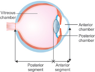
These fluid-filled spaces are crucially important for maintaining intraocular pressure (giving the eye its firm shape) and nourishing the avascular (bloodless) lens and cornea.
1. Aqueous Humor
- Production: Continuously produced by the ciliary processes (specifically the non-pigmented epithelium) of the ciliary body.
- Circulation Pathway:
- From the ciliary processes, it is secreted into the posterior chamber (the tiny space between the back of the iris and the front of the lens).
- It then flows forward, passing directly through the pupil into the anterior chamber (the larger space between the back of the cornea and the front of the iris).
- It circulates here to provide nutrients, then drains into the spongy trabecular meshwork, located deep in the angle between the iris and cornea.
- From the trabecular meshwork, it is filtered into the Canal of Schlemm (scleral venous sinus).
- Finally, it drains out of the eye entirely into the episcleral veins, returning to the systemic blood circulation.
2. Clinical Significance: Glaucoma
Definition: A devastating group of eye conditions that permanently damage the optic nerve, most often due to abnormally high intraocular pressure (IOP) crushing the nerve fibers.
Mechanism: Increased IOP is almost always caused by a plumbing failure—an imbalance between the production and drainage of aqueous humor. The faucet is on, but the drain is clogged (usually at the trabecular meshwork or Canal of Schlemm).
- Open-angle glaucoma: The most common form. The anatomical angle looks wide open, but microscopic blockages deep in the trabecular meshwork impair drainage. It is a slow, painless, silent thief of peripheral vision.
- Angle-closure glaucoma: A medical emergency. The iris is physically pushed or pulled forward, instantly slamming shut the angle and completely blocking the trabecular meshwork, halting all drainage instantly. Pressure spikes violently.
6. Innervation of the Eye
A master summary of the complex nervous supply controlling every aspect of the eye and its associated structures.
1. Motor Innervation
- Oculomotor (CN III): Superior rectus, inferior rectus, medial rectus, inferior oblique, levator palpebrae superioris.
- Trochlear (CN IV): Superior oblique.
- Abducens (CN VI): Lateral rectus.
2. Sensory Innervation (The Trigeminal Nerve - CN V)
All sensory feedback (pain, touch, temperature) from the eye is carried by the Ophthalmic Division (CN V1), which supplies the cornea, conjunctiva, eyelids, forehead, and nasal bridge.
- Lacrimal Nerve: Sensory to the lacrimal gland, upper eyelid, and conjunctiva.
- Frontal Nerve: Divides into the supraorbital and supratrochlear nerves, carrying sensation from the forehead, scalp, and upper eyelid.
- Nasociliary Nerve: The crucial nerve for eyeball sensation! Sensory to the eyeball itself (cornea, iris, ciliary body), conjunctiva, and part of the nasal mucosa. Branches include the long ciliary nerves (direct sensory wires to the iris and extremely sensitive cornea) and the anterior/posterior ethmoidal nerves.
3. Autonomic Innervation
(Pupillary Constriction and Accommodation)
- Origin: Edinger-Westphal nucleus (in the midbrain).
- Pathway: Preganglionic fibers travel piggyback with CN III and synapse in the ciliary ganglion located behind the eye.
- Distribution: Postganglionic fibers (the short ciliary nerves) travel to the eye.
- Action: They strongly innervate the sphincter pupillae muscle (causing rapid miosis/pupillary constriction to block bright light) and the ciliary muscle (causing it to contract, rounding the lens for near-vision accommodation).
- Reflexes: This pathway is the absolute driving force for the pupillary light reflex and the accommodation reflex.
(Pupillary Dilation & Fight-or-Flight)
- Origin: Hypothalamus (first-order neuron) down to the Ciliospinal center of Budge at the T1-T2 level of the spinal cord (second-order neuron).
- Pathway: Preganglionic fibers ascend straight up the sympathetic chain in the neck and synapse in the superior cervical ganglion.
- Distribution: Postganglionic fibers wrap tightly around the internal carotid artery forming a plexus, then join the long ciliary nerves (via the ophthalmic artery and nasociliary nerve) to reach the eye.
- Action: They innervate the dilator pupillae muscle (causing rapid mydriasis/pupillary dilation to let in light during a crisis) and Müller's muscle (the superior tarsal muscle, which pulls the upper eyelid wide open in terror/surprise).
Clinical Significance - Horner's Syndrome
Damage anywhere along the incredibly long sympathetic pathway (from the hypothalamus, down the spinal cord, up the neck, and into the eye—such as from a Pancoast tumor in the lung apex or a carotid dissection) results in a classic triad of symptoms known as Horner's Syndrome:
- Ptosis: Mild drooping of the upper eyelid (due to paralysis of Müller's muscle, which normally holds the lid wide open).
- Miosis: A persistently constricted pupil (due to the death of the sympathetic dilator pupillae, leaving the parasympathetic sphincter unopposed).
- Anhidrosis: Complete absence of sweating on the ipsilateral (same side) face, as the sympathetic nerves to the facial sweat glands are also destroyed.
7. Arterial Supply and Venous Drainage of the Orbit
1. Arterial Supply
The entire eye and orbit survive on blood delivered by the Ophthalmic artery, a major direct branch of the internal carotid artery.
Key Branches of the Ophthalmic Artery:
- Central Retinal Artery: The most critical branch. It pierces the dura and enters the optic nerve itself, running down its center to exclusively supply the inner layers of the retina. (If this blocks, the retina dies instantly).
- Lacrimal Artery: Supplies the massive lacrimal gland, outer eyelids, and conjunctiva. It also gives off tiny zygomatic branches.
- Posterior Ciliary Arteries (long and short): Supply the choroid (the main vascular bed), ciliary body, and iris. The short posterior ciliary arteries are numerous and supply the choroid directly. The long posterior ciliary arteries run far forward to supply the ciliary body and the iris.
- Anterior & Posterior Ethmoidal Arteries: Pierce the medial wall to supply the ethmoidal air cells and the deep nasal cavity.
- Supraorbital & Supratrochlear Arteries: Exit the orbit anteriorly to supply the skin and muscles of the forehead and scalp.
2. Venous Drainage
Venous blood leaves the eye through valveless veins, which poses a unique infection risk.
- Superior Ophthalmic Vein: Drains backward directly into the massive cavernous sinus located inside the skull. Critically, it communicates anteriorly with the facial vein.
- Inferior Ophthalmic Vein: Drains backward into the cavernous sinus and/or down into the pterygoid venous plexus. It also communicates with the facial vein.
Clinical Significance: The "Danger Triangle of the Face"
Because the ophthalmic veins have absolutely no valves, blood can flow in either direction. The connections between the ophthalmic veins deep in the orbit and the superficial facial veins are clinically terrifying. A simple infection on the face (e.g., popping a highly infected pimple on the nose or upper lip) can allow bacteria to travel backward through the facial vein, through the superior ophthalmic vein, and directly into the brain's cavernous sinus. This causes Cavernous Sinus Thrombosis, a massive, life-threatening brain infection and clot.
8. Other Important Structures: The Lacrimal Apparatus
1. The Lacrimal Gland
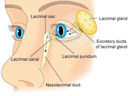
- Function: Continuously produces the watery (aqueous) component of tears, vital for washing away debris, providing oxygen to the cornea, and delivering antimicrobial enzymes (lysozyme).
- Location: Tucked safely in the superolateral part of the orbit, within the distinct lacrimal fossa of the frontal bone.
The Complex Innervation of the Lacrimal Gland
The lacrimal gland requires complex "hitchhiking" of nerves to function. It receives sensory, secretomotor (parasympathetic), and sympathetic components.
A. Sensory Innervation (Feeling pain/dryness)
- Pathway: Sensory information from the lacrimal gland (irritation, burning, pain) travels back to the CNS via the lacrimal nerve, which is a branch of the ophthalmic division (V1) of the trigeminal nerve (CN V).
B. Secretomotor (Parasympathetic) Innervation (Making you cry)
This is the primary pathway that stimulates massive fluid secretion (tear production).
- Origin: Preganglionic parasympathetic neurons originate deep in the superior salivatory nucleus in the pons of the brainstem.
- Facial Nerve (CN VII): These fibers exit the brainstem traveling inside the facial nerve (CN VII).
- Greater Petrosal Nerve: They soon branch off from the facial nerve as the greater petrosal nerve.
- Nerve of the Pterygoid Canal (Vidian Nerve): The greater petrosal nerve joins forces with the deep petrosal nerve (which carries sympathetic fibers) to form a combined cable called the nerve of the pterygoid canal.
- Pterygopalatine Ganglion: This combined nerve passes into the pterygopalatine ganglion (located in the pterygopalatine fossa). Here, the preganglionic parasympathetic fibers finally synapse with postganglionic parasympathetic neurons.
- Maxillary Nerve (V2) Hitchhike: The newly formed postganglionic parasympathetic fibers do not stay in the ganglion. Instead, they "hitchhike" by jumping onto the maxillary division (V2) of the trigeminal nerve.
- Zygomatic Nerve: They travel with the maxillary nerve until they branch off onto the zygomatic nerve.
- Zygomaticotemporal Nerve: Within the orbit, the zygomatic nerve gives off the zygomaticotemporal nerve.
- Communicating Branch: A tiny communicating branch from the zygomaticotemporal nerve (carrying the precious postganglionic parasympathetic fibers) jumps over and joins the lacrimal nerve.
- Lacrimal Gland: Finally, the fibers travel down the lacrimal nerve, reach the lacrimal gland, and trigger a flood of tears.
C. Sympathetic Innervation
- Function: Its exact role in tear production is debated. It primarily constricts blood vessels to the gland and may slightly inhibit watery secretion or stimulate mucous secretion.
- Pathway: Originates in the upper thoracic spinal cord (T1-T2) -> ascends to synapse in the Superior Cervical Ganglion -> postganglionic fibers wrap around the internal carotid artery -> branch off as the Deep Petrosal Nerve -> joins the greater petrosal nerve to form the Nerve of the Pterygoid Canal -> passes straight through the pterygopalatine ganglion (NO synapse!) -> hitchhikes along the exact same V2 -> Zygomatic -> Zygomaticotemporal -> Lacrimal nerve route to reach the gland.
2. The Lacrimal Drainage Apparatus
Tears must be constantly drained to prevent blurry vision and overflow.
- Lacrimal Puncta and Canaliculi: Tiny holes on the inner corner of your eyelids (puncta) suck up tears like a vacuum into small tubes (canaliculi).
- Lacrimal Sac: The tubes dump the tears into a holding tank called the lacrimal sac.
- Nasolacrimal Duct: A large pipe that drains tears from the lacrimal sac straight down through the bone, emptying into the inferior meatus of the nasal cavity inside your nose.
3. The Eyelids (Palpebrae)
- Orbicularis Oculi Muscle: The sphincter muscle that violently closes the eyelids (squinting/blinking). Innervated by the facial nerve (CN VII). (Damage causes Bell's Palsy, meaning the patient cannot close their eye, risking severe corneal ulcers).
- Levator Palpebrae Superioris: The primary muscle that elevates the upper eyelid. Innervated by CN III.
- Müller's Muscle (Superior Tarsal Muscle): A secondary smooth muscle that provides the extra "lift" to widen the palpebral fissure. Innervated by sympathetic fibers.
- Meibomian Glands (Tarsal Glands): Modified sebaceous (oil) glands hidden deep within the tough tarsal plates of the eyelids. They secrete the vital lipid (oil) component of the tear film. This oil layer coats the watery tears, acting as a shield to prevent the tears from evaporating into the air. (Dysfunction causes severe Dry Eye Syndrome).
9. The Eye (Structure of the Eyeball)
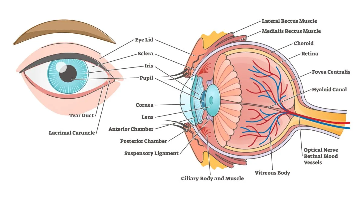
The eye is an extraordinary sensory organ responsible for capturing photons of light and converting them into electrical thought. It is constructed of three main concentric coats (tunics) and a fluid-filled interior.
A. The Fibrous Coat (Outer Layer)
This is the tough, outermost protective shell. It acts like the steel chassis of a car, providing shape, structural integrity, and resistance to internal pressure.
- Sclera (The White of the Eye):
- The posterior, highly opaque, and incredibly tough part of the fibrous coat (covers 5/6th of the eye).
- Composed of densely woven, irregular connective tissue (collagen and elastin).
- It is continuous posteriorly with the tough dura mater sheath protecting the optic nerve.
- Lamina Cribrosa: A specialized, sieve-like area of the posterior sclera that is perforated with tiny holes. The delicate axons of the retinal ganglion cells (which form the optic nerve) and central retinal vessels pass through these holes. Clinical Note: Because it is full of holes, the lamina cribrosa is the weakest point of the entire scleral shell. In Glaucoma, high pressure pushes on this weak point, bowing it backward (cupping) and crushing the nerve fibers passing through it.
- Clinical Note - Staphylomas: These are pathological, localized bulges of a dangerously thinned sclera, often appearing blue because the dark choroid underneath is showing through.
- Cornea (The Clear Window):
- The anterior, perfectly transparent, and completely avascular (no blood vessels) part of the fibrous coat.
- Because it is curved, it acts as the primary lens of the eye, refracting (bending) incoming light and contributing the vast majority of the eye's total focusing power.
- It relies entirely on the aqueous humor behind it and tears in front of it for oxygen and nutrients.
- It is densely packed with highly sensitive naked nerve endings (from CN V1), making it one of the most pain-sensitive tissues in the entire human body.
B. The Vascular Coat (Uvea - Middle Layer)
The Uveal tract is the dark, incredibly blood-rich, and heavily pigmented middle layer of the eye. Its job is nourishment and light absorption.
- Choroid:
- The highly vascular, dark brown layer sandwiched between the retina and the sclera.
- It consists of an outer pigmented layer (to absorb scattered light and prevent glare/reflections inside the eyeball) and a massive inner vascular network.
- Its primary, critical function is to pump blood to nourish the highly demanding outer layers of the retina (especially the photoreceptors).
- Ciliary Body:
- A muscular ring located anterior to the choroid, extending from the ora serrata (the jagged edge of the retina) up to the iris.
- Ciliary Ring: The flat, posterior part.
- Ciliary Processes: Highly folded, glandular structures that actively filter blood to produce the aqueous humor.
- Ciliary Muscle: A ring of smooth muscle. Its contraction and relaxation control the tension on the suspensory ligaments holding the lens, completely controlling the process of accommodation (focusing for near vision).
- Iris:
- The pigmented, contractile diaphragm that forms the beautiful colored part of the eye (blue, brown, green).
- Contains a central, adjustable hole called the pupil.
- Regulates the exact amount of light striking the retina via two competing smooth muscles:
- Sphincter Pupillae: Circular fibers that constrict the pupil like a drawstring bag (miosis) under parasympathetic control.
- Dilator Pupillae: Radial fibers pulling outward to dilate the pupil (mydriasis) under sympathetic control.
C. The Nervous Coat (Retina - Inner Layer)
This is the delicate, light-sensitive layer where the actual magic of vision happens. It is basically an extension of the brain.
- Anterior Edge: Forms the ora serrata, the jagged anterior margin where the complex nervous retina suddenly ends. The anterior ¼ of the retina past this point is entirely non-receptive (blind) and simply forms a two-layered cellular lining covering the back of the ciliary body and iris.
- Posterior ¾ (Pars Optica Retinae): The active receptor organ. It is loaded with photoreceptors (rods and cones).
- Macula Lutea: A highly specialized, yellow-pigmented oval area near the exact center of the retina. It is responsible for your sharpest, high-definition central vision.
- Fovea Centralis: A tiny, microscopic pit located dead-center within the macula. It contains only cones (no rods whatsoever) and the inner retinal layers are physically pushed aside to allow light a direct path to the cones. This pit provides the absolute highest visual acuity (sharpness) in the eye.
- Optic Disc (The Blind Spot): The bright circular area where all the retinal nerve fibers gather to exit the eyeball as the Optic Nerve (CN II), and where the central retinal artery and vein punch through to enter the eye. Because there is a massive bundle of cables here, there is absolutely no room for photoreceptors. Hence, any light falling on the optic disc is invisible to you—creating a natural "blind spot" in your visual field.
From the outermost layer (touching the choroid) to the innermost layer (touching the vitreous jelly):
- Pigment Epithelium: A single layer of melanin-rich cells that absorbs stray light, stores Vitamin A, and eats shed photoreceptor discs.
- Photoreceptor layer: The actual light-sensing outer segments of the Rods and Cones.
- External limiting membrane.
- Outer nuclear layer: Contains the cell bodies and heavy nuclei of the rods and cones.
- Outer plexiform layer: The synaptic zone where photoreceptors talk to bipolar cells.
- Inner nuclear layer: Contains the cell bodies of the middle-men (bipolar, horizontal, and amacrine cells).
- Inner plexiform layer: The synaptic zone where bipolar cells talk to ganglion cells.
- Ganglion cell layer: The cell bodies of the retinal ganglion cells (the final output neurons of the eye).
- Nerve fiber layer: The long, sweeping axons of the ganglion cells traveling across the retina to form the optic nerve.
- Internal limiting membrane: The basement membrane separating the retina from the vitreous humor.
D. Internal Contents of the Eyeball
- Aqueous Humor: A clear, watery fluid that fills the anterior and posterior chambers, maintaining pressure and nourishing the cornea/lens.
- Lens: A highly organized, transparent, biconvex, and elastic crystal structure located directly behind the iris. Its sole purpose is to dynamically change shape (accommodation) to precisely focus incoming light rays exactly onto the fovea. (As we age, the lens loses its elasticity, causing Presbyopia—the inability to focus on near objects, requiring reading glasses).
- Vitreous Humor: A massive, clear, gelatinous mass (like thick Jell-O) that fills the huge vitreous chamber (the entire space posterior to the lens). It maintains the rigid spherical shape of the eyeball and exerts pressure backward, acting like biological glue to hold the fragile retina flat against the choroid.
Blood Supply and Innervation of the Eyeball
While the orbit has a broader supply, the eyeball itself relies on a highly specific and delicate network of vessels and nerves.
1. Blood Supply of the Eyeball
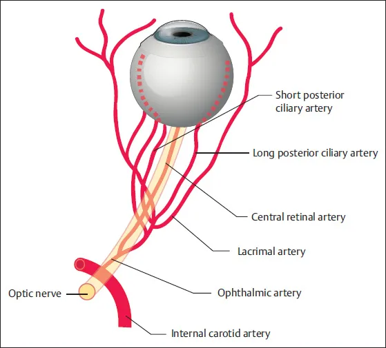
The primary arterial supply to the eyeball is derived exclusively from the ophthalmic artery, a major branch of the internal carotid artery.
- Central Artery of the Retina: Enters the eyeball directly at the center of the optic disc, running hidden entirely within the optic nerve. It exclusively supplies the inner layers of the retina. Clinical Note: Complete occlusion of this artery leads to sudden, painless, and severe vision loss.
- Ciliary Arteries:
- Anterior Ciliary Arteries: Supply the anterior structures of the eye, particularly focusing on the corneoscleral junction.
- Posterior Ciliary Arteries (Short and Long): Tasked with supplying the choroid, ciliary body, and iris.
- The short posterior ciliary arteries are highly numerous and pierce the back of the eye to supply the choroid directly.
- The long posterior ciliary arteries run far forward along the sides of the eye to supply the ciliary body and the iris.
- Cilioretinal Artery: A critical anatomical variant present in only a small percentage of individuals. It is an extra branch of the posterior ciliary arteries that specifically supplies the macula. Clinical Note: If a patient suffers a Central Retinal Artery Occlusion (CRAO), possessing this tiny artery can miraculously preserve their central macular vision!
- Central Retinal Vein: Drains the inner layers of the retina and usually perfectly accompanies the central retinal artery backward into the optic nerve. It typically drains directly into the cavernous sinus.
- Vorticose Veins: (Usually 4 to 7 in number). These unique veins are responsible for draining the massive vascular bed of the choroid. They exit the sclera obliquely (at an angle) and usually drain backward into the superior and inferior ophthalmic veins.
- No Lymph Vessels: It is highly important to note that the eyeball itself lacks any lymphatic vessels whatsoever.
2. Innervation of the Eyeball
The eyeball receives a highly complex triad of sensory, parasympathetic, and sympathetic innervation.
| Nerve Type | Pathway and Branches | Physiological Action / Target |
|---|---|---|
| Sensory Innervation | Primarily travels via the long ciliary nerves (which are direct branches of the nasociliary nerve, originating from the V1 ophthalmic division of the trigeminal nerve). The short ciliary nerves also carry some sensory fibers backward. | Provides vital general sensation (touch/pain) to the delicate cornea, the iris, and the ciliary body. |
| Parasympathetic Innervation (From Oculomotor Nerve - CN III) | Pathway: Preganglionic fibers originate deeply in the Edinger-Westphal nucleus, travel with CN III, and synapse at the ciliary ganglion. Postganglionic fibers: Travel specifically via the short ciliary nerves to enter the back of the eyeball. | Innervates the sphincter pupillae muscle (causing pupillary constriction/miosis in bright light) and the ciliary muscle (causing accommodation/thickening of the elastic lens for near vision). |
| Sympathetic Innervation | Pathway: Postganglionic fibers originate in the superior cervical ganglion high in the neck. They travel along the internal carotid artery plexus into the skull. Innervation to Eye: These fibers reach the eye primarily via the long ciliary nerves (and sometimes also hitchhike via the short ciliary nerves by passing straight through the ciliary ganglion without synapsing). | Innervates the dilator pupillae muscle (causing pupillary dilation/mydriasis in the dark or during stress) and the smooth muscle components of the levator palpebrae superioris (Müller's muscle, which contributes to elevating the upper eyelid). |
10. The Physiology of Vision
1. What is a Rod / a Cone?
These are the highly specialized photoreceptor cells living in layer 2 of the retina. They act as biological solar panels, capturing light photons and converting them into electrical brain signals.
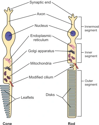
- Shape: Long, tall, and cylindrical.
- Function: Highly sensitive to even a single photon of light. Responsible for vision in near-total darkness (scotopic vision) and for detecting faint movement. They contain the pigment Rhodopsin. However, they are completely colorblind; they only see the world in shades of black, white, and gray.
- Distribution: Vastly more numerous than cones (around 120 million). They are banished to the outer peripheral retina, which is why your peripheral vision is great at sensing movement in the dark, but terrible at reading words.
- Shape: Shorter, thicker, and conical.
- Function: They require massive amounts of bright light to activate. Responsible for rich color vision and extreme high-acuity (sharp, high-definition) vision in daylight (photopic vision). There are exactly three types of cones, sensitive to different wavelengths: Red, Green, and Blue.
- Distribution: Much fewer in number (around 6 million). They are aggressively concentrated in the central macula lutea, completely dominating the fovea centralis.
2. The Visual Pathway
The incredibly complex neurological route light takes from your eye to the back of your brain where you actually "see."
- Photoreceptors: In the retina, light smashes into rhodopsin/photopsin, activating the rods and cones.
- Bipolar Neurons: Photoreceptors fire an electrical synapse to the bipolar neurons.
- Ganglion Cells: Bipolar neurons synapse with the retinal ganglion cells. The long, trailing axons of these ganglion cells gather together, leave the retina, and bundle into the massive Optic Nerve (CN II).
- Optic Chiasm: The optic nerves from both the left and right eyes travel backward and converge at the optic chiasm.
- The Decussation Rule: Fibers from the nasal (medial) half of each retina physically cross over (decussate) to the opposite side of the brain. Fibers from the temporal (lateral) half of each retina stay on the same side (uncrossed).
- The Result: This brilliant arrangement ensures that everything you see in the left half of your visual field (captured by both eyes) is sent exclusively to the right side of the brain, and vice-versa.
- Clinical Correlate: A large pituitary tumor pressing up on the optic chiasm will crush the crossing nasal fibers, causing "Bitemporal Hemianopsia" (the patient goes completely blind in their outer peripheral vision on both sides, giving them tunnel vision).
- Optic Tract: After leaving the chiasm, the newly sorted bundles of fibers are called the optic tracts. Each tract now contains complete information corresponding to the contralateral (opposite) visual field.
- Lateral Geniculate Nucleus (LGN): Located in the Thalamus. Most fibers in the optic tracts synapse here. The LGN acts as a heavy-duty relay station, sorting and organizing visual data before sending it to the cortex.
- Optic Radiations (Geniculocalcarine Tract): From the LGN, fibers fan out massively deep inside the brain, forming the optic radiations, which project backward toward the occipital lobe.
- Primary Visual Cortex: The radiations terminate in the primary visual cortex (Brodmann area 17) located at the very back of the skull in the occipital lobes. This is where the electrical signals are finally decoded, consciously perceived, and assembled into a recognized image.
3. Accommodation (Focusing the Lens)
Accommodation is the dynamic process by which the eye physically changes the optical power (thickness) of its lens to maintain a sharply focused image as an object moves closer or farther away.
| Mechanism | For FAR Vision (Object > 6 meters away) | For NEAR Vision (Object < 6 meters away) |
|---|---|---|
| Ciliary Muscle | Relaxes (it is a ring, so relaxing makes the ring wider/larger). | Contracts (the ring squeezes inward, making the hole smaller). |
| Ciliary Body | Moves backward and outward (away from the lens). | Moves forward and inward (closer to the lens). |
| Suspensory Ligaments | Become incredibly Taut (stretched tight like piano strings). | Become Relaxed and loose. |
| The Lens | Is violently pulled outward from all sides by the taut ligaments, forcing it to become thinner and flatter. This drastically reduces its refractive bending power, perfect for distant, parallel light rays. | Because the ligaments are loose, the lens (which is highly elastic) springs naturally into its default thicker, fatter, and rounder shape. This massively increases its refractive power to sharply bend diverging light rays from a close object. |
| Associated Actions | Pupils tend to dilate slightly. Eyes stare straight ahead. | 1. Pupils Constrict (Miosis) to block scattered light and increase depth of field. 2. Convergence (Both eyes instantly turn inward/adduct to keep the near object centered on the fovea). |
4. How the Light and Blink Reflexes Work
A. Pupillary Light Reflex
This is an involuntary, high-speed reflex that controls the diameter of the pupil in response to the intensity of light entering the eye. It protects the sensitive retina from being burned by overstimulation (like a camera automatically adjusting its aperture).
- Afferent Arm (The Sensor Pathway):
- Bright light smashes into photoreceptors in the retina.
- The danger signal travels rapidly down the optic nerve (CN II).
- At the optic chiasm, some fibers cross, ensuring both sides of the brain get the message.
- Crucially, these specific reflex fibers abandon the visual pathway. Instead of going to the LGN to be "seen", they exit the optic tract early and dive into the pretectal nucleus in the midbrain.
- From the pretectal nucleus, interneurons project to the Edinger-Westphal nucleus (the parasympathetic control center for CN III) on both the left and right sides of the brainstem.
- Efferent Arm (The Action Pathway):
- Preganglionic parasympathetic fibers fire from the Edinger-Westphal nuclei and travel outward inside the oculomotor nerves (CN III) of both eyes.
- They synapse in the ciliary ganglia behind the eyeballs.
- Postganglionic parasympathetic fibers (short ciliary nerves) command the sphincter pupillae muscle to contract violently.
- The Result:
- Direct Light Reflex: The pupil in the eye that the flashlight was shined into rapidly constricts.
- Consensual Light Reflex: Because the brainstem cross-wired the signal to both Edinger-Westphal nuclei, the pupil in the other eye simultaneously constricts, even though it is in total darkness.
B. Blink Reflex (Corneal Reflex)
This is an involuntary, ultra-fast protective reflex that slams the eyelids shut in response to any physical touch on the cornea, a sudden bright light, or a perceived threat flying at the face.
- Afferent Arm (The Sensor Pathway):
- A piece of dust touches the extremely sensitive cornea.
- Pain/touch sensory impulses fire down the nasociliary branch of the ophthalmic division (V1) of the trigeminal nerve (CN V).
- The warning signal crashes into the spinal nucleus of the trigeminal nerve (V) located deep in the brainstem.
- Efferent Arm (The Action Pathway):
- From the trigeminal nucleus, fast interneurons project directly to the motor nucleus of the facial nerve (CN VII) on both sides of the brain.
- Motor commands blast down the facial nerves to the face.
- The facial nerve forces the massive orbicularis oculi muscle to instantly contract.
- The Result: Rapid closure of both eyelids (a blink) before the debris can scratch the eye further.
11. Clinical Correlates and Ophthalmic Emergencies
A deep dive into severe pathological conditions affecting the eye, their mechanisms, and clinical presentations.
Cause: Damage anywhere along the massive sympathetic innervation pathway to the eye and face (e.g., from a Pancoast tumor in the lung, spinal cord injury, or carotid artery dissection).
The Triad of Symptoms:
- Ptosis (partial): Mild drooping of the upper eyelid because the sympathetic superior tarsal muscle (Müller's muscle) is paralyzed.
- Miosis: The pupil is permanently constricted because the sympathetic dilator pupillae is dead, leaving the parasympathetic constrictor unopposed.
- Anhidrosis: Total absence of sweating on the ipsilateral (same) side of the face due to denervation of facial sweat glands.
Cause: Damage specifically to the postganglionic parasympathetic innervation (often at the ciliary ganglion) controlling the pupil and ciliary muscle. It is often idiopathic or triggered by a viral infection.
Symptoms:
- Usually unilateral. The affected pupil is much larger than the normal one.
- It reacts extremely poorly and sluggishly to light (slow, tonic constriction).
- It suffers from slow, delayed re-dilation after the light stimulus is removed.
- The patient complains of blurred vision, especially for near objects, due to impaired accommodation (the ciliary muscle is partially paralyzed). Often seen in young women.
Cause: A highly specific midbrain lesion, famously associated with late-stage Neurosyphilis, and occasionally severe diabetic neuropathy.
Symptoms:
- Known clinically as the "Prostitute's pupil" because it "accommodates but does not react." (This is called light-near dissociation).
- If you shine a flashlight in the eyes, the pupils stay fixed and do not shrink. However, if you ask the patient to cross their eyes and look at their nose, the pupils successfully constrict for accommodation.
- The pupils are typically small, irregularly shaped, and often unequal in size. Involvement is usually bilateral.
Cause: A highly rare, extremely painful ophthalmoplegia caused by an idiopathic granulomatous inflammation packing into the cavernous sinus or the apex of the orbit.
Symptoms:
- Unilateral, severe, boring pain behind the eye.
- The inflammation crushes Cranial Nerves III, IV, and/or VI as they pass through the sinus, leading to complete ophthalmoplegia (total paralysis of eye movements).
- If the inflammation expands, it crushes CN V1 and V2, causing total sensory numbness across the forehead and cheek.
Cause: A catastrophic mass lesion (e.g., a massive pituitary tumor, carotid aneurysm, severe bacterial infection from the face, or a blood thrombosis) physically crushing the structures passing through the cavernous sinus.
Symptoms:
- Total Ophthalmoplegia: The eye cannot move in any direction (due to the simultaneous crushing of CN III, IV, and VI).
- Sensory loss: In the V1 and V2 distribution (forehead, cheek) due to trigeminal nerve involvement.
- Proptosis and Chemosis: The eye physically bulges out of the skull (proptosis) and the conjunctiva swells massively with fluid (chemosis) because venous blood cannot drain backward through the clotted cavernous sinus.
- Horner's syndrome may also be present as the sympathetic fibers are crushed against the carotid artery.
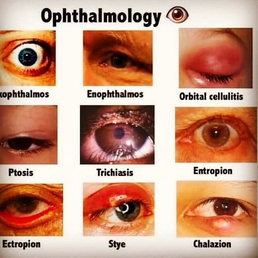
- Chalazion: A chronic, completely sterile (non-infectious), granulomatous inflammation of a Meibomian gland deep in the eyelid. It presents as a painless, firm, hard, rubbery round lump in the eyelid.
- Stye (Hordeolum): An acute, extremely painful, red, swollen bacterial infection. It can be an infection of a surface eyelash follicle (external hordeolum) or an infected Meibomian gland (internal hordeolum). It is highly tender to the touch and resembles a giant pimple on the eye.
🔴 Ophthalmic Emergencies (Immediate Action Required)
6. Closed-Angle Glaucoma (Acute Angle-Closure Glaucoma - AACG)
- Cause: A sudden, massive spike in intraocular pressure (IOP) due to the iris physically bulging forward and entirely blocking the trabecular meshwork. Aqueous humor drainage drops to zero while the ciliary body continues to pump fluid in.
- Symptoms: The patient presents screaming in agony with acute, severe eye pain. The eye is incredibly red (angry-looking). Vision is profoundly blurred, and the patient sees bright rainbow halos around lights. The pain is so severe it triggers violent nausea and vomiting. The pupil is fixed and mid-dilated. The eyeball feels as hard as a marble on palpation.
- Treatment: This is a true ophthalmic emergency requiring immediate IV pressure-lowering drugs and a laser iridotomy (shooting a hole in the iris to release the fluid) to prevent permanent blindness.
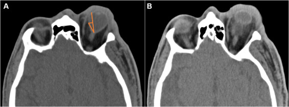
7. Orbital Blowout Fracture
- Cause: Severe blunt force trauma directly to the eyeball (e.g., getting hit with a baseball). The sudden pressure crushes the eyeball backward, causing the weakest bones of the orbit—the floor (maxilla) or medial wall (ethmoid)—to literally blow out and shatter.
- Symptoms:
- Enophthalmos: The eyeball physically sinks backward and downward into the fractured hole.
- Diplopia (double vision): Especially pronounced on upward gaze. The inferior rectus muscle and orbital fat drop through the shattered floor and get physically trapped/pinched in the broken bone. The eye cannot look up.
- Orbital emphysema: Because the bone broke into the sinus, blowing the nose forces sinus air into the orbit. The eye swells massively, and pressing on the skin feels like popping bubble wrap (crepitus).
- Infraorbital nerve anesthesia: The bone fracture slices the infraorbital nerve (V2), causing instant, total numbness in the patient's cheek, upper lip, and upper teeth.
8. Ruptured Globe (Open Globe Injury)
- Cause: Horrific penetrating trauma to the eye (e.g., a knife, a shard of glass, or a high-speed metal fragment), leading to a full-thickness breach slicing completely through the cornea or sclera. The pressurized jelly and contents of the eye begin to leak out.
- Symptoms: Severe pain and an instant, catastrophic decrease in vision.
- Hyphema: A visible pool of red blood filling the anterior chamber.
- Loss of anterior chamber depth: The eye looks deflated or flat from the side because the fluid has leaked out.
- The "Tear-drop" pupil: The pupil looks highly distorted and stretched, pointing like an arrow straight toward the site of the laceration because the iris is physically prolapsing (falling out) through the open wound.
- 360-degree Subconjunctival hemorrhage: Massive bleeding under the conjunctiva entirely encircling the cornea.
- Consequences: Irreversible visual loss and devastating endophthalmitis (total intraocular bacterial infection). Must be surgically closed immediately. A strict "NO PRESSURE" shield must be placed over the eye.
9. Central Retinal Artery Occlusion (CRAO)
- Cause: Sudden, total blockage of the central retinal artery, almost always caused by a rogue embolus (a blood clot or cholesterol plaque) breaking off from the carotid artery or heart and getting wedged in the eye. The retina suffocates instantly.
- Symptoms: Extremely sudden, entirely painless, and severe monocular vision loss. Patients often describe it terrifyingly as "a dark curtain instantly coming down over my eye."
- Fundoscopic Findings (Looking inside the eye):
- The physician sees a classic "Cherry-red spot" directly in the center of the macula. This occurs because the fovea is extremely thin and is kept alive by the vascular choroid behind it, so it stays bright red. This red dot is surrounded by a pale, white, dying, edematous retina that has lost all its blood supply.
- The arteries look like thin, empty white threads.
- Prognosis: Exceptionally poor. If blood flow is not restored within 90 minutes, the retinal ganglion cells die permanently, causing lifelong blindness.
11. Retrobulbar Hematoma / Acute Orbital Compartment Syndrome
- Cause: A severed artery causing massive, rapid hemorrhage deep into the closed, bony space behind the eyeball (the retrobulbar space), often secondary to blunt trauma or facial surgery.
- Mechanism: Because the bony orbit cannot expand, the rapidly pooling blood has nowhere to go. It violently increases the intraocular pressure (IOP) within the confined orbital space, acting like a vice grip.
- Symptoms: Acute, screaming ocular pain. Massive Proptosis (the eye is visibly being pushed out of the skull by the blood behind it). The eye is frozen tight (ophthalmoplegia). The pupil blows out, demonstrating a severe Afferent Pupillary Defect (APD). The optic nerve is crushed by the blood pressure, causing instant, rapidly progressing blindness.
- Treatment: This requires split-second action. An emergency Lateral Canthotomy and Cantholysis must be performed at the bedside (cutting the outer corner of the eyelids with scissors) to instantly pop the orbit open, decompress the tension, let the eye bulge forward, and save the optic nerve from permanent death.
References
- Moore, K. L., Dalley, A. F., & Agur, A. M. R. (2018). Clinically Oriented Anatomy (8th ed.). Lippincott Williams & Wilkins.
- Sadler, T. W. (2018). Langman's Medical Embryology (14th ed.). Wolters Kluwer.
- Kanski, J. J., & Bowling, B. (2015). Clinical Ophthalmology: A Systematic Approach (8th ed.). Elsevier.
- Snell, R. S., & Lemp, M. A. (2013). Clinical Anatomy of the Eye (2nd ed.). Wiley-Blackwell.
- Standring, S. (Ed.). (2020). Gray's Anatomy: The Anatomical Basis of Clinical Practice (42nd ed.). Elsevier.