
Topography of the Central Nervous System (CNS)
The Nervous System (NS) is indeed the most complex and highly organized system in the body, responsible for integrating and coordinating nearly all bodily functions. It operates as the master controller, ensuring survival, adaptation, and complex behaviors.
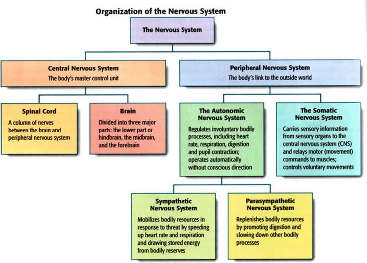
- Master Control System: It acts as the body's primary communication and control center. Every thought, action, and sensation depends on its flawless operation.
- Coordination with Endocrine System: It works in close conjunction with the endocrine system (hormonal system) to achieve global coordination.
Nervous System vs. Endocrine System
Nervous System: Functions via rapid electrical impulses (action potentials) transmitted along specialized cells called neurons, leading to immediate, highly targeted, but short-lived responses (e.g., pulling your hand away from a hot stove).
Endocrine System: Functions via slower-acting chemical messengers (hormones) transported through the bloodstream, leading to more widespread and longer-lasting effects (e.g., growth over years, or sustained adrenaline during a long exam).
Neuroendocrinology: There's significant overlap. Specialized neurons (neurosecretory cells) release hormones directly into the blood, and circulating hormones heavily influence neuronal activity. The hypothalamus is the crucial bridge connecting these two systems.
Functional Organization of the Nervous System
The Nervous System is broadly divided into two main functional components, based entirely on the type of control they exert over the body:
1. Somatic Nervous System (SNS)
This is the system you have direct, conscious control over.
- Control: Primarily controls voluntary functions of the body.
- Effectors: Targets skeletal muscles, allowing for conscious movement (walking, typing), posture maintenance, and somatic reflexes.
- Sensory Input: Receives rich sensory information from the skin, muscles, joints, and special senses (sight, hearing, touch, taste, smell).
- Pathway: Very simple and fast. It typically involves a single, heavily myelinated motor neuron extending continuously from the CNS directly to the skeletal muscle.
2. Autonomic Nervous System (ANS)
This is the "automatic" system running in the background.
- Control: Regulates involuntary (visceral) functions of the body, largely operating unconsciously to maintain homeostasis.
- Effectors: Targets smooth muscle (e.g., in walls of the intestines, blood vessels), cardiac muscle (heart), and glands (e.g., salivary, sweat, digestive).
- Sensory Input: Receives sensory information from internal organs (viscera), such as blood pressure or stomach stretch.
- Pathway: Slower. It involves a two-neuron chain to reach the effector organ: a preganglionic neuron (originating in the CNS) and a postganglionic neuron (originating in a ganglion outside the CNS).
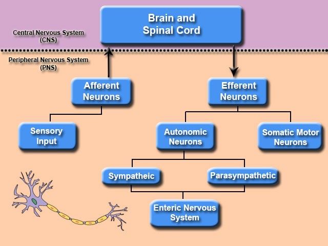
- Subdivisions: The ANS is further subdivided into two main antagonistic branches: the Sympathetic Nervous System and the Parasympathetic Nervous System.
Somatic versus Autonomic Organization: Key Differences Summarized
| Feature | Somatic Nervous System (SNS) | Autonomic Nervous System (ANS) |
|---|---|---|
| Control | Voluntary | Involuntary (visceral) |
| Effectors | Skeletal muscles | Smooth muscle, cardiac muscle, glands |
| Consciousness | Conscious perception and control | Generally unconscious control |
| Number of Neurons | One motor neuron from CNS directly to effector | Two-neuron chain: preganglionic (CNS) and postganglionic (ganglion) to effector |
| Neurotransmitter | Acetylcholine (ACh) at neuromuscular junction | Acetylcholine (preganglionic) and Norepinephrine or Acetylcholine (postganglionic) |
| Myelination | Motor neurons are heavily myelinated (very fast) | Preganglionic are myelinated; Postganglionic are unmyelinated (slower) |
| Target Response | Excitation ONLY (muscle contraction) | Excitation OR Inhibition (depending on target organ and receptor type) |
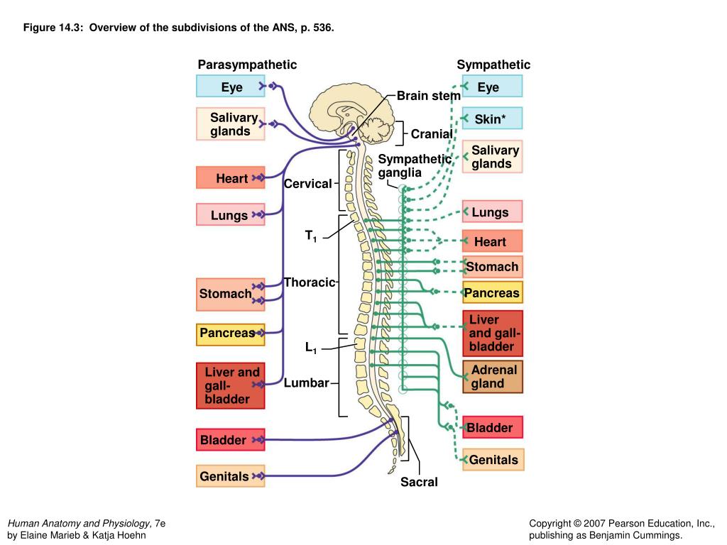
Subdivisions of the Autonomic Nervous System
The sympathetic and parasympathetic divisions typically act in opposition to each other to maintain homeostasis. Think of the Sympathetic as the accelerator and the Parasympathetic as the brake.
The Sympathetic Nervous System: "Fight or Flight"
Prepares the body for stressful situations, emergencies, or intense physical activity.
- Origin (Thoraco-lumbar Division): Preganglionic neurons originate from the lateral horns of the spinal cord gray matter in segments T1 through L2 (or L3).
- Ganglia Location:
- Paravertebral Chain Ganglia (Sympathetic Trunk): Interconnected ganglia located immediately on either side of the vertebral column. Most preganglionic fibers synapse here.
- Prevertebral (Collateral) Ganglia: Located further anteriorly, close to the abdominal aorta (e.g., celiac, superior/inferior mesenteric ganglia).
- Neurotransmitters:
- Preganglionic: Release acetylcholine (ACh) onto nicotinic receptors.
- Postganglionic: Primarily release norepinephrine (NE) at the target organ (adrenergic receptors).
- Exceptions: Postganglionic fibers to sweat glands release ACh. The adrenal medulla acts as a modified sympathetic ganglion, dumping epinephrine directly into the blood.
- Physiological Effects: Increased heart rate, massive increase in blood pressure, bronchodilation (opening airways), pupil dilation, shunting blood to skeletal muscles, complete inhibition of digestion.
The Parasympathetic Nervous System: "Rest and Digest"
Promotes body maintenance, energy conservation, and routine "housekeeping" activities.
- Origin (Cranio-sacral System): Preganglionic neurons originate from two distinct regions:
- Cranial Nerves (CN): Brainstem nuclei via CN III (eyes), CN VII (tears/saliva), CN IX (saliva), and CN X (Vagus nerve - controls heart, lungs, and most of the digestive tract).
- Sacral Spinal Cord: Segments S2, S3, S4 form pelvic splanchnic nerves to control distal colon, bladder, and reproductive organs.
- Ganglia Location: Ganglia are located very close to, or literally within the walls of, the target organs (intramural or terminal ganglia). This means preganglionic fibers are very long, and postganglionic fibers are extremely short.
- Neurotransmitters:
- Preganglionic: Release acetylcholine (ACh) onto nicotinic receptors.
- Postganglionic: Release acetylcholine (ACh) at the target organ (onto muscarinic receptors).
- Physiological Effects: Decreased heart rate, decreased blood pressure, pupillary constriction, massive increase in digestive activity, emptying of bladder and rectum.
Anatomical Organization of the Nervous System
The nervous system is anatomically divided into two major components based purely on physical location:
1. Central Nervous System (CNS)
- Composition: The CNS is composed exclusively of the brain and the spinal cord.
- Function: It is the main processing, command, and integration center of the body. It receives information from the PNS, makes decisions, and sends out commands. It is responsible for higher functions like thought, memory, emotion, and complex motor planning.
- Protection: Because it is so vital and delicate, it is heavily armored. Encased in solid bone (cranium for the brain, vertebral column for the cord), wrapped in three tough membranes (meninges), and floated in shock-absorbing cerebrospinal fluid (CSF).
2. Peripheral Nervous System (PNS)
- Composition: The PNS consists of all the neural structures located strictly outside the brain and spinal cord. This includes:
- 31 pairs of spinal nerves: Emerging from the spinal cord to innervate the trunk and limbs.
- 12 pairs of cranial nerves: Emerging directly from the brainstem to innervate the head, neck, and some visceral organs.
- Ganglia: Collections of neuron cell bodies outside the CNS.
- Plexuses: Tangled networks of nerves (e.g., brachial plexus in the shoulder, lumbar plexus in the lower back).
- Function: It serves as the physical communication cables linking the CNS to the rest of the body. It carries sensory information inward (afferent pathways) and carries motor commands outward (efferent pathways).
The Spinal Cord
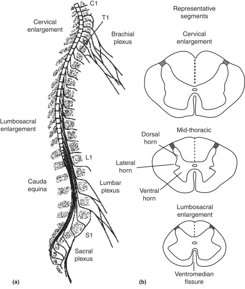
The spinal cord is a vital, primary highway of the CNS. It is an elongated, cylindrical structure extending from the foramen magnum (the hole at the base of the skull) down to roughly the level of the L1 or L2 vertebra in adults. (Note: Because bones grow faster than nerves during childhood, the spinal cord is much shorter than the vertebral column itself).
- Protection: Protected by the vertebral column, meninges (dura mater, arachnoid mater, pia mater), and CSF.
- Key Functions:
- Center for Reflex Actions: Houses neural circuits that mediate rapid, involuntary, life-saving responses to stimuli (spinal reflexes) without waiting for brain input. Example: Instantly pulling your hand off a hot stove before your brain even registers the pain.
- Pathways for Ascending Nerve Tracts: Contains bundles of axons (white matter) that transmit sensory information (touch, pain, temperature, proprioception) UP to the brain.
- Pathways for Descending Nerve Tracts: Contains bundles of axons that transmit motor commands DOWN from the brain to the muscles and glands.
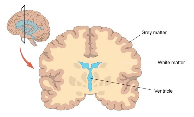
Forms and Quantity of Grey Matter
If you slice the spinal cord in half, you will see a distinct core of grey matter surrounded by white matter.
- Composition: Gray matter primarily consists of neuron cell bodies, dendrites, unmyelinated axons, and supporting glial cells.
- Shape: It has a characteristic H-shape or butterfly-shape, with projections called "horns".
- The Horns:
- Anterior (Ventral) Horns: Contain massive motor neuron cell bodies that send signals out to innervate skeletal muscles.
- Posterior (Dorsal) Horns: Receive incoming sensory input from the body via afferent fibers. They contain interneurons for processing.
- Lateral Horns: Present only in the thoracic/upper lumbar (T1-L2) and sacral (S2-S4) segments. They contain the cell bodies of preganglionic autonomic neurons (sympathetic and parasympathetic, respectively).
- Quantity: The amount of gray matter is not uniform. It bulges significantly in the cervical and lumbar regions (the cervical and lumbar enlargements). Why? Because these areas must pack in millions of extra motor neurons to control the highly complex movements of your arms and legs!
Ascending Fiber Systems (Sensory Pathways traveling UP)
| Name | Function | Origin | Ending | Location in Cord |
|---|---|---|---|---|
| Dorsal column system | Fine touch, proprioception, two-point discrimination | Skin, joints, tendons | Dorsal column nuclei. Second-order neurons project to contralateral thalamus (cross in medulla at lemniscal decussation) | Dorsal column |
| Spinothalamic tracts | Sharp pain, temperature, crude touch | Skin | Dorsal horn. Second-order neurons project to contralateral thalamus (cross in spinal cord close to level of entry) | Ventrolateral column |
| Dorsal spinocerebellar tract | Movement and position mechanisms | Muscle spindles, Golgi tendon organs, touch and pressure receptors | Cerebellar paleocortex (via ipsilateral inferior cerebellar peduncle) | Lateral column |
| Ventral spinocerebellar | Movement and position mechanisms | Muscle spindles, Golgi tendon organs, touch and pressure receptors | Cerebellar paleocortex (via contralateral and ipsilateral superior cerebellar peduncle) | Lateral column |
| Spinoreticular pathway | Deep and chronic pain | Deep somatic structures | Reticular formation of brain stem | Polysynaptic, diffuse pathway in ventrolateral column |
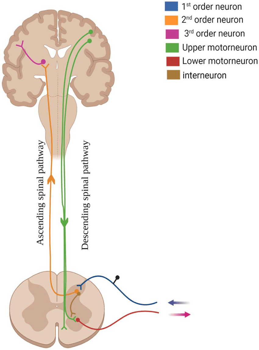
Descending Fiber Systems (Motor Pathways traveling DOWN)
| System | Function | Origin | Ending | Location in Cord |
|---|---|---|---|---|
| Lateral corticospinal (pyramidal) tract | Fine motor function (controls distal musculature like fingers), Modulation of sensory functions | Motor and premotor cortex | Anterior horn cells (interneurons and lower motor neurons) | Lateral column (crosses in medulla at pyramidal decussation) |
| Anterior corticospinal tract | Gross and postural motor function (proximal and axial musculature like the core) | Motor and premotor cortex | Anterior horn neurons | Anterior column (uncrossed until after descending, when some fibers decussate) |
| Vestibulospinal tract | Postural reflexes, keeping you upright | Lateral and medial vestibular nucleus | Anterior horn interneurons and motor neurons (for extensors) | Ventral column |
| Rubrospinal | Motor function regulation | Red nucleus (in midbrain) | Ventral horn interneurons | Lateral column |
| Reticulospinal | Modulation of sensory transmission (especially pain), Modulation of spinal reflexes | Brain stem reticular formation | Dorsal and ventral horn | Anterior column |
| Descending autonomic | Modulation of autonomic (involuntary) functions | Hypothalamus, brain stem nuclei | Preganglionic autonomic neurons | Lateral columns |
| Tectospinal | Reflex head turning (e.g., snapping your head to look at a loud noise) | Midbrain | Ventral horn interneurons | Ventral column |
| Medial longitudinal fasciculus | Coordination of head and eye movements together | Vestibular nuclei | Cervical gray | Ventral column |
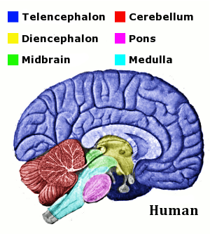
The Brain: Divisions and Topography
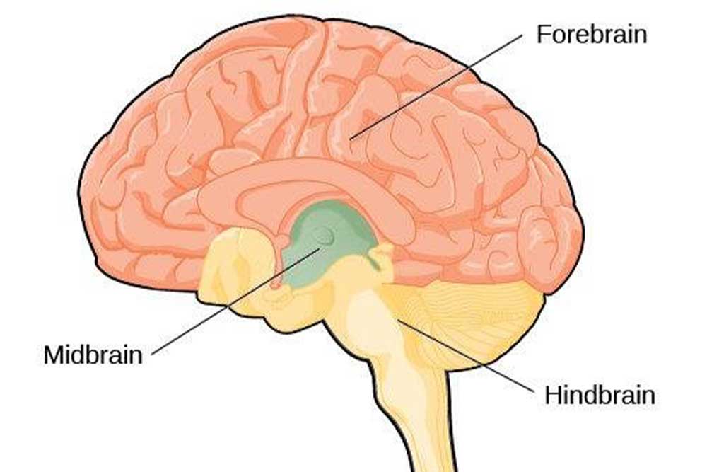
The primary divisions of the brain are crucial for understanding its organization and function. It develops from three primary embryonic vesicles: Forebrain, Midbrain, and Hindbrain.
1. The Hindbrain (Rhombencephalon)
Located at the lower back of the skull, managing vital life support and basic movement.
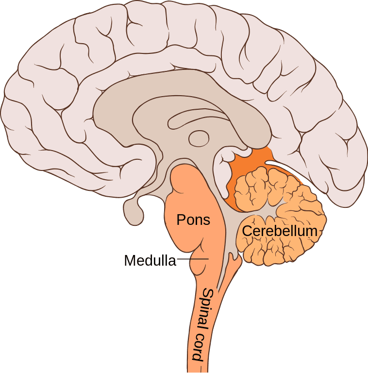
Location: Sits in the posterior cranial fossa, tucked under the occipital lobe.
Structure: Two hemispheres joined by the vermis. Surface has tightly packed folds called folia. Connects to the brainstem via three massive peduncles.
Core Functions:
- Motor Coordination: It doesn't start movement, it perfects it. It compares your intended movement with your actual movement and instantly adjusts it for smooth precision.
- Balance and Posture: Constantly receives data from the inner ear and joints.
- Motor Learning: Muscle memory (e.g., learning to ride a bike).
Disorders (Cerebellar Ataxia): Damage here causes a disastrous lack of coordination. Symptoms include Hypotonia (floppy muscles), Intention tremors (shaking only when reaching for something), Ataxic gait (staggering, drunk-like walk), Dysmetria (overshooting a target), Dysdiadochokinesia (inability to flip hands rapidly), Nystagmus (jerky eyes), and Scanning speech. Causes: Alcohol toxicity, trauma, tumors.
Location: The bulging bridge above the medulla.
Structure: Contains massive transverse fibers bridging the two sides of the cerebellum.
Core Functions:
- Relay Station: The massive bridge connecting the cerebrum down to the cerebellum.
- Respiration Control: Contains pneumotaxic and apneustic centers to smooth out breathing rhythms.
- Facial Control: Houses cranial nerve nuclei (V, VI, VII, VIII) controlling facial sensation, chewing, expressions, and eye movements.
- Sleep: Heavily involved in REM sleep regulation.
Location: The lowest stalk of the brainstem, merging into the spinal cord.
Structure: Contains the Pyramids (where major motor tracts cross over) and the Olives.
Core Functions (Life Support):
- Cardiovascular Center: Dictates heart rate and pumping force.
- Vasomotor Center: Constricts/dilates blood vessels to set blood pressure.
- Respiratory Center: Sets the absolute baseline rhythm of breathing.
- Reflexes: Vomiting, swallowing, coughing, sneezing.
- Clinical Note: Damage to the medulla is almost instantly fatal because it controls raw survival mechanics.
2. The Midbrain (Mesencephalon)
The smallest, central part of the brainstem connecting the hindbrain up to the forebrain.
- Tectum (Roof): Contains the Superior colliculi (visual tracking reflexes) and Inferior colliculi (auditory tracking reflexes).
- Tegmentum (Floor): Contains the Red Nucleus (motor control) and the highly critical Substantia Nigra (produces dopamine; its destruction directly causes Parkinson's disease).
- Cerebral Peduncles: Massive pillars of descending motor tracts running from the top of the brain down to the body.
3. The Forebrain (Prosencephalon)
The largest, most complex crown of the human brain, where all conscious thought, personality, and advanced processing occurs. Subdivided into the Telencephalon and Diencephalon.
A. Telencephalon (Cerebral Hemispheres)
Separated into left and right hemispheres by the longitudinal fissure, but constantly communicating via the massive Corpus Callosum (a bridge of 250 million axons). The outer bark is the convoluted cerebral cortex.
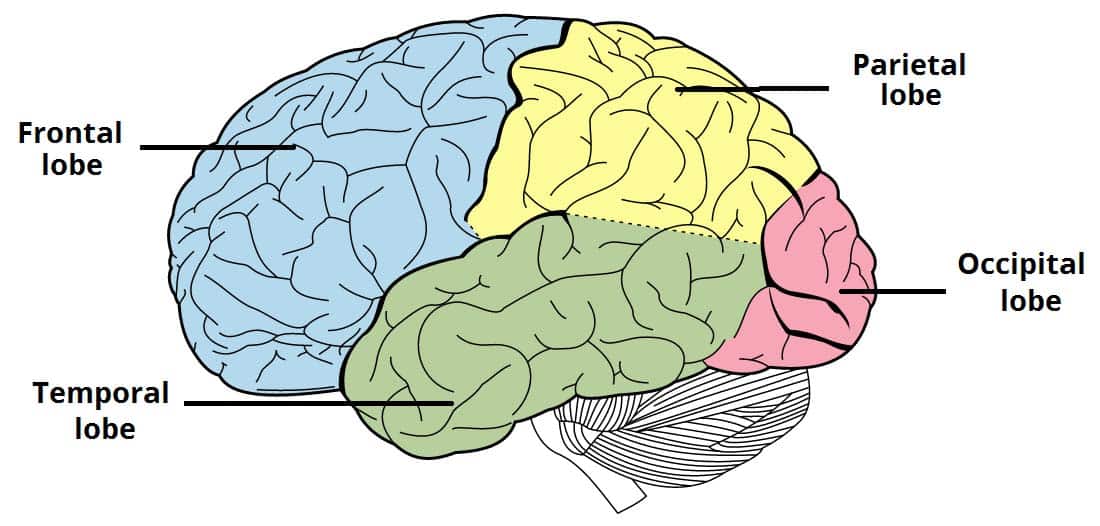
The Four Lobes of the Cortex:
- Frontal Lobe: The "Executive".
- Primary Motor Cortex: Directly commands muscles to move.
- Prefrontal Cortex: Your personality, logic, decision-making, social restraint, and working memory.
- Broca's Area: The physical production of speech.
- Parietal Lobe: The "Sensory Mapper".
- Primary Somatosensory Cortex: Feels touch, pain, temperature.
- Integrates senses to build a spatial map of where your body is in the room.
- Temporal Lobe: The "Listener & Learner".
- Primary Auditory Cortex: Processes raw sound.
- Wernicke's Area: Comprehends language (making sense of words).
- Contains the deep structures for memory (Hippocampus) and emotion (Amygdala).
- Occipital Lobe: The "Viewer".
- Primary Visual Cortex: Exclusively dedicated to processing raw visual data (color, light, motion) into recognizable images.
(Note: The Insula is a deep fifth lobe involved in taste, visceral pain, and deep bodily awareness).
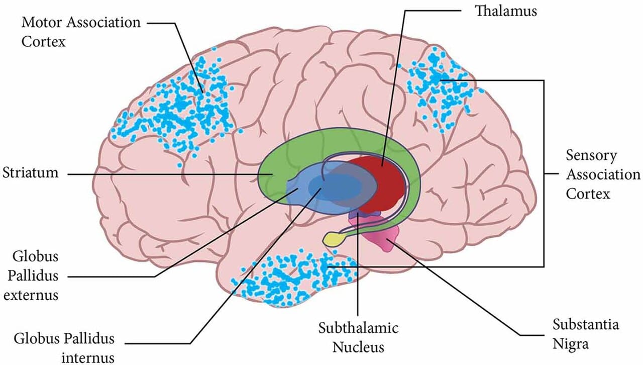
B. Basal Ganglia (Deep Telencephalon)
Deep subcortical clusters of gray matter (Caudate, Putamen, Globus Pallidus). They do not start movement; they filter and permit it.
- Function: They act as the bouncers at a club. They suppress unwanted, jerky movements and carefully select the smooth, intended movement you want to make. They also regulate habit formation.
- Disorders:
- Parkinson's Disease: Loss of dopamine breaks the basal ganglia circuit, resulting in rigidity, resting tremors, and inability to initiate movement.
- Huntington's Disease: Genetic destruction of the striatum causes the "bouncers" to fail, leading to wild, uncontrollable, flinging movements (chorea).
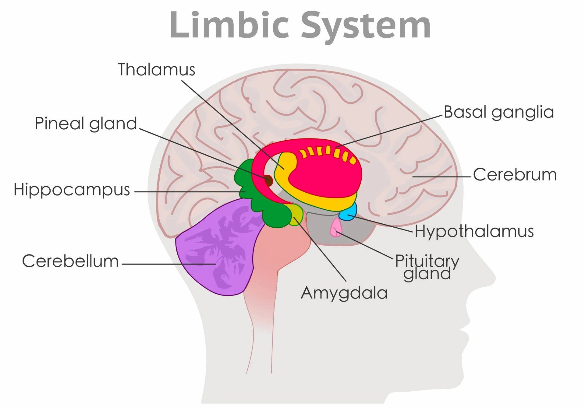
C. The Limbic System (The Emotional Brain)
Not a single organ, but a functional ring of structures on the inner edge of the cerebrum dictating our most primal drives.
- Hippocampus: The save button. Converts short-term experiences into permanent long-term memory.
- Amygdala: The alarm bell. Processes intense emotions, especially raw fear, anger, and emotional trauma.
- Cingulate Gyrus & Olfactory Bulb: Links powerful smells directly to deep emotional memories.
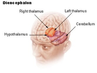
D. Diencephalon (The Deep Core)
The central hub sitting dead center in the brain, completely surrounded by the cerebral hemispheres.
Two egg-shaped masses. The ultimate Sensory Relay Station. With the sole exception of smell, every single sensory input (sight, sound, touch, pain) must pass through the thalamus. The thalamus filters the noise and decides what information is important enough to send up to the conscious cortex.
Tiny but mighty. The absolute Control Center for Homeostasis. It regulates body temperature, intense hunger/thirst, circadian rhythms (sleep/wake), and completely commands the Endocrine system by controlling the pituitary gland. It translates emotional stress into physical bodily responses.
Epithalamus: Contains the pineal gland, which secretes melatonin into the blood to enforce sleep cycles based on darkness.
Subthalamus: Works intimately with the basal ganglia to control motor function. Damage here causes violent, flinging limb movements (hemiballismus).