
Table of Contents
ToggleEndocrine Pharmacology: The Pancreas & Diabetes Mellitus Management
I. Physiological Foundation of the Pancreas
The pancreas is a unique, mixed functional gland located behind the stomach. It serves two entirely different master functions in the body:
- The Exocrine Portion: Responsible for digestion. It secretes highly active enzymes like pancrealipase (to break down fats) and chymotrypsin (to break down proteins) via ducts directly into the duodenum.
- The Endocrine Portion: Responsible for metabolic homeostasis. This portion consists of roughly 1 million Islets of Langerhans, which are highly vascularized micro-organs scattered throughout the pancreatic tissue.
| Cell Type | % of Islet | Hormone Secreted | Primary Physiological Action |
|---|---|---|---|
| A (Alpha) Cells | 20% | Glucagon / Proglucagon | Increases blood glucose (stimulates hepatic glycogenolysis). |
| B (Beta) Cells | 75% | Insulin / Pro-insulin | Decreases blood glucose (drives glucose into tissues). |
| D (Delta) Cells | 3 - 5% | Somatostatin | Inhibits the release of both insulin and glucagon. |
| F (PP) Cells | < 2% | Pancreatic Polypeptide | Regulates pancreatic exocrine and gastrointestinal secretions. |
The Extensive Physiological Effects of Insulin
Insulin is the ultimate "storage" hormone. It is released in response to high blood glucose (post-prandial state) and facilitates the transport of glucose across cell membranes into target tissues, shifting the body from a catabolic (breaking down) to an anabolic (building up) state.
The liver does not need insulin to absorb glucose, but insulin dictates what the liver does with it.
- Promotes Glycogenesis: Converts free glucose into stored glycogen.
- Inhibits Gluconeogenesis & Glycogenolysis: Stops the liver from manufacturing new glucose or breaking down stored glycogen. (Note: While some older texts may mistakenly state insulin promotes gluconeogenesis, physiologically it strictly suppresses it to prevent hyperglycemia).
Muscle requires insulin to unlock its glucose transporters (GLUT4).
- Increases massive glucose transport into the muscle cells.
- Increases amino acid transport into cells, directly stimulating protein synthesis.
- Increases Glycogenesis (muscle glycogen storage).
Insulin is highly lipogenic (fat-creating).
- Increases glucose transport into fat cells.
- Increases Triglyceride synthesis and storage.
- Strictly decreases lipolysis (fat breakdown) and decreases the release of glycerol and Free Fatty Acids (FFAs) into the blood.
II. Pathology: Diabetes Mellitus (DM)
Diabetes Mellitus is a heterogeneous group of metabolic syndromes characterized primarily by chronic hyperglycemia (elevated blood glucose). This elevation is caused by either a relative/absolute deficiency in insulin production, severe resistance to insulin's action at the receptor level, or a combination of both.
Sugar in the bloodstream acts like glass shards over time. Chronic hyperglycemia is definitively associated with long-term vascular damage, dysfunction, and failure of various vital organs. This leads to:
- Microvascular Damage: Retinopathy (blindness), Nephropathy (kidney failure), Neuropathy (nerve death and amputations).
- Macrovascular Damage: Cardiovascular disease (heart attacks), Peripheral vascular disease, and stroke.
Classic Symptoms of Diabetes
- Hyperglycemia: High blood sugar.
- Glucosuria: Sugar spilling into the urine (because the kidney's reabsorption threshold of ~180 mg/dL is exceeded).
- Polyuria: Excessive urination (glucose in urine acts as an osmotic diuretic, pulling water with it).
- Polydipsia: Excessive thirst (driven by profound dehydration from polyuria).
- Polyphagia: Excessive hunger (because cells are starving for energy despite high blood sugar, as glucose cannot enter without insulin).
The Four Clinical Classifications of Diabetes
| Feature | Type 1 Diabetes (IDDM / Juvenile) | Type 2 Diabetes (NIDDM / Maturity-onset) |
|---|---|---|
| Primary Defect | Autoimmune destruction of pancreatic beta cells. | Insulin resistance with progressive loss of beta cell function over time. |
| Insulin Levels | Absolute zero (no secretion). | Typically higher than normal initially (hyperinsulinemia), dropping as the disease progresses. |
| Insulin Resistance | No. | Yes (Receptors ignore circulating insulin). |
| Age of Onset | Typically < 30 years old. | Typically > 40 years old. |
| Nutritional Status | Undernourished / Wasting. | Typically Obese. |
| Frequency | 10 - 20% of all diabetics. | 80 - 90% of all diabetics. |
| Genetic Link | Moderate predisposition. | Strong genetic predisposition. |
| Acute Complications | Diabetic Ketoacidosis (DKA) / Wasting. | Severe Hyperglycemia (HHS). |
| Treatment | Strict Insulin Replacement. | Diet, Oral hypoglycemics, eventually Insulin. |
- Other Causes: Non-pancreatic diseases (e.g., Cushing's syndrome), genetic defects, or medication-induced (e.g., high-dose corticosteroids).
- Gestational Diabetes: Carbohydrate intolerance with onset or first recognition occurring strictly during pregnancy.
Diagnosis of Diabetes Mellitus
A diagnosis requires blood tests. It must be confirmed on a later, separate day with one of the following three methods:
- Symptomatic Criteria: Classic symptoms (thirst, polyuria, unexplained weight loss) PLUS a random plasma glucose concentration > 200 mg/dL (11.1 mmol/L).
- Fasting Plasma Glucose (FPG): Glucose > 126 mg/dL (7.0 mmol/L) after an overnight fast (strictly at least 8 hours of no caloric intake).
- Oral Glucose Tolerance Test (OGTT): Two-hour plasma glucose > 200 mg/dL (11.1 mmol/L) during a standard 75-g OGTT.
The OGTT reflects exactly how efficiently the patient's insulin can handle a massive, sudden glucose load. The procedure must be followed strictly:
- The patient undertakes a strict overnight fast.
- Basal (fasting) plasma glucose is drawn.
- The patient rapidly drinks 75 g of pure glucose dissolved in 300 ml of water over exactly 5 minutes.
- Blood is drawn every 30 minutes for 2 hours to track the spike and clearance of glucose.
- Urine is simultaneously tested for spilled sugar.
III. Pharmacotherapy: Insulin Replacement Therapy
For Type 1 Diabetics, insulin is a literal life-saver. For late-stage Type 2 Diabetics, it becomes necessary as their exhausted beta-cell mass gradually reduces to zero. Currently, human insulin is produced via recombinant DNA technology.
Insulin preparations vary primarily in two ways: their onset of activity and their duration of activity. This is manipulated by altering amino acid sequences or adding extra conjugating molecules (like protamine or zinc).
1. Types of Insulin by Duration
| Class | Generic Names | Onset | Peak Action | Total Duration |
|---|---|---|---|---|
| Rapid-Acting | Insulin Lispro, Aspart, Glulysine | 10 - 30 min | 0.5 - 2.5 hrs | 3 - 6.5 hrs |
| Short-Acting | Regular Insulin (Soluble, crystalline zinc) | 30 - 60 min | 1 - 5 hrs | 6 - 10 hrs |
| Intermediate-Acting | NPH (Isophane), Lente (Zinc susp) | 60 - 120 min | 6 - 14 hrs | 16 - 24 hrs |
| Long-Acting / Ultra-Long | Insulin Glargine, Insulin Detemir | 70 min | NONE (Flat profile) | 24+ hrs |
- Rapid-Acting: Offers highly flexible treatment regimens with a lower risk of late hypoglycemia. Because there is virtually no lag time, it is perfect for post-prandial (after meal) glucose control.
- Short-Acting (Regular): This is the exact same structure as endogenous human insulin. It is the ONLY insulin that can be injected intravenously (IV) during emergencies like Diabetic Ketoacidosis (DKA). Rapid and short-acting insulins are usually never used alone; they are paired with a long-acting background insulin.
- Intermediate (NPH): NPH stands for Neutral Protamine Hagedorn. Its duration is intermediate because conjugating insulin with the protein protamine forms a less-soluble complex, significantly delaying its absorption from the injection site.
- Long-Acting (Glargine/Detemir): These are mutated insulin analogs designed to precipitate in the tissue and slowly dissolve over 24 hours. Because they do not peak, they are perfect for mimicking continuous, natural basal insulin secretion.
- Combinations: To reduce needle pricks, premixed formulations exist (e.g., 70% NPH / 30% Regular, or 50/50 mixes).
Question: A Type 1 diabetic is prescribed two forms of insulin: an ultra-long acting form (once a day) and a fast-acting formulation (just before a meal). Which options represent this regimen?
Answer: Insulin Glargine (Ultra-long basal) and Insulin Lispro (Fast-acting bolus). This mimics natural pancreatic function perfectly.
2. Pharmacokinetics & Administration
- Route: Generally administered by Subcutaneous (SC) injection. In a hyperglycemic emergency (DKA), Regular insulin is given IV or IM.
- Inactivation: Insulin is heavily degraded by the Insulin Degrading Enzyme (Insulin Protease), which is located primarily in the liver and the kidneys.
- Clinical Rule: Diabetics with renal insufficiency (kidney failure) clear insulin much slower, so they strictly require a downward adjustment of their insulin dose to prevent fatal overdoses.
3. Adverse Effects of Insulin Therapy
- Weight Gain: Very common, especially during intensive (more frequent) insulin therapy. Insulin drives glucose into fat cells and builds triglycerides.
- Lipohypertrophy: Hypertrophy (enlargement) of subcutaneous fatty tissue occurs if insulin is injected repeatedly into the exact same site. Patients must rotate injection sites!
- Allergic Reactions: Local injection site reactions or systemic allergies (rarer now with recombinant human insulin).
- Hypoglycemia: The absolute most serious, fatal, and common adverse reaction to an overdose.
Every diabetic on insulin MUST know these symptoms. They progress in phases:
- Non-Specific: Nausea, profound tiredness, severe headache.
- Autonomic (Epinephrine Response): The body panics and dumps adrenaline to force sugar out of the liver. Symptoms: Sweating, Trembling, Pounding heart (tachycardia), intense Hunger, and severe Anxiety.
- Neuroglycopenic (Brain Starvation): The brain is running out of fuel. Symptoms: Confusion, Drowsiness, Speech difficulty, Inability to concentrate, In-coordination, progressing to coma and death.
IV. Oral Hypoglycemic Agents (For Type 2 DM)
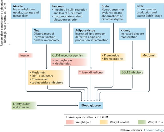
Oral agents are strictly for Type 2 Diabetes. They are absolute contraindications in Type 1 Diabetes because most require at least some functioning beta cells to work. Over time, as a Type 2 patient's beta cells naturally die off due to aging and disease progression, oral agents may fail, and insulin must be added.
1. Insulin Secretagogues (The "Squeezers")
These drugs force the pancreas to squeeze out more insulin.
A. The Sulfonylureas
- First Generation: Acetohexamide (active metabolite, requires dose reduction in renal dysfunction), Tolbutamide (very short half-life of 6-12 hours, making it the safest sulfonylurea for elderly diabetics), Chlorpropamide (long-acting, notorious for SIADH and disulfiram-like reactions with alcohol).
- Second Generation: Glipizide (requires dose reduction in hepatic dysfunction), Glyburide (active metabolite, reduce in renal dysfunction). Note: Second generation agents are vastly more potent than first generation. Glyburide has minimal transfer across the placenta, making it a reasonably safe alternative for pregnancy.
Normal Physiology: Glucose enters the beta cell → Generates ATP → ATP closes ATP-sensitive Potassium (K+) channels → K+ builds up, causing Membrane Depolarization → Depolarization opens Voltage-Gated Calcium (Ca2+) channels → Ca2+ influx triggers the exocytosis of insulin granules.
Mechanism of Sulfonylureas: They completely bypass the glucose step. They directly bind to and block the ATP-sensitive K+ channels on the beta cell. This forces immediate depolarization and massive insulin release, regardless of whether blood sugar is actually high. They also secondarily increase peripheral insulin sensitivity, reduce hepatic glucose production, and decrease glucagon release.
- Contraindications: Type 1 DM, pregnancy, lactation, significant hepatic/renal insufficiency.
- Adverse Effects: Weight gain, hyperinsulinemia, severe hypoglycemia (because they force insulin out blindly), and sulfur-based allergies. Note: As beta cells die during disease progression, sulfonylureas become completely ineffective.
B. Meglitinide Analogs (Glinides)
Examples: Repaglinide, Nateglinide.
- Mechanism: They are not sulfonylureas, but they have the exact same MOA (blocking ATP-sensitive K+ channels). However, they interact with a completely different region of the SUR1 subunit on the channel.
- Differences from Sulfonylureas: They have a very rapid onset and a much shorter duration of action. They are taken orally just 1 to 30 minutes before meals. They are also much more expensive.
- Benefit: Because they contain no sulfur in their structure, Repaglinide is the perfect alternative for Type 2 diabetics who have a severe sulfa allergy.
- Clinical Rules: Used as monotherapy or combined with metformin/glitazones. They must never be combined with sulfonylureas due to overlapping mechanisms. Adverse effects include hypoglycemia and weight gain (though slightly lower than sulfonylureas).
2. Insulin Sensitizers (Euglycemics)
These drugs do not stimulate insulin release. Therefore, when used alone, they generally do not cause hypoglycemia (they are "euglycemic"). They make the tissues listen to the insulin that is already there.
A. Biguanides (Metformin)
Metformin is the absolute 1st Line therapy for Type 2 Diabetes.
- Mechanism of Action: Its main action is a profound decrease in hepatic gluconeogenesis (it stops the liver from dumping new sugar into the blood). It also slows intestinal absorption of sugars and dramatically improves peripheral glucose uptake. It does this by stimulating a liver enzyme called AMPK, massively increasing tissue sensitivity to insulin. It does not require functioning beta cells.
- Benefits: Administered orally. It famously causes Weight Loss (due to loss of appetite/GI effects), making it perfect for obese diabetics.
- Adverse Effects: Acute side effects are aggressively gastrointestinal (anorexia, nausea, vomiting, abdominal cramping, metallic taste, severe diarrhea).
- Lactic Acidosis Risk: Rarely, it can cause a potentially fatal lactic acidosis. Because of this, it is strictly contraindicated in patients with renal disease, alcoholism, hepatic disease, or conditions predisposing them to tissue hypoxia (e.g., chronic cardiopulmonary dysfunction).
- Clinical Rule: Metformin MUST be temporarily discontinued in patients undergoing imaging diagnostics that require intravenous radiographic contrast agents (to prevent acute renal failure and subsequent lactic acidosis).
B. Thiazolidinediones (TZDs / Glitazones)
Examples: Pioglitazone, Rosiglitazone. Recommended as a second-line alternative for patients who fail or have contraindications to metformin.
- Mechanism of Action: TZDs are powerful ligands (activators) of PPAR-gamma (Peroxisome Proliferator-Activated Receptor-gamma). PPAR-g is a nuclear transcription factor. When activated, it enters the cell nucleus and literally alters the expression of multiple genes involved in lipid and glucose metabolism.
- Physiological Result: It increases GLUT4 transporters, Adiponectin, and genes involved in FFA oxidation, while down-regulating insulin-resistance cytokines like TNF-alpha and Resistin. This results in a massive increase in insulin receptor numbers and sensitivity in adipose tissue, liver, and skeletal muscle. (It is much more effective in muscle and fat than metformin).
- Delayed Efficacy: Because it relies on altering gene transcription and synthesizing new proteins, it has a very slow onset. It takes 6 to 14 weeks to achieve its maximum effect (dropping HbA1c by 0.5-1.4%).
- Severe Adverse Effects & Contraindications:
- Massive Weight Gain & Fluid Retention: It aggressively increases subcutaneous fat deposition and causes severe edema.
- Heart Failure: Because of the fluid retention, it can trigger or worsen congestive heart failure.
- Cardiac Ischemia: Rosiglitazone is associated with a highly dangerous 43% increased risk of Myocardial Infarction (MI). (This is not seen with Pioglitazone).
- Bone Fractures: Increased risk in women.
- Liver Toxicity: Rare, but liver enzyme levels (LFTs) must be measured initially and periodically.
3. Alpha-Glucosidase Inhibitors
Examples: Acarbose, Miglitol.
- Mechanism of Action: These drugs reversibly inhibit membrane-bound alpha-glucosidase enzymes located on the intestinal brush border. This enzyme normally chops up complex carbs. By inhibiting it, the hydrolysis of oligosaccharides into glucose is delayed, resulting in significantly lower postprandial (after-meal) glucose spikes. Acarbose also inhibits pancreatic amylase.
- Administration: Must be taken orally exactly at the beginning of meals.
- Safety & Efficacy: They have a relatively weak antidiabetic effect. Because they do not stimulate insulin or increase sensitivity, they do not cause hypoglycemia as monotherapy.
- Severe GI Adverse Effects: Because undigested carbohydrates pass into the colon, bacteria ferment them. This causes massive flatulence, severe diarrhea, and abdominal cramping.
- Contraindications: Absolutely contraindicated in patients with Inflammatory Bowel Disease (IBD), colonic ulceration, or intestinal obstruction.
- Clinical Rule: If a patient taking Acarbose (in combination with a sulfonylurea) experiences hypoglycemia, they MUST be treated with pure glucose (dextrose). Giving them table sugar (sucrose) will fail because the drug prevents sucrose from being broken down and absorbed!
V. Advanced Therapies: Incretins & SGLT-2 Inhibitors
1. Incretin Mimetics (GLP-1 Receptor Analogs)
Examples: Exenatide, Liraglutide, Dulaglutide.
Incretins (GLP-1 and GIP) are gut-derived hormones released naturally when we eat. They prepare the body for the incoming sugar.
- Mechanism of Action: These injected drugs mimic GLP-1. They:
- Potentiate glucose-induced insulin secretion: They only stimulate the pancreas when blood glucose is high. As glucose falls, the drug's effect diminishes (zero hypoglycemia risk alone).
- Suppress Glucagon: Stops the liver from releasing extra sugar.
- Slow Gastric Emptying: Prevents rapid sugar absorption.
- Promote Satiety: Send signals to the brain that you are full, leading to significant Weight Loss.
- Beta Cell Mass: They actively act to maintain and regenerate beta cell mass while decreasing beta cell apoptosis (death).
2. DPP-4 Inhibitors (The "Gliptins")
Examples: Sitagliptin, Saxagliptin, Linagliptin.
- Mechanism of Action: Natural GLP-1 is destroyed in the blood in under 2 minutes by an enzyme called Dipeptidyl Peptidase-4 (DPP-4). The "gliptin" drugs inhibit the DPP-4 enzyme, preventing the degradation of the patient's endogenous incretins, allowing them to stay in the blood longer to stimulate insulin and lower glucagon.
3. SGLT-2 Inhibitors (The "Gliflozins")
Examples: Canagliflozin, Empagliflozin.
- Mechanism of Action: They block the Sodium-Glucose Co-Transporter-2 (SGLT-2) receptors located in the proximal convoluted tubules of the kidneys. Normally, these receptors reabsorb 100% of filtered glucose back into the blood. By blocking them, the patient literally pees out all their excess sugar.
- Adverse Effects:
- Glucosuria: Massive sugar in the urine.
- UTIs and Yeast Infections: Bacteria and fungi thrive on the sugar-rich urine.
- Dehydration & Hypotension: The glucose acts as an osmotic diuretic, pulling water out of the body.
VI. Emergency Management: Diabetic Ketoacidosis (DKA)
DKA is a life-threatening acute complication primarily seen in Type 1 Diabetics. With zero insulin, the cells starve and the body aggressively breaks down fats into toxic, acidic ketones, leading to severe metabolic acidosis and profound dehydration.
- IV Fluids: Massive volume rehydration is the absolute first step to restore blood volume and renal perfusion.
- IV Insulin: Regular, short-acting insulin given intravenously to force glucose back into cells and instantly stop the production of ketones (halts lipolysis).
- Potassium Supplementation: Insulin forces Potassium back into the cells. If K+ is not supplemented, the patient will develop fatal hypokalemia leading to cardiac arrest.
- Sodium Bicarbonate: Used cautiously only in cases of incredibly severe acidosis (pH < 6.9) to neutralize the blood acid.
References & Further Reading
- Katzung, B. G. (2020). Basic & Clinical Pharmacology (15th ed.). McGraw-Hill Education. (Chapters on Pancreatic Hormones and Antidiabetic Drugs).
- Brunton, L. L., et al. (2017). Goodman and Gilman's The Pharmacological Basis of Therapeutics (13th ed.). McGraw-Hill. (In-depth mechanisms of SUR1 receptors and AMPK activation).
- American Diabetes Association (ADA). (2023). Standards of Medical Care in Diabetes. (Diagnostic criteria, OGTT standards, and DKA management protocols).
- Costanzo, L. S. (2018). Physiology (6th ed.). Elsevier. (In-depth physiology of the Islets of Langerhans and insulin's intracellular metabolic pathways).
Quick Quiz
Endocrine Pharmacology and DM Quiz
Pharmacology - mobile-friendly and focused practice.
Privacy: Your details are used only for quiz tracking and certificates.
Endocrine Pharmacology and DM Quiz
Pharmacology
Preparing questions...
Choose your answer and keep your streak alive.
Great effort.
Here is your quick performance summary.