
Table of Contents
ToggleIntestinal Obstruction Lecture Notes
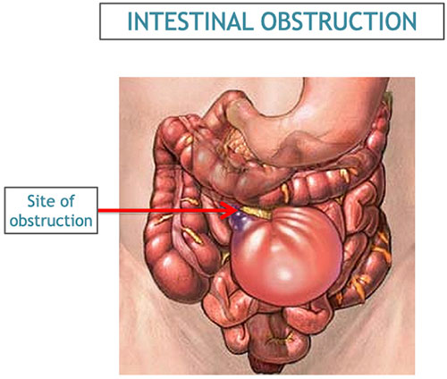
Intestinal obstruction is a potentially life-threatening interruption of the forward movement of intestinal contents. It may be mechanical, caused by a physical blockage, or functional, caused by failure of intestinal movement. Nurses should recognise dehydration, strangulation, bowel ischaemia, perforation and sepsis early, begin urgent escalation, and support assessment and treatment led by the surgical team.
Emergency warning: severe or worsening abdominal pain, repeated vomiting, progressive distension, inability to pass stool or flatus, fever, fast pulse, guarding, confusion or faintness may signal a surgical emergency. Arrange urgent assessment and referral according to local protocol. Do not delay escalation while waiting for a scan or for all classic signs to appear.
Quick Overview
| Feature | Key points |
|---|---|
| Meaning | Obstruction prevents or seriously impairs passage of intestinal contents through part of the small or large bowel. |
| Common mechanical causes | Adhesions after abdominal surgery, an incarcerated hernia, tumour, volvulus, intussusception, stricture, impacted stool or a foreign body. |
| Common functional causes | Post-operative or illness-related ileus, electrolyte disturbance, medicines that slow bowel movement, and acute colonic pseudo-obstruction. |
| Typical features | Colicky abdominal pain, nausea or vomiting, distension and reduced passage of stool or flatus. The pattern varies with level, cause and completeness. |
| First priorities | Rapid assessment, urgent surgical review, nil by mouth as directed, IV access and prescribed fluid/electrolyte correction, symptom control, close monitoring and investigations. |
| Major complications | Dehydration, electrolyte imbalance, aspiration, bowel strangulation or ischaemia, necrosis, perforation, peritonitis, sepsis, shock and death. |
Learning Objectives
By the end of this lesson, a nursing student should be able to:
- Define intestinal obstruction and distinguish mechanical obstruction from ileus and pseudo-obstruction.
- Classify obstruction by site, completeness, mechanism and whether blood supply is threatened.
- Explain common causes, risk factors, pathophysiology and clinical manifestations.
- Describe focused assessment, investigations and important differential diagnoses.
- Outline emergency, non-operative and operative management at a nursing-student level.
- Plan safe pre-operative, post-operative and ongoing nursing care and identify complications early.
Definition and Important Terms
Intestinal obstruction is a partial or complete blockage of the passage of food, fluid, digestive secretions and gas through the bowel. It can affect the small intestine, large intestine or both. Obstruction is a clinical syndrome: the underlying cause and the patient’s physiological condition determine the treatment.
| Term | Meaning | Why it matters |
|---|---|---|
| Mechanical obstruction | A physical lesion narrows, compresses or blocks the lumen, or twists/kinks the bowel. | The cause may need reduction, endoscopic treatment or surgery; bowel blood flow can become compromised. |
| Functional obstruction / ileus | Propulsive bowel movement is markedly reduced or absent without a fixed physical blockage. | Often occurs after surgery or severe illness; treatment focuses on the cause and supportive care unless another problem is found. |
| Partial obstruction | Some intestinal contents can still pass the narrowed segment. | Stool or flatus may continue early; this does not exclude obstruction. |
| Complete obstruction | Little or no content passes beyond the blockage. | Obstipation and worsening distension are common; urgent surgical assessment is required. |
| Closed-loop obstruction | A segment is blocked at two points, trapping bowel and its contents. | Pressure and impaired blood supply can progress quickly; it is a high-risk surgical emergency. |
| Strangulation | The blood supply to obstructed bowel is compromised. | May progress to ischaemia, necrosis, perforation and sepsis; urgent intervention may be needed. |
| Acute colonic pseudo-obstruction | Marked colonic dilatation without a mechanical blockage, often in a seriously ill or post-operative patient. | It resembles large-bowel obstruction and requires clinician-led evaluation because severe dilatation can perforate. |
Relevant Anatomy and Normal Bowel Function
The small intestine consists of the duodenum, jejunum and ileum. It receives digestive secretions and absorbs most nutrients and much of the fluid entering the gut. The large intestine includes the caecum, colon, rectum and anal canal; it absorbs water and stores faeces before defecation. Coordinated smooth-muscle contractions called peristalsis move bowel contents forward. The bowel wall also needs an intact blood supply and adequate fluid and electrolytes to function.
A blockage causes fluid, swallowed air and gas produced by bacteria to collect above the obstruction. The bowel dilates and its movement may initially increase as it tries to overcome the blockage. Ongoing distension can impair venous return and then arterial supply. Vomiting, reduced intake and fluid trapped inside bowel loops lead to further volume depletion. The site and duration of obstruction influence how rapidly the patient becomes unwell.
Classification of Intestinal Obstruction
| Basis | Types | Examples or notes |
|---|---|---|
| Mechanism | Mechanical; functional | Adhesion versus ileus. |
| Location | Small bowel; large bowel | Small-bowel obstruction often causes earlier vomiting; large-bowel obstruction may cause more visible distension. |
| Severity | Partial; complete | Passage of a little stool or flatus does not reliably rule out partial obstruction. |
| Blood supply | Simple; strangulated / ischaemic | Strangulation is suspected with continuous severe pain, peritonism, systemic deterioration or concerning imaging. |
| Time course | Acute; subacute; chronic or recurrent | Slowly progressive symptoms may occur with tumour or stricture; acute obstruction may follow a hernia or volvulus. |
Causes and Risk Factors
Mechanical causes
| Cause | How it obstructs the bowel | Clinical clues |
|---|---|---|
| Adhesions | Fibrous bands, commonly after abdominal or pelvic surgery, kink or tether the bowel. | A frequent cause of small-bowel obstruction in adults with prior surgery; obstruction can occur long after the operation. |
| Hernia | Bowel passes through a weak point and becomes trapped or compressed. | Examine groins, umbilicus, previous incision sites and other hernia sites. A painful, irreducible hernia with vomiting or systemic illness needs urgent surgical review. |
| Tumour or cancer | A mass narrows or blocks the lumen or compresses bowel from outside. | May present gradually with altered bowel habit, weight loss, anaemia or progressive distension, but acute presentation is possible. |
| Volvulus | A loop of bowel twists around its mesentery, obstructing the lumen and potentially its blood supply. | Often affects the sigmoid colon or caecum; abdominal distension and pain may be prominent. |
| Intussusception | One segment of bowel telescopes into an adjacent segment. | Important in children; intermittent pain and vomiting can occur. In adults, a structural lead point is more likely and needs assessment. |
| Stricture | Scarring or chronic inflammation narrows the bowel. | Possible causes include inflammatory bowel disease, previous surgery or radiotherapy, and selected infections such as intestinal tuberculosis. |
| Impacted stool / faecaloma | Hard stool obstructs the rectum or colon. | Consider in older, immobile, dehydrated or constipated patients and those taking constipating medicines. |
| Other causes | Gallstone ileus, foreign body, bezoar, congenital band, malrotation or compression from a neighbouring mass. | History, age, examination and imaging help identify the cause. |
Functional causes
- Post-operative ileus: temporary reduction in bowel motility after abdominal surgery.
- Severe illness: sepsis, major trauma, burns, severe infection or metabolic illness may suppress intestinal movement.
- Electrolyte disturbance: low potassium and other major metabolic abnormalities can contribute to ileus.
- Medicines: opioids, anticholinergic medicines and some other drugs may slow bowel activity.
- Acute colonic pseudo-obstruction: acute colonic dilatation without a mechanical lesion, often associated with severe illness, surgery or immobility.
Risk factors include previous abdominal surgery, known hernia, abdominal or pelvic tumour, inflammatory bowel disease, previous bowel obstruction, chronic constipation, reduced mobility, recent operation, severe infection, dehydration and medicines that reduce gut motility. Obstruction can also occur in someone with no known risk factor.
Pathophysiology
- Contents stop moving forward. Bowel fluid, swallowed air and gas accumulate proximal to the obstruction.
- The bowel distends. Increased pressure stretches the wall and produces colicky pain and visible or measurable distension.
- Fluid shifts into the bowel and tissues. Vomiting, poor intake and sequestration of fluid within the bowel reduce circulating volume and may cause dehydration, renal impairment and shock.
- Electrolytes and acid-base balance change. Ongoing vomiting and fluid loss can disturb sodium, potassium, chloride and bicarbonate; the pattern varies with level, duration and treatment.
- Venous drainage becomes impaired. Rising pressure can cause bowel-wall oedema and congestion, which further increases distension.
- Arterial perfusion may fall. A closed loop, tight band, twisted bowel or trapped hernia can compromise blood supply and cause ischaemia and necrosis.
- Perforation and infection may follow. A damaged bowel wall can leak bacteria and intestinal contents into the peritoneum, resulting in peritonitis, sepsis and shock.
Key point: a patient may initially have intermittent pain and active bowel sounds, then develop constant pain, tenderness, fever, tachycardia, guarding or reduced bowel sounds as the condition worsens. The trend and the whole clinical picture matter more than one sign.
Clinical Manifestations
Common symptoms and signs
- Abdominal pain: often cramping or colicky as the bowel contracts against a blockage. Sudden severe pain or pain that becomes constant may indicate ischaemia or another complication.
- Nausea and vomiting: may be early and frequent in a proximal small-bowel obstruction. Vomiting may become bilious or, in prolonged distal obstruction, foul or faeculent; appearance alone cannot establish the site.
- Abdominal distension: may be slight in a high obstruction and more marked in a distal small-bowel or large-bowel obstruction.
- Constipation or obstipation: reduced stool and flatus, especially in complete obstruction. Passage of stool or flatus early in the illness does not exclude obstruction.
- Altered bowel sounds: may be high-pitched or frequent early, then diminish with exhaustion, ileus or advanced disease. Bowel sounds are not reliable enough to rule obstruction in or out on their own.
- Dehydration: thirst, dry mucous membranes, reduced urine, dizziness, delayed capillary refill, tachycardia or hypotension.
- Systemic features: fever, fast pulse, weakness, confusion or shock may indicate dehydration, infection, ischaemia or perforation.
Patterns by site and type
| Pattern | Typical tendency | Important limitation |
|---|---|---|
| High small-bowel obstruction | Earlier and more frequent vomiting; less abdominal distension may be seen initially. | Presentation varies and can progress rapidly because of fluid losses. |
| Distal small-bowel obstruction | Greater distension may occur; vomiting can develop later. | Symptoms overlap with large-bowel obstruction. |
| Large-bowel obstruction | Progressive distension and constipation may predominate; vomiting can occur later. | Cause, site and competence of the ileocaecal valve affect the presentation. |
| Partial obstruction | Intermittent pain and some stool or flatus may continue. | Partial does not mean harmless; worsening pain or systemic signs require urgent reassessment. |
| Strangulation / ischaemia | Persistent severe pain, local or generalised tenderness, fever, tachycardia, guarding or deterioration. | Early signs can be subtle; absence of fever or a normal early lactate does not safely exclude it. |
Red flags needing immediate escalation
- Sudden severe or continuous abdominal pain, pain out of proportion, guarding, rebound tenderness or a rigid abdomen.
- Fever, persistent tachycardia, hypotension, confusion, fainting, cold clammy skin or reduced urine output.
- Repeated or bilious vomiting, aspiration risk, rapidly increasing distension or inability to pass stool and flatus.
- A painful, tender, irreducible hernia or a tense abdominal mass.
- Blood in stool or vomit, metabolic acidosis, rising lactate or imaging suggesting a closed loop, ischaemia, free air or perforation.
Assessment and Diagnosis
Diagnosis combines the history, repeated clinical assessment, laboratory results and imaging. A single symptom, physical sign or normal laboratory result cannot safely rule out obstruction or threatened bowel.
Focused history
- Onset, site, character, severity and progression of pain; whether it is colicky or now constant.
- Vomiting: onset, frequency, amount, colour, ability to keep fluids down and any blood or faeculent appearance.
- Last stool and flatus, usual bowel pattern, diarrhoea or constipation, and any blood in stool.
- Abdominal swelling, previous similar episodes and what has changed.
- Previous abdominal or pelvic surgery, radiotherapy, known hernia, bowel disease, cancer or prior obstruction.
- Current medicines, especially opioids or anticholinergic drugs; allergies; oral intake; urine output and fluid losses.
- Weight loss, appetite change, fever, chronic anaemia symptoms, trauma and relevant family or medical history.
- For children: age-appropriate feeding and stool history, episodes of drawing up the legs or unexplained lethargy, and features of dehydration.
- For patients who could be pregnant: menstrual history and pregnancy possibility, asked sensitively and privately.
Physical assessment
- Start with ABCDE and vital signs. Assess airway, breathing, circulation, disability and exposure; record temperature, pulse, blood pressure, respiratory rate, oxygen saturation, mental status and pain score. Escalate instability immediately.
- Assess hydration and perfusion. Look for dry mouth, thirst, capillary refill, cool peripheries, postural symptoms where safe, reduced urine and altered consciousness.
- Inspect the abdomen. Note distension, scars, visible peristalsis, bruising, stomas, masses and abdominal movement with breathing.
- Listen and palpate gently. Record bowel sounds if trained, then assess tenderness, guarding, rebound or rigidity. Avoid repeated forceful palpation when severe pain or peritonism is present.
- Check for hernias. Inspect and palpate appropriate hernia sites with consent, privacy and a chaperone where appropriate. Do not attempt forceful reduction; urgently escalate a painful or irreducible hernia.
- Perform other examinations only when indicated and within scope. Rectal or pelvic examination requires a clinical indication, explanation, consent, privacy and a qualified clinician.
- Reassess and document trends. Repeat vital signs, abdominal findings, pain, vomiting, urine output and fluid balance at the frequency directed by acuity and local protocol.
Investigations
| Investigation | What it may show | Nursing relevance |
|---|---|---|
| Full blood count | Leukocytosis may accompany inflammation or infection; haemoglobin may identify anaemia or bleeding. | Interpret with the clinical picture; an early normal count does not rule out a serious obstruction. |
| Urea, creatinine and electrolytes | Renal impairment, dehydration and sodium, potassium or chloride disturbance. | Support prescribed fluid and electrolyte replacement; monitor urine output and repeat tests as ordered. |
| Blood gas and lactate when indicated | Acidosis or raised lactate may suggest poor perfusion, sepsis or ischaemia. | A normal early result does not exclude bowel compromise; follow trends and escalate concern. |
| Inflammatory markers, glucose and other tests | May help assess infection, metabolic disturbance or comorbidity. | Test selection depends on severity and local protocol. |
| Group and screen / crossmatch | Prepares for transfusion if bleeding or surgery is anticipated. | Collect and label specimens correctly when ordered. |
| Urinalysis and pregnancy test when appropriate | May identify urinary causes or pregnancy relevant to imaging and differential diagnosis. | Explain tests and respect privacy and consent. |
| Abdominal radiograph | May show dilated bowel loops, air-fluid levels or a pattern suggesting obstruction; chest imaging may help identify free air. | Plain films have limitations. A normal or unclear film does not reliably exclude obstruction or ischaemia. |
| CT abdomen and pelvis | Can identify level and likely cause, a transition point, closed-loop features, free fluid, perforation or signs of compromised bowel. | Prepare the patient as instructed; check relevant contrast, allergy, renal and pregnancy considerations according to local policy. Do not delay emergency referral for CT. |
| Ultrasound | Useful in selected situations, including assessment for intussusception in children and some pelvic or pregnancy-related differentials. | Choice depends on the patient, question, equipment and expertise available. |
| Water-soluble contrast study | May help selected surgical teams assess adhesive small-bowel obstruction and likelihood of resolution. | Only use under the treating team’s protocol; assess aspiration risk and follow instructions. |
Where available and appropriate, CT is often the most informative imaging study for an adult with suspected small-bowel obstruction and an uncertain cause or possible complication. Imaging choice varies with age, pregnancy, stability, resources and the clinical question. Resuscitation and urgent surgical review take priority when the patient is unstable or peritonitic.
Differential Diagnoses
| Condition | Clues that may help distinguish it |
|---|---|
| Ileus | Reduced bowel motility without a fixed blockage; often post-operative, medicine-related or associated with severe illness or electrolyte disturbance. |
| Acute gastroenteritis | Vomiting or diarrhoea may be prominent; marked obstipation or a transition point suggests another cause, but clinical assessment is needed. |
| Severe constipation / faecal impaction | Hard stool, chronic constipation or rectal loading may be present; must be distinguished from a more proximal obstruction. |
| Appendicitis or other acute inflammatory abdomen | Localised pain, fever or peritoneal signs may occur; symptoms overlap and imaging or surgical assessment may be needed. |
| Mesenteric ischaemia | Severe pain, vascular risk factors or pain out of proportion may occur; this is another time-critical emergency. |
| Pancreatitis, biliary disease or perforated ulcer | Upper abdominal pain, relevant history and laboratory or imaging findings may point to another acute abdomen. |
| Urinary or gynaecological conditions | Urinary symptoms, pregnancy, pelvic pain or menstrual history may guide evaluation; these do not exclude bowel disease. |
Management of Intestinal Obstruction
Management depends on the cause, site, completeness, bowel viability, patient stability and available resources. It is led by the surgical and multidisciplinary team. Supportive treatment and monitoring begin while the cause is evaluated.
Immediate priorities
- Recognise and escalate. Arrange urgent surgical review or transfer to an appropriate facility. Activate emergency response for shock, peritonitis, suspected strangulation, perforation or rapid deterioration.
- Stabilise airway, breathing and circulation. Give oxygen only when indicated and prescribed or required by emergency protocol. Establish IV access and obtain urgent tests as ordered.
- Keep the patient nil by mouth when directed. Explain the reason, provide mouth care and document intake. Do not give food, drink, laxatives, enemas or oral medicines unless the treating team specifically directs it.
- Replace fluid and electrolytes as prescribed. IV isotonic fluid, electrolyte correction and blood products may be needed. The type and rate depend on clinical assessment, comorbidities, urine output and local orders.
- Relieve symptoms and reduce aspiration risk. Give prescribed analgesia and antiemetics. A nasogastric tube may be ordered for persistent vomiting or significant distension; confirm placement and maintain it according to policy.
- Monitor closely. Record vital signs, pain, abdominal findings, vomiting, NG output, intake and output, urine, fluid balance, laboratory trends and response to treatment.
- Give antibiotics when indicated and prescribed. They may be needed when ischaemia, perforation, peritonitis or sepsis is suspected, or around surgery. They are not a substitute for source control.
Non-operative treatment
A closely monitored non-operative approach may be considered by a surgical team for selected patients, especially some cases of adhesive small-bowel obstruction, when there is no peritonitis, strangulation or bowel ischaemia. It may include bowel rest, IV fluid and electrolyte management, decompression when ordered, serial assessment and selected contrast protocols. The patient must be reassessed regularly and the plan changed promptly if pain, vital signs, examination, laboratory results or imaging worsen.
Some guidance describes a time-limited trial, sometimes up to about 72 hours, for carefully selected stable patients with adhesive obstruction. This is a team-led ceiling used in context, not a bedside instruction to wait: deterioration or evidence of bowel compromise requires earlier escalation. Local surgical policy and the patient’s condition determine timing.
When surgery or another urgent procedure may be needed
- Peritonitis, suspected strangulation, ischaemia, necrosis, perforation, closed-loop obstruction or clinical deterioration.
- An incarcerated or strangulated hernia, volvulus or another cause requiring urgent correction.
- Complete obstruction, or failure of an appropriate monitored non-operative plan to resolve the obstruction.
- A tumour or other structural lesion requiring resection, bypass, stenting or another specialist plan.
Depending on the cause and bowel viability, procedures may include adhesiolysis, hernia repair, reduction of intussusception, treatment of volvulus, removal of a tumour or foreign body, bowel resection with reconnection, or formation of a stoma. The approach may be laparoscopic, open or endoscopic in selected situations. If bowel is non-viable, resection may be necessary. Explain that the exact procedure depends on operative findings and patient safety.
Nursing Management
Initial and ongoing nursing care
| No. | Nursing action | Rationale |
|---|---|---|
| 1 | Use ABCDE assessment, obtain and trend vital signs, pain score, mental state and perfusion; report deterioration immediately. | Shock, sepsis and bowel ischaemia can develop quickly and require urgent team action. |
| 2 | Assess pain pattern, abdominal distension and tenderness, vomiting, stool/flatus history and relevant hernia sites; document changes clearly. | Serial findings may reveal progression or complications; one assessment is not enough. |
| 3 | Maintain nil-by-mouth status when ordered; label the status, explain it, offer regular mouth care and check before giving oral medication. | Reduces aspiration and supports preparation for imaging or surgery while maintaining comfort and safety. |
| 4 | Maintain prescribed IV access and fluids; monitor intake, urine, vomit and NG drainage. Measure output and report low urine or high losses. | Vomiting and third-spacing can cause major volume depletion and renal impairment. |
| 5 | If an NG tube is ordered, support insertion by a trained clinician, confirm placement by approved policy before use, secure it, maintain ordered suction and record output/character. | Decompression may reduce vomiting and distension; incorrect placement or poor function can cause harm. |
| 6 | Administer prescribed analgesia, antiemetics, fluids, electrolytes and antibiotics; reassess response and observe for adverse effects. | Symptom relief and correction of losses support comfort and stabilisation; worsening pain still needs reassessment. |
| 7 | Prepare for investigations and possible surgery: verify identity, allergies, consent process by the responsible clinician, specimens, documentation and transfer checklist. | Prevents avoidable delays and supports safe, coordinated care. |
| 8 | Use aspiration precautions for active vomiting, position safely, keep suction available where indicated and seek urgent help if airway compromise occurs. | Vomiting and distension raise the risk of aspiration, especially with reduced consciousness. |
| 9 | Provide privacy, clear explanations, reassurance and family communication consistent with the patient’s wishes. | Acute pain and urgent surgery create fear; compassionate communication supports cooperation and dignity. |
| 10 | Maintain pressure-area care, safe mobilisation as appropriate, oral hygiene and infection-prevention measures. | Illness, immobility, tubes and procedures increase the risk of avoidable complications. |
Pre-operative and post-operative care
| Phase | Nursing care | Rationale / monitoring focus |
|---|---|---|
| Before surgery | Continue resuscitation and observations; maintain NPO status; check consent is completed by the responsible clinician; confirm allergies, identification, investigations, IV access, prescribed medicines, NG status and documentation. | Supports safe anaesthesia and timely surgery; report any deterioration before transfer. |
| After surgery: airway and circulation | Monitor airway, breathing, oxygenation, pulse, blood pressure, temperature, consciousness and pain according to recovery and ward protocols. | Identifies anaesthetic complications, bleeding, sepsis or shock early. |
| Wound, drain and stoma | Inspect dressings and wound; measure and describe drain output; assess stoma colour, moisture and output; report bleeding, dusky tissue, wound separation or sudden changes. | May reveal haemorrhage, infection, impaired perfusion or a surgical complication. |
| Fluids and renal function | Continue accurate intake/output; monitor IV fluids, urine, NG/drain losses and ordered laboratory tests. | Fluid shifts and ongoing losses can continue after surgery. |
| Return of bowel function | Monitor nausea, vomiting, abdominal distension, pain, passage of flatus or stool and tolerance of diet. Advance oral intake only as ordered. | Helps identify return of motility, persistent ileus or recurrent obstruction. |
| Mobility and prevention | Support deep breathing, coughing, early safe mobilisation, pressure-area care and thrombosis-prevention measures as prescribed. | Reduces respiratory, skin and thromboembolic complications of surgery and immobility. |
| Patient and family teaching | Explain medicines, wound/stoma care, follow-up, nutrition plan and danger signs using language the patient understands. | Promotes safe recovery and early presentation if complications occur. |
Examples of nursing problems and expected outcomes
| Nursing problem | Expected outcome | Key nursing measures |
|---|---|---|
| Deficient fluid volume related to vomiting and fluid sequestration | Perfusion and urine output improve; vital signs and prescribed fluid balance move toward the team’s targets. | Trend observations, measure all intake and losses, maintain prescribed IV therapy, monitor urine and report deterioration. |
| Acute pain related to distension or surgery | Pain is assessed regularly and reduced to a tolerable level without masking clinical deterioration. | Assess location, pattern and severity; give prescribed analgesia; reassess and escalate new continuous or worsening pain. |
| Nausea / risk of aspiration related to obstruction and vomiting | Vomiting is controlled where possible and airway remains clear. | Position safely, maintain NPO as directed, give prescribed antiemetic, manage an ordered NG tube and report aspiration signs. |
| Anxiety related to acute illness and possible surgery | Patient can describe the immediate plan and reports feeling supported. | Give concise updates, invite questions, protect privacy and involve family with permission. |
| Risk for infection related to bowel compromise or surgery | No new signs of wound infection, peritonitis or sepsis develop. | Use aseptic technique, monitor temperature and wound/drain findings, administer prescribed antibiotics and escalate changes. |
Complications
- Dehydration and hypovolaemic shock: caused by vomiting, poor intake and fluid sequestration.
- Electrolyte and acid-base disturbances: may cause weakness, confusion, arrhythmias or impaired bowel function.
- Acute kidney injury: may follow poor perfusion and ongoing fluid loss.
- Aspiration pneumonitis: can occur when vomit enters the airway.
- Strangulation, bowel ischaemia and necrosis: threatened bowel may become non-viable.
- Perforation and peritonitis: leakage of bowel contents can cause severe infection.
- Sepsis and multi-organ dysfunction: may follow infection, perforation or prolonged shock.
- Post-operative complications: bleeding, wound infection, ileus, anastomotic leak, stoma complications, adhesions and recurrent obstruction.
- Death: risk increases with delayed recognition, advanced age, comorbidity, ischaemia, perforation and shock.
Prevention, Discharge and Patient Education
Not every obstruction can be prevented. Risk reduction and discharge advice should match the confirmed cause, procedure and the patient’s individual needs.
- Attend follow-up appointments and seek help early for recurrent pain, vomiting, distension or constipation.
- After surgery, follow the surgeon’s instructions on activity, wound care, diet, medicines and any stoma care plan.
- Maintain fluids and a balanced diet as advised; do not start high-fibre foods, laxatives or bowel preparations after surgery without guidance.
- Take medicines only as prescribed. Discuss medicines that worsen constipation with a clinician; do not stop essential treatment independently.
- For patients with a hernia, seek urgent care if it becomes painful, tender, irreducible or is associated with vomiting or abdominal swelling.
- Explain danger signs clearly: persistent or worsening pain, repeated vomiting, fever, increasing abdominal swelling, inability to pass stool/flatus, faintness or reduced urine requires urgent assessment.
- Arrange a safe plan for medicines, transport, wound supplies and follow-up, especially after bowel resection or stoma formation.
Special Considerations
- Children: causes differ by age and include intussusception, congenital abnormalities and, in selected cases, adhesions. Young children may present with episodic crying, drawing up the legs, poor feeding, lethargy or dehydration rather than a clear pain history. Escalate bilious vomiting urgently.
- Older adults: symptoms may be less typical, dehydration can develop quickly, and malignancy, constipation, comorbidity and medication effects may contribute. Repeat assessment is important.
- Pregnancy: bowel obstruction is uncommon but potentially serious. Involve obstetric and surgical teams; imaging decisions should be made by clinicians based on urgency, pregnancy and local resources.
- No previous abdominal surgery: adhesions are less expected but obstruction remains possible from hernia, tumour, volvulus, inflammatory disease, congenital anatomy or other causes.
- Limited-resource settings: early recognition, resuscitation within scope, timely senior/surgical review and appropriate referral are vital. Follow current local guidelines and transfer pathways.
High-Yield Revision Points
- Four classic features: colicky abdominal pain, vomiting, abdominal distension and constipation/obstipation.
- Adhesions and hernias are important causes of small-bowel obstruction; tumours and volvulus are important large-bowel causes.
- Early passage of stool or flatus does not exclude partial obstruction.
- High obstruction tends to cause early vomiting; distal obstruction tends to produce more distension, but clinical patterns overlap.
- Persistent severe pain, peritonism, fever, tachycardia, shock or concerning CT findings raise concern for strangulation or ischaemia.
- CT can help locate the obstruction, identify its cause and assess for complications; no normal laboratory value alone rules out threatened bowel.
- Initial nursing priorities include escalation, ABCDE assessment, NPO as directed, prescribed IV fluids/electrolyte correction, accurate monitoring and preparation for surgery.
- Non-operative management is for selected patients under a surgical team; suspected peritonitis, strangulation, ischaemia or deterioration needs urgent action.
Common Exam Mistakes
- Assuming that absence of previous surgery rules out intestinal obstruction.
- Assuming that a patient who passed stool today cannot have a partial or early obstruction.
- Calling every dilated bowel an obstruction; ileus and pseudo-obstruction have no fixed mechanical blockage.
- Using bowel sounds alone to diagnose or exclude obstruction.
- Missing a painful hernia because the whole abdomen was assessed but hernia sites were not checked.
- Waiting for fever, raised lactate or a dramatic X-ray before escalating a deteriorating patient.
- Giving laxatives, enemas or oral intake before a clinician has assessed possible obstruction.
- Treating a trial of non-operative care as permission to delay review when the patient’s condition worsens.
Revision Questions
- Define intestinal obstruction and distinguish mechanical obstruction from ileus.
- List four common causes of small-bowel obstruction and four causes of large-bowel obstruction.
- Explain why obstruction can cause dehydration, electrolyte imbalance and acute kidney injury.
- What findings raise concern for strangulation or bowel ischaemia?
- Why does passing stool or flatus early not exclude a partial obstruction?
- What information should be included in a focused history and serial nursing assessment?
- Describe the roles and limitations of abdominal radiography and CT.
- Outline the immediate nursing priorities for a vomiting patient with suspected obstruction.
- When may a surgical team consider non-operative management, and when is urgent surgery more likely?
- List important complications to monitor for after bowel resection.
Suggested answer guide
- Mechanical obstruction is caused by a physical blockage; ileus is impaired motility without a fixed blockage.
- Small-bowel causes include adhesions, hernias, tumours, volvulus, intussusception and strictures. Large-bowel causes include colorectal cancer, volvulus, diverticular stricture and faecal impaction.
- Fluid is lost through vomiting and sequestered in bowel, reducing circulating volume and renal perfusion; electrolyte changes depend on site and duration.
- Continuous severe pain, tenderness, guarding, fever, tachycardia, hypotension, acidosis, rising lactate or concerning imaging require urgent review.
- Some contents distal to a partial or recent obstruction may still pass.
- Assess pain and symptom progression, vomiting, stool/flatus, previous surgery and hernia, medicines, hydration, abdominal findings and vital-sign trends.
- X-ray may show dilated loops or air-fluid levels but can be inconclusive; CT often defines cause and complications more clearly.
- Escalate, assess ABCDE, maintain NPO as directed, support prescribed fluids, monitor losses and prepare for investigations or surgery.
- Observation is restricted to selected stable patients under the surgical team; peritonitis, strangulation, ischaemia, perforation or deterioration calls for urgent intervention.
- Monitor for bleeding, wound infection, sepsis, persistent ileus, anastomotic leak, stoma problems and recurrent obstruction.
References and Further Reading
- Ten Broek RPG, Krielen P, Di Saverio S, et al. Bologna guidelines for diagnosis and management of adhesive small bowel obstruction (ASBO): 2017 update of the evidence-based guidelines from the World Society of Emergency Surgery ASBO working group. World Journal of Emergency Surgery. 2018;13:24. Read the full guideline.
- American College of Radiology. ACR Appropriateness Criteria: Suspected Small-Bowel Obstruction. View the imaging criteria.
- World Society of Emergency Surgery. Diagnosis and management of small bowel obstruction in virgin abdomen: a WSES position paper. Read the position paper.
- Use current Ministry of Health, hospital and surgical-service protocols for assessment, resuscitation, referral, imaging and treatment in your setting.
Related Nursing Notes
- Appendicitis: symptoms, diagnosis, treatment and nursing care
- Abdominal cysts: types, symptoms, diagnosis and nursing care
- Cholecystitis lecture notes
Return to the Diploma in Nursing Direct Curriculum
Last updated: October 2026. These notes are for nursing education and do not replace individual assessment, specialist advice, a qualified prescriber’s decision or current local protocols.