
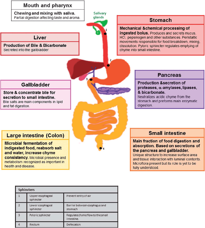
Clinical Focus & Cardinal Rule: The abdominal assessment sequence drastically differs from other body systems. You must proceed in this exact order:
HISTORY + INSPECTION ➔ AUSCULTATION ➔ PERCUSSION ➔ PALPATION
By the end of this comprehensive session, the nursing student should be able to masterfully execute the following:
- Take a focused gastrointestinal history and rapidly identify life-threatening alarm symptoms.
- Prepare and position a patient correctly for abdominal examination to ensure maximum comfort and clinical accuracy.
- Perform abdominal assessment in the correct, modified clinical sequence to avoid altering physiological findings.
- Assess thoroughly: bowel sounds, abdominal tenderness, organ enlargement (organomegaly), ascites, and selected special diagnostic signs.
- Document findings clearly using precise anatomical terminology and recognize findings requiring urgent, immediate escalation to the medical team.
Accurate anatomical mapping is crucial for identifying underlying pathology. Use either the 4-quadrant system or the more precise 9-region system when describing pain, tenderness, masses, or surgical scars.
| Quadrant | Underlying Organs & Clinical Significance |
|---|---|
| RUQ (Right Upper Quadrant) | Liver, gallbladder, duodenum, head of pancreas, right kidney, hepatic flexure of the colon. Clinical Note: Pain here strongly suggests biliary issues like cholecystitis or liver pathology like hepatitis. |
| LUQ (Left Upper Quadrant) | Stomach, spleen, left lobe of liver, body/tail of pancreas, left kidney, splenic flexure of the colon. Clinical Note: Pain here may indicate gastric ulcers, ruptured spleen, or acute pancreatitis. |
| RLQ (Right Lower Quadrant) | Caecum, appendix, terminal ileum (small bowel); right ovary/fallopian tube in females. Clinical Note: Classic site for acute appendicitis (McBurney's point) or right-sided ovarian cysts/ectopic pregnancy. |
| LLQ (Left Lower Quadrant) | Descending/sigmoid colon, small bowel; left ovary/fallopian tube in females. Clinical Note: Classic site for diverticulitis pain in older adults. |
Useful for a more precise, localized description of symptoms and examination findings. Surface regions are defined by two midclavicular lines vertically, and two horizontal planes (subcostal and intertubercular planes).
- Right hypochondriac | Epigastric | Left hypochondriac
- Right lumbar (Flank) | Umbilical | Left lumbar (Flank)
- Right iliac (Inguinal) | Hypogastric (Suprapubic) | Left iliac (Inguinal)
- Pain: Site (Where is it?), Onset (Sudden vs gradual?), Character (Burning, cramping, stabbing?), Radiation (Does it move to the back or shoulder?), Severity (0-10 scale), Timing (Constant or intermittent?), Triggers/relievers (Does eating make it worse or better?).
- Nausea/Vomiting (Emesis): Frequency, content (undigested food?), presence of blood (haematemesis - looks like coffee grounds or bright red?), presence of bile (greenish-yellow).
- Swallowing: Dysphagia (difficulty swallowing solids or liquids?), odynophagia (painful swallowing).
- Appetite: Anorexia, early satiety (feeling full after just a few bites), recent unintentional weight loss.
- Bowel habit: Frequency of movements, chronic constipation or diarrhoea, stool form/caliber (pencil-thin stools?). Presence of blood (bright red) or melaena (black, foul-smelling, tarry stool indicative of upper GI bleed).
- Jaundice: Yellowing of skin/eyes, associated severe pruritus (itching), abnormally dark urine (tea-coloured), pale or clay-coloured stools.
- Past history: Peptic ulcers, hepatitis, previous abdominal surgery (high risk for adhesions/bowel obstruction), hernia, cancer, inflammatory bowel disease (IBD).
- Medicines/substances: NSAIDs (ibuprofen, aspirin - high risk for gastric ulcers), antibiotics (risk for C. diff diarrhoea), heavy alcohol use (liver cirrhosis, pancreatitis), herbal supplements; recent diet and food safety (food poisoning).
These findings indicate potential life-threatening emergencies. They require immediate nursing interventions, cessation of routine examination, prompt senior medical review, and urgent evaluation.
| Emergency Condition | Clinical Presentation / Red Flag Signs |
|---|---|
| GI Bleeding | Haematemesis (vomiting blood), melaena (black tarry stool), haematochezia (fresh blood per rectum); accompanied by dizziness, tachycardia, hypotension, or frank signs of hypovolemic shock. |
| Bowel Obstruction | Persistent profuse vomiting (especially feculent), severe abdominal distension, severe colicky (cramping) pain, and obstipation (absolute inability to pass stool or flatus/gas). |
| Peritonitis (Acute Abdomen) | Severe, unrelenting pain, board-like guarding/rigidity of the abdominal muscles, rebound tenderness, absent bowel sounds, and a highly toxic, septic appearance (fever, chills). |
| Cancer / Systemic Disease | Progressive, worsening dysphagia (difficulty swallowing), unexplained profound anaemia, persistent unexplained vomiting, severe unintentional weight loss, palpable hard masses. |
| Liver Failure | Profound jaundice accompanied by altered mental status (hepatic encephalopathy), severe bleeding tendency (bruising easily), massive ascites, and sepsis. |
Proper preparation is crucial to relax the abdominal musculature; a tense abdomen cannot be palpated accurately.
- Consent & Dignity: Explain the examination, obtain verbal consent, ensure privacy (draw curtains), and maintain dignity.
- Bladder: Always ask the patient to empty their bladder before the exam. A full bladder is uncomfortable during palpation and can be mistaken for a pelvic mass.
- Positioning: Position the patient supine (flat on their back) with their head comfortably supported on a single pillow. Crucial Step: Ensure knees are slightly flexed (bent) or place a pillow under their knees. Rationale: Supine + knees flexed = less abdominal wall tension, relaxing the muscles for easier palpation. Keep arms down by their sides (raising arms overhead stretches and tightens the abdominal muscles).
- Exposure: Expose the abdomen fully from the xiphisternum (bottom of the breastbone) down to the symphysis pubis, while strictly keeping the chest and genital areas covered with a sheet or gown.
- Comfort: Warm your hands and the stethoscope diaphragm (cold hands cause the patient to tense up). Examine from the patient’s right side when feasible (standard medical convention).
- Pain Assessment: Ask exactly where the pain is worst — and explicitly examine that area LAST. Touching the painful area first will cause the patient to guard and tense up for the rest of the exam.
A comprehensive GIT assessment requires evaluating the entire body for signs of nutritional deficiency, chronic liver disease, or systemic illness.
- Assess appearance: Acute distress (writhing in pain vs lying completely still due to peritonitis), lethargy, cachexia (profound wasting), obesity, marked pallor, and hydration status.
- Vital signs: Temperature (fever suggests infection/inflammation), pulse (tachycardia suggests shock or severe pain), respiratory rate (shallow rapid breathing to avoid abdominal movement), and blood pressure (hypotension suggests bleeding or sepsis).
- Palms: Inspect for pallor (anemia from GI bleed) and palmar erythema (reddened palms, a classic sign of chronic liver disease).
- Nails: Check for clubbing (loss of normal nail-bed angle, seen in IBD, celiac disease, cirrhosis). Look for koilonychia (spoon-shaped nails indicating severe iron deficiency anemia from chronic bleeding) and leukonychia (white nail beds from hypoalbuminemia in liver failure).
- Tremor: Check for asterixis (the "liver flap"). Have the patient hold their arms out and extend their wrists backward; a sudden, jerky, flapping tremor indicates hepatic encephalopathy (ammonia buildup).
- Eyes: Inspect sclera for jaundice (icterus) and pull down the lower eyelid to check the palpebral conjunctiva for severe pallor.
- Face: Look for cachexia, sunken eyes (dehydration), or edema.
- Mouth/Oropharynx: Assess hydration (dry mucous membranes), oral hygiene, severe dental caries (affects chewing/digestion), mucosal pallor.
- Tongue & Lips: Inspect the tongue for thick coating, ulcers, severe inflammation, and dryness. Look for angular cheilitis (cracks at the corners of the mouth) or glossitis (smooth, beefy red tongue), which strongly suggest nutritional deficiencies (B12, Iron, Folate) due to malabsorption.
- Jaundice (yellowing of skin/sclera).
- Spider angiomas: Small, branch-like, red vascular lesions found on the face, neck, and upper chest.
- Palmar erythema and asterixis (as noted above).
- Ascites (massive fluid accumulation in the peritoneal cavity) and generalized muscle wasting.
CRITICAL NURSING PRINCIPLE
The abdominal examination sequence is uniquely different from the standard IPPA sequence. It MUST be:
- INSPECTION (Look)
- AUSCULTATION (Listen)
- PERCUSSION (Tap)
- PALPATION (Feel)
Why auscultate BEFORE percussion and palpation? Percussion and deep palpation physically manipulate the intestines. This can artificially stimulate bowel activity, creating false, hyperactive bowel sounds that alter the true physiological sounds you are trying to carefully assess.
- Contour: Get down to eye level across the abdomen. Describe it as flat, rounded, protuberant/distended, or scaphoid (sunken in, profoundly malnourished).
- Symmetry: Check for symmetrical movement with respiration. Bulging on one side could indicate an enlarged organ or massive hernia.
- Skin: Note colour, lesions, striae (stretch marks - silver is normal, deep purple suggests Cushing's syndrome), surgical scars (indicate risk for bowel adhesions), and dilated veins.
- Umbilicus: Note its position, any erythema (redness), swelling, eversion (popping out due to ascites or hernia), or purulent discharge.
- Visible peristalsis: Seeing the intestines rippling beneath the skin may indicate severe intestinal obstruction, particularly when marked and accompanied by pain.
- Abdominal distension: May result from the "6 Fs": Fat, Feces, Fluid (ascites), Flatus (gas), Fetus (pregnancy), or Fatal growth (malignant mass).
- Caput medusae: Extremely prominent, engorged, radiating umbilical veins suggesting severe portal hypertension and liver failure.
- Periumbilical bruising (Cullen's sign) or Flank bruising (Grey Turner's sign): Strongly suggests severe, life-threatening intra-abdominal bleeding or severe hemorrhagic pancreatitis.
- Stomas/Drains: Should be assessed for site, color of the stoma (should be beefy red, not black/ischemic), output quantity/quality, surrounding skin breakdown, and appliance fit.
Use the diaphragm of the stethoscope. Listen systematically in all four quadrants (RLQ ➔ RUQ ➔ LUQ ➔ LLQ). Begin in the RLQ at the ileocecal valve area because bowel sounds are almost always present there.
Assess the frequency and character of bowel sounds rather than relying only on a fixed time count.
- Normal sounds: High-pitched, intermittent clicks and gurgles occurring approximately 5 to 30 times per minute.
- Hyperactive sounds (Borborygmi): Frequent, loud, rushing, growling sounds (>30/min). May occur with severe gastroenteritis, early intestinal obstruction, or brisk GI bleeding.
- Hypoactive sounds: Infrequent, faint, fewer than 5 per minute. May occur with paralytic ileus, peritonitis, or immediately following recent abdominal surgery.
- Absent sounds: NO bowel sounds heard after prolonged, continuous auscultation (listening for up to 5 full minutes). This is a profound "Red Flag" indicating paralytic ileus or advanced peritonitis.
- Using the bell of the stethoscope, listen for bruits over the aorta, renal arteries, iliac arteries, and femoral arteries, especially when vascular disease or hypertension is present.
- A bruit is a harsh, blowing, "swooshing" sound caused by turbulent blood flow.
- An abdominal bruit may occur with severe renal artery stenosis or aortic/arterial disease (aneurysm).
- URGENT WARNING: If you hear a bruit and see a pulsatile abdominal mass, DO NOT press deeply over it. It could be an Abdominal Aortic Aneurysm (AAA) that is at high risk of rupturing!
Percussion helps map out underlying organs, masses, and fluid by analyzing the sound produced. Place the middle finger of the non-dominant hand firmly on the abdomen. Strike its distal phalanx briskly with the tip of the dominant middle finger. Compare notes across all quadrants; always percuss painful areas gently and last.
- Tympany: A hollow, drum-like sound. Typical over gas-filled stomach and bowel; it is the predominant abdominal percussion note.
- Dullness: A muffled, flat sound. Expected over solid organs (e.g., liver, spleen) or may occur with a solid tumor mass, a completely full bladder, or extensive fluid (ascites).
Used to estimate liver size and detect hepatomegaly (enlarged liver).
- Percuss down the right midclavicular line (RMCL) from resonant lung tissue until the sound changes to dullness ➔ this marks the upper liver border (usually 5th-7th intercostal space).
- Percuss upward from the RLQ (area of abdominal tympany) along the RMCL toward the dullness ➔ this marks the lower liver border (usually at the costal margin).
- Measure the distance between borders in the RMCL. (Normal is approx. 6-12 cm). Interpret in context: technique, body habitus, and disease (e.g., severe emphysema pushing the liver down) can alter estimates.
Suspect ascites when there is profound generalized abdominal distension, bulging flanks, an everted umbilicus, and visible abdominal veins.
- Shifting Dullness: With the patient supine, percuss from the center of the abdomen (tympany) outward toward the flanks to identify a change to dullness (where fluid pools). Mark the spot. Have the patient roll onto their side. Wait 1 minute for fluid to shift. Percuss again. If the area of dullness has shifted higher, ascites is present.
- Fluid Wave Test: Used when substantial ascites is suspected. Requires the patient (or assistant) to press the ulnar edge of their hand firmly down the midline of the abdomen (to stop fat transmission). The nurse taps one flank sharply and feels for the transmitted fluid wave striking the opposite hand. A positive wave supports large-volume ascites, but correlate heavily with clinical context and ultrasound.
Palpation is used to assess for tenderness, muscle guarding, masses, and organ enlargement. Remember to examine the known painful area LAST.
- Use one hand with relaxed fingers; depress the abdominal wall gently (about 1 cm deep). Move systematically through all 4 quadrants.
- Crucial: Observe the patient’s face as well as the abdomen. A grimace or wince reveals pain even if the patient denies it.
- Assess for: Localized tenderness (helps identify the site of inflammation, e.g., RLQ = appendix), superficial masses.
- Guarding vs Rigidity: Voluntary guarding is conscious tensing of muscles (patient is ticklish or anxious). Involuntary rigidity is a constant, board-like hardness of the muscles, a severe reflex response to peritoneal inflammation (peritonitis).
Use greater pressure (about 5-8 cm deep), often employing a bimanual technique (two hands) to assess deeper masses and organ enlargement. Characterize any mass by site, size, shape, consistency, tenderness, mobility, and pulsatility.
- Liver Palpation: Start low in the RLQ (so you don't miss a massively enlarged liver edge) and move upward toward the right costal margin. Ask the patient to take slow, deep breaths. Press inward and upward. You are trying to feel the firm, smooth liver edge descending to meet your fingertips during deep inspiration. Note any extreme tenderness, hard nodules, or marked enlargement.
- Spleen Palpation: The spleen is normally NOT palpable in healthy adults. Start in the RLQ and move diagonally across the abdomen toward the LUQ. Ask the patient to take deep breaths. If the spleen is palpable, it generally indicates significant enlargement (splenomegaly - seen in severe infections, malaria, leukemia).
- Kidney Palpation: Use a bimanual "duck-bill" technique. Place one hand posteriorly in the renal angle (flank) and the other anteriorly below the costal margin. Ask the patient to breathe deeply and attempt to "capture" the kidney between your hands. Assess size, tenderness, and ballotability. Particularly relevant with severe flank pain or suspected renal masses.
- Aortic Examination: Palpate the abdominal aorta gently in the upper midline, slightly left of the umbilicus. Assess the pulsation width (normally 2.5 - 3cm). A notably widened, prominent, laterally expansile pulsation suggests an Abdominal Aortic Aneurysm (AAA). Urgent assessment is required for severe abdominal/back pain with a suspected ruptured aneurysm. Do not repeatedly or forcefully palpate.
Use these specific provocative tests only when specific underlying pathology is clinically suspected.
| Special Test | Technique & Clinical Significance |
|---|---|
| Murphy's Sign (Acute Cholecystitis) | Palpate deeply at the right costal margin (gallbladder area). Ask the patient to take a deep breath. A sharp increase in pain causing a sudden halt in inspiration ("inspiratory arrest") is a positive Murphy's sign, strongly indicating an inflamed gallbladder. |
| McBurney's Point Tenderness (Acute Appendicitis) | Palpate the RLQ, specifically 1/3 of the distance from the Anterior Superior Iliac Spine (ASIS) to the umbilicus. Exquisite, localized tenderness here strongly supports appendiceal irritation. |
| Rovsing's Sign (Acute Appendicitis) | Palpate deeply and evenly in the Left Lower Quadrant (LLQ). If this action triggers referred pain in the Right Lower Quadrant (RLQ), it is a positive Rovsing's sign, indicating appendicitis. |
| Iliopsoas Sign & Obturator Sign (Acute Appendicitis) | Psoas: Have patient lift their right leg straight up against your resistance. Obturator: Flex the right hip and knee to 90 degrees, and internally rotate the leg. Pain in the RLQ during these movements indicates an inflamed appendix irritating the underlying psoas or obturator muscles. |
| CVA Tenderness (Pyelonephritis / Kidney Infection) | Have the patient sit up. Place the palm of your hand flat over the Costovertebral Angle (where the lowest rib meets the spine on the back). Strike your hand firmly with the ulnar edge of your other fist. Sharp pain indicates kidney inflammation. |
| Rebound Tenderness (Blumberg's Sign) (Peritonitis) | Press fingers deeply and slowly into the abdomen, then quickly withdraw them. Pain that is worse on the rapid release (rebound) than on the initial pressure indicates severe peritoneal irritation. Note: This is highly painful and should not be performed routinely if gentler signs of peritonitis (like rigidity) are already obvious. |
| Hernia Examination | Inspect and gently palpate the groins and surgical scars. Ask the patient to bear down or cough; look for a bulging mass. Red Flag: An irreducible, highly painful, discolored hernia associated with vomiting or bowel obstruction symptoms is a strangulated hernia—a surgical emergency! |
Interpret every sign in holistic context; single bedside findings are rarely diagnostic by themselves.
| Assessment Area | Expected / Normal Example | Possible Abnormal / Red Flag Finding |
|---|---|---|
| Contour | Flat, rounded, symmetric. | Marked distension, severe focal bulge, everted umbilicus. |
| Bowel Sounds | Intermittent clicks and gurgles (5-30/min). | Completely absent after 5 minutes of listening; high-pitched "tinkling" or rushing sounds (obstruction). |
| Percussion | Predominantly tympanic over bowels. | Diffuse generalized dullness (ascites) or shifting dullness. |
| Palpation (General) | Soft, non-tender, no masses. | Severe guarding, board-like rigidity, pulsatile mass, rebound tenderness. |
| Liver Edge | May be non-palpable, or soft, smooth, regular edge at the costal margin. | Highly tender, nodular/hard surface, markedly enlarged spanning well below the ribs. |
- Document exactly what you actually assessed — never use a pre-filled template for tasks you did not perform.
- State the exact site of tenderness or masses using specific quadrant/region terminology.
- Record stoma/drain output quantitatively (e.g., "Hemovac drained 50mL bright red blood") when relevant.
- Escalate acute deterioration separately verbally; do not hide critical emergencies inside routine electronic charting.
Example of Excellent Clinical Documentation:
"Abdomen flat and symmetrical. Skin intact; no visible masses, striae, or dilated veins. Bowel sounds present and normoactive in all four quadrants. Predominantly tympanic to percussion; no shifting dullness. Abdomen soft, non-distended, completely non-tender to light and deep palpation; no voluntary guarding or rebound tenderness. No palpable masses or organomegaly (liver and spleen not palpable). No CVA tenderness."
| Scenario | Clinical Presentation | Best Nursing Response / Action |
|---|---|---|
| Scenario A | A patient has sudden, severe abdominal pain, board-like abdominal rigidity, and a high fever. | Stop routine examination immediately. Assess ABCs and vital signs. Escalate urgently to the surgical team. Suspect acute peritonitis/ruptured viscous (acute abdomen). Prepare for IV fluids and NPO status. |
| Scenario B | A patient reports passing black, foul-smelling, tarry stool (melaena), and complains of severe dizziness and profound weakness. | Assess haemodynamic status immediately (BP and heart rate - look for hypotension and tachycardia). Recognize this as a classic presentation of a massive Upper GI bleed. Escalate urgently. Insert large-bore IVs for potential blood transfusion. |
| Scenario C | Patient has a massively distended abdomen, persistent vomiting (feculent odor), and reports absolutely no stool or flatus (gas) passed for 2 full days. | Assess for mechanical bowel obstruction. Keep the patient strictly NPO (nothing by mouth) to avoid worsening the obstruction or causing aspiration. Anticipate orders for an NG (Nasogastric) tube for decompression, IV fluids, and prompt surgical/medical review. |
- [ ] Hand hygiene; introduce self; verify patient identity (x2); explain procedure and obtain consent.
- [ ] Pain check, provide privacy, ensure appropriate exposure (xiphisternum to pubis), and correct positioning (supine, knees flexed).
- [ ] Inspect abdomen systematically (contour, skin, umbilicus, pulsations).
- [ ] Auscultate BEFORE touching the abdomen (bowel sounds x 4 quadrants, bruits).
- [ ] Percuss all quadrants; perform specific liver/ascites tests only when appropriate.
- [ ] Perform light palpation, followed by deep palpation; always examine the known painful area last.
- [ ] Assess relevant organs (liver edge, spleen) and special signs (Murphy's, Rebound) only if clinically indicated.
- [ ] Thank the patient, cover them comfortably, perform hand hygiene, document findings, and escalate red flags.
- Ask a focused history and actively identify life-threatening alarm symptoms (bleeding, obstruction, peritonitis).
- Prepare the patient well; flex the knees, warm your hands, and ensure the pain area is examined LAST.
- Abdominal sequence is absolute: INSPECTION ➔ AUSCULTATION ➔ PERCUSSION ➔ PALPATION.
- Describe findings anatomically and objectively using the 4 quadrants or 9 regions.
- Recognize red flags and escalate early — a thorough nursing assessment is a primary safety intervention, not just paperwork.
- Jarvis C, Eckhardt AL. Physical Examination and Health Assessment. 9th ed. Elsevier; copyright 2024.
- Soriano RP. Bates’ Guide to Physical Examination and History Taking. 14th ed. Wolters Kluwer; 2025.
- OpenStax. Clinical Nursing Skills. Section 27.2: Physical Assessment. Updated online resource, accessed October 2026.
- Ernstmeyer K, Christman E, eds. Nursing Skills. NCBI Bookshelf. Chapter 12: Abdominal Assessment. StatPearls/Chippewa Valley Technical College resource.
- Merck Manual Professional Edition. Evaluation of the Gastrointestinal Patient. Updated online resource, accessed October 2026.
- Merck Manual Professional Edition. Acute Abdominal Pain. Updated online resource, accessed October 2026.
- Royal Children’s Hospital Melbourne. Nursing Assessment: Gastrointestinal assessment section. Updated online guideline, accessed October 2026.
Quick Quiz
GIT Examination Quiz
Health Assessment
Privacy: Your details are used only for quiz tracking and certificates.
GIT Examination Quiz
Health Assessment
Preparing questions...
Choose your answer and keep your streak alive.
Great effort.
Here is your quick performance summary.