
Diseases of the respiratory system account for up to a third of deaths in most countries. The key to successful diagnosis is a clear and carefully recorded history. Symptoms may seem trivial or extremely distressing, but either may indicate serious and life-threatening disease. Most patients will present with breathlessness, cough, excess sputum, haemoptysis, wheeze, or chest pain.
Dyspnoea is breathlessness that is inappropriate to the level of physical exertion, or occurring at rest. The mechanisms are complex and relate to lowered blood oxygen (hypoxia), raised carbon dioxide (hypercapnia), cardiac disease, anaemia, thyrotoxicosis, or metabolic acidosis.
- Exercise Tolerance: Assess how far the patient can walk at a normal pace on the level. Ask about their daily activities to estimate their mobility accurately.
- Variability: Ask if symptoms have good and bad days, or specific worsening times. Asthma is often worse at night/early morning. COPD patients often report feeling fine sitting in bed, but become severely distressed upon exertion.
| Grade | Description of Breathlessness |
|---|---|
| 1 | Not troubled by breathlessness except on strenuous exercise. |
| 2 | Short of breath when hurrying or walking up a slight hill. |
| 3 | Walks slower than contemporaries on the level because of breathlessness, or has to stop for breath when walking at own pace. |
| 4 | Stops for breath after about 100 m or after a few minutes on the level. |
| 5 | Too breathless to leave the house, or breathless when dressing or undressing. |
Acute: Anaphylaxis, Asthma, Pneumothorax, Pulmonary embolus, Metabolic acidosis, Myocardial infarction, Pulmonary oedema.
Subacute: Pneumonia, Exacerbation of COPD, Pleural effusion, Malignancy, Cardiac tamponade.
Chronic: COPD, Interstitial lung disease, Chronic pulmonary emboli, Congestive heart failure, Valvular dysfunction, Severe anaemia, Obesity, Neuromuscular disorders.
Cough is defined chronologically: Acute (lasting less than 3 weeks) or Chronic (lasting more than 8 weeks). Acute coughs are mostly viral/bacterial. Any chronic cough (>8 weeks) requires a baseline CXR and spirometry.
- Duration: A cough lasting weeks in a middle-aged smoker has major significance (possible malignancy).
- Timing: A dry nocturnal cough, or one coming in long spasms, may be an early sign of asthma.
- Triggers: Dust, animals, pollen, cold air, or exercise.
- Associations: Severe coughing may trigger vomiting. Haemoptysis accompanying any cough requires urgent investigation.
- Post-viral upper respiratory tract infection (URTI)
- Smoking
- Asthma (including cough-variant asthma and non-asthmatic eosinophilic asthma)
- Postnasal drip (e.g., from hay fever or sinusitis)
- Gastro-oesophageal reflux disease (GORD)
Children and some adults swallow sputum, but always ask for a description:
- Appearance: Yellow or green implies purulence/infection. Asthmatics may produce thick, jelly-like casts or yellow sputum heavily laden with eosinophils (even without infection). Clear, sticky sputum indicates chronic bronchitis without active infection. Frothy pink/white sputum is classical for pulmonary oedema. Rusty sputum suggests pneumococcal lobar pneumonia.
- Amount: Large daily volumes (e.g., a cupful) strongly suggest bronchiectasis (though rarer now). Foul-smelling sputum indicates anaerobic infection or a lung abscess.
This is a critical red-flag symptom that must never be dismissed. Fear often causes patients to hide it.
- Differentiate if the blood is fresh (bright red) or altered, the volume, and frequency.
- Ask about epistaxis (nosebleeds) or melaena (dark, tarry stools) to rule out upper airway or gastrointestinal bleeding masquerading as haemoptysis.
- Major Causes: Lung malignancy/metastasis, TB, bacterial pneumonia, pulmonary emboli, bronchiectasis, severe congestive heart failure, and anticoagulant drugs.
- Wheeze: A high-pitched, musical sound typically heard in expiration due to small airway narrowing (asthma, COPD). Ask if the patient or their partner hears it at night.
- Stridor: A harsh, high-pitched inspiratory noise indicating critical narrowing of the larynx, trachea, or main bronchi (upper airway). This is a medical emergency.
Lung parenchyma itself has no pain receptors; respiratory pain arises from the pleura or chest wall.
- Pleuritic Pain: Sharp, stabbing, and distinctly worsened by taking a deep breath or coughing. Usually implies pleural inflammation from underlying pneumonia or pulmonary infarction (PE).
- Constant Aching Pain: May be due to tumor invasion into the chest wall.
- Aching/Tearing Pain: Seen in spontaneous pneumothorax. A massive pulmonary embolus can cause central, crushing, cardiac-type chest pain due to hemodynamic collapse.
- Upper Airway/ENT: Rhinosinusitis coexists with asthma. Postnasal drip causes chronic cough. A hoarse voice lasting >4 weeks requires laryngoscopy (rule out left recurrent laryngeal nerve invasion by a lung apex tumour, or vocal cord carcinoma).
- Smoking & Recreational Drugs: Ask sympathetically. Get the age started/stopped and average consumption. Remember that smoking cannabis or crack cocaine can cause severe, early-onset emphysema.
- Occupational History: Take a chronological work history from the time they left school.
- Asthma triggers: Paint sprayers (isocyanates), bakers (flour dust), hairdressers, laboratory workers (animal dander).
- Asbestos exposure: Plumbers, shipbuilders, demolition workers, mechanics (brake linings). Exposure causes mesothelioma decades later, often presenting as a pleural effusion.
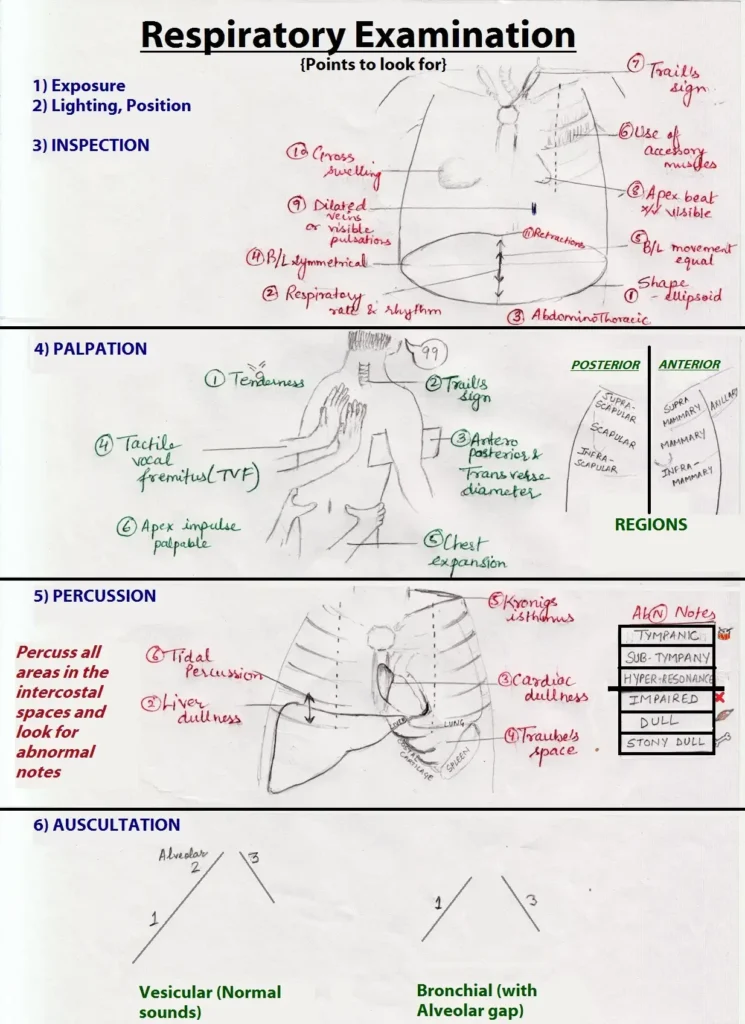
A respiratory examination is incomplete without a simultaneous general physical assessment. Gather maximum information using the classic sequence: Look, Feel, Percuss, Listen.
- Posture & Effort: Is the patient breathless at rest? Are they using accessory muscles (sternomastoid)? Patients with severe COPD often breathe out through pursed lips to keep airways open.
- Noises: Listen for an audible wheeze, stridor, or a weak/non-existent cough effort.
- Bedside Clues: Look for inhalers, peak flow meters, oxygen masks, CPAP machines, or sputum pots.
| Area | Clinical Sign to Look For | Possible Respiratory Cause |
|---|---|---|
| Hands | Clubbing of fingers | Lung cancer, pulmonary fibrosis, bronchiectasis, lung abscess. |
| Hands | Fine tremor | Side effect of inhaled Beta-2 agonists (e.g., salbutamol). |
| Hands | CO2 Flap (Asterixis) & Warm palms | Hypercapnia (Carbon dioxide retention in Type 2 Respiratory Failure). |
| Hands | Tobacco staining (Tar) | Heavy, chronic smoking. |
| Neck | Raised Jugular Venous Pressure (JVP) | Cor pulmonale (right heart failure due to chronic lung disease). If accompanied by a swollen, red face, suspect Superior Vena Cava Obstruction (SVCO). |
| Face / Eyes | Central cyanosis (blue lips/tongue) | Hypoxia / poor blood oxygenation by the lungs. |
| Face / Eyes | Horner's syndrome (Ptosis, miosis) | Pancoast tumour at the lung apex invading the sympathetic chain. |
Ensure the patient is resting comfortably at a 45-degree angle. Look for:
- Surgical Scars: Thoracotomy scars (wrap from the scapula to the axilla), VATS scars (small axillary incisions), or midline tracheostomy scars.
- Chest Shape:
- Barrel Chest: Overinflated lungs due to severe COPD (increased anteroposterior diameter).
- Spinal distortion: Kyphosis (forward bending) or Scoliosis (lateral bending) which restrict lung movement.
- Movement Symmetry: If chest movement is diminished on one side, that is the side of the pathology. Watch for intercostal recession (sucking in of spaces on inspiration) indicating severe upper airway obstruction.
- Lymph Nodes: Palpate supraclavicular, cervical, and axillary nodes. Enlargement heavily alters cancer staging and treatment.
- Trachea Position: Place the 2nd and 4th fingers on the edges of the sternal notch and use the 3rd (middle) finger to feel the tracheal rings.
- Pushed Away (Contralateral): Tension pneumothorax, large pleural effusion.
- Pulled Towards (Ipsilateral): Lobar collapse, massive pulmonary fibrosis.
- Apex Beat: Displacement without tracheal shift may indicate scoliosis or left ventricular enlargement.
- Chest Expansion: Place thumbs in the midline hovering over the spine/sternum and fingers gripping the lateral ribcage. Ask the patient to take a deep breath. Thumbs should move apart equally. Asymmetry means unilateral restriction (e.g., effusion, consolidation).
- Tactile Vocal Fremitus: Use the flat of the hand/fingertips while the patient says "ninety-nine". Vibrations are increased over solid consolidation and decreased over fluid/air (effusion/pneumothorax).
Press the middle finger of the left hand firmly flat on the chest. Strike the middle phalanx sharply with the tip of the right middle finger, pivoting strictly from the wrist. Always compare side-to-side symmetrically.
- Resonant: Normal air-filled lung tissue.
- Dull: Solid tissue replacing air. Seen in lobar consolidation (pneumonia) or collapse.
- Stony Dull: A flat, hard resistance resembling percussing a solid wall. Extremely characteristic of a pleural effusion.
- Hyper-resonant: Excess air in the pleural space, classical for a pneumothorax.
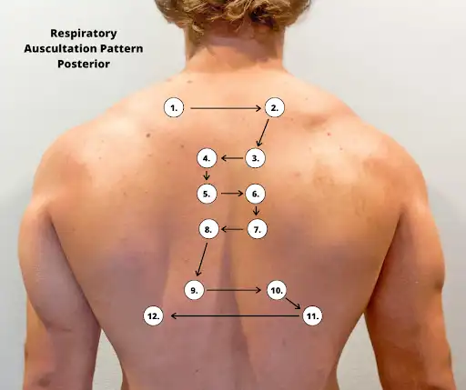
Listen using the diaphragm of the stethoscope. Ask the patient to take deep breaths in and out through their open mouth. Compare side-to-side.
- Vesicular (Normal): Quiet, filtered sounds with no distinct pause between inspiration and expiration. Intensity is reduced over effusions or emphysema.
- Bronchial (Abnormal): Harsh, loud, high-frequency sounds resembling listening over the trachea. The expiratory phase is hissing and prolonged, with a distinct pause between inspiration and expiration. Classical sign of consolidation (e.g., pneumonia) where the solid lung transmits airway sounds perfectly without filtering.
- Wheezes: Musical sounds due to airway narrowing. Polyphonic (multiple pitches) expiratory wheezes signify widespread obstruction (Asthma, COPD). Monophonic (single pitch) wheezes signify localized blockage (tumor, foreign body).
- Crackles: Short, explosive, bubbling or clicking sounds from sudden opening of closed small airways. Coarse crackles are heard in bronchiectasis or severe pulmonary oedema. Fine, late-inspiratory crackles (sounding like rolling hair near the ear) are classical for diffuse interstitial pulmonary fibrosis.
- Pleural Rub: A creaking, crunching sound (like walking on fresh snow) linked to pleuritic pain. Caused by inflamed pleural surfaces grating together.
Listen with the stethoscope while the patient says "ninety-nine".
- Increased Resonance: Over consolidation. The sounds are loud and clear. If they whisper and you hear it perfectly, it is called whispering pectoriloquy.
- Decreased/Absent: Over fluid (pleural effusion) or air (pneumothorax) which block sound transmission.
- Aegophony: A nasal, bleating sound heard at the upper margin of a pleural effusion.
Why check Breath Sounds, Vocal Resonance, AND Tactile Fremitus?
Because they all test the same physics: sound transmission.
- Consolidation (Solid): Transmits sound better. ➔ Bronchial breathing, Increased resonance, Increased fremitus.
- Effusion/Pneumothorax (Fluid/Air): Blocks sound. ➔ Decreased breathing, Decreased resonance, Decreased fremitus.
Spirometry assesses lung size and airflow efficiency. The patient takes a maximal inspiration and exhales as hard and fast as possible to measure FVC (Forced Vital Capacity) and FEV1 (Forced Expiratory Volume in 1 second).
| Defect Type | FEV1 | FVC | FEV1/FVC Ratio | Classic Conditions |
|---|---|---|---|---|
| Normal | Normal | Normal | > 70% | Healthy lungs |
| Obstructive | Significantly Reduced | Normal or mildly reduced | < 70% | Asthma, COPD, Bronchiectasis |
| Restrictive | Reduced | Significantly Reduced | Normal (>70%) | Pulmonary Fibrosis, Kyphoscoliosis |
Peak Expiratory Flow Rate (PEFR): Evaluates the steepest slope of expiration. Easily monitored at home by asthmatics to detect early deterioration (noted by a massive drop in morning readings).
Assesses the partial pressures of Oxygen (PaO2) and Carbon Dioxide (PaCO2).
| Type | Typical ABG Values (On Room Air) | Interpretation |
|---|---|---|
| Type 1 Resp Failure | pH: 7.43 | PaCO2: 3.8 kPa | PaO2: 7.5 kPa | Sats: 91% | Hypoxia with low/normal CO2. The lungs are failing to oxygenate, but the patient is hyperventilating, blowing off CO2 successfully. |
| Type 2 Resp Failure (Decompensated) | pH: 7.25 | PaCO2: 9.3 kPa | PaO2: 7.5 kPa | Sats: 92% | Hypoxia with HIGH CO2 & Acidosis. The patient's alveolar ventilation has completely failed. They are retaining toxic levels of carbon dioxide. |
- Chest X-Ray (CXR): Systematically check the patient identity, alignment (rotation alters mediastinal silhouettes), bony skeleton, trachea position, cardiac outline, diaphragm height, and lung fields (zones). Lobar collapse shows distinct silhouette losses (e.g., middle lobe collapse obscures the right heart border).
- CT Scan: Cross-sectional slices ideal for staging lung cancer, finding bronchiectasis, or detailing interstitial lung disease (High-Resolution CT).
- V/Q Scan (Isotope Imaging): Combined Ventilation and Perfusion scan. A "matched defect" (no air, no blood) suggests lung parenchyma disease. An "unmatched defect" (lung gets air, but no blood reaches it) strongly points to a Pulmonary Embolism.
- PET Scan: Uses a radioactive sugar (FDG) to locate highly metabolic tissues, presenting as 'hot spots'. Crucial for staging lung cancer metastasis.
- Ultrasound: Excellent bedside tool for identifying and safely guiding drainage of pleural effusions and assessing diaphragmatic paralysis.
- Flexible Bronchoscopy: Direct visualization of the bronchial tree to spot tumors, take biopsies, and wash out samples for TB/cytology. Endobronchial Ultrasound (EBUS) allows transbronchial needle aspiration of mediastinal lymph nodes for precise cancer staging.
- Pleural Aspiration: Drawing fluid from an effusion. Differentiate a transudate from an exudate using Light's Criteria:
- Exudate if: Pleural fluid protein/Serum protein > 0.5
- Exudate if: Pleural fluid LDH/Serum LDH > 0.6
- Exudate if: Pleural fluid LDH > two-thirds the upper limit of normal serum LDH.
- Note: Pleural pH < 7.2 or very low glucose implies severe infection/empyema requiring urgent chest tube drainage.
- VATS (Video-Assisted Thoracoscopic Surgery): Minimally invasive surgical approach to visually inspect the pleural cavity, biopsy the lung, perform lobectomies, or strip infected linings (decortication).
- TB Testing: Use specialized Zeihl-Neelsen or auramine stains to find Acid-Fast Bacilli (AFB). GeneXpert (PCR) rapidly diagnoses active TB and Rifampicin resistance. Mantoux testing and IGRA blood tests check for latent TB exposure.