
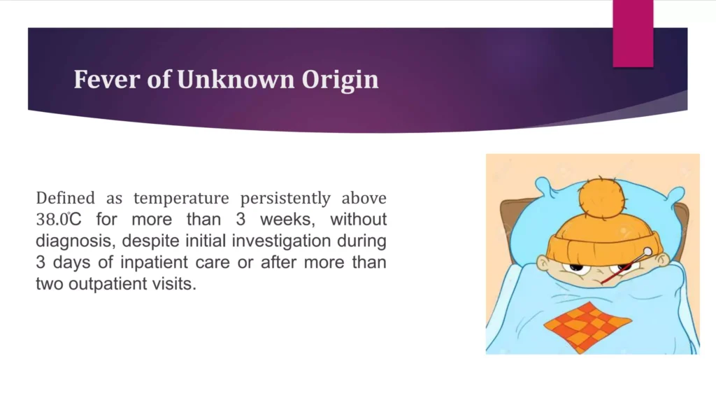
Pyrexia of Unknown Origin (PUO) is synonymous with Fever of Unknown Origin (FUO). While PUO is sometimes used, FUO is the term most commonly utilized in medical literature and international guidelines.
Historically, the diagnosis of FUO required three strict criteria to be met:
- Duration: Illness of more than 3 weeks duration. (Rationale: This extended timeframe helps to naturally eliminate self-limiting viral infections, which usually resolve within 1 to 2 weeks).
- Temperature: Fever greater than 38.3°C (101°F) on several occasions.
- Investigation: Cause remains uncertain after 1 week of in-hospital investigation.
With the advent of advanced outpatient diagnostics, keeping a patient hospitalized for a week simply for observation became impractical and costly. Furthermore, specific vulnerable patient populations required tailored definitions. Thus, FUO was redefined into four distinct categories:
- Classical FUO:
- Duration: ≥ 3 weeks.
- Fever: ≥ 38.3°C (101°F).
- Cause uncertain after 3 days despite appropriate in-hospital investigation OR after 3 out-patient visits.
- Nosocomial FUO:
- Occurs in hospitalized patients.
- Fever ≥ 38.3°C (101°F) on several occasions.
- The infection was not present or incubating on admission.
- Diagnosis remains uncertain after 3 days despite appropriate investigations.
- Neutropenic FUO:
- Neutrophil count is <0.5 x 10⁶/L (or is expected to fall to that level in 1 to 2 days).
- Fever ≥ 38.3°C (101°F) on several occasions.
- Diagnosis remains uncertain after 3 days despite appropriate investigations. (Note: These patients lack the white blood cells to mount a normal inflammatory response, making them highly susceptible to rapid, overwhelming sepsis).
- HIV-Associated FUO:
- Confirmed HIV infection.
- Fever ≥ 38.3°C (101°F) on several occasions.
- Duration of > 4 weeks for out-patients, or > 3 days for hospitalized patients.
- Diagnosis remains uncertain after 3 days despite appropriate investigations.
Neither the original definition nor the currently-used definitions of FUO clearly define the word "workup" or its required intensity. Because of this, a patient labeled as having "FUO" in a resource-limited rural hospital (without access to advanced imaging or molecular tests) might easily and quickly be diagnosed with tuberculosis or a hidden abscess if transferred to a well-equipped tertiary hospital.
Clinical Argument: Reasonably, only those medical centers with access to specified, modern required diagnostic techniques (like CT, MRI, advanced serology) should be fully authorized to firmly label a patient's condition as a true FUO.
When investigating a prolonged fever, clinicians categorize the potential causes into major groups. Statistically, the breakdown of Classical FUO causes is as follows:
| Disease Category | Percentage of FUO Cases |
|---|---|
| Infections | 22 - 58% (Most common globally) |
| Neoplasms (Cancers) | Up to 30% |
| Non-infectious Inflammatory / Autoimmune Diseases | Up to 25% |
| Miscellaneous Causes | Up to 25% |
| Undiagnosed | Up to 30% (Despite exhaustive workups, some fevers spontaneously resolve without a definitive diagnosis). |
Infections remain the leading cause of FUO. They can be deeply sequestered (hidden) or present atypically.
- Bacterial Infections:
- Abscesses: Dental, intra-abdominal (liver, spleen, psoas muscle), or pelvic. These can be clinically silent except for fever.
- Pyomyositis (bacterial infection of skeletal muscle).
- Loculated effusions or obstructions that are not drained.
- Osteomyelitis (bone infection) and spine infections (discitis).
- Prosthetic joint infections or catheter-related bloodstream infections.
- Infective endocarditis (heart valves) and pericarditis.
- Mycotic aneurysm (infection of an arterial wall).
- Biliary tract infections (cholangitis).
- Occult (hidden) urinary tract infections, renal or perinephric abscesses.
- Mycobacterial: Extrapulmonary tuberculosis, miliary (disseminated) tuberculosis, MOTT (Mycobacteria Other Than Tuberculosis).
- Specific organisms: Salmonella infections, Clostridium difficile colitis, Cat scratch disease (Bartonella), Tularemia, Yersiniosis, Brucellosis, Melioidosis, Nocardiosis, Rhodococcosis.
- Atypical/Intracellular: Q fever (Coxiella burnetii), Spirochaetal, Rickettsial, Mycoplasma, and Chlamydial infections (including Psittacosis).
- Fungal Infections:
- Candidiasis, Cryptococcosis, Aspergillosis, Pneumocystis jiroveci, Mucormycosis, Histoplasmosis, Penicilliosis, and other endemic fungi.
- Viral Infections:
- Acute HIV infection, HHV8-associated Castleman’s Disease, CMV (Cytomegalovirus) infection, EBV (Epstein-Barr Virus / Infectious mononucleosis), Parvovirus B19 infection.
- Parasitic Infections:
- Malaria, Amoebic liver abscess.
Cancers can cause fever in two primary ways:
1) Tumours may cause fever in their own right (by releasing pyrogenic cytokines like IL-1 and TNF-alpha).
2) Solid tumours may cause luminal obstruction, leading to a secondary bacterial infection behind the blockage (e.g., a tumor in the bronchus leading to post-obstructive pneumonia, or a bile duct tumor leading to cholangitis).
- Hematologic Malignancies: Hodgkin’s lymphoma, Non-Hodgkin’s lymphoma, Leukaemias (including the aleukemic type where abnormal cells don't spill into the blood), Myelodysplasia, Multiple myeloma.
- Solid Tumors: Renal cell carcinoma (classic cause of neoplastic fever), Adenocarcinomas of the breast, Hepatocellular carcinoma (HCC) or metastases to the liver, Colonic cancer, Lung cancer, Pancreatic tumor, Sarcoma, Kaposi sarcoma, Atrial myxoma (a benign heart tumor that mimics endocarditis), and disseminated carcinomatosis.
Systemic inflammation without an infectious trigger.
- Systemic lupus erythematosus (SLE) and Rheumatoid arthritis.
- Rheumatic fever.
- Adult Onset Still’s disease (characterized by high spiking fevers, salmon-colored rash, and joint pain).
- Macrophage Activation Syndrome / Hemophagocytosis Syndrome.
- Temporal arteritis (giant cell arteritis) and Polymyalgia rheumatica (common in the elderly).
- Polyarteritis nodosa.
- ANCA-sensitive vasculitides: Microscopic polyangiitis, Churg-Strauss Syndrome, Wegener’s granulomatosis.
- Other vasculitides: Takayasu arteritis, Hypersensitivity vasculitis, Cryoglobulinemic vasculitis, Behcet’s disease.
- Relapsing polychondritis, Antiphospholipid syndrome, Mixed connective tissue disease.
- Hypersensitivity pneumonitis.
ANCA testing is crucial for diagnosing specific vasculitides that cause FUO.
| Disease Entity | C-ANCA Sensitivity (anti-proteinase 3 / PR3) | P-ANCA Sensitivity (anti-myeloperoxidase / MPO) |
|---|---|---|
| Wegener’s Granulomatosis | 85% (Highly sensitive) | 10% |
| Microscopic Polyangiitis | 45% | 45% |
| Idiopathic Crescentic Glomerulonephritis | 25% | 65% |
| Churg-Strauss Syndrome | 10% | 60% |
| Polyarteritis Nodosa | 5% | 15% |
(Data derived from Kallenberg CGM. The vasculitides: Science and Practice, 1996)
- Kikuchi’s Disease (Histiocytic necrotizing lymphadenitis).
- Sarcoidosis (Granulomatous disease).
- Inflammatory bowel disease (Ulcerative colitis, Crohn’s disease).
- Granulomatous hepatitis & Alcoholic hepatitis.
- Vascular/Thrombotic: Thrombophlebitis, Deep vein thrombosis (DVT), Recurrent pulmonary emboli (PE), Thrombotic thrombocytopenic purpura (TTP).
- Endocrine: Subacute thyroiditis (de Quervain's), Adrenal insufficiency.
- Crystal Arthropathies: Gout, Pseudogout.
- Hidden Bleeding: Hematoma (e.g., retroperitoneal space, aortic dissection/aneurysm, hip, pelvis). As the body breaks down a large volume of blood, it generates heat and fever.
- Hereditary periodic fever syndromes (e.g., Familial Mediterranean Fever).
- Immune reconstitution inflammatory syndrome (IRIS) - especially common with TB and MAC in HIV patients starting antiretrovirals.
- Sweet’s Syndrome (Acute febrile neutrophilic dermatosis).
It is clinically vital to distinguish true fever (where the hypothalamus resets the body's core temperature higher in response to pyrogens) from Hyperthermia (where the body's temperature regulation fails and heat generation exceeds heat loss, independent of pyrogens). Antipyretics (like paracetamol) will not lower hyperthermia.
- Heat Stroke: Induced by intense exercise or impaired sweating (often exacerbated by anticholinergic drugs).
- Drug-Induced: Overdoses or toxicity from Cocaine, Amphetamines, or MAO inhibitors.
- Neuroleptic Malignant Syndrome (NMS): A life-threatening reaction to antipsychotic drugs (like Phenothiazines or Haloperidol), causing "lead-pipe" muscle rigidity and extreme temperatures.
- Malignant Hyperthermia: A severe genetic reaction triggered by inhalational anesthetics during surgery.
- Endocrinopathy: Severe Thyrotoxicosis (thyroid storm) or Pheochromocytoma (adrenaline-secreting tumor).
- Hypothalamic Fever: Direct damage to the brain's thermostat due to cerebral trauma, massive hemorrhage, massive stroke, or anoxic brain injury.
Drug fever is a febrile response that coincides temporally with the administration of a drug and disappears after discontinuation of the offending agent.
- Contamination: Contamination of the IV drug/fluid with an exogenous pyrogen or microorganism during manufacturing or administration.
- Pharmacologic Action: Related to the direct action of the drug itself (e.g., Amphotericin B causing cytokine release, or massive bacterial die-off causing a Jarisch-Herxheimer reaction).
- Allergic (Hypersensitivity): The most common mechanism; an immune-mediated allergic reaction to the drug or its excipients.
- Presentation: The fever is often completely out of proportion to the clinical picture (the patient looks surprisingly well despite a very high temperature).
- Pattern: The pattern of fever is highly variable (can be continuous, spiking, or remittent).
- Onset: Typically occurs 7 to 10 days (can be up to 21 days) after the initiation of the drug.
- Resolution: Usually resolves rapidly, within 48 hours after discontinuation of the drug (though this depends on the half-life of the specific medication).
- Associated Findings:
- Rigor (43%), Myalgia (25%), Rash (18%), Headache (18%).
- Lab findings: Leukocytosis (22%), Eosinophilia (22%), Proteinuria, Abnormal liver function tests.
- Serum sickness (fever, tissue swelling, rash, lymph node enlargement).
Almost any drug can cause drug fever, but the classic offenders include:
- Antimicrobials: Sulfa drugs, Dapsone, Pentamidine, Clindamycin, Penicillins, Cephalosporins, Macrolides, Aminoglycosides, Vancomycin, Nitrofurantoin, Amphotericin.
- Anti-TB/Antiretrovirals: Isoniazid, Rifampicin, Abacavir, Nevirapine, Fosamprenavir, NRTIs (can cause lactic acidosis presenting with fever).
- Cardiovascular/Neuro: Phenytoin, Barbiturates, Carbamazepine, Hydralazine, Methyldopa, Procainamide, Quinidine, Haldol, Amphetamines.
- Others: Interferons, Iodide, Allopurinol, Antihistamines, NSAIDs.
Factitious fever is a self-induced or artificially manipulated temperature elevation.
- When to suspect: The diagnosis should be strongly considered in any puzzling FUO, especially in:
- Young women.
- Persons with medical training or healthcare backgrounds.
- Patients who appear clinically completely well despite charting extremely high temperatures.
- Clinical Clues:
- Disparity between temperature and pulse: Normally, heart rate rises ~10 bpm for every 1°C increase in temperature. In factitious fever (e.g., dipping the thermometer in hot water), the pulse remains normal while the recorded temperature is sky-high.
- Absence of the normal diurnal pattern of body temperature.
- Many patients possess underlying psychiatric features of Munchausen syndrome (factitious disorder imposed on self).
To standardize the approach to FUO, Dr. Tso proposes a systematic "Fever Checklist" for patient management.
- Fever is officially present if the temperature in the mouth (oral) is over 37.5°C (99.5°F).
- Institution Specific: At UCH, the Genius 2 Infrared Tympanic Electronic Thermometer (set at Oral mode) is used. A temperature > 37.5°C on this device triggers the FUO pathway.
The diagnostic approach to adults with prolonged unexplained fever demands a rigorous, exhaustive history.
- Inquire about symptoms from all major organ systems. Look for general complaints (weight loss, profound night sweats, headaches, transient rashes).
- Record all complaints, even if they completely disappeared before the current examination. Previous illnesses (including long-past surgeries and psychiatric illnesses) are vital clues.
- Detailed Evaluation Areas:
- Family history (genetic periodic fevers, autoimmune diseases).
- Immunization status.
- Occupational history (e.g., abattoir worker exposed to Brucellosis).
- Travel history (crucial for Malaria, Dengue, Typhoid).
- Nutrition and weight changes.
- Comprehensive drug history (including all over-the-counter supplements, prescribed meds, and illicit substances).
- Sexual history (HIV, Syphilis).
- Recreational habits (hiking/camping exposing to tick-borne diseases).
- Animal contacts (exposure to ticks, birds/Psittacosis, cats/Bartonella).
- Ensure definitive documentation of the fever (rule out factitious fever or faulty thermometers).
- Fever Patterns: The pattern of fever (continuous, remittent, intermittent) is usually of little help in the modern evaluation. In general, the correlation between specific fever patterns and specific diseases is very weak.
- Notable Exceptions: Tertian and quartan fever patterns in Malaria.
- Other diseases (Brucellosis, Borreliosis, Hodgkin's disease/Pel-Ebstein fever) tend to cause recurrent, undulating episodes of fever.
- Repeat the Exam: A regular physical examination must be repeated daily while the patient is hospitalized. The body changes, and physical signs may evolve late in the disease course.
- Pay special attention to: Rashes, new or changing cardiac murmurs (endocarditis), signs of arthritis/swollen joints, subtle abdominal tenderness or rigidity, lymph node enlargement, fundoscopic changes in the eye (Roth spots, tubercles), and new neurologic deficits.
| Body System | Diagnostic Clues / Associated Diseases |
|---|---|
| Eye Involvement | Sarcoidosis, SLE, Rheumatoid disease, Giant-cell arteritis. |
| Lymphadenopathy | Lymphoma, Castleman's disease, Still's disease, Brucellosis, Filariasis, Tuberculosis, Whipple's disease. |
| Hepatomegaly (Liver) | Lymphoproliferative/myeloproliferative disorders, Granulomatous hepatitis, Q fever, Relapsing fever. |
| Splenomegaly | Lymphoproliferative disorders, Brucellosis, Malaria, Q-fever, Tuberculosis, Rheumatoid disease, SLE, Sarcoidosis. |
| Abdominal Pain | Familial Mediterranean Fever (FMF), Crohn's disease, Hyper-IgD syndrome. |
| Skin Rash | Secondary syphilis, Filariasis, Whipple's, Still's disease, SLE, Sarcoidosis, FMF, Sweet's syndrome. |
| Joint Pain | Rheumatoid disease, SLE, Ankylosing spondylitis, Still's disease, Brucellosis, Rat-bite fever, FMF. |
Testing must be based on clinical presentation and the specific system involved. A "shotgun" approach is discouraged; targeted screening is preferred.
- Baseline Bloods: CBC (with differential count), ESR, CRP, Liver/Renal Function Tests (LRFT), Glucose, PT/APTT, CK, LDH, D-dimer, Amylase.
- Urine: Routine urinalysis. Early Morning Urine (EMU) x 3 for Acid-Fast Bacilli (AFB/TB). Urine antigen testing for Legionella pneumophila and Pneumococcus.
- Respiratory: NPS/NPA (Nasopharyngeal aspirate) for Influenza A/B Ag, PCR, viral culture. Sputum/gastric lavage for AFB smear and culture (x3). Sputum for fungal culture (Nocardia/Rhodococcus) and Pneumocystis jiroveci cytology. PCR for TB DNA or Chlamydia psittaci.
- Fluid & Tissue: Relevant body fluid (joint, pleural, peritoneal, pericardial) for cell count, protein, glucose, pH, Gram stain, cultures, AFB, and cytology. Lumbar puncture for CSF examination.
- Blood Smear: Peripheral blood smear for morphology. Thick/thin blood smear specifically for Malaria screen.
- Blood Cultures (C/ST): Standard cultures for blood, sputum, urine, wound. Crucial: Prolong blood cultures to 14 days for suspected endocarditis or fungemia (must specify this on the lab request).
- Differential time to positivity: Taking blood cultures simultaneously from a peripheral vein and a central catheter lumen to determine if the catheter is the source of sepsis.
- Fresh blood for CMV pp65. Blood for cryptococcal Ag. Blood for AFB culture (Bactec bottle). Clotted blood for galactomannan or (1->3)-beta-D-Glucan (fungal markers).
- Stool Exam: Viral study, culture, Ova & Cysts, Amoeba, Clostridium difficile toxin.
- Immunology & Tumor Markers: ANA, anti-ds-DNA, C3, C4, anti-ENA, RF, ANCA, Serum immunoelectrophoresis (Ig pattern), Cryoglobulin, Ferritin, TFT, Tumor markers (CEA, AFP, PSA).
- Infection Serology: HIV Ab (and CD4 count if positive), Atypical pneumonia paired sera, Mycoplasma IgM, VDRL, Widal test, Weil-Felix, Rickettsial, Coxiella (Q fever), Brucella, Monospot, EBV (VCA IgM, EBNA IgG), Hepatitis, Dengue, Leptospirosis, Parvovirus B19, Hantavirus, CMV, JEV, HTLV1, Burkholderia, Bartonella, Aspergillus, Histoplasma, Toxoplasma, Amoebic, Cysticercosis.
Note: The Mantoux Test (MT2) can yield false negatives in disseminated TB or severely immunocompromised patients; a negative test does not exclude TB.
- First Line: Chest X-Ray (CXR) and targeted X-rays of symptomatic regions. Ultrasonography (abdomen/pelvis). Echocardiogram (to rule out endocarditis or atrial myxoma). High-Resolution CT (HRCT) thorax (plain).
- Advanced CT/MRI: CT with contrast of suspected regions (thorax, abdomen, pelvis).
- Caution: Avoid contrast CT or MRI if the patient has severe renal failure to prevent contrast-induced nephropathy or Nephrogenic Systemic Fibrosis (NSF from MRI Gadolinium).
- For mild renal failure, contrast CT may proceed with strict precautions: Encourage aggressive fluid intake/Normal Saline infusion before the scan, and administer Acetylcysteine 600mg twice daily orally for 2 days (the day before and the day of the CT).
- Vascular Imaging: CT pulmonary angiogram for suspected pulmonary embolism. Venous Doppler Ultrasound of the leg for suspected DVT.
- Nuclear Medicine: Bone scan, Gallium SPECT/CT, and PET-CT.
Positron Emission Tomography-Computed Tomography (using [18F]FDG) is increasingly vital in modern FUO workups.
- It expertly guides further investigation by pinpointing the exact metabolic site of the lesion for targeted biopsy.
- It is exceptionally good at detecting hidden inflammatory lesions (both infective and non-infective) and solid tumors (e.g., hidden caecal adenocarcinomas).
- It has a very high negative predictive value (NPV); if the PET scan is negative, it reliably rules out major inflammatory or malignant solid lesions.
- It can easily detect large vessel vasculitis (which is notoriously difficult to diagnose with standard imaging).
- Limitations: It may not detect microscopic lesions isolated entirely within the bone marrow. Negative findings can also be present if the FUO is caused by a systemic viral infection or drug fever (which lack localized inflammatory masses).
- Advantage in Renal Disease: PET-CT (plain, without IV iodine contrast) can be safely performed in patients with severe renal impairment.
- It is generally more sensitive than a Gallium SPECT/CT scan.
When non-invasive tests fail, direct visualization and tissue sampling are required.
- Endoscopy: Oesophago-Gastro-Duodenoscopy (OGD), Colonoscopy, ERCP (biliary tract), Bronchoscopy, Mediastinoscopy.
- Biopsies: Crucial for histology, routine culture/sensitivity, AFB culture, and fungal culture.
- Bone marrow biopsy (especially if the Complete Blood Picture is abnormal).
- Pleural or Pericardial biopsy.
- Skin lesion biopsy.
- Lymph node biopsy: Note: Fine Needle Aspiration (FNA) may be considered first, but a full excisional biopsy usually provides a much more definitive diagnosis because it preserves the tissue architecture (crucial for lymphoma staging).
- Liver biopsy (if LFTs are abnormal and undiagnosed).
- Temporal artery biopsy (gold standard for Giant Cell Arteritis).
- Bone biopsy (for osteomyelitis).
A major dilemma in treating a patient with prolonged, undiagnosed fever is the temptation to simply "treat blindly" out of frustration.
- For unexplained, prolonged fever in a clinically stable patient who has already failed a standard empiric antimicrobial agent, conducting empirical trials or switching to "big-gun" broad-spectrum antibiotics without identifying a clear cause is strongly discouraged.
- Why? It breeds antibiotic resistance, exposes the patient to drug toxicity (or drug fever), and often partially suppresses an infection, making it impossible to culture and identify the true organism.
- Once an empirical anti-TB trial is started, there is very little potential for ever obtaining a positive TB culture afterward, robbing the clinician of the ability to test for drug resistance.
- Furthermore, medications in the TB regimen (specifically Rifampicin, Aminoglycosides, or Fluoroquinolones) have broad antibacterial activity and can transiently suppress fever caused by other non-TB bacterial infections, leading to a dangerous false diagnosis of Tuberculosis.
- Routine use of antipyretics (Paracetamol, NSAIDs) is discouraged during the diagnostic phase because they mask the true temperature curve and fever pattern.
- They should be indicated only for patients experiencing substantial physical discomfort, extreme tachycardia, or metabolic stress directly due to the fever itself.
Based on the presentation by Dr. Eugene Tso, Division of Infectious Diseases, Department of Medicine & Geriatrics, United Christian Hospital