Gynaecology

Cancers of Reproductive Health Organs

Cancers of Reproductive Health Organs

Breast Cancer

Breast cancer occurs when cells in the breast grow and divide uncontrollably, forming a mass of tissue known as a tumour.

Breast cancer can invade nearby tissues and travel to other body parts, forming new tumours, a process called metastasis.


Clinical Manifestations

Early Signs of Breast Cancer

  1. Asymptomatic: Sometimes, breast cancer shows no symptoms at all, especially in the early stages. This means you might not notice anything unusual.
  2. Size and Shape Changes: A noticeable change in the size or shape of the breast.
  3. Lump: A mass or lump that may be as small as a pea.
  4. Persistent Lump or Thickening: A lump or thickening in or near the breast or underarm that persists through the menstrual cycle.
  5. Skin Changes: Dimpling, wrinkling, scaliness, or inflammation of the skin on the breast or nipple.
  6. Redness: Redness of the skin on the breast or nipple.
  7. Distinct Area: An area distinctly different from other areas on either breast.
  8. Nipple Discharge: Blood-stained or clear fluid discharge from the nipple.

Others;

  1. Unilateral nipple discharge: When fluid, which could be clear, bloody, or another color, leaks from only one nipple.
  2. Change in breast size: One breast might become noticeably larger or smaller than the other, or there could be a change in the overall size of the breast.
  3. Nipple or skin retraction: The nipple may become inverted or pulled inward, or there may be dimpling or puckering of the skin on the breast.
  4. Local lymphadenopathy: Swollen or enlarged lymph nodes in the armpit or collarbone area, indicating possible spread of cancer.
  5. Skin changes-orange-like appearance (Peau d’orange): The skin on the breast might take on an orange peel-like appearance, due to changes caused by cancer cells blocking lymph vessels.
  6. Nipple or skin ulceration: Sores or ulcers on the breast or nipple that do not heal or go away.
  7. Breast pain: Persistent or unusual pain in the breast, although breast cancer does not cause pain in its early stages.
  8. Symptoms of metastasis: If the cancer has spread to other parts of the body, symptoms may include bone pain, shortness of breath, jaundice, or neurological symptoms like headaches or seizures.

Risk Factors

  • Age: Being 55 or older increases the risk of breast cancer.
  • Sex: Women are much more likely to develop breast cancer than men.
  • Family History and Genetics: A family history of breast cancer increases the risk especially if close relatives like mother, sister, or daughter have had it. About 5% to 10% of breast cancers are due to inherited abnormal genes like the BRCA1 and BRCA2 genes.
  • Smoking: Tobacco use is linked to many cancers, including breast cancer.
  • Alcohol Use: Drinking alcohol increases the risk of certain types of breast cancer.
  • Obesity: Obesity increases the risk of breast cancer and recurrence.
  • Radiation Exposure: Prior radiation therapy, especially to the head, neck, or chest, increases risk.
  • Early onset menarche: Starting menstruation at a young age, usually before age 12.
  • Late menopause: Continuing menstruation later in life, usually after age 55.
  • Delayed first pregnancy (after 30 years of age): Not becoming pregnant for the first time until after the age of 30.
  • Null parity: Never having given birth to a child.
  • Family history (maternal or paternal) BRCA1 and BRCA2 genes: A family history of breast cancer, 
  • History of breast biopsy: Previous biopsies or other breast procedures may indicate increased risk.
  • Use of Hormonal therapy for more than 4 years: Long-term use of hormone replacement therapy (HRT), which involves taking oestrogen and progesterone to relieve symptoms of menopause.
breast cancer staging

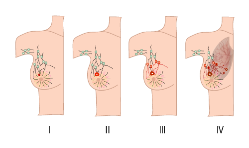
Stages of Breast Cancer

Staging helps describe the extent of cancer by determining the size, location, and spread of the tumour.

  • Stage 0: Non-invasive; cancer has not broken out of the breast ducts.
  • Stage I: Cancer cells have spread to nearby breast tissue.
  • Stage II: Tumour is smaller than 2 cm and has spread to underarm lymph nodes or is larger than 5 cm without spreading to underarm lymph nodes.
  • Stage III: Cancer has spread beyond the breast to nearby tissues and lymph nodes but not to distant organs (locally advanced breast cancer).
  • Stage IV: Cancer has spread to distant organs such as bones, liver, lungs, or brain (metastatic breast cancer).

Diagnosis/Investigations:

  • History Taking: About family history of breast cancer,medical history, and any other symptoms.
  • Self Breast Examination and Breast Examination: For any lumps, changes in size or shape, or other abnormalities.
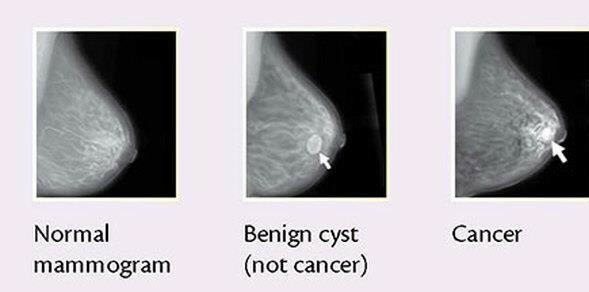
  • Mammogram: Special X-ray of the breasts that can detect changes or abnormal growths, even before they can be felt. It’s a common screening tool for breast cancer.
  • Ultrasonography: Also known as ultrasound, Uses sound waves to create images of breast tissues. It helps in diagnosing lumps or other abnormalities found during a physical examination or mammogram.
  • Positron Emission Tomography (PET) Scan: This test uses special dyes to highlight areas of the body with abnormal metabolic activity, which can indicate the presence of cancer cells.
  • Magnetic Resonance Imaging (MRI): MRI uses magnets and radio waves to produce detailed images of breast structures. It’s especially useful for evaluating the extent of the disease in the breast.
  • TNM System: This is a staging system used to describe the extent of the cancer based on the size of the tumour (T), whether it has spread to nearby lymph nodes (N), and whether it has metastasized to other parts of the body (M).
  • Full Blood Count: This blood test helps assess the overall health and can indicate if there are any abnormalities or signs of infection or inflammation.
  • Renal and Hepatic Profile: Blood tests to assess the function of the kidneys and liver, as metastatic breast cancer can spread to these organs.
  • Chest X-Ray: This test may be done to check for any signs of metastasis to the lungs.
  • Biopsy (Preferably Fine Needle Aspiration): A biopsy is the definitive way to diagnose breast cancer by analyzing a sample of breast tissue under a microscope. Fine needle aspiration is a less invasive biopsy method often used for initial diagnosis.

Management of breast Cancer

Management depends on the tumour’s location and size, lab test results, and whether the cancer has spread.

Stage 0 (Cancer in situ):

  • Young Women: Conservative surgery only, such as lumpectomy.
  • Advanced Age: Mastectomy only.

Early Stage (Stage I and II):

  • Surgery: Modified radical mastectomy and lymphadenectomy for advanced age, and simple mastectomy or wide local lumpectomy for young age.
  • Hormonal Therapy: Tamoxifen 20 mg orally daily for 5 years, but may cause retinal damage.  Blocks hormones that fuel certain cancers.
  • Chemotherapy:
    • Cyclophosphamide: 30 mg/kg IV single dose.

    • Fluorouracil: 300-1000 mg/m2 IV, given every 4 weeks based on patient response.

    • Paclitaxel: 6mg/ml in combination with Cisplatin 1mg/ml.

Late Cancer (Stage III and IV):

  • Hormonal Therapy: Same as for early stage, Tamoxifen 20mg orally daily for 5 years, but may cause retinal damage.
  • Chemotherapy: same as for early stage.
  • Radiation Therapy: Uses high-energy rays to kill cancer cells.
  • Immunotherapy: Helps the immune system fight cancer.
  • Targeted Drug Therapy: Uses drugs to target specific cancer cells.
Types of Breast Cancer Surgery
  • Lumpectomy (Partial Mastectomy): Removal of the tumour and some surrounding tissue, often followed by radiation therapy.
  • Mastectomy: Removal of the entire breast.
  • Axillary Lymph Node Dissection: Removal of multiple lymph nodes.
  • Modified Radical Mastectomy: Removal of the entire breast, underarm lymph nodes, and chest wall muscles if the cancer has spread. Reconstruction may be an option.

Mastectomy

Mastectomy is a planned surgical procedure involving the removal of the breast tissue

There are different types of mastectomy:

  • Partial Mastectomy: Removal of lumps with surrounding normal tissue.
  • Simple Mastectomy: Removal of breast tissue with node biopsy.
  • Extended Simple Mastectomy: Removal of breast tissue, axillary tail, and nodes.
  • Total Mastectomy: Complete removal of the breast leaving the pectoralis muscle intact.
  • Radical Mastectomy: Removal of breast, skin, muscle, and nodes.
  • Modified Radical Mastectomy: Removal of breast, skin, muscles, and nodes with subsequent skin grafting.

Pre-operative Care

  • Admission: Patient admitted to a surgical ward.
  • History Taking: Record medical, surgical, and gynaecological history.
  • Observations: Vital signs monitoring, general examination. And the doctor is informed.
  • Investigations: Various tests including urinalysis, blood tests, and imaging.
  • Patient Education: Inform the patient about the surgery, its purpose, complications, and anaesthesia side effects.
  • Informed Consent: Obtain consent from the patient.
  • Preparation: IV line, blood booking, catheterization, pre-medications administration, and changing into hospital gown
  • Feeding: No feeds or drinks on the day of the operation
  • Rest and sleep: Ensure enough rest and sleep i.e. minizing noise, reducing bright light.

Morning at the day of the operations

  • IV line is put up
  • Booking for blood in the laboratory
  • Catheterisation of the patient
  • Administration of pre-medications
  • Helping the patient to change into hospital gown
  • Removal of all ornaments from the patient and keep them properly.
  • Continuous counselling to relieve anxiety
  • Preparation of patients medical document
  • Taking the patient to the theatre and handing her over to the theatre.

Post-operative Management

When the operation is finished, the information from the theatre will be sent to the ward and 2 nurses will go and collect the patient, reports are received from the surgeon, recovery room nurses and anaesthetists and then the patient is wheeled to the ward.

  • Patient Reception: Patient is received in a warm bed, flat position and turned to one side. As soon as she gains consciousness sit her in the bed leaning on the affected side to aid drainage.
  • Arm Care: Elevation and positioning as per surgeon’s orders.
  • Observation: Regular monitoring of temperature, pulse, respiration, blood pressure, bleeding, and edema.
  • Medical Treatment: Pain relief, antibiotics, vitamins, and supportives.
  1. Pethidine 100mg 8 hourly for 3 doses on change to panadol to complete 5 days
  2. Antibiotics: ampicillin or gentamicin as ordered
  3. Supportives: vitamins like vitamin c, Iron, folic acid, diazepam
  • Wound Care: Inspection, dressing, drainage management, and stitch removal. Stitches are removed on the 8th – 10th day.
  • Wound and Drainage Care
  1. Aseptic Care: Avoid unnecessary touching; inspect for tension or edema.
  2. First Dressing: Done 48-72 hours post-surgery.
  3. Drain Management: Monitor and remove drainage when discharge ceases.

Nursing Care After Mastectomy

  • Initial Care: Patient received in a warm bed in a flat position with head turned to one side.
  • Positioning: Once conscious, position the patient upright to aid drainage.
  • Vital Observations: Check vitals every 15 minutes in the first hour, then every 30 minutes for the next hour until stable.
  • Site Observation: Monitor for bleeding and edema.
  • IV and Blood Transfusion: Ensure correct flow rates.
  • Welcome and Explanation: Explain the procedure and provide comfort.
  • Analgesics and Antibiotics: Administer as prescribed (e.g., Pethidine, Ampicillin).
  • Supportive Care: Provide vitamins and minerals.
  • Hygiene: Provide bed baths and oral care until self-sufficient.
  • Diet: Encourage fluids and nutritious food.
  • Elimination: Promote regular bowel and bladder emptying.
  • Exercise: Begin chest, arm, and leg exercises to prevent deformity and contractures.
  • Psychotherapy: Reassure and counsel on using artificial breasts.

Advise on discharge

  • Radiotherapy: Start when the wound heals (6-8 weeks), lasting 2 months.
  • Follow-Up: Every 2 months for up to 2 years.
  • Chemotherapy: Continue as prescribed.
  • Regular Checkups: Monitor for metastasis.
  • Cancer Institute Visits: Attend radiotherapy.
  • Artificial Breast Use: Educate on proper use.
Complication
  • Necrosis: Death of suture line tissue.
  • Nerve Damage: Potential paralysis of the arm.
  • Contractures: Tightening of muscles and joints.
  • Sloughing: Shedding of dead tissue.
  • Infections: Risk of infection at the wound site.
  • Gaping: Opening of the wound.
  • Chronic Sinus: Persistent drainage site.
  • Oedema: Swelling of the arm.
  • Thrombosis: Blood clots in the axillary vein.
  • Cosmetic Deformity: Changes in appearance post-surgery
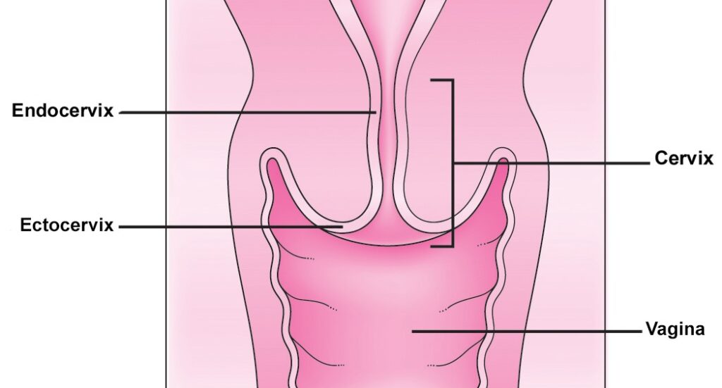
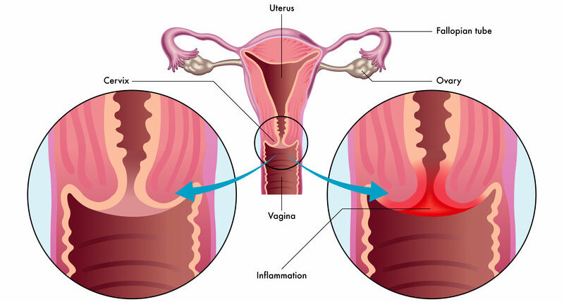
CEERVIX

The Cervix

The cervix is a vital part of the female reproductive system, connecting the uterus to the vagina. It has two main types of cells:

  • Squamous cells: Flat, thin cells found in the outer layer of the cervix (ectocervix).
  • Glandular cells: Column-shaped cells that produce cervical mucus and are found in the cervical canal (endocervix).
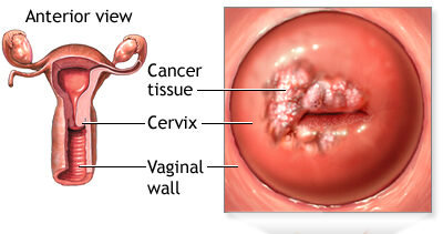
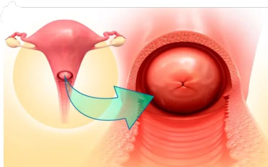
Glandular cells from the cervical canal frequently migrate outside the canal and undergo changes to become squamous cells. This transformation process is known as squamous metaplasia, occurring in a region called the transformation zone.
CERVICAL CANCER

Cervical Cancer

Cervical cancer is a malignant tumor found in the tissues of the cervix, occurring when abnormal cells in the cervix turn into cancer cells. These cancer cells can invade the surface cells (epithelium) and the underlying tissue (stroma) of the cervix, most commonly beginning in the transformation zone.

Epidemiology

  • Around 3,100 women are diagnosed with cervical cancer each year in the UK.
  • In Australia, about 780 women are diagnosed annually.
  • In Uganda, 2,464 women die from cervical cancer annually, with over 3,577 new cases diagnosed each year, making it a leading cause of death among women in the country.

Types of Cervical Cancer

  1. Squamous cell carcinoma: Begins in the flat, thin cells lining the bottom of the cervix; accounts for 80-90% of cervical cancers.
  2. Adenocarcinoma: Develops in the glandular cells lining the upper portion of the cervix; accounts for 10-20% of cervical cancers.
  3. Mixed carcinomas: Involve both types of cells.

Causes of Cervical Cancer

The exact cause is unknown, but several risk factors have been identified:

  1. Human papillomavirus (HPV): A major cause, with types 16 and 18 being the most oncogenic.
  2. Smoking: Chemicals in cigarette smoke can damage cervical cells.
  3. Immunosuppression: Conditions like HIV/AIDS weaken the immune system.
  4. Oral contraceptives: Long-term use increases risk, which decreases after stopping the pill.
  5. Other STIs: Infections like herpes and chlamydia can increase risk.
  6. Circumcision: Women with uncircumcised partners are at higher risk.
  7. Early sexual intercourse: Exposure to sperm can promote cell division in the transformation zone.
  8. High parity: Multiple pregnancies can cause cervical trauma.
  9. Repeated induced abortions: Can cause cervical trauma.
  10. Exposure to chemicals: Occupational exposure to substances like tetrachloroethylene.

Symptoms of Cervical Cancer

Early symptoms may not be noticeable but can include:

  • Vaginal bleeding (between periods, after intercourse, post-menopausal)
  • Unusual vaginal discharge (watery, pink, foul-smelling)
  • Pelvic pain (during intercourse or otherwise)

Advanced stages can present with:

  • Severe weight loss, anemia, dehydration
  • Fatigue
  • Back pain
  • Pain or swelling in the legs
  • Urinary or fecal incontinence
  • Bone fractures
  • Hematuria
  • Enlarged organs
  • Rectal bleeding
  • Tenesmus (desire to defecate)
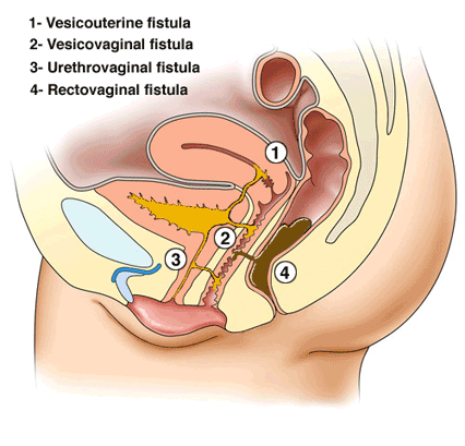
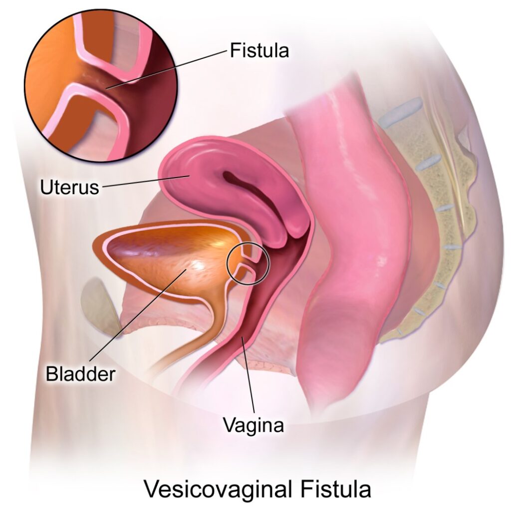
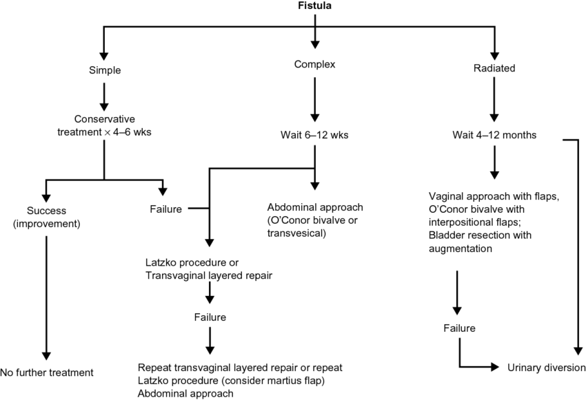
  • Fistulas
cervical cancer staging

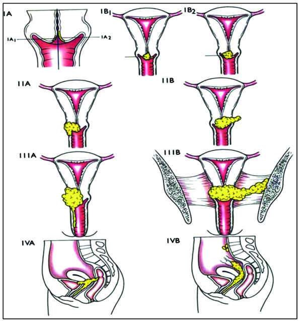
Staging of Cervical Cancer

Staging describes the extent of cancer spread. Federation for International Gynecology and Obstetrics (Figo) staging.

Stage 0: Carcinoma in situ (pre-invasive)

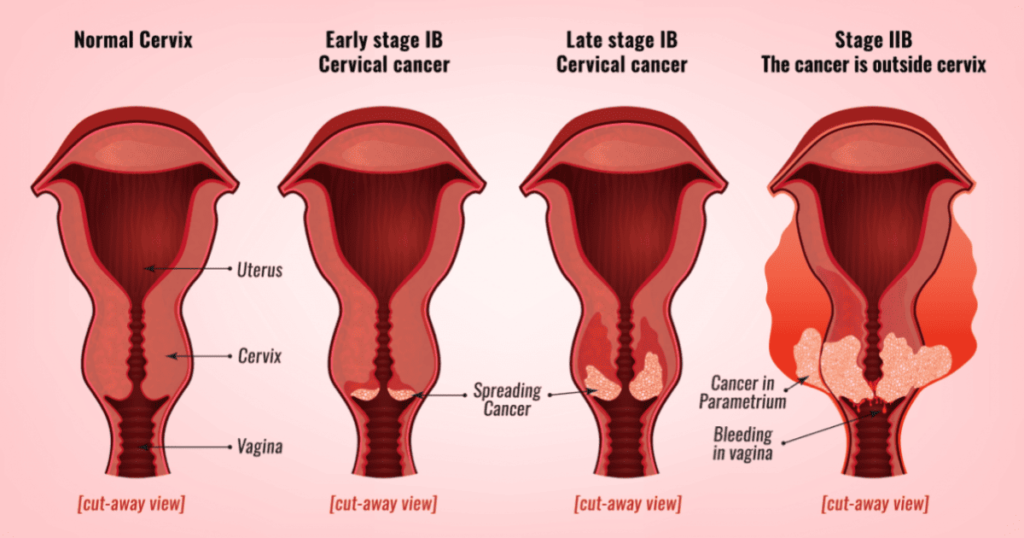
Stage I: Confined to the cervix

  • 1a: Microscopic invasion
  • 1b: Clinically visible lesion confined to the cervix

Stage II: Beyond the uterus but not to pelvic wall or lower third of vagina.

  • IIa: Limited to 2/3 of the vagina
  • IIb: Parametrial invasion(Cancer cells found outside the smooth muscles of the cervix.

Stage III: To pelvic wall, involves lower third of vagina, or causes hydronephrosis

  • IIIa: Invasion of lower 1/3 of the vagina.
  • IIIb: Invasion of pelvic sidewall +/- hydronephrosis

Stage IV: Invades bladder/rectum mucosa or distant metastasis.

Can spread by:

  • Direct spread to parametria on both sides, upper part of cervix, uterus, vaginal wall, bladder.
  • Lymphatic spread to lymph nodes in parametria, obturator nodes, external and internal iliac nodes, inguinal nodes, sacral nodes, hypogastric glands and rarely aortic and lumbar glands.
  • Blood spread to the lungs, liver, bone and intestines implantation.
Parametrium

Diagnosis of Cervical Cancer

1. History and Examination: Includes speculum and colposcopic examination.

2. Colposcopy-directed biopsy: Examination and tissue sample collection. Cervix-lesion may be in the form of an ulcer, Cauliflower growth.

3. Pap smear(papanicolau): Detects early-stage cancer or precancerous changes. The doctor scrapes a sample of cells from the cervix. For a Pap test, the lab checks the sample for cervical cancer cells or abnormal cells that could become cancer later if not treated.

4. Acetic Acid Test: This is of two types;

  • Unaided Visual Inspection (UVI): 3% acetic acid is painted on to the cervix. The abnormal area stains white and is biopsied to find out what type of lesion it is.
  • Aided Visual Inspection(AVI): cervix is painted with 3% acetic acid using a magnifying instrument to find the lesions present.

5. HPV testing: Identifies high-risk HPV strains.

6. Other tests: Full blood count, urea and electrolyte levels, liver function tests.

7. Biopsy: This is a surgical removal of tissue to look for cancer cells and usually done under local anesthesia. This may be done if cervical smear reveals evidence of cervical intraepithelial neoplasia. The tissue sample obtained is sent to the pathologist for histology and for confirmation.

Treatment and Prevention of Cervical Cancer

  • Pre-invasive: Pre invasive- lesions are destroyed using methods like liquid carbon dioxide, laser beam(leep)loop, electric excision procedure where the doctor uses an electric wire loop to slice off a thin, round piece of cervical tissue. Lesions destroyed using cryotherapy, laser beam, or LEEP.
  • Invasive carcinoma: Treatment is by wertheim’s hysterectomy. This involves total hysterectomy with removal of the upper 1/3 of the vagina as well as dissection of the lymph nodes including Para-aortic nodes plus salpingo-oophorectomy and this can be followed by radiotherapy. Treated with surgery (e.g., hysterectomy), chemotherapy, and radiotherapy.
  • Radiation therapy: This is the use of high-energy rays to kill cancer cells.
  • It’s an option for women with any stage of cervical cancer and may prefer radiation therapy to surgery.
  • It may also be used after surgery to destroy any cancer cells that remain in the area.
  • For women with cancer that extends beyond the cervix may need to combine radiation therapy and chemotherapy.

Surgical Management

Surgery is an option for women with Stage I or II cervical cancer. If you have a small tumor, the type of surgery may depend on whether you want to get pregnant and have children later on. Some women with very early cervical cancer may decide with their surgeon to have only the cervix, part of the vagina, and the lymph nodes in the pelvis removed (radical trachelectomy).

Prevention

  • Primary Prevention: Includes vaccination, health education, promoting safe sexual practices, reducing drug abuse, and regular screening.
  • Secondary Prevention: Early detection through regular screening and prompt treatment of precancerous lesions.

Primary Prevention

Since cervical cancer is often caused by a sexually transmitted infection (STI), steps can be taken to prevent its incidence. Primary prevention involves reducing or eliminating risk factors.

Vaccination: Encourage HPV vaccination to prevent cervical cancer.

Community Health Education

  • Promote awareness about the importance of early marriages and safe sexual practices.
  • Conduct educational programs to reduce drug abuse and promote the use of condoms.
  • Advocate for reducing the number of sexual partners.
  • Encourage behavior change and improved hygiene.

Men Involvement: Involve men in educational programs to promote understanding and support for prevention measures.

Income Generating Activities: Support income-generating activities to improve community well-being and reduce risk factors associated with poverty.

Secondary Prevention

Secondary prevention involves methods to detect cancer in its earliest stages so that treatment can begin as soon as possible.

Screening

  • Promote regular Pap smear tests to detect early cervical changes.
  • Ensure early referral to higher levels of care for further evaluation and treatment if needed.

Awareness and Training

  • Create awareness among health workers about the importance of early detection.
  • Train healthcare providers to perform screenings effectively.

Cost and Accessibility

  • Reduce the cost of screening to make it more accessible to the population.
  • Provide additional radiotherapy units in the country to extend services closer to the people.

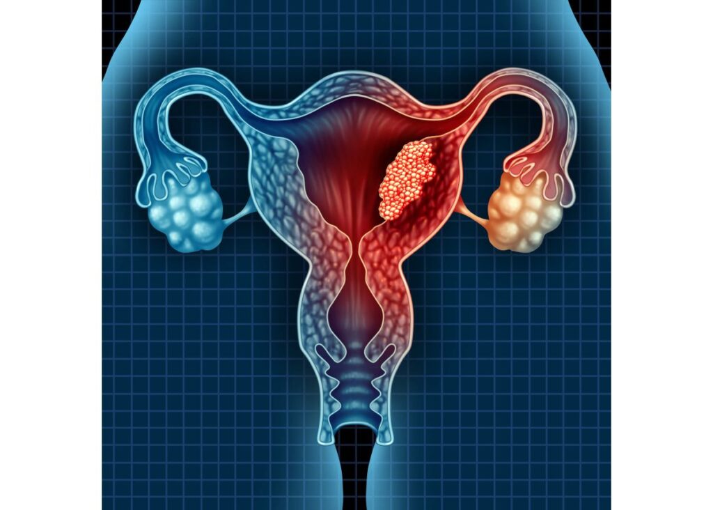
Endometrial Cancer/Uterine Cancer

Endometrial cancer is a malignant tumor within the endometrium, resulting in abnormal cell growth that can invade or spread to other parts of the body. 

Incidence/Epidemiology

  • It is the sixth most common cancer in women globally.
  • More common in developed countries, with a lifetime risk of 1.6% compared to 0.6% in developing countries.
  • Occurs in 12.9 out of 100,000 women annually in developed countries.
  • Most frequently appears during peri-menopause (ages 50-65).
  • 75% of cases occur after menopause.
  • Women younger than 40 make up 5% of cases; 10-15% occur in women under 50.

Causes/Risk Factors

The exact cause is idiopathic, but it is associated with:

  • High blood pressure
  • Diabetes
  • Excessive or long-term estrogen exposure
  • Polycystic ovary syndrome (PCOS)
  • Functioning ovarian tumors
  • Anovulation
  • Infertility
  • Family history or genetic factors
  • Obesity
  • Late menopause
  • Early menarche
  • Age above 55 years
  • Excessive use of tamoxifen
  • Nulliparity (never having had children)

Classifications of Endometrial Cancer

  • Type 1 Endometrial Carcinoma: Estrogen-related, occurs in younger, obese, premenopausal women, usually low-grade and endometrioid.
  • Type 2 Endometrial Carcinoma: High-grade, usually serous or clear cell, affects older women.
  • Type 3 Endometrial Carcinoma: Hereditary or genetic types, some related to Lynch II syndrome.

Clinical Presentation

  • Vaginal bleeding or spotting in postmenopausal women (90% of cases).
  • Abnormal menstrual cycles or heavy, frequent bleeding in premenopausal women.
  • Thin white or clear vaginal discharge in postmenopausal women.
  • Enlarged uterus on physical examination.
  • Lower abdominal pain, pelvic cramping, painful sexual intercourse, painful or difficult urination (with metastasis).

Diagnosis

  • History and physical examination.
  • Dilation and curettage.
  • Transvaginal ultrasound to examine endometrial thickness in postmenopausal bleeding.
  • Endometrial biopsy.
  • CT scan.

Differential Diagnosis

  • Senile endometritis/vaginitis.
  • Dysfunctional uterine bleeding.
  • Submucous myoma/endometrial polyps.
  • Cervical cancer.
  • Uterine sarcoma.
  • Primary carcinoma of the fallopian tube.

Management

Surgery

  1. Stage I: Total Abdominal Hysterectomy and Bilateral Salpingo-Oophorectomy (TAH-BSO).
  2. Stage II: Radical Hysterectomy.
  3. Stage III: Radical surgery with maximal debulking followed by radiotherapy.
  4. Stage IV: Radical radiotherapy, with or without hormonal therapy and/or chemotherapy.

Radiotherapy

  • Most patients with early-stage disease receive a combination of surgery and radiotherapy based on histopathological findings.
  • Surgery alone is limited to patients with endometrioid type carcinoma confined to less than 50% of the myometrial thickness.

Hormonal Therapy

  • Progestogens are the most commonly used form of hormonal therapy in endometrial cancer.

Chemotherapy

Chemotherapy is uncommon but should be considered in fit patients with systemic disease. Commonly used medications include:

  • Doxorubicin (Anthracycline) and Cisplatin
  • Carboplatin (Platinum Medicines): Use is limited by the patient’s advanced age and poor performance status.
  • Typical regimen: Cisplatin 50 mg/m² IV, Adriamycin 45 mg/m² IV on Day 1, followed by Paclitaxel 160 mg/m², repeated every 21 days.
  • Alternative regimen: Carboplatin and Paclitaxel as for ovarian cancer.

Ovarian Cancer

Ovarian cancer is a malignant growth within the ovarian tissue.

Etiology and Pathogenesis

There is a link between ovulation and epithelial ovarian cancer. Combined hormonal contraception reduces the risk by approximately 50%. Risk factors include having a first-degree relative with ovarian cancer.

Risk Factors

  • Postmenopausal women

  • Family history of ovarian cancer (mother, sister)

  • Abnormal ovarian development (e.g., Turner’s syndrome)

  • Nulliparity

  • BRCA1 and BRCA2 gene mutations

  • Smoking and alcoholism

  • Ovulatory stimulant drugs

  • High-fat diet

  • Fertility drugs

  • Hormonal replacement therapy

  • Increased number of ovulatory cycles (early menarche, late menopause)

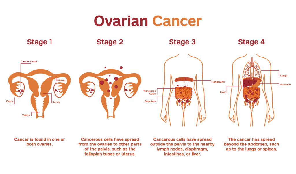
Stages of Ovarian Cancer

Stage I: Confined to the ovaries

  • 1a: One ovary involved
  • 1b: Both ovaries involved
  • 1c: Positive cytology, ascites, or capsule breach

Stage II: Confined to the pelvis

Stage III: Confined to the peritoneal cavity

  • 3a: Micronodular disease outside the pelvis
  • 3b: Macroscopic tumor deposits <2 cm
  • 3c: Tumor >2 cm or retroperitoneal node involvement

Stage IV: Distant metastases

Clinical Manifestations

Ovarian cancer often lacks early symptoms. Advanced disease may present with:

  • Pain
  • Bloating or fullness
  • Abdominal distention
  • Lower abdominal pain
  • Pelvic mass
  • Menstrual disturbances
  • Gastrointestinal symptoms
  • Pressure symptoms (dyspareunia, urinary frequency, constipation)
  • Ascites
  • Metastasis symptoms (nausea, tiredness, shortness of breath)

Investigations

  • Abdominal ultrasound
  • Intravenous urogram
  • Ascitic tap for cytology
  • Laparotomy/laparoscopy for biopsy and histology
  • CT scan and/or MRI
  • CA-125
  • Chest X-ray, FBC, liver function, renal function

Management

Surgery

  • Laparotomy with large debulking
  • Peritoneal cavity washings or ascitic fluid for cytology
  • Total abdominal hysterectomy, bilateral salpingo-oophorectomy, and infracolic omentectomy (if stage <3c)

Chemotherapy
Given to all patients post-surgery, with a 70-80% response rate:

  • Carboplatin AUC 5-7 IV and Paclitaxel 175 mg/m² IV every 21 days for 3-6 cycles
  • Cisplatin 75 mg/m² IV and Paclitaxel 135 mg/m² IV infusion over 24 hours (neurotoxic)
  • Carboplatin and Cyclophosphamide 750 mg/m² IV

Hormonal Therapy

  • Tamoxifen may be used if other treatments are inappropriate.

Radiotherapy

  • Not commonly used, but may be applied postoperatively in early-stage cancer or as palliative care in advanced cancer.

Recommendations

  • Manage pelvic pain and abdomino-pelvic mass, especially with vaginal bleeding.
  • Perform annual pelvic examinations and ultrasounds for reproductive and advanced-age women.
  • Encourage oral contraceptives for high-risk women.
  • Consider prophylactic bilateral laparoscopic oophorectomy for women not desiring fertility but at high risk.
  • CA-125 is useful for follow-up but not for screening.

Complications
Ovarian cancer often presents with complications, including:

  • Ascites
  • Bowel obstruction/intestinal occlusion
  • Bladder infiltration causing hematuria
  • Secondary deposits in liver or lung
  • Severe weight loss
  • Metastasis to other organs

Cancers of Reproductive Health Organs Read More »

OBSTETRIC/VAGINAL FISTULA

OBSTETRIC/VAGINAL FISTULA

OBSTETRIC/VAGINAL FISTULA

Vaginal Fistula is an abnormal communication (opening) of the vagina and the neighbouring -pelvic organs as a result of obstetrical causes e.g. delivery. 

Urogenital Fistula: Abnormal communication between the urinary (ureters, bladder, urethra) and genital (uterus, cervix, vagina) systems.

A fistula is an abnormal communication between two or more epithelial surfaces.

Types of vaginal/obstetric fistula

Vaginal Fistula: A general term for a fistula formed within the vaginal wall.

  1. Vesicovaginal Fistula (VVF): When a vaginal fistula extends into the urinary tract, it is specifically referred to as vesicovaginal fistula.  The most common type of urogenital fistula, occurring between the bladder and vagina.
  2. Rectovaginal Fistula (RVF): If the vaginal fistula opens into the rectum, it is termed a rectovaginal fistula.
  3. Colovaginal Fistula: An occurrence where a vaginal fistula communicates with the colon.
  4. Enterovaginal Fistula: When the opening of a vaginal fistula connects with the small bowel.

Anatomical Communications

Organ

Ureter

Bladder

Urethra

Vagina

Ureterovaginal

Vesicovaginal

Urethrovaginal

Cervix

Ureterocervical

Vesicocervical

Urethrocervical

Uterus

Ureterouterine

Vesicouterine

Not reported

common causes of fistula

General Causes of Urogenital Fistula

Obstetric Conditions/Procedures
  • Prolonged, Obstructed Labor: Prolonged pressure of the foetal head against the pelvic tissues during obstructed labour can cause ischemia and necrosis of the vaginal wall and bladder. This necrosis can create a fistula, typically between the bladder and vagina (vesicovaginal fistula).
  • Caesarean Section (Especially Repeat Cesareans): Surgical incisions through the bladder or close to the bladder during caesarean sections can cause direct injury or lead to ischemia. This can result in a vesicovaginal fistula if the bladder is inadvertently cut or damaged.
  • Bladder is Cut: Accidental incision into the bladder while performing the surgery.

  • Bladder Wall Sutured: Suturing the bladder wall during closure of the uterus can cause damage.

  • Adherent Bladder: In women with previous caesarean scars, the bladder may adhere to the uterine scar and tear during separation.

  • Caesarean Hysterectomy: This procedure involves the removal of the uterus following a caesarean delivery. The close proximity of the bladder to the uterus increases the risk of bladder injury. Damage to the bladder during this surgery can create a vesicovaginal fistula.
  • Operative Vaginal Delivery: Use of forceps or vacuum during delivery can cause trauma to the vaginal and bladder tissues. This trauma can lead to tissue necrosis and the development of a fistula.
  • Ruptured Uterus: Uterine rupture can involve the bladder, especially in patients with previous scars where the bladder is adherent.
  • Bladder Cut or Sutured: During repair of the uterus or hysterectomy, the bladder may be inadvertently cut or sutured.
  • Symphysiotomy: During the procedure to widen the pelvis, the bladder and urethra, if not properly displaced, can be damaged. This damage can lead to a vesicovaginal or urethrovaginal fistula.
  • Cervical Cerclage: Placement of a stitch around the cervix to prevent premature birth can sometimes cause damage to surrounding tissues if not properly placed. This damage can lead to a fistula between the cervix and bladder.
Gynaecological and Urological Procedures
  • Hysterectomy: Removal of the uterus can sometimes damage the bladder or ureters due to their proximity. This can result in vesicovaginal or ureterovaginal fistulas.
  • Myomectomy: Removal of fibroids from the uterus can inadvertently damage the bladder or ureters. This can lead to vesicovaginal or ureterovaginal fistulas.
  • Loop Excision of Cervix: Treatment for cervical dysplasia involves removing abnormal cervical tissue, which can sometimes damage nearby structures. This can create a fistula if the bladder is unintentionally injured.
  • Voluntary Interruption of Pregnancy: Procedures to terminate pregnancy can sometimes cause trauma to the bladder or ureters. This trauma can result in fistula formation.
  • Anterior Colporrhaphy: Surgery to repair a cystocele (bladder prolapse) can sometimes damage the bladder. This can lead to a vesicovaginal fistula.
  • Periurethral Bulking: Injection of materials around the urethra to treat incontinence can sometimes cause trauma. This can result in a urethrovaginal fistula.
  • Urethral Diverticulum Repair: Surgery to remove a diverticulum from the urethra can cause damage to surrounding tissues. This can result in a urethrovaginal fistula.
  • Ureteral Wall Stent: Placement of stents in the ureters can sometimes cause trauma to the ureters or bladder.This trauma can result in a ureterovaginal or vesicovaginal fistula.
  • Insertion of Shirodkar Stitch: Placement of a cervical stitch to prevent preterm birth can damage the bladder if not done carefully. This can create a vesicocervical fistula
  • Dilatation and Curettage (D&C): The procedure, especially during pregnancy termination, can cause trauma to the bladder or urethra. This trauma can result in the formation of fistulas.
  • Manchester Operation: Surgery for uterine prolapse can cause damage to the bladder or urethra. This can result in a vesicovaginal or urethrovaginal fistula.
Pelvic/Medical Conditions
  • Endometriosis: Abnormal growth of endometrial tissue can invade the bladder or ureters. This invasion can create fistulas due to chronic inflammation and tissue damage.
  • Gynecologic Cancers: Tumours from cancers like cervical, uterine, or ovarian cancer can invade the bladder or ureters. Surgical removal or the tumour itself can cause fistula formation.
  • Cervical Cancer (Stage 4): Advanced cancer can invade the bladder tissues. This invasion can create a vesicocervical fistula
  • Pelvic Irradiation: Radiation therapy for pelvic cancers can cause tissue necrosis in the bladder and surrounding areas. This necrosis can lead to vesicovaginal fistulas.
  • Infections (Tuberculosis, Lymphogranuloma Venereum): These infections can cause chronic inflammation and tissue damage in the urinary and genital tracts. This damage can lead to the formation of fistulas.
  • Intrauterine Device (IUD): IUDs can sometimes perforate the uterus and migrate, causing damage to the bladder. This can lead to a vesicovaginal fistula.
  • Retention of Vaginal Foreign Object: Forgotten or unrecognized foreign objects such as tampon, diaphragm, cervical cap, pessary in the vagina can cause chronic inflammation and tissue damage. This can result in fistula formation.
  • Accidental Trauma: Blunt or penetrating trauma to the pelvic region can cause direct injury to the bladder or urethra. This injury can lead to the formation of a fistula.
  • Sexual Trauma: Violent or forced sexual activity can cause severe trauma to the vaginal and bladder tissues. This trauma can result in vesicovaginal fistulas.
  • Mitomycin C Instillation: Chemotherapy agent used for bladder cancer can cause severe bladder irritation and necrosis. This necrosis can lead to fistula formation.
  • Bladder Stone: Large bladder stones can cause chronic irritation and erosion into the bladder wall. This erosion can create a vesicovaginal fistula.

Risk Factors of Fistula

  1. Poverty: Limited access to healthcare can lead to poor management of obstetric and gynecological conditions, increasing the risk of fistulas.
  2. Malnutrition: Poor nutritional status can weaken tissues, making them more susceptible to damage during childbirth or surgery.
  3. Lack of Education: Deficient knowledge about prenatal care and safe childbirth practices increases the risk of complications leading to fistulas.
  4. Early Childbirth: Young mothers often have smaller pelvic dimensions, increasing the risk of obstructed labour and subsequent fistula formation.
  5. Lack of Healthcare: Inadequate access to skilled medical care during childbirth can result in prolonged obstructed labour or mismanaged surgical procedures.
  6. High Parity: Multiple pregnancies can increase the risk of uterine and bladder prolapse, leading to a higher risk of fistula formation during childbirth or surgical procedures.
  7. Prolonged Labour without Medical Assistance: Lack of timely medical intervention can lead to obstructed labor, increasing the risk of ischemic injury to the bladder and adjacent tissues.
  8. Inadequate Prenatal Care: Poor prenatal care can result in undiagnosed or poorly managed conditions like fetal macrosomia or malpresentation, which can complicate delivery and increase fistula risk.
  9. Pre Existing Medical Conditions: Conditions such as diabetes or hypertension can impair wound healing and tissue resilience, increasing susceptibility to fistulas.
  10. Previous Pelvic Surgeries: Scar tissue from prior surgeries can complicate new procedures and increase the risk of bladder or urethral injury.
  11. Use of Harmful Traditional Practices: Practices like female genital mutilation or the Gishiri cut can cause direct injury to the urinary and genital tracts, leading to fistula formation.

Symptoms of Urogenital Fistula

  1. Continuous Urinary Leakage: Persistent and unexplained leakage of urine from the vagina following recent surgery, a difficult vaginal delivery, or local trauma. The continuous passage of urine through the vaginal opening is due to an abnormal connection (fistula) between the bladder or urethra and the vagina.
  2. Recurrent Cystitis or Pyelonephritis: Frequent bladder infections or kidney infections. The abnormal passage allows urine to stagnate and become infected, leading to recurrent urinary tract infections.
  3. Unexplained Fever: Persistent fever without an obvious cause. Chronic infections related to the fistula can cause systemic symptoms such as fever.
  4. Hematuria: Presence of blood in the urine. Trauma or infection around the fistula site can lead to bleeding into the urinary tract.
  5. Flank, Vaginal, or Suprapubic Pain: Pain in the sides (flank), vagina, or above the pubic bone (suprapubic). Inflammation, infection, and ongoing leakage of urine can cause significant pain in these areas.
  6. Abnormal Urinary Stream: Changes in the usual pattern of urination. The fistula can disrupt the normal flow of urine, leading to an abnormal urinary stream.
  7. Vaginal, Vulvar, and Perineal Irritation: Irritation or discomfort in the vaginal, vulvar, and perineal areas. Constant exposure to urine can irritate these tissues, leading to inflammation and discomfort.
  8. Foul Ammoniacal Odour: A strong, unpleasant smell resembling ammonia. Bacterial activity in the urine leads to the production of ammonia, causing a foul odour.
  9. Severe Perineal Dermatitis: Severe skin irritation and inflammation in the perineal area. Continuous contact with urine can lead to dermatitis, characterized by redness, swelling, and irritation of the skin.
  10. Greenish-Gray Phosphate Crystals in the Vagina and Vulva: Presence of greenish-gray deposits on vaginal and vulvar surfaces.Bacterial action on urea in urine leads to an alkaline environment, causing phosphate crystals to precipitate and deposit in the affected areas.
  11. Social Isolation, Disrupted Sexual Relations, Depression, Low Self-Esteem, Insomnia: Emotional and psychological distress due to the condition. The constant leakage of urine and associated symptoms can lead to significant social and emotional impacts, including isolation, difficulties in sexual relationships, depression, low self-esteem, and sleep disturbances.

Diagnostic Signs and Examination Findings

Patient History

  • History of Prolonged and Obstructed Labor: Key indicator of potential fistula development.
  • Mother Reports Leakage of Urine: Continuous leakage without control is a classic sign of fistula.

Physical Examination

  • No Palpable Bladder on Abdominal Palpation: Indicates that urine does not accumulate in the bladder but leaks out.
  • Urine Smell: The patient often has a characteristic smell of urine.
  • Signs of UTI and Low-Grade Fever: Recurrent infections due to urine leakage.
  • Vulva Inspection: Visible dribbling of urine from the vagina.
  • Speculum Examination: Visible defect with urine escaping through it.

Diagnostic Tests

  • Methylene Blue or Gentian Violet Test: Injection of methylene blue or gentian violet dye into the bladder via a catheter. The presence of dye leaking into the vagina confirms the fistula.
  • Soft Tissue X-ray and Cystography: Show defects and injuries in the bladder.
  • Creatinine Content in Vaginal Fluid: High levels indicate urine leakage.
  • Cystoscopy: This endoscopic examination allows direct visualization of the bladder and urethra, helping to locate the exact anatomical origin of the fistula.
  • Soft Tissue X-ray: Helps to visualize the defect and confirm the presence of a fistula.
  • Speculum Examination: Direct visual inspection using a speculum to identify and assess the fistula.
  • Digital Examination: Manual examination to feel the fistula and surrounding tissues.
  • Subtraction Magnetic Resonance Fistulography: A specialized imaging technique that can provide detailed visualization of the fistula.
  • Endocavitary Ultrasound: Transrectal or transvaginal ultrasound, potentially with Doppler or contrast agents, to visualize the fistula. Transvaginal sonography can clearly show the exact site, size, and course of the fistula.
  • Biopsy: If malignancy is suspected, a biopsy of the affected tissue is taken for histologic examination to rule out cancer.
VESICO VAGINAL FISTULA VVF

VESICO-VAGINAL FISTULA

Vesicovaginal fistula or VVF is an abnormal fistulous tract extending between the bladder (vesico) and the vagina that allows the continuous involuntary discharge of urine into the vaginal vault. 

OR: It is the abnormal opening of the vagina and the urinary bladder.

Pathology of a urinary fistula

  • If the cause is a tear, urine escapes at once but the wound may not become infected immediately and primary union can occur in one week or two provided the urinary stream is diverted.
  • If the cause is pressure necrosis, the affected area will form a slough which eventually drops out leaving a fistula.
  • If the fistula is large (over 2 cm diameter) spontaneous healing is unlikely and scar tissue gradually forms a dense white ring round the edge of the fistula even fixing it to the pubic ramus.
  • Urinary fistula has a natural tendency to close by granulation, fibrosis, and contraction.
types of VESICO-VAGINAL FISTULA

Types of VVF

  • Simple fistula: Only about 20% of obstetric fistulas can be defined as simple. Simple fistulas are less than 3 cm in diameter with no or only mild scarring and do not involve the urethra.
  • Complex fistula: A complex obstetric fistula can be described as being larger than 3 cm, involving the urethra and associated with reduced vaginal capacity from significant scarring and/or a reduced bladder volume. Sometimes the defect may be urethrovaginal, but more commonly both the urethra and bladder are involved and therefore the fistula is called a urethro-vesicovaginal.

Management of vesico – vaginal fistula

If the woman is very ill and the fistula is small and does not involve the urethra: she can be managed conservatively while treating the cause of the illness.

  • Small fistula: Small vesico-vaginal fistulas can often be repaired with a high chance of success. A catheter is passed into the bladder and left in place for several days to keep the bladder empty while the tissues heal.
  • For larger fistulas: If the tissues are badly damaged or if the fistula involves the urethra or if the tissues are very scarred and inflexible, delayed repair or reconstructive surgery may be needed.
  • With the conservative treatment of the large fistulae, a catheter is passed into the bladder through the urethra and left in place for several days to keep the bladder empty while the tissues heal.
  • Any indwelling catheter should be left in place for 2 to 3 weeks following the repair.
  • Insertion of the catheter: A catheter is inserted through the urethra to continuously drain urine from the bladder.
  • If urine is not draining, the catheter may be blocked. If the bladder is not emptying because the bladder muscle is not contracting, the catheter may be blocked or the catheter may have been inserted through the side of the urethra. If the catheter is in the vagina and not in the bladder, urine will not be draining from the catheter.

In the Health Center

  • Mother is encouraged on personal hygiene and referred to the hospital.

In Hospital

  • Mother is admitted to a gynaecological ward.
  • Doctor is informed and will carry out a gynaecological examination:
    • Genital examination with fingers, no instruments used for fear of enlarging the opening.

    • May carry out a speculum examination.

  • A self-retaining catheter is passed and the mother is kept on continuous bladder drainage as dripping of urine prevents healing.
  • Give a balanced diet including iron, vitamin supplements, and if necessary, blood transfusion to restore her general health.
  • Most fistulas will close spontaneously within 6 weeks as long as there is continuous bladder drainage, good health, and control of infections.
  • Use of antiseptic vaginal douches to clear any smell.
  • At the end of puerperium, a patient is assessed by means of speculum examination.
  • Previously, enough time was to be given to allow the tissue to heal and strengthen sufficiently.
  • Thereafter, a mother would be asked to go home and return for surgery after 3 months. Today, it can be repaired as soon as it is diagnosed.
  • During the resting and waiting time for the surgery, the following are necessary:
    • Reassurance

    • Plenty of rest

    • Good diet with high protein and vitamins for quick healing

    • Hygiene/vulva toilet

    • Wearing pads at all times and frequently changing them.

    • Use of a barrier cream to prevent excoriation of the skin, e.g., Zinc and Castor oil

    • Mother is put on continuous bladder drainage.

Actual Treatment

  • Repair the fistulae as soon as the patient is first seen.
  • Perform the necessary examination under anaesthesia to establish where urine is coming from and the appropriate position for repair.
  • This can be done together with the injection of dye through a catheter into the urinary bladder to observe where the opening is, as the dye will be seen coming out of it.
  • Then the fistulae can be repaired surgically.

Care After Repair

  • Care is similar to that for any mother after an operation or obstructed labour.
  • Mother is nursed in a supine position to prevent excessive pressure on the suture site.
  • Continuous bladder drainage to rest the bladder and allow proper healing.
  • Plenty of fluids to flush the bladder and prevent pressure on the wound. Any blood clot or debris is washed out, preventing urine stasis and urinary tract infections.
  • Maintain a fluid balance chart.
  • Observe the amount of urine passed and its colour, especially for blood clots which may block the catheter.
  • Bed is observed daily for wetness.
  • Remove the catheter after 2 weeks if the bed is dry. If the catheter remains in place, it might prevent a small area from healing or closing yet, and with time it might close.
  • Continuous bladder drainage to prevent the bladder from over-distending, ensuring proper healing. The catheter is kept in place for at least 2 weeks or until there is no more leakage of urine.
  • If urine continuously leaks onto the bed and very little or no urine is draining into the bag, chances are that the bladder repair has almost completely broken down, necessitating a repeat repair.
  • Inspect the bed to ensure it is dry.
  • Ensure there are no blood clots or debris blocking the catheter, ensuring free drainage of urine.
  • Plenty of fluids to prevent the formation of debris that could block the catheter.
  • Bladder training to release urine at increasing intervals to allow the bladder to regain its capacity and muscles to regain their tone.
  • If after 2 weeks all urine is draining into the catheter and the bed is dry, continue bladder training for 5 days.

Post-Operative Catheter Care

  • Catheter must drain freely at all times; if it becomes blocked, the operation may fail.
  • Catheter strapped to the mother’s thighs.
  • Patients must not lie on the catheter.
  • Catheter or tubing must not be twisted.
  • Drainage tubing must go into a basin or bucket at the side of the bed. Urine must be draining at all times.
  • Patient must drink fluids freely as soon as she has recovered from the anaesthetic.
  • Urine should be very pale, almost like water; if not, the patient should drink more.
  • If the catheter stops draining or the patient complains of a full bladder, the catheter must be removed immediately.
  • It must be irrigated to unblock it.
  • If irrigation fails, the catheter must be changed, usually by the doctor.
  • Apply Vaseline around the thighs.

Advice on Discharge

  • No coitus for at least 3-6 months.
  • Rest and take prescribed drugs.
  • Maintain vulva hygiene.
  • Come back for review.
  • Continue feeding well.
  • Next mode of delivery should be ELECTIVE C/S.
Complications of VVF
  • Recurrent fistula: If a fistula is closed and urine is allowed to accumulate in the bladder, the pressure may tear the repair and a new fistula may develop.
  • Sepsis: If a woman develops fever or sepsis, she must be given antibiotics.
  • Social problems: The social stigma attached to a woman with a VVF can be severe and prolonged. The constant wetness and odour of urine are offensive to the woman and those around her. She may be abandoned by her husband and family and may become an outcast.
  • Permanent conditions: Despite surgery, the woman may still leak urine. This can be because the tissues are scarred and cannot stretch, because the urethra is damaged, or because the bladder cannot empty.
  • If the woman has been leaking urine for months or years, the bladder may be too small or damaged to store the normal amount of urine.
  • Fertility: damage to the cervix and the uterus and if there is an infection in the uterus, the woman may not be able to conceive and carry a pregnancy to term.
Prevention of VVF
  • Community health education.
  • Emphasis on antenatal care.
  • Training traditional birth attendants: they should learn how to recognize prolonged labour and refer the woman for emergency care.
  • Timely referral of the woman: to a hospital for an emergency c/s if needed.
  • Government support: it should be provided to improve facilities and personnel.

RECTO-VAGINAL FISTULA 

Recto-vaginal fistula is the connection between a woman’s rectum and vagina. The opening allows stool and gas to leak from the bowel into the vagina.

Causes

  • Complications during childbirth: During difficult delivery, the perineum can tear, or when performing an episiotomy to deliver the baby.
  • Inflammatory bowel disease (IBD): Conditions such as Crohn’s disease and ulcerative colitis cause inflammation in the digestive tract and can increase the risk of developing a fistula in rare cases.
  • Cancer or radiation to the pelvis: Cancer in the vagina, cervix, rectum, uterus, or anus can cause a recto-vaginal fistula. Radiation to treat these cancers can also create a fistula.
  • Surgery: Surgery on the vagina, rectum, perineum, or anus can cause an injury or infection that leads to an abnormal opening.
  • Infections due to HIV.
  • Sexual assault.

Signs and Symptoms

  • Passing stool or gas from the vagina.
  • Trouble controlling bowel movements.
  • Smelly discharge from the vagina.
  • Repeated vaginal infections.
  • Pain in the vagina or the area between the vagina and anus (perineum).
  • Dyspareunia (painful intercourse).

Risk Factors

  • Mother with prolonged labor.
  • Mother with obstructed labor.
  • Episiotomy during labor.
  • Women with infections such as an abscess or diverticulitis.
  • Women having cancer of the vagina, cervix, rectum, uterus, or anus, or radiation to treat these cancers.
  • Women who have undergone a hysterectomy or other surgeries to the pelvic area.

Diagnosis

  • History taking: The doctor will ask about symptoms and perform a physical examination.
  • Physical examination: With a gloved hand, the doctor will check the vagina, anus, and perineum. A speculum may be inserted into the vagina to open it up so the doctor can see the area more clearly. A proctoscope can help the doctor see into the anus and rectum.
  • Tests:
    • Anorectal or transvaginal ultrasound: A wand-like instrument is inserted into the anus and rectum, or into the vagina. An ultrasound uses sound waves to create a picture from inside the pelvis.

    • Methylene enema: A tampon is inserted into the vagina, then a blue dye is injected into the rectum. After 15 to 20 minutes, if the tampon turns blue, one has a fistula.

    • Barium enema: A contrast dye helps a doctor see the fistula on an X-ray.

    • CT scan: Uses powerful X-rays to make detailed pictures inside the pelvis.

    • MRI: Uses strong magnets and radio waves to make pictures from inside the pelvis. It can show a fistula or other problems with the organs, such as a tumor.

Management

Surgery: The main treatment for a fistula is surgery to close the abnormal opening. However, surgery can’t be performed if there is an infection or inflammation. The tissues around the fistula need to heal first.

  • The doctor might decide to wait for three to six months for an infection to heal and to see if the fistula closes on its own. Antibiotics are given to treat an infection or infliximab (Remicade) to bring down inflammation if the patient has Crohn’s disease.

While waiting for surgery:

  • Take antibiotics and analgesics.
  • Keep the area clean. Wash the vagina gently with warm water if you pass stool or a foul-smelling discharge. Use only gentle, unscented soap. Pat the area dry.
  • Use unscented wipes  instead of toilet paper after visiting the bathroom.
  • Apply talcum powder or a moisture-barrier cream to prevent irritation in the vagina and rectum.
  • Wear loose, breathable clothing made from cotton or other natural fabrics.
  • If leaking stool, wear disposable underwear or an adult diaper to keep the feces away from the skin.

Surgery options:

  • Vaginal repair: Usually done when the fistula is in the lower half of the vagina or near the perineum.
  • Abdominal repair: Used by a general surgeon when repairing a recto-vaginal fistula arising in the vault after hysterectomy or radiotherapy.
  • During surgery, the doctor will take a piece of tissue from somewhere else in the body and make a flap or plug to close the opening.
  • The surgeon will also fix the anal sphincter muscles if they are damaged.
  • Some women will need a colostomy if a fistula is large and if continuing malignant tissue is suspected.

Complications

  • Recto-vaginal fistula can affect sex life.
  • Trouble controlling the passage of stool (faecal incontinence).
  • Repeated urinary tract or vaginal infections.
  • Inflammation of the vagina or perineum.
  • Abscess in the fistula.
  • Another fistula after the first one is treated.

Prevention

  • Health education to women on regular ANC services.
  • Early detection of associated risks and appropriate referral should be made.
  • Proper monitoring of labour using the partograph.
  • Skilled attendance at all births.

RELATED QUESTION

Objectives

  • Define obstetrical fistula.
  • Define fistula.
  • General causes of fistula.
  • General signs and symptoms of fistula.
  • Classifications of fistula.
  • Define VVF and RVF.
  • Investigations of VVF.
  • Management of VVF and RVF.
  • Prevention of VVF.
  • Complications of VVF.

Fistula

  • Obstetrical fistula: An opening or passage between organs of the genital tract and the urinary tract.
  • Fistula: An abnormal communication between two organs.

Classifications of Fistula

  • Vesico-vaginal fistula: Between bladder and vagina.
  • Recto-vaginal fistula: Between rectum and vagina.
  • Vesico-uterine fistula: Between bladder and uterus.
  • Urethro-cervical fistula: Between urethra and cervix.
  • Uretero-cervical fistula: Between ureter and cervix.

General Causes of Fistula

Obstetrical causes:

  • Poorly performed episiotomy.
  • Instrumental delivery (e.g., vacuum extractor and forceps delivery).
  • Operations such as caesarean sections.
  • Prolonged labor due to narrowing of pelvis.
  • Obstructed labor due to compressions from fetal head and symphysis pubis.

Gynecological causes:

  • Injuries caused during operations (e.g., hysterectomy and myomectomy).
  • Criminal abortion (e.g., use of sticks and other sharp objects).

Traumatic causes:

  • Direct trauma on the bladder or rectum due to road traffic accidents.
  • Falls on sharp pointed objects.

Radiation:

  • Common during treatment of cancer of the genital organs by radiotherapy rays.

Infections:

  • Infections like tuberculosis (e.g., tuberculosis of vagina may infiltrate normal tissues or cells).

Malignancy:

  • Abnormal growth of tissues in the vagina, cervix, bladder, and vagina.

General Signs and Symptoms of Fistula

History taking:

  • A mother may give a history of prolonged labor or obstructed labor leading to the rupture of the uterus.

Passage of urine:

  • Passage is kept open by chronic inflammations leading to continuous passage of urine in the vagina.
  • No bladder is felt on abdominal palpation since all urine escapes as soon as it reaches the bladder.

On vulva inspection:

  • Urine is seen dribbling from the vagina.
  • On speculum examination (e.g., Casco’s), the bladder mucosa may be seen prolapsed through fistula.
  • Complete wetness of the underwear due to continuous dribbling of urine.
  • Signs of urinary tract infections (e.g., syphilis, candida, and gonorrhea) may occur.

Other signs and symptoms:

  • Stool: Faeces will be seen in the vagina during recto-vaginal fistula.
  • Pain: The mother will feel pain during fistula.
  • Excretion of the vagina: Offensive smell which may be due to infections and itching of the vulva.

Vesico-Vaginal Fistula

  • Definition: An abnormal communication between the bladder and the vagina.

Investigations of VVF

  • Retrograde pyelography: Used to visualize the ureter.
  • Intravenous urography: Used to visualize the abnormalities of ureter and bladder.
  • Ultrasound scanning: Used to examine the interior of hollow organs.
  • Cystography: In complex fistula where lateral view of uterine cavity may be seen.

Management of VVF

Surgery:

  • Done after 3 months following delivery in case of old VVF.
  • If fistula is recognized within 24 hours, it may be repaired immediately if it is small.

Aims of management:

  • To relieve pain.
  • To relieve anxiety.
  • To promote quick recovery.
  • To prevent complications.

Management in the health center:

  • Receive the mother and relatives.
  • Offer a seat for them.
  • Take personal history.
  • Take gynecological history and call for an ambulance if it is a surgical condition.
  • Fill the referral form while reassuring the mother and accompanying her to the hospital, observing the vitals to rule out other bacterial infections.
  • Hand over the mother to the nurse/midwife on duty and give a report about the mother.

Hospital management:

  • The nurse on duty receives the mother and takes vital observations to rule out other abnormalities.
  • Admit the mother to the gynecological ward, have her sign a consent form, and call the doctor.
  • The nurse takes a brief history and continues taking vitals as the doctor comes.
  • The doctor carries out a genital examination and speculum examination.
  • Self-retaining catheter is passed, and she is kept on continuous bladder drainage as dripping urine prevents wetting of the linen.
  • The mother is put on appropriate antibiotics to treat any bacterial infection as prescribed by the doctor.
  • Encourage the mother to eat nutritious foods which are light and can easily be digested.
  • Most fistulas will close spontaneously within 6 weeks as long as there is continuous bladder drainage, good health, and control of infections.
  • In case of swelling or vaginal discharge due to sloughing of necrotic tissue, antiseptic vaginal douches are given (e.g., vaginal douche syringe to suck the fluid).
  • At the end of the puerperium, the patient is assessed by use of speculum.
  • Allow enough time for tissue healing and strengthening before scheduling surgery after 3 months.
  • Remove the catheter at 6 weeks if it is no longer needed; if urine continues to drip, prepare the mother for surgery.
  • Reassure the mother before taking her to the theater for operation.

Pre-Operative Management

  • The mother should have plenty of rest and sleep.
  • Encourage a nutritious diet with light, high-protein, and vitamin-rich foods to aid healing.
  • Maintain hygiene with vulva toileting three times a day.
  • Teach the patient about the condition, signs, and symptoms, and to wear a pad at all times, changing frequently to avoid infections.
  • Reassure the mother to relieve anxiety.
  • Administer drug therapy (antibiotics for bacterial infections and pain killers for pain relief).
  • Take vital observations to rule out other abnormalities.
  • Prepare the mother for operation by bathing, shaving, dressing in theater gown, and informing the theater nurse.
  • Accompany the mother to the theater and hand her over to the theater nurse with the necessary forms and details.
  • Prepare a post-operative bed for the patient’s return from the theater.

Post-Operative Management

  • The patient is received by qualified nurses from the theater with all necessary information.
  • Vital observations are taken to confirm the mother’s condition.
  • The mother is taken to the gynecological ward and placed on a post-operative bed.
  • Position the mother in a comfortable position (e.g., prone position) to relieve pressure on the bladder.

Special Nursing Care

  • Record vital observations (TPR/BP) every 15 minutes, 1 hour, 2 hours, and 4 hours for the first 24 hours.
  • Keep the mother on complete bladder drainage for 14 days.
  • Maintain a fluid balance chart, ensuring the correct rate and amount of ordered fluids.
  • Check the level, color, and care of the urine drainage bag and catheter.
  • Administer antibiotics (e.g., Ampicillin 1g every 6 hours and Gentamycin 160mg once daily) for prophylaxis.
  • Encourage plenty of oral fluids to prevent stenosis.

General Nursing Care

Hygiene:

  • Change linens daily and perform vulva swabbing twice a day.
  • Pay attention to perineal hygiene and provide bed baths.
  • Rest the bowel for 4 days to prevent pressure on the pelvic floor, using roughages and suppositories to ease defecation.

Bladder training:

From the 15th day post-operation, perform dye tests and release the catheter alternately if the test is negative:

  • 15th day: 30 minutes
  • 16th day: 1 hour
  • 17th day: 2 hours
  • 18th day: 3 hours
  • 19th day: 4 hours

If all is well within 8 hours, the catheter is completely removed if the bed is dry.

Diet:

  • Encourage a nutritious diet to repair worn-out tissues.

Psychological care:

  • Counsel the mother about the operation and provide psychological support.
  • On discharge, advise the mother to avoid sex for 3-6 months and to seek antenatal care if she becomes pregnant again.
  • Ensure perineal hygiene.
  • Educate the mother about family planning, nutritious diet, and bowel and bladder care.

Prevention of Vesico-Vaginal Fistula

  • Early referral of a mother identified with obstructed labor to a hospital.
  • Encourage all primigravidae to deliver in properly supervised maternity units.
  • Identify complete perineal tears promptly.
  • Do not allow any mother to go into the second stage of labor with a full bladder.
  • Administer broad-spectrum antibiotics to prevent bacterial infections.

Complications of Vesico-Vaginal Fistula

  • Psychological trauma due to stress.
  • Divorce leading to breakage of marriage.
  • Necrosis of the skin around the thigh.
  • Keloids (tumors in scars).


Bladder Training Post-Operatively for Vesico-Vaginal Fistula

Bladder training is a critical part of post-operative care for patients who have undergone surgery for vesico-vaginal fistula. The goal is to gradually restore normal bladder function and control.

Bladder Training Schedule:

Starting from the 15th day after the operation, a structured bladder training program is implemented. This involves intermittent removal of the catheter to allow the bladder to fill and empty on its own, helping to strengthen the bladder muscles and improve control.

  • 15th Day:
    • Remove the catheter for 30 minutes.

    • During this time, the patient should try to void naturally.

    • Reinsert the catheter after 30 minutes to drain any residual urine and prevent over-distension of the bladder.

  • 16th Day:

    • Remove the catheter for 1 hour.

    • Encourage the patient to drink fluids and attempt to void naturally.

    • Reinsert the catheter after 1 hour.

  • 17th Day:

    • Remove the catheter for 2 hours.

    • Continue to monitor the patient’s ability to void and ensure adequate fluid intake.

    • Reinsert the catheter after 2 hours.

  • 18th Day:

    • Remove the catheter for 3 hours.

    • Observe the patient’s ability to control urination and the amount of urine voided.

    • Reinsert the catheter after 3 hours.

  • 19th Day:

    • Remove the catheter for 4 hours.

    • This extended period allows the patient to test their bladder control for a longer duration.

    • Reinsert the catheter after 4 hours.

  • 20th Day Onwards:

    • If the patient is able to control urination and the bed remains dry, the intervals without the catheter can be gradually increased.

    • Eventually, if there are no issues with leakage or retention, the catheter can be removed completely.

  • Patient Monitoring: Throughout the bladder training process, closely monitor the patient for signs of urinary retention, infection, or discomfort.
  • Fluid Intake: Encourage the patient to drink plenty of fluids to promote regular urination and prevent dehydration.
  • Hygiene: Maintain good perineal hygiene to prevent infections, especially during periods when the catheter is removed.
  • Reassurance: Provide reassurance and support to the patient, as they may experience anxiety or discomfort during the initial stages of bladder training.
  • Documentation: Keep detailed records of the patient’s fluid intake, urine output, and any symptoms or issues that arise during the training period.

Purpose of Bladder Training:

  • Strengthen Bladder Muscles: Gradually increasing the time the bladder holds urine helps strengthen the bladder muscles, improving control.
  • Prevent Incontinence: Regular intervals of voiding help in regaining control over urination and reduce the risk of incontinence.
  • Help in Recovery: Bladder training is a key part of the overall recovery process, ensuring the patient can return to normal bladder function as soon as possible.


OBSTETRIC/VAGINAL FISTULA Read More »

Pelvic Inflammatory Diseases (PID)

Pelvic Inflammatory Diseases (PID)

Pelvic Inflammatory Diseases (PID)

Pelvic inflammatory disease (PID) refers to various inflammatory conditions affecting the upper genital tract in females.

Pelvic inflammatory diseases are diseases of the upper genital tract.

It is a spectrum of infection and inflammation of the upper genital tract organs involving the endometrium, fallopian tubes, ovaries, pelvic peritoneum and surrounding structures.


Infections, often ascending from the vagina, can lead to salpingitis, endometritis, pelvic peritonitis, or the formation of tubo-ovarian abscesses.

Aetiology of Pelvic Inflammatory Diseases

Exact cause is unknown but PID is often attributed to multiple pathogens, including

  • Neisseria Gonorrhoeae: A bacterium that causes the sexually transmitted infection gonorrhoea. If left untreated, gonorrhoea can ascend from the cervix to the upper reproductive organs, leading to PID.
  • Chlamydia Trachomatis: The bacterium responsible for chlamydia, another common sexually transmitted infection. Chlamydia can infect the cervix and ascend to the uterus and fallopian tubes, leading to PID.
  • Mycoplasma: Certain species of Mycoplasma, such as Mycoplasma genitalium, have been implicated in PID. These bacteria can cause inflammation and infection in the reproductive tract.
  • Gardnerella Vaginalis: An overgrowth of Gardnerella vaginalis can lead to bacterial vaginosis, an imbalance of vaginal bacteria that can contribute to the development of PID.
  • Bacteroides: Bacteroides species are anaerobic bacteria that can be involved in the polymicrobial infection associated with PID.
  • Gram-Negative Bacilli like Escherichia Coli: Certain gram-negative bacteria, including Escherichia coli, commonly found in the gastrointestinal tract, can cause infections in the reproductive organs, contributing to the development of PID.

Risk Factors/Other Factors.

The aetiology of pelvic inflammatory diseases (PIDs) can be attributed to several other factors, including:

  1. Sexually Transmitted Infections (STIs): Infections such as chlamydia and gonorrhoea are common causes of PID. These bacteria can travel from the cervix to the upper genital tract, leading to inflammation and infection.
  2. Bacterial Vaginosis: Imbalance of normal vaginal bacteria can increase the risk of developing PID. The overgrowth of harmful bacteria can lead to inflammation and infection of the reproductive organs.
  3. Postpartum or Post-Abortion Infections: Infections following childbirth or abortion can lead to inflammation of the reproductive organs, increasing the risk of PID.
  4. IUD Insertion: Insertion of intrauterine devices (IUDs) for contraception can introduce bacteria into the reproductive tract, potentially leading to PID.
  5. Endometrial Procedures: Certain medical procedures, such as endometrial biopsy or dilation and curettage (D&C), can introduce bacteria into the uterus, increasing the risk of PID.
  6. Unprotected Sexual Activity: Engaging in unprotected sexual activity with multiple partners can increase the risk of acquiring STIs, which can lead to PID.
  7. Douching: Douching is the practice of washing or flushing the vagina with water or other fluids. It can disrupt the natural balance of bacteria in the vagina, increasing the risk of developing PID.
  8. Previous PID Infections: Individuals with a history of pelvic inflammatory disease are at an increased risk of developing recurrent episodes of PID.
  9. Multiple or New Sexual Partners: Engaging in sexual activity with multiple partners or having a new sexual partner can elevate the risk of acquiring sexually transmitted infections (STIs) that can lead to PID.
  10. History of STIs in the Patient or Her Partner: A history of sexually transmitted infections, such as chlamydia or gonorrhea, in either the patient or her partner can increase the likelihood of developing PID.
  11. History of Abortion: Previous induced abortions can be a risk factor for PID, particularly if the procedure leads to infections in the reproductive tract.
  12. Young Age (Less Than 25 Years): Younger individuals, particularly adolescents, are at a higher risk of PID, possibly due to increased sexual activity and immature cervix, which may facilitate the spread of infections.
  13. Postpartum Endometritis: Infections following childbirth, particularly involving the lining of the uterus (endometritis), can increase the risk of developing PID.
Clinical Manifestations of Pelvic Inflammatory Diseases (PID)

Clinical Manifestations of Pelvic Inflammatory Diseases (PID)

  1. Lower Abdominal Pain (usually <2 weeks): Patients with PID commonly experience pain in the lower abdominal region, usually lasting for less than two weeks. This pain is often a result of the inflammation and infection affecting the pelvic organs.  The nature of the pain is bilateral, affecting both sides of the lower abdomen.
  2. Dysuria, Fever: Dysuria (painful or difficult urination) and fever are indicative symptoms of PID. These manifestations result from the inflammatory response and the body’s attempt to combat the infection.
  3. Smelly Vaginal Discharge Mixed with Pus: PID can lead to an alteration in vaginal discharge, which may become malodorous and contain pus. This change is a consequence of the infection affecting the reproductive organs and the discharge’s composition.
  4. Painful Sexual Intercourse (Dyspareunia): Dyspareunia, or pain during sexual intercourse, is a common symptom of PID. Inflammation and infection can make sexual activity uncomfortable or painful.
  5. Cervical Motion Tenderness: Cervical motion tenderness is a clinical sign observed during a pelvic examination. It involves pain or discomfort when the cervix is moved, indicating inflammation in the pelvic region, specifically around the cervix.
  6. Abnormal Uterine Bleeding: PID may cause irregular or abnormal uterine bleeding. The inflammatory processes can disrupt the normal menstrual cycle, leading to unusual bleeding patterns.
  7. Palpable Swellings in Severe Cases: In severe cases of PID, palpable swellings may be detected, indicating the presence of pus in the fallopian tubes or the development of a pelvic abscess. Signs of peritonitis, such as rebound tenderness (pain upon release of pressure), suggest an advanced and serious stage of the disease.
  8. Urinary Symptoms: PID can sometimes affect the nearby urinary structures, leading to symptoms like increased frequency or urgency of urination. This occurs due to the proximity of the reproductive and urinary organs in the pelvic region.
  9. Gastrointestinal Symptoms: PID’s inflammatory processes can extend to the gastrointestinal tract, causing symptoms such as nausea, vomiting, or diarrhoea. These symptoms may result from the proximity of the reproductive and digestive organs in the pelvic cavity.
  10. Painful Bowel Movements: PID can cause inflammation around the pelvic organs, leading to pain during bowel movements. This symptom is a consequence of the infection affecting the nearby structures.
  11. Adnexal Mass:  The presence of an adnexal mass, indicating swelling or enlargement in the region near the uterus and ovaries, can be detected in PID cases. This mass is a clinical finding associated with pelvic inflammation.
  12. Speculum Examination: A speculum examination may reveal a congested cervix with purulent discharge, providing visual evidence of cervical involvement in PID.
  13. Intermenstrual Bleeding: Intermenstrual bleeding, occurring between regular menstrual cycles, is another symptom associated with PID, contributing to the spectrum of abnormal bleeding patterns.
  14. Post-coital Bleeding: Post-coital bleeding, or bleeding following sexual intercourse, is highlighted as a distinctive symptom of PID, reflecting the impact of inflammation on the reproductive organs.
Diagnosis and Investigations for Pelvic Inflammatory Diseases (PID)

Diagnosis and Investigations for Pelvic Inflammatory Diseases (PID)

  1. Gram Staining: Gram staining to detect intracellular diplococci, providing microscopic evidence of bacterial presence. This method aids in identifying pathogens like Neisseria gonorrhoeae.
  2. Cervical Culture and Sensitivity: Collecting pus samples for culture and sensitivity from the cervix helps identify the specific microorganisms causing the infection and their sensitivity to antibiotics.
  3. Abdominal Pelvic Ultrasound Scan: An ultrasound scan assesses the abdominal and pelvic regions. While it may appear normal in some cases, it is crucial for detecting complications such as pelvic tubo-ovarian abscess or hydrosalpinx.
  4. Pelvic Tubo-Ovarian Abscess: Visualization of a pelvic tubo-ovarian abscess is a diagnostic indicator, revealing a localized collection of pus and inflammatory tissue within the pelvic region.
  5. Physical Examination – Must Include:
  • Lower Abdominal Pain (LAP): Assessment of lower abdominal pain,, as PID commonly presents with pelvic discomfort.
  • Cervical Motion Tenderness: Tenderness observed during movement of the cervix is a clinical sign of PID.
  • Adnexal Tenderness: Tenderness in the adnexal region (near the uterus and ovaries).

6. Speculum Examination: A speculum examination assists in assessing the cervix and vaginal canal.

7. Pregnancy Test: Conducting a pregnancy test is essential to rule out pregnancy-related causes of pelvic symptoms.

Management of Pelvic Inflammatory Diseases

Management of Pelvic Inflammatory Diseases

Aims of Management 

  • To eliminate the infection.
  • To relieve symptoms.
  • To prevent complications.

Medical Management:

Outpatient treatment involves a combination of medications covering multiple microorganisms.

  • Ceftriaxone 250 mg IM (or cefixime 400 mg stat if ceftriaxone is not available)
  • Doxycycline 100 mg orally every 12 hours for 14 days
  • Metronidazole 400 mg twice daily orally for 14 days
  • In pregnancy, erythromycin 500 mg every 6 hours for 14 days replaces doxycycline.

  • Do not use doxycycline during pregnancy and breastfeeding.

For severe Cases, Admission is considered.

  • Severe cases or those not improving after 7 days require referral for ultrasound scan and parenteral treatment.
  • Patients with severe PID should be admitted and injectable antibiotics should be given for at least 2 days then switch to the oral antibiotics.
  • IV Clindamycin 900 mg 8 hourly plus gentamycin 2 mg/kg loading dose then 1.5mg/kg 8 hourly. OR Ceftriaxone 1 g IV daily plus metronidazole 500mg IV every 8 hours until clinical improvement, then continue oral regimen.
  • Note: A number of patients have repeated infection resulting from inadequate treatment or re-infection from untreated partners.
  • Therefore: Male sexual partners should be treated with drugs that cover N.gonorrhoeae and C. Trachomatis to avoid reinfection.ie. Cefixime 400 mg stat Plus Doxycycline 100mg 12 hourly for 7 days.

Nursing Interventions for Pelvic Inflammatory Disease (PID):

  1. Assessment (History and Physical Examination): Thorough assessment, including a detailed history and physical examination, helps identify specific symptoms, risk factors, and the extent of pelvic involvement.
  2. Fever Management: Effective management of fever involves monitoring temperature regularly and implementing interventions such as antipyretic medications and cooling measures to ensure patient comfort and prevent complications.
  3. Pain Management: Over-the-counter nonsteroidal anti-inflammatory drugs (NSAIDs), like ibuprofen, can alleviate pelvic pain and inflammation. Prescription pain medications may be considered for severe cases.
  4. Anxiety Alleviation:  Addressing emotional well-being is crucial. Provide support and information to alleviate anxiety related to the diagnosis, treatment, and potential complications of PID.
  5. Health Education: Patient education focuses on understanding PID, its causes, and the importance of compliance to prescribed medications. Information on preventive measures, symptom recognition, and follow-up care is also provided.
  6. Rest and Sleep Promotion: Encouraging adequate rest and sleep aids in the body’s recovery process. Assist in creating a conducive environment for rest, addressing discomfort and promoting relaxation.
  7. Hygiene (Bowel and Bladder Care):  Maintaining proper hygiene, especially regarding bowel and bladder care, is emphasized to prevent infections and promote overall well-being during the recovery phase.
  8. Dietary Guidance: Provide dietary recommendations to support healing. Adequate nutrition is essential for recovery, and guidance may include hydration, balanced meals, and nutritional supplements if necessary.
  9. Discharge Advice: Comprehensive discharge instructions cover post-treatment care, prescribed medications, and potential signs of complications. Patients are educated on when to seek medical attention and the importance of completing the entire course of antibiotics.
  10. Sexual Partners: Educating and treating sexual partners exposed to the same STIs is A MUST. This preventive measure aims to interrupt the cycle of reinfection and reduce the transmission of STIs.
  11. Follow-up Care:  Post-treatment follow-up ensures the effectiveness of antibiotic therapy. Recommend additional tests or visits to confirm resolution and assess for any complications.
  12. Prevention: Emphasize preventive measures, including safe sex practices, consistent condom use, regular STI testing, and limiting sexual partners. Vaccination against specific STIs, such as HPV and hepatitis B, is promoted to reduce the risk of PID. Education on maintaining a healthy sexual lifestyle is also provided.
Complications of Pelvic Inflammatory Disease (PID)

Complications of Pelvic Inflammatory Disease (PID)

  •  
  1. Infertility: PID poses a high risk of infertility by causing scarring and damage to the reproductive organs. This can impair fertility by obstructing the fallopian tubes, disrupting normal ovulation, or affecting the uterus.
  2. Ectopic Pregnancy: The increased likelihood of scarring in the fallopian tubes from PID raises the risk of ectopic pregnancies. An ectopic pregnancy occurs when a fertilized egg implants outside the uterus, usually in the fallopian tubes, posing a serious medical emergency.
  3. Chronic Pelvic Pain: Persistent or recurrent pelvic pain may develop as a long-term consequence of PID. 
  4. Pelvic Abscess: In some cases, untreated or severe PID can lead to the formation of a pelvic abscess—a collection of pus within the pelvic cavity. 
  5. Pelvic Peritonitis: Pelvic peritonitis refers to inflammation of the peritoneum, the lining of the pelvic cavity. It can result from the spread of infection within the pelvis, leading to severe abdominal pain, tenderness, and potential complications.
  6. Tubo-ovarian Abscess (TOA): A tubo-ovarian abscess is a localized collection of infected fluid involving the fallopian tubes and ovaries. This serious complication may necessitate surgical intervention, such as drainage or removal of the abscess.
  7. Adhesions and Scarring: PID can contribute to the formation of adhesions and scarring within the pelvic organs. These adhesions may lead to structural changes, increasing the risk of complications such as bowel obstruction or chronic pain.

Pelvic Inflammatory Diseases (PID) Read More »

CERVICAL ECTROPION (CERVICAL EROSION)

CERVICAL ECTROPION (CERVICAL EROSION)

CERVICAL ECTROPION (CERVICAL EROSION)

Cervical ectropion is a condition where cells from inside the cervix form a red, inflamed patch on the outside the cervix.

Cervical ectropion is a condition in which there is a raw-looking area on the cervix. 

Cervical ectropion happens when cells from inside the cervical canal grow onto the outside of the cervix. These cells are called glandular cells. Glandular cells are red, so the area may look red. Cervical ectropion is sometimes called cervical erosion or cervical ectopy.


This is a benign (non-cancerous) condition and does not lead to cervical cancer.

The cervix is the lower portion of the uterus. It is composed of two regions; the ectocervix and the endocervical canal.

  1. Endocervical canal (endocervix) – the more proximal, and ‘inner’ part of the cervix. It is lined by a mucus-secreting simple columnar epithelium.
  2. Ectocervix – the part of the cervix that projects into the vagina. It is normally lined by stratified squamous non-keratinized epithelium. A cervical ectropion is the presence of everted endocervical columnar epithelium on the ectocervix. This change is thought to be induced by high levels of oestrogen and does not represent metaplasia.
cervical ectropion erosion

Etiology

The most common cause of a Cervical Ectopy is hormonal changes. Women who are taking oral contraceptives often have cervical ectopy. This is thought to be a response to high levels of oestrogen in the body. The cells which line the inside surface of the cervix often travel and sit on the exterior surface of the cervix. This can be seen when examination with a speculum is performed. 

It is thought that cervical ectropion is induced by high levels of oestrogen. Therefore, factors that increase the risk of ectropion are related to those that increase levels of oestrogen:

  • Use of the combined oral contraceptive pill
  • Pregnancy
  • Adolescence
  • Menstruating age (it is uncommon in postmenopausal women)
Clinical Features of Cervical Erosion s

Clinical Features of Cervical Erosion

Cervical ectropion is most commonly asymptomatic. It can occasionally present with post-coital bleeding, intermenstrual bleeding, or excessive discharge (non-purulent). On speculum examination, the everted columnar epithelium has a reddish appearance – usually arranged in a ring around the external os.

  1. Unexpected Vaginal Bleeding: Cervical ectropion can lead to unexpected vaginal bleeding, which may occur spontaneously or be triggered by factors like sexual intercourse. The raw area on the cervix, exposed due to ectopy, is more susceptible to irritation and bleeding.
  2. Spotting or Blood-Streaked Discharge: Women with cervical ectropion may experience spotting or a discharge with streaks of blood. This is a result of the fragile blood vessels in the exposed glandular cells, which can rupture easily, causing small amounts of bleeding.
  3. Common Occurrence During or After Sexual Intercourse: Bleeding is often associated with sexual intercourse. The friction and pressure exerted during penetration can irritate the raw area on the cervix, leading to bleeding. This may not happen every time but can be a recurring issue for some women.
  4. Association with Vaginal Infections (Thrush or Bacterial Vaginosis): Presence of vaginal infections such as thrush or bacterial vaginosis can exacerbate the symptoms of cervical ectropion. These infections cause additional irritation to the already sensitive area, leading to increased risk of bleeding.
  5. Possibility of Asymptomatic Cases: Some women may have cervical ectropion without experiencing noticeable symptoms. The condition may only be identified during routine examinations, such as Pap smears.
Clinical Features of Cervical Erosion

Investigations of Cervical Erosion.

Cervical erosion/ectropion is diagnosed through a clinical examination. The main role of any investigation is to exclude other potential diagnoses:

  • Pregnancy test: To rule out pregnancy as a cause of cervical erosion
  • Triple swabs – if there is any suggestion of infection (such as purulent discharge), endocervical and high vaginal swabs should be taken. (A triple swab refers to the collection of three separate swab samples from different areas of the reproductive system during a medical examination)
  • Cervical smear – to rule out cervical intraepithelial neoplasia. If a frank lesion is observed, a biopsy should be taken (note that biopsies are not performed as routine).
Management of Cervical Erosion

Management of Cervical Erosion

Cervical ectropion is not a harmful condition and does not usually require treatment unless symptomatic.

  1. First-line treatment is to stop any oestrogen-containing medications – most commonly the combined oral contraceptive pill. This is effective in the majority of cases.
  2. If symptoms persist, the columnar epithelium can be ablated, using cryotherapy or electrocautery. This will result in significant vaginal discharge until healing is completed.
  3. Medication to acidify the vaginal pH has been suggested, such as boric acid pessaries.
  4. Mostly cervical erosion is present in women who do not have any symptoms and thus no specific treatment is advised.

There are three different versions of cauterization therapy:

  • Diathermy: This uses heat to cauterize the affected area.
  • Cryotherapy: This uses very cold carbon dioxide to freeze the affected area. A 2016 Study found this to be an effective treatment for women with cervical ectropion who were experiencing a lot of discharge.
  • Silver nitrate: This is another way to cauterize the glandular cells.

After the treatment, the doctor may recommend that a woman avoids sexual activity and using tampons for up to 4 weeks. After this time, her cervix should have healed.

If a woman experiences any of the following after the treatment, she should go back to the doctor:

  • Discharge that smells bad
  • Heavy bleeding (more than a average period)
  • Ongoing bleeding
CERVICAL POLYPS

CERVICAL POLYPS

Cervical polyps are benign growths protruding from the inner surface of the cervix. 

They are benign tumours arising from the endocervical epithelium and may be seen as smooth reddish protrusion in the cervix.

They are usually asymptomatic, but a very small minority can undergo malignant change. They are estimated to be present in 2-5% of women.

Types of Cervical Polyps

Ectocervical Polyps:

  • Location: Ectocervical polyps originate from the outer surface of the cervix, protruding into the vaginal canal.
  • Characteristics: These polyps typically emerge from the stratified squamous non-keratinised epithelium of the ectocervix. Due to their external location, they are readily visible during routine gynecological examinations.
  • Appearance: Ectocervical polyps often present as finger-like or grape-like growths extending from the cervix. Their appearance may vary in size, and they are usually distinguishable by their pedunculated or stalk-like structure.
  • Symptoms: While ectocervical polyps are frequently asymptomatic, they can cause abnormal vaginal bleeding, spotting, or post-coital bleeding when symptoms are present. Their visibility facilitates diagnosis during speculum examinations.

Endocervical Polyps:

  • Location: Endocervical polyps develop within the endocervical canal, the more proximal and inner part of the cervix.
  • Characteristics: These polyps arise from the mucus-secreting simple columnar epithelium lining the endocervical canal. Unlike ectocervical polyps, their location within the canal makes them less visible during routine examinations.
  • Appearance: Endocervical polyps are less noticeable externally but may be detected through imaging techniques like ultrasound or hysteroscopy. They may obstruct the cervical canal, leading to symptoms like infertility or irregular bleeding.
  • Symptoms: Similar to ectocervical polyps, endocervical polyps may cause abnormal bleeding or spotting. However, their impact on fertility or interference with cervical smears may be more pronounced due to their location.

Clinical Features of Cervical Polyps

Often asymptomatic, identified only through routine cervical screening.

Abnormal vaginal bleeding:

  • Menorrhagia (heavy menstrual bleeding)
  • Intermenstrual bleeding (bleeding between periods)
  • Post-coital bleeding (bleeding after sex)
  • Post-menopausal bleeding
  • Increased vaginal discharge
  • Rarely, large polyps can block the cervical canal, causing infertility.
  • On speculum examination, cervical polyps are usually visible as polypoid growths projecting through the external os.

Aetiology

The aetiology of cervical polyps remains unknown. Some of the risk factors are:

  • Premenopausal women
  • Multigravida
  • Sexually transmitted infections
  • Previous history of cervical polyps
  • Chronic cervicitis.
  • Chronic inflammation
  • Abnormal response to oestrogen (cervical polyps are associated with endometrial hyperplasia)
  • Localized congestion of the cervical vasculature

Investigations

The definitive diagnosis for a cervical polyp is histological examination after its removal. Therefore, the main role of any other investigations is to exclude alternative causes of the symptoms:

  • Triple swabs – if there is any suggestion of infection (such as purulent discharge), endocervical and high vaginal swabs should be taken.
  • Cervical smear – to rule out cervical intraepithelial neoplasia (CIN). Sometimes the polyp can prevent the smear being taken, in which case the smear should be repeated after the polyp has been removed.

Management

  1. Cervical polyps have a small (less than 0.5%) risk of malignant transformation – and so it is common practice to remove them whenever they are identified (even if asymptomatic).
  2. Polyps are easily removed in the doctor’s office, without anaesthesia.
  3. They’re simply held and twisted off gently or taken off with polypectomy forceps or  ring forceps. Any resulting bleeding can be cauterized with silver nitrite. They’re then sent to the laboratory to make sure that there’s no sign of cancer. polypectomy forceps or ring forceps.
  4. If the polyps is infected antibiotics may be prescribed.
  5. Larger polyps, or those that are more difficult to access can be removed by diathermy loop excision in the colposcopy clinic, or under general anaesthesia if the base of the polyp is broad.
  6. Any excised polyps should be sent for histological examination to exclude malignancy. They have a recurrence rate of 6-12%.
CERVICAL TRAUMA

CERVICAL TRAUMA

Cervical trauma refers to any injury occurring on the cervix.

Etiology

It is caused by

  • Childbirth: Trauma during childbirth, especially prolonged or difficult deliveries, can result in cervical injuries.
  • Rough Sexual Intercourse: Forceful or rough sexual activities may cause trauma to the cervix.
  • Surgical Procedures: Gynaecological surgeries or procedures involving the vaginal canal can lead to cervical trauma.
  • High Vaginal Fluid Acidity: Elevated acidity levels in vaginal fluids can contribute to irritation and potential trauma.
  • Tampon Usage: Improper or forceful tampon insertion and removal may cause cervical injuries.
  • Criminal Abortion: Unregulated and unsafe abortion practices, including the use of inappropriate instruments, can result in cervical trauma.
  • Gynaecological Procedures: Certain medical interventions, such as dilation and curettage (D&C), may pose a risk of cervical trauma.

Clinical Features

  • Dyspareunia: Pain during sexual intercourse is a common symptom of cervical trauma.
  • Postcoital Bleeding: Bleeding following sexual activity is a notable clinical feature.
  • Vaginal Bleeding: Unexplained or persistent vaginal bleeding may indicate cervical trauma.
  • Lower Abdominal Pain: Discomfort or pain in the lower abdominal region can be associated with cervical injuries.

Investigations

  • Speculum Examination: A thorough examination using a speculum helps visualize any visible signs of trauma.
  • High Vaginal Swab: Swabs may be taken to assess for infections or abnormal discharge.
  • Cryotherapy: In some cases, cryotherapy may be used to evaluate and treat cervical trauma.
  • History Taking: Understanding the patient’s medical history and the context of the symptoms is crucial.

Management

  • Antibiotics: Prescribe broad-spectrum antibiotics to prevent or address potential infections resulting from cervical trauma.
  • Analgesics: Provide analgesic medications to manage pain associated with cervical trauma. Nonsteroidal anti-inflammatory drugs (NSAIDs) or other suitable pain relievers may be recommended.
  • Restrictions for Sexual Intercourse: Emphasize the importance of abstaining from sexual activity until the cervical trauma has adequately healed. Educate the patient on the potential risks of premature resumption of sexual intercourse.
  • Rest and Sleep: Advice on sufficient rest and sleep to support the body’s natural healing processes. Stress the importance of avoiding activities that could strain the pelvic region during the recovery period.
  • Follow-up Examinations: Schedule regular follow-up examinations to monitor the progress of cervical healing. Adjust the management plan based on the findings.
  • Pelvic Floor Exercises: Recommend simple pelvic floor exercises to promote muscle tone and support the healing of cervical tissues. 
  • Hygiene Practices: Emphasize proper hygiene practices to prevent infections in the healing cervix.
  • Avoidance of Vaginal Products: Instruct the patient to refrain from using irritant vaginal products, such as douches, tampons or harsh soaps, during the recovery phase.
  • Psychological Support: Acknowledge the potential psychological impact of cervical trauma and provide emotional support. Encourage open communication about any concerns or anxieties related to the injury.
  • Patient Education: Educate the patient about the causes of cervical trauma and preventive measures. Provide information on recognizing warning signs that may necessitate immediate medical attention.
  • Monitoring for Complications: Monitor for any signs of complications, such as persistent bleeding, worsening pain, or signs of infection.

CERVICAL ECTROPION (CERVICAL EROSION) Read More »

HABITUAL ABORTION (RECURRENT ABORTION)

HABITUAL ABORTION (RECURRENT ABORTION)

HABITUAL ABORTION (RECURRENT ABORTION)

Habitual abortion refers to the occurrence of spontaneous abortion in three or more consecutive pregnancies

  • Recurrent abortion is the consecutive loss of 3 or more fetuses weighing less than 500g.

It usually occurs before 20 weeks of gestation and the risk of further abortion increases with further pregnancy loss.

Approximately 1% of women experience this, with an increased risk of further abortion with each pregnancy loss. The high incidence suggests underlying causes.

Causes of Habitual Abortion

Causes of Habitual Abortion

  1. Genetic causes: Abnormal parental karyotype, commonly translocation.
  2. Immunological factors: Women with a history of pregnancy loss lack immunological G (1gG) locking agent (rhesus incompatibility).
  3. Endocrine factors: Hypersecretion of Luteinizing hormone may affect the oocyte or endometrium, leading to errors in implantation.
  4. Polycystic ovaries in mothers increase the risk of early pregnancy loss.
  5. Infections (TORCHES): Toxoplasmosis, Rubella, Syphilis, Herpes Simplex Virus, and Cytomegalovirus.
  6. Structural abnormalities:
  • Uterine abnormalities like bicornuate uterus.
  • Cervical incompetence.

Management of Habitual Abortion

  1. Mothers should be referred to specialized clinics for screening services.
  2. The treatment of recurrent abortion depends on the cause.
  3. Recurrent abortion due to cervical incompetence is treated with cervical suture/ cerclage at the 14th week of pregnancy and remains in place until the 38th week of pregnancy.
  4.  Specific treatment for any identified cause, e.g., cervical cerclage at 14 weeks using Shirodkar’s or McDonald’s method.
  5. An absorbable suture is inserted at the level of the cervical os, remaining until 38 weeks or the onset of labour when it is removed.

CRIMINAL ABORTIONS

Criminal abortions are intentionally performed to end pregnancy, violating the law. 


Implements like knives, sticks, and oxytocin drugs are used, often leading to septic abortion.

Treatment

  • Treatment follows the protocol for septic abortion.

At the medical Centre.

  • Mother is received and put in bed.
  • Counselling but she must be sent to the hospital
  • She should be started on Antibiotics for example ceftriaxone 1g stat IV or any other antibiotics available but in large doses.
  • Resuscitate the mother depending on her condition
  • Refer to hospital for further management
  • A full report will be received plus the general examination of the mother.
  • Re-assurance is necessary

At the Hospital.

  • The mother is admitted and preferably isolation done due to the fear of infection
  • Doctor is informed, Meanwhile the following should be done:
  • Histories are obtained from the mother.
  • General examination will be done and Vaginal examination too.
  • If sepsis has set in, she will be put on IV drugs immediately (antibiotics) like Gentamycin 160 mg o.d for 5/7 and metronidazole 500 mg 8 hourly for 5/7 then the evacuation of the products.

Dangers of Criminal Abortions:

  • Death due to haemorrhage.
  • Pelvic Sepsis.
  • Pelvic peritonitis.
  • General peritonitis.
  • Sterility.
  • Acute renal failure.

THERAPEUTIC ABORTION

Therapeutic abortion consists of evacuating the uterus and is undertaken as a treatment to save the life of the mother.

 It is performed only by a doctor, with the consent of the woman and her husband.

Indications for Therapeutic Abortion

  • Chronic nephritis.

  • Severe hypertension.

  • Heart defects.


HABITUAL ABORTION (RECURRENT ABORTION) Read More »

SEPTIC ABORTION

SEPTIC ABORTION

SEPTIC ABORTION

Septic abortion is characterized by micro-organisms invading the retained products of conception in the uterus.


 It commonly arises as a complication of induced or incomplete abortion.

Causative Organisms of Septic Abortion

Causative Organisms of Septic Abortion

  • Escherichia coli.
  • Non-hemolytic streptococci.
  • Staphylococcus aureus.
  • Streptococcus pyogenes.
  • Streptococcus pneumoniae.

Clinical Features of Septic Abortion

  • History of amenorrhea and incomplete abortion.
  • Pyrexia (fever).
  • Tender uterus on palpation.
  • Rapid pulse.
  • Patient’s awareness of interference with the pregnancy.
  • Headache.
  • General malaise.
  • Severe lower abdominal pain.
  • Profuse offensive brownish discharge from the vagina.
  • Mental confusion and endotoxic shock.

Management of Septic Abortion

In the Maternity Center:

  • Arrange transport to the hospital.
  • While waiting for transport, keep the patient isolated.
  • Examine the patient and record all particulars.
  • Send the patient to the hospital with a written note.

In the Hospital:

  1. Admit the mother to a gynaecological ward in isolation.
  2. Take a detailed history from the patient.
  3. Nurse the patient in a sitting-up position to aid drainage of liquor or pus from the uterus.
  4. Perform a general examination to rule out anaemia, shock, etc.
  5. Monitor vital observations.
  6. Inform the doctor.
  7. Conduct necessary investigations, e.g., blood for HB, grouping, and cross-match, high vaginal swab for culture and sensitivity.
  8. CBC indicates elevation of white blood cell count.
  9. Vaginal swab may be recommended to identify the causative bacteria 
  10. Administer intravenous fluids for rehydration and electrolyte replacement.
  11. Conduct blood transfusion if the patient is anaemic.
  12. Provide a highly nutritious diet.
  13. Administer broad-spectrum antibiotics based on laboratory results.
  14. Avoid urine retention and oliguria indicating tubular necrosis.
  15. Perform evacuation after the course of antibiotics.
  16. Medical treatment includes injection penicillin, gentamycin, and hydrocortisone to counteract shock.
  17. Treatment should start with IV antibiotics and then switch to oral antibiotics. Amoxicillin 500mg 8 hourly for 7 days plus metronidazole 400mg 8 hourly for 7 days.
  18. Give IV fluids and tetanus toxoid.

Complications of Septic Abortion

  • Septicemia: Septic abortion can lead to the development of septicemia, a condition characterized by the systemic spread of infection, resulting in high fever, rapid heart rate, and altered mental status.
  • Renal Failure: In severe cases of septic abortion, the systemic infection can lead to acute renal failure, a condition marked by the loss of kidney function, resulting in decreased urine output, fluid retention, and electrolyte imbalances.
  • Uterine Perforation: Instrumentation or medical procedures associated with septic abortion can lead to uterine perforation, a serious complication that may result in internal bleeding, infection.
  • Pelvic Thrombophlebitis: Septic abortion can increase the risk of developing pelvic thrombophlebitis, a condition characterized by the formation of blood clots in the pelvic veins, leading to pain, swelling, and the risk of pulmonary embolism.
  • Anaemia: Prolonged or heavy bleeding associated with septic abortion can lead to anaemia, a condition marked by a low red blood cell count, resulting in fatigue, weakness, and shortness of breath.
  • Disseminated Intravascular Coagulation (DIC): In severe cases of septic abortion, the body’s response to infection can lead to disseminated intravascular coagulation, a condition characterized by abnormal blood clotting and bleeding.

MISSED ABORTION

Missed abortion occurs when the embryo dies or fails to develop, and the products of conception are retained in the uterus for weeks or months.

Missed abortion also refers to fetal death without expulsion of products of conception.

Death of the embryo usually occurs before 8 weeks gestation.


Cessation of symptoms of pregnancy usually prompts the mother to seek medical attention from a health facility. Symptoms include vaginal bleeding, abdominal pain, brown vaginal discharge and the cervix is usually closed.

Clinical Features of Missed Abortion

  • History of amenorrhea.
  • Symptoms of threatened abortion occur and cease.
  • Absence of usual signs of pregnancy progress.
  • Reduction in breast size.
  • Fundus loss for dates due to fetal non-growth.
  • Complete separation of products of conception from uterine walls without expulsion.
  • Uterus ceases to enlarge, and the cervix remains tightly closed.
  • After several weeks, brown discharge precedes bleeding, lower abdominal pain, and expulsion of a reddish-brown mass.
Management of Missed Abortion

Management of Missed Abortion

In the Maternity Center:

  1. Prepare transport to the hospital.
  2. While waiting, obtain the patient’s history and conduct an examination.
  3. Send the patient to the hospital with a written note.

In the Hospital:

  • Admit the patient to a gynaecological ward.
  • If spontaneous abortion doesn’t occur, intravenous infusion of prostaglandins or oxytocin may be given.
  • If the mole is not expelled, surgical emptying of the uterus using a suction curette is performed after cervix dilation.
  • Administer analgesics for pain relief.
  • Monitor vital observations: temperature, pulse, respiration, and blood pressure.

SEPTIC ABORTION Read More »

INCOMPLETE ABORTION

INCOMPLETE ABORTION

INCOMPLETE ABORTION

Incomplete abortion occurs when some products of conception, that is the placental tissues (chorionic membranes), are retained within the uterus.
Clinical Features of Incomplete Abortion

Clinical Features of Incomplete Abortion

  • Heavy and Excessive Vaginal Bleeding: Incomplete abortion presents with heavy and profuse vaginal bleeding, which may be accompanied by the passage of blood clots and tissue fragments.
  • Abdominal Pain and Backache: Individuals experiencing incomplete abortion commonly report abdominal pain, which may be crampy or persistent, and backache, indicative of ongoing uterine contractions and tissue expulsion.
  • Cervical Changes: Physical examination may reveal a partially open and soft cervix, reflecting the incomplete nature of the abortion process and the presence of retained products of conception within the uterus.
  • Bulky Uterus: A palpable enlargement of the uterus may be observed, indicating the presence of retained tissue and blood within the uterine cavity.
  • Products of Conception Felt on Abdominal Palpation: May detect the presence of retained products of conception during abdominal palpation.
  • Signs of Anaemia: Symptoms such as fatigue, weakness, and pallor may indicate the development of anaemia due to prolonged or heavy bleeding associated with incomplete abortion.
  • Signs of Shock: In severe cases, incomplete abortion can lead to signs of shock, including rapid heart rate, low blood pressure, and cold, clammy skin, reflecting the body’s response to significant blood loss and compromised circulation.
  • Fever and Chills: The presence of fever and chills may indicate an infection, which can complicate incomplete abortion and necessitate immediate medical attention.
  • Foul-Smelling Vaginal Discharge: In cases of infection, individuals may experience a foul-smelling vaginal discharge, suggestive of uterine or pelvic involvement requiring evaluation and treatment.
  • Emotional Distress: Coping with the physical symptoms and emotional impact of incomplete abortion can lead to psychological distress, including feelings of grief, anxiety, and depression.
Management of Incomplete Abortion

Management of Incomplete Abortion

  1. Admit the mother to a gynaecological ward.
  2. Take the patient’s history.
  3. Reassure the patient and relatives to allay anxiety.
  4. Notify the doctor for investigations such as HB, grouping, and cross-match.
  5. Resuscitate the patient with intravenous fluids.
  6. If in shock, keep the patient warm.
  7. Monitor vital observations.
  8. Obtain informed consent.
  9. Administer oxytocin or misoprostol to contract the uterus and expel retained products, controlling bleeding.
  10. If products are seen in the vagina, perform manual evacuation with sterile gloves.
  11. Monitor airway, breathing, and circulation.
  12. Transfuse according to laboratory HB results.
  13. Shave and dress the patient in a clean theatre gown, informing theatre staff.
  14. Evacuate the uterus under general anaesthesia in theatre when the mother is stable. Manual vacuum aspiration is recommended to remove retained products
  15.  Misoprostol 400-800 mcg every 6 hours orally OR Oxytocin may be recommended to control blood loss.
  16.  Rhesus negative mothers should receive Anti-D immunoglobulin 300 μg IM single dose.
  17. Provide prophylactic antibiotics.
  18. Administer ferrous/folic acid.
  19. Ensure a good nutritious diet.

Provide advice on discharge:

  • Adequate rest at home.
  • Nutritious diet.
  • Report for scheduled reviews.
  • Take prescribed medications.
  • Attend antenatal care clinics as required.
Complications of Incomplete Abortion

Complications of Incomplete Abortion

  • Hemorrhagic Shock: Excessive bleeding associated with incomplete abortion can lead to hemorrhagic shock, a critical condition characterized by inadequate blood flow to vital organs. This can result in symptoms such as rapid heart rate, low blood pressure, and organ dysfunction.
  • Anaemia: Prolonged or heavy bleeding during incomplete abortion can lead to anaemia, a condition characterized by a low red blood cell count, which can result in fatigue, weakness, and shortness of breath.
  • Sepsis: Incomplete abortion can increase the risk of uterine infection, potentially leading to sepsis, a life-threatening condition characterized by the body’s extreme response to an infection. Symptoms of sepsis may include fever, rapid breathing, elevated heart rate, and altered mental status.
  • Uterine Perforation: In rare cases, instrumentation or medical procedures during incomplete abortion can lead to uterine perforation.
  • Retained Tissue: Incomplete abortion may result in the retention of fetal or placental tissue within the uterus, increasing the risk of infection, haemorrhage, and ongoing symptoms.
  • Future Fertility Concerns: In cases of recurrent or severe incomplete abortion, there may be concerns about its impact on future fertility and reproductive health.
  • Emotional and Psychological Impact: Coping with the physical and emotional aspects of incomplete abortion can lead to psychological distress, including feelings of grief, guilt, and anxiety.

COMPLETE ABORTION

Complete abortion occurs when all products of conception have been expelled spontaneously.

Clinical Features of Complete Abortion

  • Resolution of Pain: Following a complete abortion, patients experience a cessation of the previously reported abdominal pain and cramping, indicating the successful expulsion of all products of conception from the uterus.
  • Minimal Blood Loss: Scanty or minimal vaginal bleeding is commonly observed after a complete abortion, reflecting the natural cessation of uterine bleeding as the pregnancy-related tissues are fully expelled.
  • Well-Contracted Uterus: Physical examination may reveal a well-contracted uterus, indicating that the uterine muscles have effectively expelled all fetal and placental tissues, leading to the restoration of its normal size and tone.
  • Regression of Signs of Pregnancy: As the pregnancy-related tissues are expelled during a complete abortion, signs and symptoms of pregnancy, such as breast tenderness, nausea, and fatigue, usually regress, reflecting the resolution of the pregnancy.
  • Emotional Relief: Following a complete abortion, individuals may experience a sense of emotional relief and closure as they no longer experience the physical symptoms and uncertainty associated with an ongoing pregnancy complication.
  • Negative Pregnancy Test: A pregnancy test may return negative following a complete abortion, indicating the absence of pregnancy hormones in the bloodstream.

Management of Complete Abortion.

  1. Evacuation of the uterus is NOT necessary.
  2. Observe for heavy bleeding.
  3. Ensure follow-up of the woman after treatment.
  4. Recommend bed rest
  5. Rhesus negative mothers should receive Anti-D immunoglobulin 300 μg IM single dose within 72 hours.
  6. Advice the mother to come back if bleeding reoccurs or she develops fever which may be a sign of infection.

Note: If no active bleeding and ultrasound shows an empty uterine cavity, no further treatment is required, and hospital admission is unnecessary.

INCOMPLETE ABORTION Read More »

THREATENED ABORTION

THREATENED ABORTION

THREATENED ABORTION

Threatened abortion occurs when products of conception tend to be expelled before 28 weeks of gestation, but the disturbance is minor enough that the fetus can continue to term.

Clinical Features of Threatened Abortion

Clinical Features of Threatened Abortion

Symptoms:

  • History of amenorrhea.
  • Painless vaginal bleeding.
  • Slight or no abdominal pain.
  • Patient may complain of backache and abdominal discomfort.

Examination (Signs):

  • General condition is good; on Vaginal examination, the OS is closed.
  • No uterine contractions.
  • Membranes remain intact.
  • Slight bleeding per vagina.
  • Signs of pregnancy.
  • The size of the uterus corresponds with the weeks of amenorrhea.
  • On abdominal palpation, the height of the fundus usually corresponds to the period of amenorrhea.

NB: No vaginal examinations must be done unless the bleeding is severe with clots.

Management of Threatened Abortion

Management of Threatened Abortion

Maternity:

  • Admit the patient and ensure complete bed rest.
  • Take personal and obstetrical histories, including the last normal menstrual period.
  • Monitor vital signs: temperature, pulse, respiration, and blood pressure.
  • Conduct a general examination to rule out anaemia, dehydration, and jaundice.
  • Investigations include, Blood smear for malaria parasites, Urine for urinalysis
  • Aseptic vulval scrubbing, provide a clean pad, and save used pads.
  • Administer Phenobarbital tablets 30mg – 60 mg 8 hourly.
  • Observe for 4-6 hours.
  • Paracetamol 1 g every 6-8 hours prn for 5 days.
  • Provide lubricants such as liquid paraffin (30 ml) to lubricate the faeces.
  • No enema is provided.
  • Change vulval scrubbing pads and examine used ones for blood loss.
  • Record the amount of blood loss, including clots and membranes.
  • Ensure daily bath and oral hygiene.
  • Pay special attention to the bladder; ensure regular urination and bowel movements.
  • If bleeding stops, avoid strenuous activity and abstain from sex for at least 14 days.
  • Follow up in 2 days in the ANC clinic.
  • If bleeding persists, refer to the hospital.

Hospital:

  • Admit the mother in the gynaecological ward for complete bed rest.
  • Take personal and obstetrical histories, including the last normal menstrual period.
  • Monitor vital signs: temperature, pulse, respiration, and blood pressure.
  • Conduct a general examination to rule out anaemia, dehydration, and jaundice.
  • Order investigations: blood for HB, grouping and cross-match, blood smear for malaria parasites, urine for urinalysis.
  • Reassure and calm the mother.
  • Conduct vaginal inspection, clean the vulva with normal saline, and apply a clean pad.
  • Encourage frequent urination to avoid urine retention.
  • Provide roughages to prevent constipation.
  • Ensure a highly nutritious diet.
  • Administer prescribed mild sedatives if the patient is restless and anxious.
  • Treat the identified cause of abortion (e.g., malaria).
  • Administer Phenobarbital tablets 30mg – 60 mg 8 hourly as prescribed.
  • Observe for 4-6 hours.
  • Paracetamol 1 g every 6-8 hours prn for 5 days as prescribed.
  • Provide lubricants such as liquid paraffin (30 ml) to lubricate the faeces.
  • No enema is provided.
  • Change vulval scrubbing pads and examine used ones for blood loss.
  • Record the amount of blood loss, including clots and membranes.
  • Ensure daily bath and oral hygiene.
  • Pay special attention to the bladder; ensure regular urination and bowel movements.
  • Maintain hygiene by changing soiled linen, carrying out bed baths, and ensuring oral hygiene.
  • Provide clean clothing to the patient.

Advice on discharge:

  • Continue bed rest at home.
  • Avoid sexual intercourse for 3-6 weeks.
  • Avoid heavy work such as lifting heavy things.
  • Report immediately if bleeding reoccurs.
  • Attend antenatal clinics.
  • Take only prescribed drugs.

Note: If threatened abortion is not attended to properly, it may lead to inevitable abortion.

Causes of Inevitable Abortion

INEVITABLE ABORTION

Inevitable abortion occurs when no measures can be taken to stop the abortion, leading to the cessation of pregnancy.

Causes of Inevitable Abortion

  • Maternal Infections: Infections such as syphilis, especially during the mid-trimester, can significantly increase the risk of inevitable abortion. Syphilis can lead to complications that affect the developing fetus and the health of the pregnancy, potentially resulting in unavoidable pregnancy loss.
  • Congenital Abnormalities: Fetal congenital abnormalities, which may arise due to genetic factors or environmental influences, can contribute to inevitable abortion. These abnormalities can impact the fetus’s viability and development, leading to pregnancy complications that cannot be averted.
  • History of Induced Abortion: A previous history of induced abortion can be a risk factor for inevitable abortion in subsequent pregnancies. The uterine scarring or damage resulting from a previous abortion procedure may increase the likelihood of pregnancy loss in future gestations.
  • Incompetent Cervix: Also known as cervical insufficiency, this condition involves the cervix opening too early during pregnancy, potentially leading to inevitable abortion. The weakened cervix is unable to support the growing fetus, resulting in premature dilation and pregnancy loss.
  • Uterine Anomalies: Structural abnormalities of the uterus, such as a septate or bicornuate uterus, can predispose women to inevitable abortion. These anomalies can interfere with the implantation and development of the fetus, increasing the risk of pregnancy loss.
  • Hormonal Imbalances: Fluctuations in hormone levels, particularly progesterone, can impact the maintenance of a healthy pregnancy. Hormonal imbalances may lead to inadequate support for the developing fetus, contributing to inevitable abortion.

Clinical Features of Inevitable Abortion

  • History of amenorrhea.
  • Lower abdominal pain and backache.
  • Heavy vaginal bleeding with clots.
  • Dilated cervix.
  • Painful uterine contractions.
  • Rupture of membranes, with liquor visible, especially after 16 weeks.
  • On speculum examination, membranes and other products of conception may protrude through the cervix or vagina.
  • Signs and symptoms of shock in the mother.
  • Palpable uterus may be smaller than expected.

NB: Inevitable abortion may be either complete or incomplete.

Management of Inevitable Abortion

In the Maternity Center:

  1. Considered a gynaecological emergency requiring swift actions.
  2. Admit the patient and provide reassurance.
  3. Take a history, including presenting complaints.
  4. Perform a physical examination, including vital signs and general examination.
  5. Rule out signs of shock.
  6. Examine to determine the level of the fundus and estimate gestation.
  7. For pregnancies above 12 weeks, conduct a speculum examination to remove blood clots or visible products of conception.
  8. If bleeding is heavy, administer ergometrine 0.5mg IM or oxytocin IV to induce uterine contractions and expel products of conception.
  9. Administer pethidine injection 100 mg if Blood Pressure is 100/80 mm/Hg or more.
  10. Keep the mother on IV fluids to prevent shock.
  11. After complete expulsion of products of conception, check the uterus for adequate contraction.
  12. Measure and record all blood loss and observations accurately.
  13. Allow the mother to rest comfortably.
  14. Assess if abortion is complete or incomplete and manage accordingly.

In the Hospital:

  1. Admit the mother to a well-equipped gynaecological ward.
  2. Take a complete history, focusing on the onset and amount of bleeding and any history of infection or disease.
  3. Reassure the patient and relatives.
  4. Conduct a brief general examination to assess the mother’s condition and rule out anaemia, dehydration, and shock.
  5. Palpate the mother’s abdomen to estimate weeks of gestation.
  6. Take baseline vital observations.
  7. Clean the vulva, prepare for a vaginal examination, and apply a sterile pad.
  8. Attempt to remove parts of the placenta or fetus visible through the cervical os or vagina.
  9. Inform the doctor.
  10. Carry out investigations as requested by the doctor.
  11. Doctor’s treatment includes IV oxytocin for pregnancies 16 weeks and below; blood loss control with oxytocin/ergometrine injection; possible blood transfusion according to lab results; administration of intravenous fluids to prevent shock.
  12. Prescribe analgesics to reduce pain, haematinics such as ferrous, and ensure good hygiene.
  13. Provide a nutritious diet to the patient.

Prevention of Inevitable Abortion

  • Regular Antenatal Care: Attending antenatal clinics is crucial for the early identification and management of potential risk factors for inevitable abortion. Regular prenatal check-ups allow healthcare providers to monitor the pregnancy closely and address any emerging issues promptly.
  • Prompt Reporting of Symptoms: Early detection of symptoms such as bleeding is vital in preventing inevitable abortion. Individuals experiencing any signs of potential pregnancy complications, such as abnormal bleeding, should promptly report to maternity centres for thorough evaluation and appropriate intervention.
  • Timely Medical Consultation: Seeking prompt medical advice and treatment in the event of any concerning symptoms or risk factors can significantly contribute to preventing inevitable abortion. Timely intervention by healthcare professionals can help mitigate potential complications and support the continuation of a healthy pregnancy.
  • Lifestyle Modifications: Adopting a healthy lifestyle, including proper nutrition, regular exercise, and avoiding harmful substances such as alcohol and tobacco, can contribute to reducing the risk of inevitable abortion. Maintaining a healthy weight and managing pre-existing medical conditions can also play a role in preventing pregnancy loss.
  • Addressing Underlying Health Conditions: Managing pre-existing health conditions such as diabetes, hypertension, and thyroid disorders through appropriate medical care and lifestyle modifications can help minimize the risk of inevitable abortion.
  • Genetic Counselling: For individuals with a history of genetic abnormalities or recurrent pregnancy loss, genetic counselling can provide valuable insights and guidance on family planning, prenatal testing, and potential interventions to reduce the risk of inevitable abortion.

Complications of Inevitable Abortion

  • Hemorrhagic Shock: Excessive bleeding associated with inevitable abortion can lead to hemorrhagic shock, a life-threatening condition characterized by inadequate blood flow to vital organs. This can result in symptoms such as rapid heart rate, low blood pressure, and organ dysfunction.
  • Anaemia: Prolonged or heavy bleeding during inevitable abortion can cause anaemia, a condition characterized by a low red blood cell count. Anaemia can lead to fatigue, weakness, and shortness of breath, impacting the overall health and well-being of the individual.
  • Dehydration: Significant blood loss and prolonged bleeding can lead to dehydration, potentially resulting in electrolyte imbalances and compromised organ function. Dehydration can manifest as dizziness, dry mouth, decreased urine output, and in severe cases, may necessitate medical intervention.
  • Infection: Incomplete evacuation of the products of conception during inevitable abortion can increase the risk of uterine infection. This can lead to symptoms such as fever, pelvic pain, and abnormal vaginal discharge, requiring prompt medical attention to prevent complications.
  • Psychological Distress: Coping with the emotional impact of inevitable abortion can lead to psychological distress, including feelings of grief, guilt, and anxiety. Providing appropriate emotional support and counselling is essential to address the mental health implications of pregnancy loss.
  • Uterine Perforation: In rare cases, instrumentation or medical procedures during inevitable abortion can lead to uterine perforation, a serious complication that requires immediate medical evaluation and intervention.
  • Long-term Reproductive Health Implications: In some instances, inevitable abortion may be associated with long-term reproductive health implications, including scarring of the uterus, which can impact future pregnancies.

THREATENED ABORTION Read More »

ABORTIONS

ABORTIONS

ABORTIONS

Abortion is defined as the  expulsion of products of conception before 28 weeks of gestation.

Abortion is also defined as the termination of pregnancy prior to 28 weeks of gestation or delivery of a fetus weighing less than 500g.

It may be early or late abortion. 

  • Early abortion is the termination of pregnancy before 12 weeks of gestation
  • Late abortion is the termination of pregnancy between 12-24 weeks of gestation.
causes of abortions

Causes of Abortion

Abortion can be categorized into 

  • Fetal, Maternal, Uterine and Local causes. 

Fetal Causes:

  • Malformation of the Zygote in Chromosomal Disorders: Abnormalities in the zygote’s chromosomal structure such as trisomy 21 (Down syndrome) or monosomy X (Turner syndrome) are examples of conditions that can result in fetal malformation and contribute to abortion.
  • Abnormal Implantation in the Uterus: Includes conditions like placenta previa, where the placenta attaches near the internal os, can impact normal fetal development and lead to complications that may result in abortion. Abnormal implantation can disrupt the supply of nutrients and oxygen to the fetus, affecting its growth and development.
  • Diseases of the Fertilized Ovum: Disorders affecting the fertilized ovum such as genetic or metabolic abnormalities, can compromise the viability of the embryo and contribute to spontaneous abortion. These diseases may interfere with the embryo’s ability to develop and thrive in the early stages of pregnancy.
  • Chromosome Abnormalities of the Fetus (30% – 40%): Genetic irregularities in the fetus including numerical and structural chromosomal abnormalities, are significant contributors to spontaneous abortion. These abnormalities can disrupt normal fetal development and increase the likelihood of pregnancy loss.

Maternal Causes:

  • Acute Illness with High Temperatures: Conditions like malaria, typhoid, rubella, etc., which can raise body temperatures.
  • Chronic Illnesses: Persistent conditions like anaemia, chronic nephritis, diabetes mellitus (DM), syphilis, etc.
  • Cervical Incompetence: Impaired cervical function leading to the inability to maintain a pregnancy.
  • Severe Malnutrition: Inadequate nutritional support impacting maternal health and fetal development.
  • Oxytocic Drugs: Medications that stimulate uterine contractions.
  • Hormonal Insufficiency: Such as insufficient production of progesterone before placental formation, affecting decidua development. Thyroid Deficiency and Hyperthyroidism also increase the risk.
  • Effects of Drugs Taken: Such as Cytotoxic Drugs which are toxic to cells, Radiation therapy, Overdose of malaria drugs, etc.
  • Uterine Abnormalities: Such as Retroverted Uterus, Divided Uterus (Bicornuate) or Fibroids (Submucosal): Noncancerous growths in the uterus affecting implantation.
  • Trauma: Severe Trauma on the Uterus informs of  Impact injuries or falls or Insertion of Instruments or Foreign Bodies into the cervix, Operations Like Myomectomy.
  • Immunological Factors: Antibodies Crossing the Placenta in maternal blood attacking fetal erythrocytes (rhesus incompatibility).
  • Acute Emotional Disturbances like Severe Fright or Sudden Bereavement triggering contractions and potential abortions.

Predisposing Factors to Abortion

Unwanted Pregnancy:

  • Too early (adolescence): Teenage pregnancies are often unplanned and may result in abortion due to lack of resources, support, and education.
  • Too frequent: Pregnancies that occur too close together may strain a woman’s physical and emotional resources, increasing the risk of abortion.
  • Too late: Pregnancies that occur later in a woman’s reproductive life may carry increased health risks, leading some women to consider abortion.

Problem of Teenage Sexuality and Pregnancy:

  • Lack of sex education and access to contraception can contribute to high rates of teenage pregnancy and abortion.
  • Social and cultural factors may also influence teenage sexual behavior and the likelihood of unplanned pregnancy.

Low Preference Use of Family Planning:

  • Inconsistent or incorrect use of family planning methods can lead to contraceptive failure and unplanned pregnancy.
  • Lack of access to affordable and effective contraception can also contribute to unplanned pregnancy and abortion.

Sexual Coercion or Rape:

  • Unwanted pregnancy resulting from sexual coercion or rape may lead to abortion, as the woman may not have consented to the pregnancy.

Unstable Relationship:

  • Unstable or abusive relationships may contribute to unplanned pregnancy and abortion, as the woman may feel unsafe or unsupported in continuing the pregnancy.

Financial Constraints:

  • Financial difficulties may make it difficult for a woman to afford the costs of raising a child, leading her to consider abortion.

Need to Continue with Education or Job:

  • Some women may choose abortion in order to continue their education or maintain their job, as they may not have the resources or support to balance pregnancy and these other responsibilities.

Unfaithfulness:

  • In some cases, a woman may choose abortion if she discovers that her partner has been unfaithful, as she may not want to raise a child with someone she no longer trusts.

PREVENTION OF ABORTIONS.

  1. Health educate the community about the dangers of unsafe abortion.
  2. Talk about the importance of family planning to the community.
  3. Provide family planning services to school girls.
  4. The government should strengthen the rule governing unsafe abortion.
  5. Good upbringing of children by the parents.
  6. Strengthening youth friendly services at all health facilities.
  7. Strengthening community based organization to teach the community on the dangers of unsafe abortion.
Classifications of abortions

Types of Abortion

Abortions are broadly classified into spontaneous and induced types. 

Spontaneous Abortion
Spontaneous abortion, also known as miscarriage, is the unintentional loss of a pregnancy before 20 weeks of gestation. 

It is the most common type of pregnancy loss, occurring in about 10-20% of all pregnancies.

Types of Spontaneous Abortion:

  1. Threatened Abortion: Bleeding occurs in early pregnancy without the opening of the cervix or evacuation of the products of conception (POC). Resolves on its own with no medical intervention.
  2. Inevitable Abortion: The cervix is open and POC are visible indicating an unavoidable termination of pregnancy. The pregnancy will not continue and will proceed to incomplete or complete abortion.
  3. Incomplete Abortion: POC are partially expelled.
  4. Complete Abortion: POC are completely expelled.
  5. Habitual Abortion: Three or more recurrent spontaneous abortions
Induced Abortion
Induced abortion is the intentional termination of a pregnancy before the fetus is viable outside the uterus. It is a legal medical procedure in many countries, but it is illegal in some countries.

Types of Induced Abortion:

  1. Legal or Therapeutic Abortion: This type of abortion is performed to protect the life or health of the mother, or in cases of rape or incest.
  2. Illegal or Criminal Abortion: This type of abortion is performed outside the law and is considered a crime in many countries. Poses serious health risks to the woman due to non-professional and unsafe procedures hence increased likelihood of Septic abortion.

General Nursing Interventions and Actions for Patients with Abortion

1. Helping Patient Through Anxiety and Providing Emotional Support

Assess and Encourage Expression of Feelings:

  • Assess the client’s anxiety and facilitate the expression of her emotions.
  • Recognize potential feelings of guilt in both the client and her partner.
  • Encourage grieving and acknowledge that the process may differ for each individual.

Consider Cultural Beliefs:

  • Assess the client’s and her partner’s cultural beliefs regarding abortion.
  • Establish a therapeutic relationship by demonstrating empathy and unconditional positive regard.
  • Provide compassionate care, acknowledging the significance of the pregnancy.

Provide Psychological Comfort:

  • Offer psychological and mental support to the client and her partner.
  • Utilize comfort measures such as breathing and relaxation techniques to reduce anxiety.
  • Explain procedures, stay with the client, and provide information for informed decision-making.

Support Person and Spiritual Guidance:

  • Facilitate the presence of a support person, especially during second-trimester procedures.
  • Explore spiritual support as a resource for coping.
  • Encourage questions, allowing the client to express fears and concerns.

2. Providing Pain Relief and Comfort:

Assess and Monitor Pain:

  • Evaluate the severity and location of discomfort, considering variations in pain perception.
  • Systematically monitor the client for verbal reports and objective cues of pain every two hours.

Educate About Expected Discomfort:

  • Explain the nature of expected discomfort associated with the termination process.
  • Provide information about the use of prescription or nonprescription analgesics.

Administer Analgesics and Comfort Measures:

  • Administer narcotic/non-narcotic analgesics, sedatives, and antiemetics as prescribed.
  • Offer comfort measures, including relaxation and breathing techniques.
  • Position the client for comfort and encourage position changes.

Assist with Pain Management Procedures:

  • Assist with the administration of paracervical block before surgical termination.
  • Support the client through pain management strategies during the termination process.

3. Promoting Maternal Safety and Preventing Injuries:

Assess and Monitor Methods Used:

  • Evaluate if the abortion is self-managed and assess for any additional methods used.
  • Monitor for excessive nausea and vomiting before and after elective termination.

Evaluate Discomfort and Vital Signs:

  • Assess for dyspnea, wheezing, or agitation, which may indicate complications.
  • Evaluate the level of discomfort, addressing abdominal pain and tenderness.
  • Stress the importance of returning for a follow-up examination.

Ensure Proper Procedures and Support:

  • Determine cervical status before the procedure and assist with the insertion of Laminaria tent or prostaglandin.
  • Administer RhoGAM to Rh-negative clients after termination.
  • Assist with additional treatment or procedures to control complications.

4. Preventing Hypovolemic Shock:

Monitor Vital Signs and Blood Loss:

  • Monitor vital signs, noting increased pulse rate and signs of shock.
  • Monitor and assess blood loss, counting and weighing peri pads.

Educate and Provide Emergency Contacts:

  • Educate the client on reporting signs of haemorrhage and adherence to prescribed medications.
  • Provide emergency contact information for immediate assistance.

Determine Cervical Status and Administer Medications:

  • Determine cervical status before the procedure and assist with the insertion of Laminaria tent or prostaglandin.
  • Administer antiemetic agents and draw blood specimens for blood typing and crossmatch.

Administer Oxygen and Intravenous Fluids:

  • Administer oxygen to increase oxygen tension and intravenous fluids as ordered.
  • Assist with surgical procedures to mitigate haemorrhage.

5. Preventing Infection:

Monitor and Assess for Signs of Infection:

  • Monitor for signs of infection, including fever, crampy abdominal pain, and tender uterus.
  • Regularly assess vital signs, particularly temperature.

Perform Hand Hygiene and Educate on Perineal Hygiene:

  • Practice hand hygiene before and after each care activity.
  • Educate the client on proper perineal hygiene to prevent the spread of bacteria.

Encourage Universal STD Screening:

  • Educate the client about the importance of universal STD screening for sexually active women.
  • Instruct the client to report any signs or symptoms of infection promptly.

ABORTIONS Read More »

PREMENSTRUAL SYNDROME

PREMENSTRUAL SYNDROME 

PREMENSTRUAL SYNDROME 

Premenstrual syndrome is a group of symptoms both physical and psychological that occur before menstruation.

Symptoms can range from mild to severe and occur during the luteal phase (begin 7-14 days before the onset of menses) and disappear after the onset of menses.


Majority of women experience premenstrual syndrome but it is considered severe if it impairs work, relationships or usual activities.

Causes of Premenstrual Syndrome


The cause of premenstrual syndrome remains unknown/unclear although changes in the hormonal level, Vitamin B6 and calcium deficiency have been suspected.

PREMENSTRUAL SYNDROME signs and symptoms (1)

Signs and symptoms of Premenstrual Syndrome

Premenstrual syndrome presents with both physical and psychological symptoms as outlined below.

Psychological Symptoms: These are emotional and mental aspects affected by PMS, including irritability, depression, tension, anxiety, fatigue, and difficulties in concentration.

  • Irritability: Feelings of annoyance, impatience, and mood disturbances.
  • Depression: Persistent feelings of sadness, hopelessness, and despair.
  • Tension: Increased stress levels and heightened emotional response.
  • Anxiety: Experiencing nervousness, unease, or a sense of impending doom.
  • Fatigue: A general feeling of tiredness, weakness, and lack of energy.
  • Inability to Concentrate: Difficulty focusing, poor attention, and mental fog.

Physical Symptoms: Physical manifestations of PMS, such as abdominal bloating, perceived weight gain, breast swelling, acne, headache, and migraines.

  • Abdominal Bloating: Swelling or feeling of fullness in the abdominal area.
  • Feeling of Weight Gain: Perceived increase in body weight, often due to fluid retention.
  • Swelling of Breasts: Breast tenderness and enlargement due to hormonal changes.
  • Acne: Skin breakouts and increased oiliness.
  • Headache: Pain or discomfort in the head.
  • Migraine: Severe headaches often accompanied by other symptoms like nausea and sensitivity to light.

Management of Premenstrual Syndrome (PMS)

A woman is considered to have PMS if her symptoms interfere with the activities of daily living.

Non-pharmacological Strategies

Diet:

  • Increase intake of vitamin B6, carbohydrates, fruits, and vegetables, such as legumes and cereals.
  • Avoid consumption of caffeine.
  • Refrain from smoking and limit alcohol intake.
  • Reduce overall salt intake.
  • Educate patients about the benefits of a diet rich in omega-3 fatty acids and low in saturated fats.
  • Encourage the consumption of fruits and vegetables.

Physical Exercises:

  • Engage in regular physical activities, including aerobic exercises and walking.
  • Encourage exercises to relieve bloating, irritability, and insomnia.

Education and Counseling:

  • Provide education about the causes, treatment, and prevention of PMS.
  • Offer counselling to address emotional aspects and coping mechanisms.

Stress Management:

  • Implement relaxation techniques and mental imagery to manage stress.

General Measures:

  • Educate the patient about premenstrual syndrome.
  • Encourage regular exercises to relieve bloating, irritability, and insomnia.
  • Advise a diet rich in carbohydrates, calcium, omega-3 fatty acids, and low in saturated fats.
  • Avoid caffeine to reduce breast tenderness and irritability.
  • Reduce the consumption of sugar, alcohol, and salt.
  • Restrict salt intake to decrease abdominal bloating and fluid retention.
  • Encourage the consumption of fruits and vegetables.
  • Advise patients who smoke to quit.

Pharmacological Strategies

Combined Oral Contraceptives (COCs): Use hormonal contraceptives to regulate hormonal fluctuations.

Antidepressants

  • Consider selective serotonin reuptake inhibitors (SSRIs) like fluoxetine (20 mg once daily) or paroxetine (20 mg once daily) if PMS presents with depression and anxiety symptoms.
  • Start SSRIs at the time of ovulation and stop on the first day of menses.
  • Monitor for side effects such as insomnia, fatigue, and loss of libido.

Diuretics:

  • Recommend diuretics, such as furosemide (20-40 mg), for patients with weight gain.
  • Spironolactone (50-100 mg daily for 7 days) may help reduce fluid retention.

Vitamins:

  • Consider Vitamin B6 (50-100 mg once daily) and calcium (600 mg twice daily) to reduce physical symptoms.

PREMENSTRUAL SYNDROME  Read More »

Want this in PDF?

Copy the link

Send it to 0726113908 on WhatsApp

Prepare Shs. 5000 (1.3$)

And you will get the full PDF sent to you on WhatsApp.

Scroll to Top