Nurses Revision

Physiology

Control of Respiration (Neural and Chemical Regulation)

Control of Respiration (Neural and Chemical Regulation)

Control of Respiration: Neural and Chemical Regulation

Control of Respiration (Neural and Chemical Regulation)

Respiration is controlled by both the involuntary and voluntary nervous systems. Involuntary control, which governs automatic breathing, is managed by the respiratory centers in the brainstem (medulla oblongata and pons), which respond to blood levels of oxygen, carbon dioxide, and pH. Voluntary control comes from the cerebral cortex and allows you to control your breathing for activities like speaking or holding your breath.

Overall Objective: To understand the neural pathways and chemical factors that regulate the rate and depth of breathing, ensuring appropriate gas exchange to meet metabolic demands and maintain blood gas homeostasis.

Objective 1: Identify and describe the key neural control centers for respiration in the brainstem.

The control of breathing is a complex process involving both voluntary and involuntary mechanisms. The involuntary, rhythmic control of breathing primarily originates in the brainstem, specifically in the medulla oblongata and the pons. These areas contain specialized groups of neurons that generate and modulate the respiratory rhythm.

I. Medullary Respiratory Centers

The medulla oblongata houses the most crucial respiratory control centers, responsible for setting the basic rhythm of breathing. These are broadly divided into two main groups: the Dorsal Respiratory Group (DRG) and the Ventral Respiratory Group (VRG).

A. Dorsal Respiratory Group (DRG)

Location:

Located in the posterior portion of the medulla, near the nucleus of the tractus solitarius.

Primary Function:

The DRG is the most fundamental and active group involved in controlling the basic rhythm of breathing, especially during quiet (eupneic) respiration. It primarily controls inspiration.

Neuronal Activity:
  • Contains inspiratory neurons that fire rhythmically.
  • These neurons generate a ramp-like signal: they start weakly and increase in intensity over approximately 2 seconds, then abruptly cease for about 3 seconds, allowing for elastic recoil and exhalation. This gradual increase helps to ensure a smooth, progressive filling of the lungs.
Innervation:
  • Sends efferent (motor) signals via the phrenic nerves to the diaphragm.
  • Sends signals via the intercostal nerves to the external intercostal muscles.
  • Activation of these muscles causes the diaphragm to contract and flatten, and the rib cage to expand, leading to inspiration.
Afferent Input:

Receives sensory input (afferent signals) from:

  • Peripheral chemoreceptors: via glossopharyngeal (CN IX) and vagus (CN X) nerves, detecting changes in PO2, PCO2, and pH.
  • Lung receptors: via vagus (CN X) nerve, detecting stretch and irritation in the lungs and airways.

This sensory input allows the DRG to modify the basic respiratory rhythm in response to physiological demands.

B. Ventral Respiratory Group (VRG)

Location:

Located in the anterior portion of the medulla, extending from the brainstem to the upper spinal cord, including the pre-Bötzinger complex.

Primary Function:

The VRG is largely inactive during quiet breathing. It becomes active and crucial for generating the respiratory rhythm only when there is an increased ventilatory demand, such as during forceful (active) inspiration and expiration.

Neuronal Activity:

Contains both inspiratory and expiratory neurons.

  • Inspiratory neurons: When stimulated (e.g., by intense DRG signals or strong chemoreceptor input), they send signals to accessory muscles of inspiration (e.g., sternocleidomastoid, scalenes).
  • Expiratory neurons: When stimulated, they send signals to the internal intercostals and abdominal muscles, which are primarily active during forceful exhalation.
Rhythm Generation (Pre-Bötzinger Complex):

Current research suggests that a small area within the VRG, known as the pre-Bötzinger complex, is the primary site responsible for generating the basic respiratory rhythm. It acts as the pacemaker for breathing, relaying signals to the DRG.

Innervation:
  • Inspiratory neurons: Innervate accessory muscles of inspiration.
  • Expiratory neurons: Innervate internal intercostal and abdominal muscles (for active expiration).
Role in Forced Breathing:

During exercise or respiratory distress, the DRG activates the VRG. The VRG then significantly increases the strength of both inspiratory and expiratory signals, leading to a deeper and more rapid breathing pattern.

II. Pontine Respiratory Centers (Pontine Respiratory Group - PRG)

The pons contains centers that modify and fine-tune the activity of the medullary respiratory centers, ensuring smooth transitions between inspiration and expiration. These are often collectively referred to as the Pontine Respiratory Group (PRG) and include the Pneumotaxic and Apneustic centers.

A. Pneumotaxic Center

Upper Pons (Nucleus Parabrachialis)

Primary Function: Primarily acts to limit inspiration and fine-tune the respiratory rate. It essentially "switches off" the inspiratory ramp signal from the DRG.

Effect: By shortening the inspiratory phase, it leads to:
  • Decreased tidal volume (shallower breaths).
  • Increased respiratory rate.
Analogy: Think of it as an "off switch" or a "brake" for inspiration. A strong pneumotaxic signal reduces the duration of inspiration.

Clinical Significance: Damage to this center can lead to prolonged inspiration and decreased respiratory rate.

B. Apneustic Center

Lower Pons

Primary Function: Has an excitatory effect on the medullary inspiratory neurons, particularly the DRG. It essentially prolongs inspiration.

Effect: If unopposed by the pneumotaxic center, it would lead to:
  • Prolonged, gasping inspirations followed by brief, insufficient expirations (a breathing pattern called apneusis).

Interaction with Pneumotaxic Center: Normally, the pneumotaxic center overrides the apneustic center, preventing prolonged inspiration and ensuring rhythmic breathing.

Clinical Significance: Damage to the pneumotaxic center or vagal nerves (which also inhibit inspiration) can allow the apneustic center to dominate, leading to apneustic breathing.

Summary of Brainstem Control

  • Medulla (DRG & VRG): Generates the basic rhythm of breathing. DRG for quiet inspiration; VRG for forceful inspiration/expiration and contains the pacemaker (pre-Bötzinger complex).
  • Pons (Pneumotaxic & Apneustic): Modulates the medullary centers. Pneumotaxic center limits inspiration and increases rate; Apneustic center prolongs inspiration.

This intricate interplay of neural centers ensures that breathing is a continuous, rhythmic process that can be finely adjusted to meet the body's changing metabolic demands.

Objective 2: Explain the roles of central chemoreceptors in regulating breathing.

The chemical control of respiration is paramount for maintaining arterial blood gas homeostasis (PCO2, PO2, and pH). Chemoreceptors are specialized sensory receptors that detect changes in the chemical composition of the blood and cerebrospinal fluid (CSF) and send signals to the respiratory centers in the brainstem to adjust ventilation accordingly.

Central chemoreceptors are the most potent and important regulators of ventilation under normal physiological conditions.

I. Location of Central Chemoreceptors

Primary Location: Strategically located in the ventrolateral surface of the medulla oblongata, very close to the DRG and VRG respiratory centers. This proximity allows for a rapid and direct influence on breathing patterns.

II. Primary Stimulus: Changes in Cerebrospinal Fluid (CSF) pH

(Largely driven by arterial PCO2)

Not directly sensitive to blood CO2: Central chemoreceptors are not directly sensitive to changes in arterial PCO2, but rather to the pH of the cerebrospinal fluid (CSF).

The Crucial Link: Arterial PCO2 and CSF pH

1. CO2 freely crosses the Blood-Brain Barrier (BBB):

Unlike H+ and HCO3- ions, CO2 is lipid-soluble and readily diffuses across the blood-brain barrier from the systemic circulation into the CSF.

2. Conversion to Carbonic Acid in CSF:

Once in the CSF, CO2 reacts with water to form carbonic acid (H2CO3), a reaction facilitated by carbonic anhydrase (though less prevalent than in RBCs, it still occurs spontaneously).

CO2 + H2O ↔ H2CO3 ↔ H+ + HCO3-
3. CSF Lacks Significant Buffering Capacity:

The CSF has a very low protein concentration, meaning it has a much weaker buffering capacity compared to blood plasma. Therefore, even small changes in CO2 entering the CSF can cause a significant change in CSF H+ concentration, and thus, a notable change in CSF pH.

4. H+ Stimulates Chemoreceptors:

It is these increased H+ ions (decreased pH) in the CSF that directly stimulate the central chemoreceptors.

Relationship Summary:

Increased arterial PCO2 → Increased CO2 in CSF → Increased H+ in CSF → Decreased CSF pH → Stimulation of central chemoreceptors → Increased ventilation.

Decreased arterial PCO2 → Decreased CO2 in CSF → Decreased H+ in CSF → Increased CSF pH → Inhibition of central chemoreceptors → Decreased ventilation.

III. Mechanism of Action

  • Detection: Central chemoreceptors detect changes in CSF H+ concentration (pH).
  • Signal Transmission: When stimulated, these receptors send excitatory signals directly to the medullary respiratory centers (DRG and VRG).
  • Ventilatory Response: The respiratory centers respond by increasing the rate and depth of breathing (hyperventilation).
  • Restoration of Homeostasis: This increased ventilation leads to a more rapid "blowing off" of CO2 from the blood. As arterial PCO2 decreases, less CO2 diffuses into the CSF, allowing CSF H+ concentration to fall and CSF pH to normalize. This, in turn, reduces the stimulation of the central chemoreceptors, completing the negative feedback loop.

IV. Significance in Long-Term Control of Breathing

Dominant Regulator: Under normal physiological conditions, arterial PCO2 (and thus CSF pH) is the most powerful and closely regulated chemical stimulus for breathing. Even small changes in PCO2 (e.g., a 1-2 mmHg increase) can significantly alter ventilation.

Acute vs. Chronic Changes

Acute Hypercapnia

Central chemoreceptors respond quickly (within seconds to minutes) to acute changes in PCO2, causing a robust increase in ventilation.

Chronic Hypercapnia (e.g., COPD)

If high PCO2 levels persist for several days, the kidneys compensate by retaining bicarbonate ions (HCO3-) in the blood. These HCO3- ions eventually diffuse into the CSF, buffering the excess H+ ions. This "normalizes" the CSF pH, even though arterial PCO2 remains high.

Clinical Relevance: COPD & Oxygen Administration

In such patients, the central chemoreceptors become desensitized or "reset" to the chronically high PCO2. Their primary respiratory drive then shifts from PCO2 to the hypoxic drive (detected by peripheral chemoreceptors).

The Danger: If supplemental oxygen is administered at high concentrations to these patients, their arterial PO2 may increase significantly, which can then depress the hypoxic drive from the peripheral chemoreceptors. Without the strong PCO2 drive (due to desensitization) or the hypoxic drive (due to O2 administration), the patient's respiratory drive can diminish, leading to hypoventilation, further CO2 retention, and potentially respiratory acidosis and coma.

This is why oxygen administration in COPD patients needs to be carefully monitored and typically delivered at lower flow rates.

Insensitivity to Hypoxia: Central chemoreceptors are essentially insensitive to changes in arterial PO2. This role is primarily handled by the peripheral chemoreceptors.

In essence, central chemoreceptors are the body's primary "CO2 sensors," indirectly monitoring arterial CO2 levels by sensing CSF pH, and they are crucial for maintaining CO2 homeostasis.

Objective 3: Explain the roles of peripheral chemoreceptors in regulating breathing.

Peripheral chemoreceptors provide an additional layer of chemical control, primarily acting as the body's emergency sensors for oxygen levels and as a secondary sensor for CO2 and pH.

I. Location of Peripheral Chemoreceptors

These are specialized sensory organs located in specific arteries outside the brain.

Carotid Bodies

Location: Small, highly vascularized structures located bilaterally at the bifurcation of the common carotid arteries (where they split into internal and external carotid arteries).

Innervation: Send afferent (sensory) signals to the medulla oblongata via the glossopharyngeal nerve (CN IX).

Significance: Because they sample blood going to the brain, they are particularly important for ensuring adequate oxygen supply to the brain.

Aortic Bodies

Location: Scattered along the aortic arch.

Innervation: Send afferent signals to the medulla oblongata via the vagus nerve (CN X).

Significance: Monitor the arterial blood that will be distributed to the rest of the body.

II. Primary Stimuli: Severe Decreases in Arterial PO2 (Hypoxia)

Oxygen Sensitivity: Peripheral chemoreceptors are the body's primary and most important sensors for detecting changes in arterial oxygen levels.

The Critical Threshold

They are relatively insensitive to changes in PO2 until arterial PO2 falls below a critical threshold, typically around 60-70 mmHg.

Below this level, their firing rate increases sharply and exponentially. This means they act more as an "emergency" oxygen sensor rather than a fine-tuner of normal PO2.

Why 60-70 mmHg?

This corresponds to the steep part of the oxygen-hemoglobin dissociation curve. Below this point, a small drop in PO2 leads to a significant decrease in hemoglobin saturation and oxygen content, which could rapidly become life-threatening.

Response to Hypoxia: When activated by low PO2, they send strong excitatory signals to the DRG, leading to a significant increase in ventilation (hyperventilation).

III. Secondary Stimuli: Increases in Arterial PCO2 and H+ (Decreased pH)

PCO2 Sensitivity

While central chemoreceptors are the dominant sensors for PCO2, peripheral chemoreceptors also respond to increases in arterial PCO2.

Their response to CO2 is faster but quantitatively less powerful (about 20-30%) than that of the central chemoreceptors. This means they contribute to the overall ventilatory response to hypercapnia, particularly in its initial, acute phase.

pH Sensitivity

Peripheral chemoreceptors are directly sensitive to changes in arterial H+ concentration (pH), independent of PCO2.

This is especially important in metabolic acidosis (e.g., diabetic ketoacidosis), where H+ levels rise without a primary increase in PCO2. In such cases, the peripheral chemoreceptors are crucial for stimulating hyperventilation to "blow off" CO2, thereby attempting to raise blood pH.

IV. Mechanism of Action

  • Detection: Peripheral chemoreceptors monitor the arterial blood for changes in PO2, PCO2, and pH.
  • Signal Transmission: Upon stimulation (e.g., significant drop in PO2, rise in PCO2 or H+), they generate action potentials that are transmitted via the glossopharyngeal (carotid bodies) and vagus (aortic bodies) nerves to the medullary respiratory centers (primarily the DRG).
  • Ventilatory Response: The medullary centers, receiving this input, increase the firing rate of inspiratory neurons, leading to an increased rate and depth of breathing (hyperventilation).
  • Integrated Response: The overall ventilatory response to hypercapnia and acidosis is a combined effect of both central and peripheral chemoreceptor activity.

V. Significance in Immediate, Emergency Responses and Clinical Relevance

Hypoxic Drive & COPD Oxygen Therapy

Hypoxic Drive: As mentioned in our previous discussion on central chemoreceptors, in individuals with chronic hypercapnia (e.g., severe COPD), the central chemoreceptors become desensitized to high PCO2. In these patients, the hypoxic drive (stimulation of peripheral chemoreceptors by low PO2) becomes the primary stimulus for breathing.

Clinical Point Revisited:

If a COPD patient with chronic hypercapnia is given high concentrations of supplemental oxygen, their arterial PO2 rises significantly. This rise in PO2 removes the hypoxic stimulus from the peripheral chemoreceptors, thus diminishing their main remaining drive to breathe.

This can lead to severe hypoventilation, worsening hypercapnia, respiratory acidosis, and potentially coma or death. Therefore, oxygen therapy in these patients must be carefully managed to avoid suppressing their crucial hypoxic drive.

Acute Hypoxemia:

The peripheral chemoreceptors are vital for triggering a rapid ventilatory response to acute hypoxemia (e.g., at high altitude, during suffocation).

Metabolic Acidosis:

They are the sole chemoreceptors to respond to changes in pH that are not caused by changes in PCO2 (i.e., metabolic acidosis), driving the compensatory hyperventilation (Kussmaul breathing) seen in conditions like diabetic ketoacidosis.

In summary, while central chemoreceptors are the primary sensors for CO2 and pH via CSF, peripheral chemoreceptors are indispensable for detecting critically low oxygen levels and for responding to metabolic acid-base disturbances, making them vital for acute and emergency respiratory regulation.

Objective 4: Describe the various lung and airway receptors that influence breathing.

Beyond the central control in the brainstem and chemical feedback from chemoreceptors, a variety of mechanoreceptors and irritant receptors located within the lungs and airways provide sensory input that modifies the breathing pattern. These receptors relay information predominantly via the vagus nerves (CN X) to the medullary respiratory centers.

I. Pulmonary Stretch Receptors (Slowly Adapting Receptors)

  • Location: Found in the smooth muscle of the airways (trachea, bronchi, and bronchioles).
  • Stimulus: Activated by distension or stretching of the lung tissue during inspiration. As the lungs inflate, these receptors fire with increasing frequency.
Reflex: The Hering-Breuer Reflex

Mechanism: When these receptors are significantly stimulated (i.e., during deep inspiration, or in infants even during normal inspiration), they send inhibitory signals to the inspiratory neurons of the DRG.

Effect: This inhibition terminates inspiration and therefore prolongs the expiratory phase. It acts as a protective mechanism to prevent overinflation of the lungs, particularly important in newborns and during exercise in adults. In resting adults, it may not play a major role until tidal volume exceeds approximately 1.5 liters.

Adaptation: They are "slowly adapting" because they continue to fire as long as the stretch is maintained.

II. Irritant Receptors (Rapidly Adapting Receptors)

  • Location: Located in the epithelium of the entire airway, from the trachea to the terminal bronchioles.
  • Stimulus: Activated by a wide variety of noxious stimuli:
    • Mechanical irritants (e.g., dust, foreign particles)
    • Chemical irritants (e.g., smoke, fumes, sulfur dioxide, ammonia)
    • Cold air
    • Inflammatory mediators (e.g., histamine, prostaglandins)
Protective Reflexes:
  • Bronchoconstriction: Narrows the airways, limiting further entry of irritants.
  • Coughing: A forceful expulsion of air to clear the airways.
  • Sneezing: Similar to coughing, but typically for irritants in the nasal passages.
  • Hyperpnea/Shallow Breathing: Increased rate or shallow pattern depending on the irritant.

Adaptation: They are "rapidly adapting" because they respond vigorously to the onset of a stimulus but then quickly decrease their firing rate even if the stimulus persists.

III. J-Receptors (Juxtacapillary Receptors)

  • Location: Located in the alveolar-capillary walls, in the interstitial space between the pulmonary capillaries and the alveoli.
  • Stimulus: Activated by an increase in interstitial fluid volume or pressure (e.g., pulmonary edema, pneumonia, left heart failure) and by chemical agents such as histamine.
Reflexes & Response:
  • Rapid, Shallow Breathing (Tachypnea): Increases the respiratory rate but with reduced tidal volume.
  • Bronchoconstriction (sometimes).
  • Dyspnea: Sensation of shortness of breath. Thought to be a major contributor to the feeling of breathlessness in conditions like pulmonary edema.
Physiological Role: Their precise physiological role is still debated, but they are thought to be important in sensing pathological changes in the lung interstitium.
Receptor Type Function/Reflex
Pulmonary Stretch Prevent overinflation, modulate inspiratory duration (Hering-Breuer).
Irritant Protect airways from noxious stimuli, trigger cough/bronchoconstriction.
J-Receptors Respond to interstitial fluid changes, contribute to dyspnea and rapid shallow breathing.

These receptors act as sophisticated sensors within the respiratory system, providing essential feedback to the brain to adjust ventilation and activate protective reflexes, ensuring both efficient gas exchange and the integrity of the airways.

Objective 5: Identify other factors that influence respiratory control.

Beyond the primary medullary and pontine centers, chemoreceptors, and pulmonary reflexes, several other physiological and psychological factors can exert significant influence over the rate and depth of respiration. These often involve higher brain centers or specialized sensory receptors throughout the body.

I. Voluntary Control (Cerebral Cortex)

Mechanism: The cerebral cortex, particularly the motor cortex, can temporarily override the brainstem's automatic respiratory centers. This allows for conscious control over breathing.

Examples:
  • Holding Breath (diving).
  • Talking, Singing, Playing Wind Instruments.
  • Breath-holding for medical procedures (X-ray).
  • Voluntary Hyperventilation/Hypoventilation.
The "Breaking Point":

This voluntary control is ultimately limited. If CO2 levels rise too high (or O2 levels fall too low) during breath-holding, the involuntary drive from the medullary centers (primarily via central chemoreceptors sensing CO2) will eventually become so strong that it overrides voluntary inhibition, forcing a breath.

II. Hypothalamic Influence (Emotion, Pain, Temperature)

Mechanism: The hypothalamus, a key brain region for regulating homeostatic functions and emotional responses, can influence the respiratory centers.

Emotion:

Strong emotions (e.g., fear, anxiety, anger, excitement) can cause changes in breathing patterns (e.g., gasping, hyperventilation, sighing). Mediated by pathways from the limbic system to the hypothalamus.

Pain:

Sudden severe pain often causes a brief period of apnea followed by rapid, shallow breathing. Prolonged pain typically leads to an increase in respiratory rate.

Temperature:
  • Increased Body Temperature (Fever): Increases respiratory rate (hyperpnea). Mechanism to increase heat loss.
  • Decreased Body Temperature (Hypothermia): Generally decreases respiratory rate and depth.

III. Proprioceptors & V. Muscle Stretch Receptors (Exercise)

Location: Sensory receptors located in muscles, tendons, and joints throughout the body.

Mechanism: The "Anticipatory Response"

When movement begins (e.g., at the start of exercise), these proprioceptors send excitatory signals to the medullary respiratory centers, causing an immediate increase in ventilation. This anticipatory response ensures that ventilation increases before there are significant changes in blood gases or pH due to increased metabolic activity.

Significance: This "neurogenic drive" is a significant contributor to the rapid increase in breathing observed at the onset of exercise.

IV. Baroreceptors (Blood Pressure)

Location: Carotid sinuses and aortic arch.

  • Increased BP: Stimulation inhibits respiratory centers → temporary decrease in rate/depth.
  • Decreased BP: Reduced stimulation excites respiratory centers → increase in rate/depth.

Significance: Plays a role in integrated cardiovascular/respiratory homeostasis, though less powerful than chemoreceptors.

VI. Irritation of Upper Airways

Receptors: Free nerve endings in nose, pharynx, larynx, trachea.

Reflexes: Sneezing, coughing, bronchoconstriction, temporary apnea. Similar to lung irritant receptors but specific to the upper tract.

These diverse influences demonstrate that respiration is not merely an automatic process driven by basic chemical needs, but a highly adaptable system integrated with our emotional state, physical activity, and protective reflexes.

Objective 6: Explain how the body responds to changes in PCO2, PO2, and pH to maintain respiratory homeostasis.

The respiratory system works tirelessly to maintain arterial partial pressures of carbon dioxide (PCO2) and oxygen (PO2), and arterial pH within very narrow physiological limits. This is achieved through a sophisticated negative feedback system involving chemoreceptors and medullary respiratory centers.

I. Response to Hypercapnia (Increased Arterial PCO2)

Definition: Hypercapnia is an abnormally high level of CO2 in the arterial blood (PaCO2 > 45 mmHg). It typically occurs due to hypoventilation (inadequate removal of CO2).

Consequences:
  • Respiratory Acidosis: As CO2 combines with water to form carbonic acid (H2CO3) which then dissociates into H+ and HCO3-, the H+ concentration in the blood increases, causing a drop in pH.
  • Direct effect on tissues: High CO2 can have narcotic effects on the brain at very high levels.

Body's Response:

1. Central Chemoreceptors (Dominant Role):

Increased PaCO2 readily diffuses across the blood-brain barrier into the CSF. In the CSF, CO2 is converted to H+, leading to a decrease in CSF pH. This decreased CSF pH strongly stimulates the central chemoreceptors in the medulla.

2. Peripheral Chemoreceptors (Secondary, Faster Role):

Increased PaCO2 also directly stimulates the peripheral chemoreceptors (carotid and aortic bodies). This response is faster but less powerful than the central chemoreceptor response to CO2.

Physiological Outcome:
  • Overall Effect: Both sets of chemoreceptors send strong excitatory signals to the medullary respiratory centers (DRG and VRG).
  • Ventilatory Outcome: The respiratory centers respond by dramatically increasing the rate and depth of breathing (hyperventilation).
  • Restoration of Homeostasis: This increased ventilation "blows off" excess CO2 from the lungs, reducing PaCO2 back towards normal. As PaCO2 falls, CSF pH rises, and the stimulation of chemoreceptors decreases, completing the negative feedback loop.

Summary: Increased PaCO2 is the most powerful ventilatory stimulus. A rise of just 1-2 mmHg in PaCO2 can double ventilation.

II. Response to Hypoxemia (Decreased Arterial PO2)

Definition: Hypoxemia is an abnormally low level of O2 in the arterial blood (PaO2 < 80 mmHg).

Body's Response:

Peripheral Chemoreceptors (Exclusive Role):
  • Central chemoreceptors are insensitive to changes in PO2.
  • The peripheral chemoreceptors (carotid and aortic bodies) are the only chemoreceptors that directly sense arterial PO2.
  • They become significantly stimulated when PaO2 drops below approximately 60-70 mmHg. Their firing rate increases exponentially below this threshold.
Physiological Outcome:
  • Overall Effect: Stimulated peripheral chemoreceptors send excitatory signals to the medullary respiratory centers.
  • Ventilatory Outcome: The respiratory centers respond by increasing the rate and depth of breathing (hyperventilation).
  • Restoration of Homeostasis: This increased ventilation brings more oxygen into the alveoli, raising PaO2 back towards normal.

Summary: Decreased PaO2 is a potent ventilatory stimulus, but only when it falls significantly below normal levels. It acts primarily as an emergency mechanism.

III. Response to Acidosis/Alkalosis (Changes in Arterial pH)

Definition: Acidosis (pH < 7.35) or alkalosis (pH > 7.45) refers to an imbalance in arterial blood pH.

Metabolic Acidosis

Decreased pH, Normal/Decreased PaCO2

Primary Role: Peripheral chemoreceptors are directly sensitive to increased H+.

Secondary Role: Central chemoreceptors play a delayed, indirect role if acidosis is prolonged.

Outcome: Marked increase in ventilation (hyperventilation), often characterized by deep, rapid breaths known as Kussmaul respiration.

Restoration: "Blowing off" CO2 reduces H+ concentration, raising pH back toward normal.

Metabolic Alkalosis

Increased pH, Normal/Increased PaCO2

Primary Role: Decreased H+ (increased pH) inhibits peripheral chemoreceptors.

Outcome: Decreased ventilation (hypoventilation).

Restoration: Hypoventilation leads to CO2 retention, increasing H+ concentration and lowering pH back toward normal.

Objective 7: Identify and describe common abnormal breathing patterns and their physiological basis.

Understanding normal quiet breathing (eupnea) is essential, but equally important is the ability to recognize and interpret deviations from this pattern.

I. Eupnea (Normal Breathing)

Quiet, effortless, rhythmic breathing (12-20 breaths/min). Generated by medullary centers maintaining homeostasis.

II. Apnea

Cessation of breathing.

  • Voluntary: Breath-holding.
  • Reflexive: Pain, cold shock.
  • Pathological: Sleep apnea, brainstem lesions, opioid overdose.
III. Dyspnea

Subjective sensation of "shortness of breath." Complex sensation involving increased effort, ventilatory demand mismatch, and chemoreceptor stimulation. Common in heart failure, COPD, anxiety.

IV. Tachypnea

Rapid breathing (>20/min). Caused by acidosis, hypoxemia, fever, anxiety, pain.

V. Bradypnea

Slow breathing (<12/min). Caused by CNS depression (opioids), hypothermia, metabolic alkalosis.

VI. Hyperpnea & VII. Kussmaul Respiration

Hyperpnea: Increased depth/rate to meet metabolic demand (exercise, altitude).

Kussmaul: Deep, rapid, labored breathing. Specific compensatory mechanism for severe metabolic acidosis (DKA) to blow off CO2.

VIII. Cheyne-Stokes Respiration

Cyclical pattern: gradual increase in volume/rate, then decrease, then apnea. Repeats.

Mechanism (Unstable Feedback Loop): Heart failure/brain injury → Slow blood flow → Delayed signal to brain → Overshoot (hyperventilation) → Undershoot (apnea) → Cycle repeats.
IX. Biot's (Ataxic)

Irregular shallow breaths followed by irregular apnea. Indicates severe medullary damage (stroke, trauma). Pre-terminal.

X. Apneustic

Prolonged inspiratory pauses, short expirations. Indicates pontine damage disrupting the pneumotaxic center.

Physiology: Control of Respiration Exam
Logo

Control of Respiration Exam

Test your knowledge with these 30 questions.

Control of Respiration (Neural and Chemical Regulation) Read More »

Gas Exchange and Transport

Gas Exchange and Transport

Gas Exchange &: Transport

Gas Exchange and Transport

Gas exchange is the process by which oxygen and carbon dioxide move between the lungs and the bloodstream, driven by simple diffusion along partial pressure gradients. This is coupled with the transport of these gases throughout the body via the circulatory system, primarily using hemoglobin in red blood cells. The entire process involves two main stages:

I. External Respiration (in the Lungs)

This is the exchange of gases between the air in the alveoli and the blood in the pulmonary capillaries.

  • Oxygen uptake: Inhaled air has a high partial pressure of oxygen (PO2 ≈ 100 mmHg) in the alveoli, while the deoxygenated blood in the capillaries has a low PO2 ≈ 40 mmHg. This gradient causes oxygen to diffuse rapidly from the alveoli into the blood.
  • Carbon dioxide release: The deoxygenated blood in the capillaries has a higher partial pressure of carbon dioxide (PCO2 ≈ 45 mmHg) compared to the air in the alveoli (PCO2 ≈ 40 mmHg). This causes carbon dioxide to diffuse from the blood into the alveoli to be exhaled.

II. Internal Respiration (in the Tissues)

This is the exchange of gases between the blood in systemic capillaries and the body's tissue cells.

  • Oxygen release: Oxygenated blood arriving at the tissues has a high PO2 ≈ 100 mmHg, while the metabolizing tissue cells have a low PO2 < 40 mmHg due to continuous consumption for cellular respiration. This gradient causes oxygen to dissociate from hemoglobin and diffuse into the cells.
  • Carbon dioxide uptake: Tissue cells produce carbon dioxide as a waste product, resulting in a high PCO2 > 45 mmHg compared to the blood in the capillaries (PCO2 ≈ 40 mmHg). Carbon dioxide diffuses from the cells into the blood.

Objective 1: Describe the partial pressures of oxygen and carbon dioxide in atmospheric air, alveoli, arterial blood, and venous blood.

To understand gas exchange, we first need to grasp the concept of partial pressure.

1. Dalton's Law of Partial Pressures

Dalton's Law states that in a mixture of non-reacting gases, the total pressure exerted is equal to the sum of the partial pressures of the individual gases.

Ptotal = P1 + P2 + P3 + ... Pn

Where Ptotal is the total pressure of the gas mixture and P1, P2, etc. are the partial pressures of each individual gas.

The partial pressure of an individual gas in a mixture is the pressure that gas would exert if it alone occupied the volume. It is directly proportional to its percentage concentration in the mixture.

Partial Pressure of Gas (Px) = % Concentration of Gas x Total Pressure
Example Calculation (Atmospheric Air at Sea Level):

Total Pressure = 760 mmHg. Composition:

  • Nitrogen (N2): ~79%
  • Oxygen (O2): ~21%
  • Carbon Dioxide (CO2): ~0.04%

PO2 = 0.21 x 760 mmHg = ~160 mmHg

2. Partial Pressures in Different Locations

Gases always diffuse down their partial pressure gradients from an area of higher partial pressure to an area of lower partial pressure. This is the driving force for gas exchange.

Let's examine the typical partial pressures of Oxygen (O2) and Carbon Dioxide (CO2) in four key locations:

A. Atmospheric (Inspired) Air (at sea level, dry)

This is the air we breathe in.

PO2 (Atmospheric):
Percentage: ~21%
0.21 x 760 = ~160 mmHg
PCO2 (Atmospheric):
Percentage: ~0.04%
0.0004 x 760 = ~0.3 mmHg
(often rounded to 0 mmHg)

B. Alveolar Air

As atmospheric air enters the lungs, it mixes with the air already present in the dead space and alveoli, and it becomes saturated with water vapor. This significantly alters the partial pressures.

Influencing Factors:
  • Water Vapor: At 37°C, water vapor pressure is 47 mmHg. This dilutes other gases (Effective pressure: 760 - 47 = 713 mmHg).
  • Gas Diffusion: CO2 continuously enters from blood; O2 continuously leaves into blood.
PO2 (Alveolar - PAO2):
~104 mmHg
Lower than atmospheric due to water vapor dilution and O2 diffusion into blood.
PCO2 (Alveolar - PACO2):
~40 mmHg
Higher than atmospheric due to CO2 diffusion from blood.

C. Arterial Blood

This is the blood leaving the pulmonary capillaries (oxygenated blood) and traveling to the systemic tissues.

PO2 (Arterial - PaO2):
~95-100 mmHg
Slightly lower than alveolar PO2 due to physiological shunts (bronchial circulation).
PCO2 (Arterial - PaCO2):
~40 mmHg
Same as alveolar PCO2; high solubility allows rapid equilibration.

D. Venous Blood (Mixed Venous Blood)

This is the blood returning to the lungs from the systemic tissues, carrying metabolic waste products.

PO2 (Mixed Venous - PvO2):
~40 mmHg
Lower than arterial because O2 was delivered to tissues.
PCO2 (Mixed Venous - PvCO2):
~45 mmHg
Higher than arterial because CO2 was picked up from tissues.

Summary of Partial Pressures (Approximate Values at Sea Level)

Location PO2 (mmHg) PCO2 (mmHg)
Atmospheric Air 160 0.3
Alveolar Air 104 40
Arterial Blood 95-100 40
Mixed Venous Blood 40 45

Key Gradients for Gas Exchange

  • O2 gradient for diffusion (Alveoli to Pulmonary Capillaries):
    104 mmHg (alveolar) - 40 mmHg (venous) = 64 mmHg
  • CO2 gradient for diffusion (Pulmonary Capillaries to Alveoli):
    45 mmHg (venous) - 40 mmHg (alveolar) = 5 mmHg

Note: Notice the much larger gradient for O2 compared to CO2. This is important because CO2 is much more soluble than O2, allowing it to diffuse efficiently even with a smaller pressure gradient.

Checkpoint Question:

Why is the partial pressure of oxygen in the alveoli (PAO2) significantly lower than the partial pressure of oxygen in atmospheric air (PO2)?

Objective 2: Explain the principles governing gas exchange across the alveolar-capillary membrane (e.g., Dalton's Law, Henry's Law, Fick's Law of Diffusion).

Gas exchange, both between the alveoli and pulmonary capillaries, and between systemic capillaries and tissues, is driven by fundamental physical laws. We've already touched upon Dalton's Law of Partial Pressures, which establishes the pressure gradient for individual gases. Now let's integrate Henry's Law and Fick's Law of Diffusion to understand how these gases actually move and dissolve.

1. Dalton's Law of Partial Pressures (Recap)

  • Principle: The total pressure exerted by a mixture of gases is the sum of the partial pressures of the individual gases. The partial pressure of a specific gas is proportional to its concentration in the mixture.
  • Relevance to Gas Exchange: This law explains why gases move. Gases diffuse from an area where their partial pressure is higher to an area where it is lower. This partial pressure gradient is the primary driving force for gas exchange.
    • O2: High PO2 in alveoli, low PO2 in venous blood → O2 moves into blood.
    • CO2: High PCO2 in venous blood, low PCO2 in alveoli → CO2 moves into alveoli.

2. Henry's Law

Principle:

When a gas is in contact with a liquid, the amount of gas that dissolves in the liquid is directly proportional to its partial pressure above the liquid, and its solubility coefficient in that liquid, at a given temperature.

  • The higher the partial pressure of a gas above a liquid, the more of that gas will dissolve into the liquid.
  • The higher the solubility of a gas in a specific liquid, the more of that gas will dissolve at a given partial pressure.
Amount of dissolved gas = Px * Solubility Coefficient

Where Px is the partial pressure of the gas.

Relevance to Gas Exchange:

  • Loading and Unloading: Henry's Law explains how O2 and CO2 move between the gaseous phase (alveoli) and the liquid phase (blood plasma) and vice versa.
  • Solubility Differences: It highlights a critical difference between O2 and CO2:

    CO2 is about 20-24 times more soluble in plasma than O2. This is extremely important because even though the partial pressure gradient for CO2 across the alveolar-capillary membrane (typically 5 mmHg) is much smaller than for O2 (typically 64 mmHg), CO2 can still diffuse across the membrane very rapidly and efficiently due to its high solubility. This ensures efficient CO2 elimination despite the small gradient.

3. Fick's Law of Diffusion

Fick's Law quantifies the rate at which a gas diffuses across a membrane.

V gas = (A * D * ΔP) / T
V gas: Rate of gas diffusion.
A (Area): Surface area of the membrane. (Larger area = faster diffusion).
D (Diffusion Coefficient): Depends on solubility/molecular weight. (D ∝ Solubility / √MW).
ΔP (Pressure Gradient): Difference in partial pressure. (Larger gradient = faster diffusion).
T (Thickness): Membrane thickness. (Thicker membrane = slower diffusion).

Relevance to Gas Exchange:

This law combines the key anatomical and physiological factors that determine how effectively gas moves between the alveoli and blood.

  • Surface Area (A): The human lungs have an enormous alveolar surface area (estimated 50-100 m², about the size of a tennis court). Diseases like emphysema reduce this area, impairing diffusion.
  • Diffusion Coefficient (D): As mentioned with Henry's Law, CO2 has a much higher diffusion coefficient than O2 due to its greater solubility, meaning it diffuses much faster than O2 for a given partial pressure gradient.
  • Partial Pressure Gradient (ΔP): This is the driving force from Dalton's Law. Maintaining appropriate partial pressure differences is crucial.
  • Thickness (T): The alveolar-capillary membrane is incredibly thin (0.2-0.6 µm). Diseases like pulmonary fibrosis or pulmonary edema increase this thickness, significantly impairing gas diffusion.

In Summary:

  • Dalton's Law explains the direction and driving force (gradients).
  • Henry's Law explains solubility and dissolving into liquid.
  • Fick's Law describes the rate of diffusion integrating area, thickness, gradients, and solubility.
Checkpoint Question:

Given that the partial pressure gradient for O2 across the alveolar-capillary membrane is much larger (64 mmHg) than for CO2 (5 mmHg), why do O2 and CO2 still diffuse across the membrane at roughly equal rates under normal physiological conditions?

Objective 3: Discuss the factors affecting the efficiency of gas exchange at the alveolar-capillary membrane.

The efficiency of gas exchange across the delicate alveolar-capillary membrane is paramount for maintaining proper blood gas levels. Several interconnected factors, derived directly from the laws we just discussed (especially Fick's Law), determine this efficiency.

1. Partial Pressure Gradients of O2 and CO2

How it affects efficiency: This is the most fundamental driving force (Dalton's Law). The steeper the gradient for a gas, the faster it will diffuse.

  • For O2: PO2 alveoli (~104 mmHg) >> PO2 venous blood (~40 mmHg). Large gradient ensures rapid uptake.
  • For CO2: PCO2 venous blood (~45 mmHg) >> PCO2 alveoli (~40 mmHg). Smaller gradient sufficient due to high solubility.
Factors influencing gradients:
  • Alveolar ventilation: Maintains PAO2 and PACO2. Hypoventilation reduces gradients.
  • Perfusion: Brings deoxygenated blood to maintain gradients.
  • Altitude: Low atmospheric PO2 reduces alveolar PO2 and the gradient.

2. Thickness of the Respiratory Membrane

How it affects efficiency: Per Fick's Law, diffusion is inversely proportional to thickness. A thicker membrane slows down diffusion.

Normal state: Extremely thin (0.2-0.6 µm).

Pathological conditions causing increased thickness:
  • Pulmonary Edema: Fluid accumulation in interstitial space.
  • Pulmonary Fibrosis: Scarring/thickening of tissue.
  • Pneumonia: Inflammatory exudates.

These conditions primarily impair O2 diffusion (less soluble) more than CO2.

3. Surface Area of the Respiratory Membrane

How it affects efficiency: Rate of diffusion is directly proportional to surface area.

Normal state: Immense surface area (50-100 m²).

Pathological conditions causing decreased surface area:
  • Emphysema: Destruction of alveolar walls, merging alveoli.
  • Lung Resection: Surgical removal.
  • Tumors/Atelectasis: Reduced functional area.

4. Ventilation-Perfusion (V/Q) Matching

How it affects efficiency: Requires a close match between ventilation (V) and perfusion (Q).

Ideal V/Q ratio: Around 0.8-1.0.

High V/Q Ratio ("Dead Space")

Ventilation exceeds perfusion (e.g., pulmonary embolism). Ventilated air doesn't exchange gas effectively.

Low V/Q Ratio ("Shunt")

Perfusion exceeds ventilation (e.g., pneumonia, atelectasis). Blood remains poorly oxygenated, reducing arterial PO2.

5. Diffusion Coefficient of Gases

How it affects efficiency: Depends on solubility and molecular weight.

  • CO2 vs. O2: CO2 is ~20-24 times more soluble. Its diffusion coefficient is ~20 times greater than O2.
  • Result: CO2 diffuses much more rapidly despite the smaller gradient.
Clinical Relevance:

When diffusion capacity is impaired (e.g., thick membrane), O2 diffusion is affected much more severely than CO2. A patient may present with hypoxemia (low O2) but a relatively normal PCO2.

Checkpoint Question:

A patient with severe pulmonary edema (fluid in the interstitial space) is likely to experience more significant problems with oxygenation (hypoxemia) than with carbon dioxide elimination (hypercapnia) in the initial stages. Explain why, using the factors discussed above.

Objective 4: Explain the mechanisms of oxygen transport in the blood, including the role of hemoglobin and the oxyhemoglobin dissociation curve.

Once oxygen diffuses from the alveoli into the blood, it needs to be transported efficiently to the metabolically active tissues. Oxygen is transported in two main forms:

1. Oxygen Dissolved in Plasma (Small Amount)

  • Mechanism: A small percentage of oxygen (~1.5%) dissolves directly into the blood plasma.
  • Amount: For every mmHg of PO2, about 0.003 mL of O2 dissolves in 100 mL of blood.
Significance:

While small in quantity (at an arterial PO2 of 100 mmHg, only ~0.3 mL O2/100 mL blood), this fraction is critically important because:

  • It's the only form of oxygen that exerts a partial pressure.
  • It creates the partial pressure gradient for diffusion into the tissues.
  • It serves as the "gateway" for O2 to bind to hemoglobin.

2. Oxygen Bound to Hemoglobin (Major Amount)

Mechanism: The vast majority of oxygen (~98.5%) is transported bound reversibly to the iron atoms within the heme groups of hemoglobin (Hb) inside red blood cells.

Hemoglobin Structure

  • Composed of four subunits (2 alpha, 2 beta).
  • Each subunit contains a heme group with an iron atom (Fe2+).
  • Each iron atom binds one O2 molecule (Max 4 O2 per Hb).

Definitions & Capacity

  • Oxyhemoglobin (HbO2): Hb with bound oxygen.
  • Deoxyhemoglobin (HHb): Hb without bound oxygen.
  • Capacity: Each gram of Hb carries ~1.34 mL O2. (Normal 15 g/dL = ~20 mL O2/100 mL blood).

3. The Oxyhemoglobin Dissociation Curve

This S-shaped (sigmoidal) curve represents the relationship between partial pressure of oxygen (PO2) and hemoglobin saturation (%).

Plateau (High PO2 - Lungs)

At Lung PO2 (100 mmHg): Hb is ~97-98% saturated.

Significance: The flat upper part provides a "safety margin." Large drops in PO2 (e.g., to 60 mmHg) result in only small decreases in saturation, ensuring loading.

Steep Slope (Low PO2 - Tissues)

At Tissue PO2 (40 mmHg): Saturation drops to ~75%.

Significance: Small drops in tissue PO2 cause large unloading of O2. Crucial for active tissues (PO2 < 20 mmHg) to receive massive O2 release.

4. Factors Shifting the Curve

Releases O2

Right Shift (Decreased Affinity)

"Bohr Effect" - Favors unloading to tissues.

  • PCO2
  • Acidity (H+) / Low pH
  • Temperature
  • 2,3-BPG
Holds O2

Left Shift (Increased Affinity)

Favors loading in lungs.

  • PCO2
  • Acidity / High pH
  • Temperature
  • 2,3-BPG
  • HbF (Fetal Hemoglobin)
Checkpoint Question:

During intense exercise, a person's muscle tissue produces more CO2 and generates more heat. How do these changes affect the oxyhemoglobin dissociation curve, and what is the physiological advantage of this shift?

Objective 5: Explain the mechanisms of carbon dioxide transport in the blood.

Carbon dioxide (CO2) is a metabolic waste product constantly produced by body cells. It is transported in the blood in three main forms:

Form Percentage
Dissolved in Plasma 7-10%
Carbaminohemoglobin 20-23%
Bicarbonate Ions (HCO3-) 70%

1. Dissolved in Plasma

Creates the PCO2 gradient for diffusion. It is the only form that can diffuse across membranes.

2. Carbaminohemoglobin

CO2 binds to protein (globin), not heme. Favored by deoxygenated Hb (Haldane Effect).

3. As Bicarbonate Ions (HCO3-) (Major Amount)

This is the most significant mechanism (70%) and is crucial for buffering blood pH.

A. Process in Systemic Capillaries (Loading)

  1. CO2 Entry: Diffuses from tissues into RBCs.
  2. Conversion: CO2 + H2O ↔ H2CO3 (Catalyzed by Carbonic Anhydrase).
  3. Dissociation: H2CO3 ↔ H+ + HCO3-.
  4. Buffering: H+ is buffered by hemoglobin (H+ + Hb → HHb).
  5. Chloride Shift: HCO3- diffuses out to plasma; Cl- enters RBC to maintain electrical neutrality.

B. Process in Pulmonary Capillaries (Unloading)

  1. Reversal of Chloride Shift: HCO3- re-enters RBC; Cl- moves out.
  2. Reformation: HCO3- + H+ (released from Hb as O2 binds) → H2CO3.
  3. Conversion: H2CO3 → CO2 + H2O (Catalyzed by CA).
  4. Diffusion: CO2 diffuses into plasma and then into alveoli for exhalation.
The Haldane Effect

Describes the relationship between O2 binding and CO2 transport.

  • Principle: Deoxygenated Hb (systemic) has greater affinity for CO2 and H+. Oxygenated Hb (pulmonary) has reduced affinity.
  • Significance: Enhances CO2 loading in tissues (where Hb is deoxygenated) and CO2 unloading in lungs (where Hb becomes oxygenated).
Checkpoint Question:

A person is experiencing severe metabolic acidosis (excess H+ in the blood). How might the body's mechanisms for CO2 transport respond to help compensate for this acidosis?

Objective 6: Describe the concept of ventilation-perfusion (V/Q) matching and its importance for efficient gas exchange.

For optimal gas exchange, it is not enough to simply ventilate the lungs and perfuse them with blood. The amount of air delivered to the alveoli (ventilation, V) must be appropriately matched with the amount of blood flowing through the pulmonary capillaries (perfusion, Q). This relationship is known as Ventilation-Perfusion (V/Q) Matching.

1. Defining Ventilation (V) and Perfusion (Q)

Ventilation (V)

The volume of fresh air reaching the alveoli per minute.

Normal ≈ 4-5 L/min
Perfusion (Q)

The volume of blood flowing through the pulmonary capillaries per minute (Cardiac Output).

Normal ≈ 5 L/min

2. The Ideal V/Q Ratio

  • Ideal: In a perfectly ideal lung, every alveolus would be perfectly ventilated and perfectly perfused, resulting in a V/Q ratio of 1.0.
  • Healthy/Actual: However, in a healthy lung, the overall V/Q ratio is approximately 0.8 (e.g., 4 L/min ventilation / 5 L/min perfusion). This slight mismatch is normal and due to physiological differences in ventilation and perfusion throughout the lung.

3. Physiological Variations in V/Q Ratio

Due to gravity, both ventilation and perfusion are not uniform throughout the lung, especially in an upright person.

Apex (Top) of Lung

  • Ventilation: Lower than at the base (alveoli are stretched/less compliant).
  • Perfusion: Significantly lower than at the base (harder to flow against gravity).
  • V/Q Ratio: High (> 1.0)
  • Note: Ventilation is relatively better than perfusion. Referred to as having "physiological dead space".

Base (Bottom) of Lung

  • Ventilation: Higher than at the apex (alveoli less stretched/more compliant).
  • Perfusion: Significantly higher than at the apex (gravity assists flow).
  • V/Q Ratio: Low (< 1.0, approx 0.6)
  • Note: Perfusion is relatively better than ventilation. Referred to as having "physiological shunt".

Despite these regional differences, the overall V/Q matching is remarkably efficient in a healthy lung.

4. Consequences of V/Q Mismatch

V/Q mismatch is the most common cause of hypoxemia (low arterial PO2) in many lung diseases.

Low V/Q Ratio (Perfusion > Ventilation)

"Shunt-like" effect

  • Definition: Alveoli are well-perfused but poorly ventilated (e.g., airway obstruction, fluid).
  • Result: Blood passes without picking up O2. "Venous admixture" lowers arterial PO2.
  • Examples: Pneumonia, atelectasis, pulmonary edema, asthma.
  • Effect on Blood Gases: Low PO2, normal/elevated PCO2.

High V/Q Ratio (Ventilation > Perfusion)

"Dead Space-like" effect

  • Definition: Alveoli are well-ventilated but poorly perfused (e.g., reduced blood flow).
  • Result: Ventilated air does not contact enough blood. Increases "physiological dead space".
  • Examples: Pulmonary embolism, emphysema (capillary destruction), low cardiac output.
  • Effect on Blood Gases: Increased PCO2 (if severe), normal PO2 or mild hypoxemia.

5. Body's Compensatory Mechanisms

The body has local regulatory mechanisms to optimize V/Q matching:

Hypoxic Pulmonary Vasoconstriction
  • Mechanism: If an alveolus is poorly ventilated (low PAO2), the pulmonary arterioles supplying it constrict.
  • Purpose: Diverts blood flow away from poorly ventilated areas to better-ventilated areas.
  • Unique to pulmonary circulation; systemic hypoxia causes vasodilation.
Bronchoconstriction (Low PCO2)
  • Mechanism: If an area is poorly perfused (high V/Q), leading to low local PCO2, bronchioles constrict.
  • Purpose: Reduces ventilation to poorly perfused areas, redirecting air to better-perfused areas.

6. Importance of V/Q Matching

  • Efficient Gas Exchange: Ensures O2 is loaded and CO2 is removed effectively.
  • Homeostasis: Critical for maintaining arterial PO2 and PCO2 within limits.
  • Clinical Relevance: Hallmark of many respiratory diseases.

Conclusion of Module 7, Section III

We have now covered the complete journey of oxygen from the atmosphere to the blood, and carbon dioxide from the blood to the atmosphere, detailing the physical laws, anatomical features, and physiological mechanisms that govern this vital process.

Final Review Question:

Consider a patient who has experienced a severe acute asthma attack, leading to widespread bronchoconstriction. How would this primarily affect their V/Q ratio, and what would be the immediate impact on their blood gas levels (PO2 and PCO2)? How would the body attempt to compensate?

How V/Q Ratios are Determined and Why They Vary

The V/Q ratio represents the ratio of alveolar ventilation (A) to pulmonary blood flow (c).

V/Q =
Alveolar Ventilation (L/min)
Pulmonary Blood Flow (L/min)
Formula used to calculate the efficiency of matching air to blood.

1. Overall V/Q Ratio (Typically ~0.8)

Measured Values at Rest:
  • Total Alveolar Ventilation (A): Approximately 4-5 L/min.
    Calculated from minute ventilation (tidal volume x respiratory rate) minus anatomical dead space ventilation.
  • Total Pulmonary Blood Flow (c): Approximately 5 L/min.
    Equal to the cardiac output of the right ventricle.
Calculation:
V/Q = 4 L/min / 5 L/min = 0.8
V/Q = 5 L/min / 5 L/min = 1.0

So, the overall V/Q ratio of 0.8-1.0 is simply a division of the average measured total alveolar ventilation by the average measured total pulmonary blood flow.

2. Regional V/Q Variations (Apex vs. Base)

This is where it gets more complex and is based on experimental observations, primarily due to the effects of gravity in an upright lung.

Perfusion (Q) Gradient

  • Gravity: Significantly affects blood flow. In an upright person, blood tends to pool at the bottom (base).
  • Hydrostatic Pressure: The pressure of the blood column is highest at the base and lowest at the apex. This means pulmonary arterial pressure is highest at the base, leading to greater distension of capillaries and more blood flow.
  • Result: Blood flow (Q) is much higher at the base than at the apex.
    • Apex: Perfusion can be almost zero (Zone 1 of West's Lung Zones).
    • Base: Perfusion is highest (Zone 3).
  • Quantification: Perfusion at the base might be 5-10 times higher than at the apex.

Ventilation (V) Gradient

  • Pleural Pressure: Gravity makes pleural pressure more negative at the apex and less negative (or more positive) at the base.
  • Alveolar Size & Compliance:
    • At Apex: The more negative pressure stretches alveoli more at rest. They are larger but less compliant (stiffer). They are "full" at the start, so they expand less with each breath.
    • At Base: The less negative pressure means alveoli are smaller and less stretched at rest. They are more compliant. They expand more with each breath.
  • Result: Ventilation (V) is higher at the base than at the apex, though the gradient is less steep than for perfusion.
  • Quantification: Ventilation at the base might be 2-3 times higher than at the apex.

3. Calculating Regional V/Q (Conceptual)

The Apex

High Ratio

Since V is relatively low and Q is very low (even lower than V):

Low V
Very Low Q
= High V/Q (> 1.0)
Hypothetical Example:

V_apex = 0.2 L/min
Q_apex = 0.05 L/min
Ratio = 4.0

The Base

Low Ratio

Since V is relatively high and Q is very high (even higher than V):

High V
Very High Q
= Low V/Q (< 1.0)
Hypothetical Example:

V_base = 0.8 L/min
Q_base = 1.2 L/min
Ratio = 0.67

Physiology: Gas Exchange and Transport Quiz
Logo

Gas Exchange and Transport

Test your knowledge with these 30 questions.

Gas Exchange and Transport Read More »

Mechanics of Breathing (Pulmonary Ventilation)

Mechanics of Breathing (Pulmonary Ventilation)

Mechanics of Breathing (Pulmonary Ventilation)

Module Learning Objectives

By the conclusion of this comprehensive master guide, you will be deeply conversant with:

  • The precise pressure changes in the thoracic cavity (Intrapleural, Intrapulmonary, Transpulmonary) that drive pulmonary ventilation.
  • The application of Boyle's Law to the mechanics of active inspiration and passive/active expiration.
  • The diagnostic definitions and clinical significance of all Lung Volumes and Capacities (including FEV1/FVC ratios).
  • The pathophysiological impact of Airway Resistance (Poiseuille's Law) and Pulmonary Compliance (Surfactant and the Law of Laplace).
  • The distinction between Dead Spaces (Anatomical vs. Alveolar) and the calculation of true Alveolar Ventilation (VA).
  • The neurophysiological basis of Pulmonary Reflexes (Hering-Breuer, Cough, Sneeze, J-receptors).

I. Introduction to Pulmonary Mechanics

The mechanics of breathing, technically termed pulmonary ventilation, involve creating highly orchestrated pressure changes within the sealed thoracic cavity. These pressure changes act as a biological pump, moving air from the atmosphere into the lungs (inspiration) and pushing it back out (expiration). This continuous bulk flow of air is driven entirely by the contraction of skeletal muscles acting against the natural elastic recoil properties of the lung tissues.

Precisely, pulmonary ventilation is not just "sucking in air." It is a volumetric manipulation of a sealed container (the chest) that forces air to move down a pressure gradient. To master this, we must first understand the specific physiological pressures at play.


II. Objective 1: The Key Pressures of the Respiratory Cycle

To understand exactly how air is forced into and out of the lungs, it is crucial to master the various pressure gradients that drive the process. In respiratory physiology, these pressures are always described relative to each other, and most importantly, relative to the atmospheric pressure.

1. Atmospheric Pressure (Patm)

  • Definition: Atmospheric pressure is the pressure exerted by the massive column of air surrounding the Earth's surface. It is the literal "weight" of the atmosphere pushing down on your body.
  • Typical Value: At sea level, Patm is universally recognized as exactly 760 millimeters of mercury (mmHg) or 1 atmosphere (atm). This is also equivalent to approximately 1033 cm H2O.
  • Reference Point: In clinical respiratory physiology, atmospheric pressure is set as the baseline reference point of 0 mmHg (or 0 cm H2O). This brilliant simplification allows us to discuss all other internal body pressures easily as either positive values (higher than atmosphere) or negative values (lower than atmosphere).
  • Significance: Air, acting as a fluid, will always obey the laws of physics and move from an area of high pressure to an area of low pressure. Therefore, manipulating internal respiratory pressures to be above or below this baseline Patm is what drives the bulk flow of breathing. (Extra Example: If you travel to the top of Mount Everest, Patm drops drastically to around 250 mmHg, making the pressure gradient extremely weak, which is why it is incredibly difficult to ventilate your lungs effectively at high altitudes without supplemental oxygen.)

2. Intrapulmonary Pressure (Ppul) / Alveolar Pressure (Palv)

Definition: This is the precise pressure contained within the alveoli—the millions of microscopic air sacs deep within the lung parenchyma where actual gas exchange with the blood occurs. It directly represents the pressure of the air deep inside the lungs.

Characteristics and Dynamic Changes during Breathing:

  • Between Breaths (End-Expiration or End-Inspiration): At the very end of a normal breath, there is a split second where airflow momentarily ceases. Because the airway is open to the outside world, Ppul perfectly equilibrates with Patm. Therefore, Ppul = 0 mmHg.
  • During Inspiration: For air to flow from the outside world into the lungs, Ppul MUST become lower than Patm.
    • As the thoracic cavity physically expands (due to diaphragm and intercostal muscle contraction), the lung volume increases.
    • According to Boyle's Law, this massive increase in volume causes the pressure within the alveoli to drop slightly below atmospheric pressure (e.g., Ppul drops to -1 or -3 mmHg).
    • This resulting negative pressure gradient (Patm > Ppul) acts as a vacuum, drawing air rushing into the lungs.
  • During Expiration: For air to flow out of the lungs and back into the atmosphere, Ppul MUST become higher than Patm.
    • As the thoracic cavity decreases in volume (due to the passive elastic recoil of the lungs and relaxation of muscles), the lung volume violently shrinks.
    • This sudden decrease in volume compresses the trapped air, causing the pressure within the alveoli to rise slightly above atmospheric pressure (e.g., Ppul rises to +1 or +3 mmHg).
    • This positive pressure gradient (Ppul > Patm) pushes the air out of the lungs.

3. Intrapleural Pressure (Pip)

Definition: This is the pressure within the pleural cavity—the microscopically narrow, serous fluid-filled potential space existing between the visceral pleura (the membrane shrink-wrapped around the lungs) and the parietal pleura (the membrane lining the inside of the ribcage/thoracic wall).

Key Characteristic: It is ALWAYS Negative

During normal breathing in a healthy human, Pip is absolutely always negative relative to both Patm and Ppul.

  • At rest (between breaths), Pip is typically around -4 mmHg (-5 cm H2O).
  • During active inspiration, as the chest aggressively expands outward, the pleural space is pulled wider, making the pressure even more profoundly negative (e.g., dropping to -6 to -8 mmHg).
  • During expiration, as the chest collapses back inward, it becomes less negative, returning to the baseline of -4 mmHg.

Why is Pip always negative? This is a critical foundational concept. It results from a permanent "tug-of-war" between two massive opposing elastic forces:

  1. The Lungs' Natural Tendency to Recoil Inward: The lung parenchyma is packed with highly elastic connective tissue (elastin). It constantly wants to shrink and collapse down to the size of a fist. This creates a permanent, relentless inward-pulling force on the visceral pleura.
  2. The Chest Wall's Natural Tendency to Expand Outward: The structural anatomy of the ribcage (ribs, sternum, cartilage) has its own natural spring-like elasticity. If left alone, the ribcage wants to spring outward and expand. This creates a permanent outward-pulling force on the parietal pleura.

Because the lungs pull completely inward while the chest pulls completely outward, they create a permanent "suction" effect on the microscopic layer of pleural fluid between them, generating sub-atmospheric (negative) pressure. The surface tension of this pleural fluid acts like superglue between two wet panes of glass—allowing them to slide, but preventing them from being pulled apart.

Clinical Pathology

Pneumothorax (Collapsed Lung)

The persistent negative intrapleural pressure is the only thing keeping the lungs inflated against their will. If a patient suffers a stab wound to the chest, or a weakened alveolar blister (bleb) ruptures inside the lung, atmospheric air rushes into the pleural cavity. The Pip instantly equalizes with Patm (going from -4 mmHg to 0 mmHg). The suction is broken. Without the negative suction holding it open, the lung instantly obeys its natural elastic recoil and collapses entirely into a tiny ball. This life-threatening condition is a Pneumothorax, and it requires a chest tube hooked to a vacuum pump to artificially recreate the negative pressure and re-inflate the lung.

4. Transpulmonary Pressure (Ptp)

Definition: Transpulmonary pressure is the exact pressure difference measured across the wall of the lung. It is the mathematical difference between the intrapulmonary pressure (Ppul) and the intrapleural pressure (Pip).

Formula: Ptp = Ppul - Pip
  • Key Characteristic - Always Positive: Because Pip is always a negative number, subtracting a negative number yields a positive result. (Example: If Ppul is 0 mmHg and Pip is -4 mmHg, then Ptp = 0 - (-4) = +4 mmHg).
  • Significance: Transpulmonary pressure represents the distending pressure across the lung wall. This is the exact force that keeps the microscopic air spaces of the lungs open. A greater transpulmonary pressure dictates a more deeply stretched and expanded lung. It is the direct physiological antagonist to the lung's inward elastic recoil.
Summary of Pressure Relationships during a Respiratory Cycle
Phase Patm (relative) Ppul (relative) Pip (relative) Ptp (Ppul - Pip) Airflow Direction
Start of Insp. 0 0 -4 +4 None
Mid-Inspiration 0 -1 to -3 -6 to -8 +5 to +8 Into lungs
End of Insp. 0 0 -6 to -8 +6 to +8 None
Mid-Expiration 0 +1 to +3 -4 to -6 +5 to +7 Out of lungs
End of Exp. 0 0 -4 +4 None

III. Objective 2: Inspiration, Expiration, and Boyle's Law

Pulmonary ventilation is fundamentally a mechanical engineering process driven by changes in thoracic volume. The physical law that governs this entire relationship is Boyle's Law.

Boyle's Law

States: At a constant temperature, the pressure of a gas is inversely proportional to the volume of its container.

P ∝ 1/V

  • If container Volume INCREASES, internal Pressure DECREASES.
  • If container Volume DECREASES, internal Pressure INCREASES.

A. Inspiration (Inhalation)

Inspiration is ALWAYS an active process. It requires the expenditure of cellular energy (ATP) to fire motor neurons and contract skeletal muscles.

1. Muscular Contraction

Primary Muscles of Quiet Breathing:

  • The Diaphragm: The supreme muscle of respiration. It is a large, dome-shaped parachute of muscle forming the floor of the thoracic cavity (innervated by the Phrenic nerve: C3, C4, C5). When it contracts, its central tendon is pulled rigidly downward. It flattens out, massively increasing the vertical (top-to-bottom) dimension of the chest.
  • External Intercostal Muscles: Located between the ribs. Upon contraction, they pull the rib cage upwards and outwards. This acts like lifting a "bucket handle," drastically increasing the lateral (side-to-side) and anteroposterior (front-to-back) dimensions of the chest.

Accessory Muscles (Forced/Deep Inspiration):

When running from a lion or fighting for air, the body recruits heavy backup muscles to rip the chest open even further:

  • Sternocleidomastoid: Violently elevates the sternum.
  • Scalenes: Elevate the first and second ribs.
  • Pectoralis Minor: Elevates ribs 3 through 5.

2. The Sequence of Events (Applying Boyle's Law)

  1. Thoracic Volume Increases: The muscles contract, ripping the ribcage outward and downward.
  2. Lung Volume Increases (via Ptp): Because the parietal pleura is glued to the visceral pleura by pleural fluid, the expanding chest wall pulls the lungs entirely open with it. The transpulmonary pressure increases, distending the lung tissue.
  3. Intrapulmonary Pressure Drops: The alveoli are now much larger. Following Boyle's Law, this massive volume increase causes the internal Ppul to drop to roughly -2 mmHg.
  4. Airflow: The Patm (0) is now heavier than Ppul (-2). A gradient is established. Air violently rushes down the trachea into the lungs until the pressure fills the space and equilibrates back to 0.

B. Expiration (Exhalation)

Unlike inspiration, normal resting expiration is a completely passive process requiring zero muscle contraction and zero energy.

1. Quiet Expiration (Passive Process)

  • Muscular Relaxation: The phrenic nerve stops firing. The diaphragm and external intercostals simply relax. The diaphragm balloons back upward into its dome shape.
  • Thoracic Volume Decreases: Gravity pulls the ribcage down.
  • Lung Volume Decreases (Elastic Recoil): The millions of elastin fibers in the lungs, which were stretched tight like rubber bands during inspiration, now snap back to their resting size.
  • Intrapulmonary Pressure Rises: The alveolar volume shrinks. Following Boyle's Law, compressing the trapped gas forces the Ppul to spike to +2 mmHg.
  • Airflow Out: Ppul (+2) is now higher than Patm (0). Air is forcibly pushed out of the mouth until pressure equilibrates.

2. Forced Expiration (Active Process)

Occurs during singing, shouting, blowing out candles, exercising, or in severe lung diseases like COPD where passive recoil is destroyed.

  • Internal Intercostal Muscles: Contract aggressively to rip the rib cage further downward and inward.
  • Abdominal Muscles (Rectus Abdominis, Obliques): Contract powerfully. This turns the abdomen into a hydraulic press, shoving the liver and intestines violently upward into the diaphragm, forcing the diaphragm high into the thoracic cavity.
  • Effect: This causes a rapid, massive compression of thoracic volume, spiking Ppul to +30 or +40 mmHg, creating a hurricane-force pressure gradient to expel air rapidly.

IV. Objective 3: Lung Volumes and Capacities

To diagnose respiratory diseases, pulmonologists measure the exact quantities of air a patient can move using a technique called spirometry. These are strictly categorized as Volumes (single measurements) and Capacities (the mathematical addition of two or more volumes).

A. The 4 Distinct Lung Volumes
  • 1. Tidal Volume (VT or TV): The volume of air inhaled or exhaled with each normal, quiet, resting breath. Typical value is exactly 500 mL.
  • 2. Inspiratory Reserve Volume (IRV): The absolute maximum volume of air that can be forcibly, desperately inhaled AFTER a normal tidal inspiration is finished. Typical value: 3000 mL. Reduced IRV indicates weak diaphragm or stiff lungs.
  • 3. Expiratory Reserve Volume (ERV): The maximum volume of air that can be forcibly pushed out AFTER a normal passive tidal exhale. Typical value: 1200 mL.
  • 4. Residual Volume (RV): The volume of air permanently trapped in the lungs even after blowing out as hard as physically possible. Typical value: 1200 mL.
    Clinical Note: RV prevents the wet alveoli from collapsing and sticking closed between breaths. RV cannot be measured by a spirometer because you can never blow it out; it requires helium dilution tests. Massively increased RV is the hallmark of Emphysema due to chronic "air trapping."
B. The 4 Lung Capacities
  • 1. Inspiratory Capacity (IC): (TV + IRV). The absolute total amount of air you can breathe in starting from the bottom of a normal exhale. (Approx. 3500 mL).
  • 2. Functional Residual Capacity (FRC): (ERV + RV). The total air resting in your lungs during the pause between breaths. This is the crucial functional reserve where continuous gas exchange happens while you are between breaths.
  • 3. Vital Capacity (VC): (TV + IRV + ERV). The absolute maximum amount of usable, exchangeable air you can manipulate. Take the deepest breath possible, then blow out every ounce of air you can. (Approx. 4800 mL).
  • 4. Total Lung Capacity (TLC): (VC + RV). The absolute maximum volume the lungs can physically hold fully inflated. (Approx. 6000 mL or 6 Liters).

C. Dynamic Tests: FEV1 and the FEV1/FVC Ratio

The most important diagnostic numbers in pulmonology. These are measured by having the patient take a maximal breath and blast it out as fast and hard as humanly possible.

  • FEV1 (Forced Expiratory Volume in 1 Second): The exact volume of air blasted out during the very first second of the test.
  • FVC (Forced Vital Capacity): The total volume of air pushed out during the entire maximal expiration test.
  • The FEV1/FVC Ratio: Healthy adults can effortlessly blow out 70% to 80% of their total lung capacity in just 1 second. (Normal ratio = 0.8).

Diagnostic Mastery: Obstructive vs. Restrictive Diseases

Obstructive Diseases (Asthma, COPD, Emphysema, Bronchitis):

The airway pipes are narrowed, filled with mucus, or collapsing. It is incredibly difficult to push air OUT. Therefore, it takes them forever to empty their lungs. FEV1 drops massively (e.g., they only blow out 1.5 L in the first second). Their total FVC also drops slightly, but FEV1 drops so severely that the FEV1/FVC Ratio plummets below 70%. Due to air getting trapped behind floppy airways, their Residual Volume (RV) and Total Lung Capacity (TLC) skyrocket (Hyperinflation / Barrel Chest).


Restrictive Diseases (Pulmonary Fibrosis, Severe Scoliosis, Asbestosis):

The airways are perfectly wide open and clear, but the lung tissue itself has turned into stiff, rigid scar tissue (or the rib cage is crushed). The lungs simply cannot inflate. They hold very little air. Their FVC is tiny (e.g., 2.0 L total capacity). However, because the pipes are wide open, they can easily blast out 90% of that tiny volume in the first second. Therefore, FEV1 and FVC drop equally, leaving the FEV1/FVC Ratio normal or beautifully HIGH (>80%). All lung volumes (TLC, RV, VC) are universally shrunken.


V. Objective 4: Factors Affecting Pulmonary Ventilation

The efficiency of air flowing into the lungs is fiercely governed by two major physical barricades: Airway Resistance and Pulmonary Compliance.

A. Airway Resistance (The Friction of Flow)

Definition: The aerodynamic drag and friction encountered by air molecules scraping against the walls of the respiratory tree.

Poiseuille's Law of Fluid Dynamics:

This law proves that resistance to flow is universally governed by the radius of the tube. Specifically, Resistance is inversely proportional to the radius raised to the 4th power (R ∝ 1/r4).

Clinical Translation: If a patient suffers an asthma attack and their airway radius shrinks by just half (1/2), the resistance to breathing doesn't double—it mathematically skyrockets by 16 times (24 = 16)! This is why asthma is so rapidly deadly. A microscopic change in airway diameter requires immense, exhausting muscular effort to overcome.

Sites of Resistance:

  • Upper Airway: The nose, pharynx, and larynx naturally account for the highest baseline resistance due to turbulent flow around nasal conchae.
  • Medium Bronchi: The most heavily regulated site of resistance.
  • Terminal Bronchioles: Counterintuitively, resistance here is practically zero! Although each bronchiole is microscopic, there are hundreds of millions of them. Their combined, massive cross-sectional area brings resistance to near zero, allowing smooth, laminar flow into the alveoli.

Neurological Control of Airway Radius:

  • Sympathetic Nervous System (Bronchodilation): During stress, the adrenal glands dump Epinephrine into the blood. It binds to Beta-2 (β2) receptors on the bronchial smooth muscle, causing profound relaxation and opening the pipes wide to maximize oxygen for running. (This is how Albuterol inhalers work).
  • Parasympathetic Nervous System (Bronchoconstriction): Vagus nerve releases Acetylcholine onto Muscarinic (M3) receptors, causing the muscle to squeeze shut during rest. Inflammatory chemicals like Histamine and Leukotrienes also cause severe, pathological bronchoconstriction.

B. Pulmonary Compliance (The Stretchability)

Definition: Compliance is the exact mathematical measure of how easily the lungs stretch and distend. Formula: C = ΔV / ΔP. High compliance means the lungs inflate effortlessly like a thin plastic grocery bag. Low compliance means they are incredibly stiff and hard to inflate, like a thick rubber tire.

Factors Dictating Compliance:

  1. Tissue Elasticity: The balance of collagen (stiff) and elastin (stretchy). Pulmonary Fibrosis deposits massive amounts of stiff collagen scar tissue, dropping compliance drastically. Emphysema destroys elastin completely; compliance becomes pathologically high (easy to fill, impossible to empty because recoil is gone).
  2. Alveolar Surface Tension & Surfactant: This is the most crucial factor. The inside of an alveolus is coated with a microscopic layer of water. Water molecules are intensely attracted to each other (hydrogen bonding). This surface tension creates a violent inward force that constantly tries to crush and collapse the spherical alveolus.

    The Miracle of Surfactant: To prevent the lungs from crushing themselves, specialized Type II Pneumocyte cells secrete Surfactant (a complex detergent made mostly of the phospholipid DPPC). Surfactant molecules insert themselves between the water molecules, breaking their hydrogen bonds, and vastly lowering the surface tension.
    Law of Laplace (P = 2T/r): This law states that smaller spheres have a higher collapsing pressure. Therefore, smaller alveoli should theoretically collapse and empty their air into larger ones. Surfactant uniquely concentrates heavily in small alveoli, lowering their tension dramatically more than in large ones, perfectly stabilizing the entire lung architecture.
Pathology Highlight

Infant Respiratory Distress Syndrome (IRDS)

Human fetuses do not begin producing adequate amounts of surfactant until roughly the 28th to 32nd week of gestation. If a baby is born highly premature, they lack surfactant. Their alveolar surface tension is catastrophic. Every time they exhale, their alveoli glue themselves completely shut. The baby must expend superhuman muscular effort to rip the alveoli open for every single breath. The infant rapidly exhausts to death. We treat this today by intubating the baby, instilling artificial bovine/porcine surfactant directly down their trachea, and placing them on CPAP machines to force the airways open.


VI. Objective 5: Dead Space and Alveolar Ventilation (VA)

Breathing 6 Liters of air into your face does not mean 6 Liters reaches your blood. To survive, air must reach the alveolar capillaries. Any air that fails to reach the blood is called Dead Space.

1. The Types of Dead Space

  • Anatomical Dead Space (Vdanat): The physical volume of the conducting pipes (nose, pharynx, trachea, bronchi). No gas exchange can physically occur through the thick cartilage walls of the trachea. The volume of this space is a constant: roughly 1 mL per pound of ideal body weight (e.g., 150 lbs = 150 mL of dead space). This air is essentially wasted.
  • Alveolar Dead Space (Vdalv): This is deeply pathological. These are perfectly healthy alveoli that are full of fresh oxygen, but there is absolutely no blood flowing past them to pick the oxygen up! (Example: A massive blood clot in the lung—a Pulmonary Embolism—cuts off blood flow. The alveoli are ventilated, but unperfused. This is a severe V/Q mismatch).
  • Physiological Dead Space (Vdphys): The total mathematical sum of Anatomical + Alveolar dead space. In a healthy human, Alveolar dead space is zero, so Physiological Dead space perfectly equals Anatomical dead space.

The Train Analogy

Imagine the respiratory system is a massive train moving people (Oxygen).
- Tidal Volume: Every single person who boards the train.
- Anatomical Dead Space: People who sat in the engine and the baggage car; there are no doors to exit (no gas exchange).
- Alveolar Dead Space: People who successfully sat in the passenger cars, but the train stopped at a broken station with no platform (no blood flow).
- Alveolar Ventilation: The only people who actually stepped off the train at a working station and reached the city (the blood).

2. Alveolar Ventilation (VA) - The True Measure of Breathing

Definition: The exact volume of fresh atmospheric air that actually reaches the functional, perfused alveoli per minute. It represents the true life-saving efficiency of your breathing.

The Formula:

VA = (Tidal Volume - Physiological Dead Space) × Respiratory Rate

Clinical Implication: Shallow vs. Deep Breathing

If two patients both breathe a total of 6 Liters per minute, they are not equally healthy. Consider:

  • Patient A (Yoga breathing): Breathes deeply (1000 mL) and slowly (6 times/min). Dead space is 150 mL.
    VA = (1000 - 150) x 6 = 5100 mL/min of fresh oxygen hitting the blood. Highly efficient!
  • Patient B (Broken ribs / Panting): Breathes shallowly (200 mL) and rapidly (30 times/min). Dead space is 150 mL.
    VA = (200 - 150) x 30 = 1500 mL/min of fresh oxygen hitting the blood. Lethally inefficient!

Because dead space is a fixed tax of 150 mL per breath, taking shallow 200 mL breaths means almost nothing but dead space air is being sloshed back and forth. This patient will rapidly suffer from Hypercapnia (toxic buildup of CO2 in the blood, leading to severe respiratory acidosis) despite breathing 30 times a minute!


VII. Objective 6: Pulmonary Reflexes and Neural Protection

The respiratory system is continuously guarded by unconscious neurological reflexes processed in the medulla oblongata to prevent mechanical destruction and chemical poisoning.

1. Hering-Breuer (Inflation) Reflex

Mechanism: Mechanosensitive stretch receptors embedded deep within the visceral pleura and bronchiole smooth muscle monitor lung volume. If you take a massive, dangerously deep breath, these receptors fire. Signals rip up the Vagus Nerve (CN X) to the medulla, violently slamming the brakes on the Dorsal Respiratory Group (DRG).

Purpose: It instantly terminates inspiration to physically prevent the lungs from popping or over-inflating like a cheap balloon. It is highly active in infants regulating rhythm, but in adults, it only activates during extreme exercise or mechanical ventilation.

2. The Cough Reflex

Mechanism: Irritant receptors heavily concentrated at the Carina (the split of the trachea) sense dust, acid, or mucous. Signals travel via the Vagus and Glossopharyngeal nerves to the medullary cough center. The body takes a deep 2.5 L breath. The vocal cords (glottis) slam tightly shut. The abdominal muscles violently contract, driving intrathoracic pressure to an insane 100+ mmHg. The glottis suddenly rips open, and air blasts out at over 100 mph.

Purpose: To act as a ballistic cannon, forcibly clearing the lower respiratory tract of lethal obstructions, aspiration, and bacterial mucus.

3. The Sneeze Reflex

Mechanism: Almost identical to the cough reflex, but triggered by irritant receptors specifically in the nasal mucosa. The afferent signal travels via the Trigeminal Nerve (CN V). During the ballistic blast, the uvula and soft palate violently depress, blocking the mouth and forcing the 100 mph blast of air exclusively out through the nose.

Purpose: To clear the upper respiratory tract of foreign pollens, dust, and viral particles.

4. Peripheral Receptors
  • J-Receptors (Juxtacapillary): Located in the alveolar walls right next to the blood vessels. They sense fluid backing up in the lungs (Pulmonary Edema from Left Heart Failure). When they sense drowning fluid, they trigger rapid, shallow panting (tachypnea) and the horrible feeling of shortness of breath (dyspnea).
  • Proprioceptors: Located in your skeletal joints. The absolute millisecond you start running, joint movement signals the brain to increase breathing instantly, even before blood oxygen levels drop.

VIII. References

  • Guyton and Hall: Textbook of Medical Physiology. (Chapters strictly focusing on Pulmonary Ventilation, Mechanics, and Circulation).
  • John B. West: Respiratory Physiology: The Essentials. (The globally recognized gold standard for V/Q mismatching and dead space ventilation).
  • Linda S. Costanzo: Physiology. (For integrated application of Boyle's Law, Poiseuille's Law, and Laplace's relationships).
  • Bates' Guide: To Physical Examination and History Taking. (For clinical correlations of obstructive vs. restrictive spirometry findings).

Quick Quiz

Mechanism of Breathing Quiz

Physiology - mobile-friendly and focused practice.

Privacy: Your details are used only for quiz tracking and certificates.

Mechanics of Breathing (Pulmonary Ventilation) Read More »

Platelets and Hemostasis

Blood Related Pathophysiology

Physiology of Red Blood Cells & Comprehensive Anemia Pathology

Module Learning Objectives

By the conclusion of this exhaustive master guide, you will be deeply conversant with:

  • The complete physiological journey of Erythropoiesis, from stem cell to senescence.
  • The complex biochemical pathways of Hemoglobin Synthesis and Red Blood Cell Metabolism.
  • The precise morphological and pathophysiological Classification of Anemias.
  • In-depth clinical presentations, diagnostic criteria, and management of Iron Deficiency Anemia (IDA), Megaloblastic Anemias, and the Thalassemia Syndromes.

Part I. Physiology of Red Blood Cells


I. Erythropoiesis: The Journey of a Red Blood Cell

Erythropoiesis is the highly regulated, continuous, and dynamic process of red blood cell (RBC) production. Its primary goal is to maintain a stable red blood cell mass and an optimal oxygen-carrying capacity in the blood, balancing perfectly with the rate of RBC destruction. In a healthy adult, the body produces roughly 2 million new RBCs every single second.

A. Sites of Erythropoiesis (Ontogeny)

The location of RBC production shifts dramatically as a human develops from an embryo to an adult.

Embryonic & Fetal Life
  • Yolk Sac (0-3 months): This is the initial primitive site of erythropoiesis (mesoblastic phase). Cells produced here are large, nucleated, and express embryonic hemoglobins.
  • Liver (3-7 months): The hepatic phase takes over. The liver becomes the absolute primary peak activity center for RBC production during the mid-trimester.
  • Spleen (3-6 months): Contributes to a lesser extent alongside the liver.
  • Bone Marrow (5 months to birth): The medullary (myeloid) phase begins, gradually taking over entirely by the time the child is born.
Adult Life
  • Red Bone Marrow: In normal adults, this is the exclusive site of production. In children, all bones contain red marrow. By age 20, the marrow in the shafts of long bones turns into yellow (fatty) marrow. Adult erythropoiesis is restricted to the flat bones and axial skeleton: Vertebrae, sternum, ribs, pelvis (iliac crest), and proximal epiphyses of the humerus and femur.
  • Extramedullary Hematopoiesis: In severe pathological states (e.g., severe thalassemia, myelofibrosis), the bone marrow fails or is overwhelmed. The body reverts to fetal mechanisms, and the liver and spleen enlarge massively as they resume producing RBCs.

B. Stages of Erythropoiesis

The maturation process progresses from a master stem cell to a mature RBC through distinct morphological changes. This occurs within an "erythroblastic island" in the marrow, where a central macrophage acts as a "nurse cell," providing iron and consuming extruded nuclei.

  1. Pluripotent Hematopoietic Stem Cell (HSC): The "master cells" capable of self-renewal. They differentiate into Common Myeloid Progenitors (CMPs).
  2. Erythroid Progenitors (BFU-E & CFU-E):
    • BFU-E (Burst-Forming Unit-Erythroid): Primitive, sensitive to Erythropoietin (EPO) but not strictly dependent on it.
    • CFU-E (Colony-Forming Unit-Erythroid): More mature, highly sensitive, and absolutely dependent on EPO for survival to avoid apoptosis (programmed cell death).
  3. Pronormoblast (Proerythroblast): The first microscopically recognizable precursor. It is large (20-25 µm), features a deeply basophilic (blue) cytoplasm due to massive numbers of ribosomes, and has a large nucleus with prominent nucleoli. It actively begins globin chain synthesis.
  4. Basophilic Normoblast: Smaller in size. The nucleus condenses slightly (nucleoli disappear). The cytoplasm remains intensely basophilic. Active hemoglobin (Hb) synthesis accelerates here.
  5. Polychromatophilic Normoblast: The cytoplasm turns a grayish-blue (polychromatophilic) because it now contains a mix of blue ribosomes and pink/red hemoglobin. This is the last stage capable of cell division (mitosis).
  6. Orthochromatophilic Normoblast: The smallest nucleated precursor. The nucleus becomes dense, small, and inactive (pyknotic). The cytoplasm is heavily pink because it is massively packed with Hb. At the end of this stage, the cell violently extrudes (spits out) its nucleus, which is immediately eaten by a bone marrow macrophage.
  7. Reticulocyte (Polychromatophilic Erythrocyte): An anucleated (no nucleus) cell that still contains a residual network of ribosomal RNA (the "reticulum"). It is released from the marrow into the peripheral blood, where it matures for 1-2 days. It constitutes 0.5-2.5% of circulating RBCs.
    Clinical Note: Reticulocytosis (an elevated count) indicates the marrow is working overtime to produce RBCs (e.g., bleeding or hemolysis). A low count during anemia indicates bone marrow failure.
  8. Mature Erythrocyte: A highly flexible, biconcave disc (7-8 µm). It is anucleated, has absolutely no organelles (no mitochondria, no endoplasmic reticulum), and is completely packed with Hemoglobin for maximum O2 transport. Lifespan is approximately 120 days.

C. Regulation of Erythropoiesis

The body uses a highly sensitive feedback loop to ensure oxygen delivery matches tissue demand.

Regulatory Factor Mechanism and Clinical Impact
Erythropoietin (EPO)
(The Key Hormone)
Source: Kidneys (90% via peritubular interstitial cells), Liver (10%).
Stimulus: Renal Hypoxia (Low O2 tension) due to anemia, high altitude, or severe lung disease. Hypoxia stabilizes Hypoxia-Inducible Factor 1α (HIF-1α), which translocates to the nucleus to trigger EPO gene transcription.
Action: Binds to receptors heavily concentrated on CFU-E progenitors. It promotes their massive proliferation, prevents their apoptosis (survival), speeds up Hb synthesis, and causes the early release of reticulocytes into the blood.
Nutritional Requirements Iron: Essential for building the Heme ring. Deficiency = Microcytic Anemia.
Vitamin B12 & Folic Acid: Crucial cofactors for DNA synthesis and rapid cell division. Deficiency = Macrocytic (Megaloblastic) Anemia.
Proteins & Trace Elements: Amino acids for globin synthesis; Vitamin C, B6 (pyridoxine), Copper, and Zinc for enzyme optimization.
Hormonal Influences Androgens (Testosterone): Stimulate EPO production and have a direct stimulatory effect on the bone marrow. (This is why adult males naturally have a higher RBC count and Hemoglobin level than adult females).
Thyroid & Growth Hormones: Exert broad stimulatory effects on tissue metabolism and oxygen demand. Severe hypothyroidism often presents with mild to moderate anemia.

II. Hemoglobin Synthesis

Hemoglobin (Hb) is the primary, vital protein within red blood cells, responsible for oxygen transport from the lungs to the tissues, and carbon dioxide transport from the tissues back to the lungs. It is a massive, complex molecule, and its synthesis is a highly coordinated, multi-compartment process.

A. Structure of Hemoglobin

A mature hemoglobin molecule is a tetramer (four subunits). Each individual subunit has two integral parts:

  1. Heme (The Non-Protein Core): A porphyrin ring structure featuring a central Ferrous Iron (Fe2+) atom.
    Function: This is the exact site where molecular oxygen binds reversibly. Since there are 4 Heme groups per Hb molecule, one Hb molecule carries exactly 4 O2 molecules.
  2. Globin (The Protein Chain): Four polypeptide chains (typically 2 identical pairs). In an adult, standard Hb consists of two alpha (α) and two beta (β) chains. Each globin chain wraps tightly around a heme group to protect it from oxidation. The specific combination of these chains determines the type of hemoglobin.

B. The Synthesis Process

Synthesis occurs primarily in the cytoplasm and mitochondria of developing RBCs (from the pronormoblast stage through the reticulocyte stage). Once the RBC loses its nucleus and ribosomes, it can no longer synthesize hemoglobin.

  • 1. Globin Chain Synthesis: Occurs entirely on the ribosomes in the cytoplasm through standard gene transcription and translation.
    • Alpha (α) chains: Encoded by four genes on Chromosome 16.
    • Beta (β), Gamma (γ), Delta (δ), Epsilon (ε): Encoded by genes clustered on Chromosome 11.
  • 2. Heme Synthesis: A complex, multi-step enzymatic pathway that begins in the Mitochondria, moves to the Cytoplasm, and finishes back in the Mitochondria.
    • Start: Succinyl CoA (from the Krebs cycle) + Glycine.
    • Rate-Limiting Step: The formation of delta-aminolevulinic acid (ALA) by the enzyme ALA synthase (requires Vitamin B6/Pyridoxine).
    • Intermediates: Porphobilinogen → Uroporphyrinogen → Coproporphyrinogen → Protoporphyrin IX.
    • Final Step: The insertion of Ferrous Iron (Fe2+) into the center of the Protoporphyrin IX ring by the mitochondrial enzyme Ferrochelatase (Heme synthase). (Clinical Correlate: Lead poisoning directly inhibits both ALA dehydratase and Ferrochelatase, halting heme synthesis and causing severe anemia).
  • 3. Assembly: Heme and Globin rapidly combine in the cytoplasm. One Globin chain grabs one Heme disk to form a Globin-Heme Monomer. Four monomers assemble seamlessly to form the Final Hemoglobin Tetramer.

C. Types of Normal Hemoglobin & Developmental Changes

The body alters its globin chain production at different stages of life to perfectly adapt to different oxygen environments (e.g., extracting oxygen from maternal blood vs. breathing atmospheric air).

1. Embryonic Hemoglobins

Produced in the yolk sac during the first 8-10 weeks of gestation. They have an extremely high O2 affinity to aggressively extract oxygen from the mother's primitive circulation.

  • Gower 12ε2): Zeta + Epsilon.
  • Gower 22ε2): Alpha + Epsilon.
  • Hb Portland2γ2): Zeta + Gamma.
2. Fetal Hemoglobin (HbF)

Predominant from 10 weeks gestation until birth (produced by the liver and marrow).

  • Composition: 2 Alpha (α) + 2 Gamma (γ) chains (α2γ2).
  • Function: It binds to 2,3-BPG poorly, meaning it has a significantly higher O2 affinity than adult Hb. This allows the fetus to literally strip oxygen away from the mother's blood across the placenta.
  • Post-Birth: Constitutes 60-90% of Hb at birth; it gradually declines and is almost entirely replaced by HbA by 6 months of age.
3. Adult Hemoglobins

The standard hemoglobins found in a healthy adult outside the womb.

  • Hemoglobin A (HbA - α2β2): Constitutes 95-97% of adult Hb. Its oxygen affinity is tightly regulated by 2,3-BPG for efficient oxygen delivery to exercising tissues.
  • Hemoglobin A2 (HbA2 - α2δ2): Constitutes 1.5-3.5% (A minor variant). It is diagnostically important because it becomes notably elevated in patients with Beta-thalassemia trait.

III. Red Blood Cell Metabolism

Unlike most cells in the human body, mature red blood cells are completely anucleated and lack mitochondria, rough endoplasmic reticulum, and lysosomes. This means they cannot synthesize new proteins or carry out oxidative phosphorylation (the normal way cells make mass amounts of ATP using oxygen). Ironically, the cell that carries all the oxygen uses none of it.

Their metabolism is highly specialized and focuses on two main survival goals:

  1. Generating energy (ATP): To maintain membrane integrity, fuel ion pumps, and preserve the biconcave shape.
  2. Protecting hemoglobin from oxidative damage: Hemoglobin acts as a magnet for oxidative stress, which easily damages the cell.

A. Energy Production (ATP Generation)

RBCs rely absolutely exclusively on Anaerobic Glycolysis (the Embden-Meyerhof pathway). Glucose enters the RBC freely via the GLUT-1 transporter (insulin-independent).

  • 1. Embden-Meyerhof Pathway: Converts 1 molecule of Glucose → Pyruvate → Lactic Acid.
    • Yield: A net gain of only 2 ATP per glucose molecule.
    • Key Functions of this ATP: Powers the massive Na+/K+ ATPase pump on the membrane (preventing sodium from rushing in, which would cause the cell to swell and burst/osmotic lysis). It also powers the phosphorylation of cytoskeletal proteins (like spectrin) to keep the cell squishy and deformable.
  • 2. Rapoport-Luebering Shunt: A unique offshoot of the glycolysis pathway specifically designed to produce 2,3-Bisphosphoglycerate (2,3-BPG).
    • Significance: 2,3-BPG wedges itself into the center of the hemoglobin tetramer. This physical wedging stabilizes the "T-state" (Tense state) of hemoglobin, drastically lowering its affinity for oxygen.
    • High BPG (e.g., at high altitude or in anemia): Decreases Hb's hold on oxygen (causes a Right Shift on the oxygen dissociation curve), forcing the RBC to dump more oxygen into starving tissues.
    • Cost: Running this shunt bypasses an ATP-generating step, meaning the RBC sacrifices 1 ATP just to make 2,3-BPG.

B. Protection Against Oxidative Damage

Oxygen naturally creates highly destructive free radicals. RBCs have dedicated antioxidant systems to neutralize Reactive Oxygen Species (ROS) that would otherwise convert Hemoglobin into useless Methemoglobin (Fe3+) or denature it into clumps called Heinz bodies.

  1. Hexose Monophosphate (HMP) Shunt: The most important protective pathway. It diverts 10% of glucose to reduce NADP+ to NADPH. NADPH is the absolute primary reductant required by the enzyme Glutathione Reductase.
    Clinical Correlate: G6PD Deficiency. If a patient lacks Glucose-6-Phosphate Dehydrogenase (the rate-limiting enzyme of this shunt), they cannot produce NADPH. Under oxidative stress (from infections, fava beans, or antimalarial drugs), their RBCs are destroyed by free radicals, leading to episodic, severe hemolytic anemia.
  2. Glutathione System:
    • Glutathione Reductase: Uses the NADPH from the HMP shunt to recycle Oxidized Glutathione (GSSG) back into active Reduced Glutathione (GSH).
    • Glutathione Peroxidase: Uses the active GSH to chemically neutralize highly toxic Hydrogen Peroxide (H2O2) into harmless water.
  3. Methemoglobin Reductase Pathway: Uses NADH (produced from standard glycolysis) to actively reduce any Methemoglobin (oxidized Fe3+ which cannot carry oxygen) back to functional, normal Hemoglobin (Fe2+).

C. Maintenance of Cell Membrane Integrity

The RBC membrane is a miracle of bioengineering. It is a highly flexible lipid bilayer supported underneath by a dynamic protein cytoskeleton (made of Spectrin, Ankyrin, Band 3, and Band 4.1).

ATP is required to constantly phosphorylate these proteins. This maintains the unique biconcave shape. Why biconcave? It provides an extremely high surface-area-to-volume ratio, which is mathematically perfect for rapid gas exchange, and it allows the cell to fold and squeeze (deformability) through tiny capillaries that are half its diameter without rupturing. If ATP drops, the cell turns into a rigid sphere and is immediately destroyed by the spleen.

D. Red Blood Cell Lifespan and Destruction

After approximately 120 days of circulating through the body (traveling roughly 300 miles total), the RBC reaches the end of its life.

  • 1. Senescence (Aging): The older the cell gets, the more its enzymes degrade. Decreased ATP leads to failure of ion pumps and loss of the flexible biconcave shape. Decreased antioxidant enzymes lead to massive oxidative damage. The rigid, damaged membrane exposes "eat me" signals (like phosphatidylserine) on its outer surface.
  • 2. Extravascular Hemolysis (The Primary Method): 90% of RBCs die this way. As rigid, old RBCs try to squeeze through the microscopic fenestrations in the Spleen (the RBC Graveyard), they get stuck. Resident macrophages identify the "eat me" signals and phagocytose (devour) the RBC.

    The Breakdown Products:
    • Globin Chains: Digested and completely recycled into free amino acids to build new proteins.
    • Heme (Iron - Fe2+): Salvaged entirely. It binds to the transport protein Transferrin, which carries it back to the bone marrow for immediate reuse, or to the liver for storage as Ferritin.
    • Heme (Porphyrin Ring): The body cannot recycle the toxic porphyrin ring. It is catabolized into green Biliverdin, which is rapidly reduced to yellow Unconjugated Bilirubin.
    • Bilirubin Pathway: Unconjugated bilirubin is fat-soluble and toxic, so it binds to Albumin to travel safely to the Liver. In the liver, the enzyme UGT1A1 conjugates it with glucuronic acid, making it water-soluble. It is excreted in the Bile into the intestines. Gut bacteria convert it to Urobilinogen, which oxidizes into Stercobilin (giving feces its brown color) and Urobilin (absorbed into blood and gives urine its yellow color).
  • 3. Intravascular Hemolysis: Less common (10%) and usually highly pathological (e.g., severe physical trauma from artificial heart valves, toxic snake venom, or complement-mediated attack). The RBC violently ruptures directly inside the blood vessel.
    • It releases mass amounts of highly toxic free Hemoglobin directly into the plasma.
    • The body rushes to bind this free Hb using a scavenger protein called Haptoglobin.
    • Clinical Correlate: In severe intravascular hemolysis, Haptoglobin levels drop to ZERO because it is all consumed. The excess free Hb is then filtered by the kidneys, resulting in Hemoglobinuria (dark, cola-colored urine) and renal damage.

Part II. Classification and Differentiation of Anemia

Anemia is clinically characterized by a significant decrease in the total RBC count, hemoglobin concentration, or overall oxygen-carrying capacity of the blood. It is critically important to understand that Anemia is NOT a diagnosis in itself; it is a clinical sign of a deeper underlying condition or disease.

I. Defining Anemia

  • Clinical Definition: Reduced O2 carrying capacity leading directly to tissue hypoxia.
  • Laboratory Reference Ranges (General):
    • Men: Hb < 13.5 g/dL; Hematocrit (Hct) < 40%.
    • Women: Hb < 12.0 g/dL; Hematocrit (Hct) < 36%.
    • Children & Pregnant Women: Lower age- and trimester-dependent thresholds.

II. Clinical Manifestations

Symptoms are entirely related to reduced oxygen delivery to tissues. Severity depends heavily on the rate of onset. (A slow-bleeding ulcer over 6 months allows the heart to compensate; a sudden massive hemorrhage causes immediate shock).

General / Non-Specific Signs

Common to all anemias due to hypoxia and sympathetic compensation:

  • Profound fatigue and generalized weakness.
  • Pallor (paleness of the skin, mucous membranes, and conjunctiva).
  • Dyspnea (shortness of breath) upon mild exertion.
  • Dizziness, lightheadedness, and headache (brain hypoxia).
  • Palpitations and tachycardia (the heart racing to compensate for low O2 delivery).
Specific / Etiological Signs

These point to the exact cause of the anemia:

  • Jaundice & Dark Urine: Indicates Hemolytic anemias (massive bilirubin release).
  • Glossitis (smooth tongue) & Cheilitis (cracked lips): Iron, Folate, or B12 deficiency.
  • Pica (craving to eat ice, dirt, clay): Highly specific for Iron Deficiency.
  • Neurological Signs (Paresthesias, ataxia): Exclusive to Vitamin B12 deficiency.
  • Bone Pain: Due to massive marrow expansion in severe inherited hemolysis (like Thalassemia).

III. Classification of Anemia

Anemias are grouped logically in two ways: by how they look under a microscope (Morphological) and by what caused them (Pathophysiological).

A. Morphological Classification (Based on MCV)

The initial diagnostic classification is strictly determined by the Mean Corpuscular Volume (MCV), which measures the average size of the red blood cells.

Category Pathophysiology & Mechanisms Key Causes & Examples
1. Microcytic Anemia
(MCV < 80 fL)
Small Cells: Result from deep defects in Hemoglobin synthesis (either lacking heme or lacking globin chains). Because there is too little Hb to fill the cell, the RBC undergoes extra cell divisions in the marrow to normalize the internal concentration, resulting in tiny, pale (hypochromic) cells. Mnemonic: T.I.C.S.
Thalassemia: Defective globin chain production.
Iron Deficiency (IDA): The most common. Insufficient iron to build heme.
Chronic Disease (ACD): The body actively hides iron away to starve infectious bacteria.
Sideroblastic Anemia: Iron is available, but a mitochondrial defect prevents it from entering the protoporphyrin ring. Lead Poisoning is an acquired cause of this.
2. Normocytic Anemia
(MCV 80-100 fL)
Normal Size, Reduced Number: The cells being produced are perfectly healthy and normal in size, but there simply aren't enough of them. This occurs from acute physical loss of blood, or when the factory (bone marrow) shuts down. Acute Blood Loss: Hemorrhage from trauma or GI bleed.
Chronic Kidney Disease: Kidneys fail to produce EPO; the marrow falls asleep.
Marrow Failure: Aplastic Anemia, Leukemia replacing healthy marrow.
Hemolysis: Intravascular or extravascular destruction of normal cells (G6PD, Sickle Cell).
Pregnancy: A dilutional anemia (plasma volume increases faster than RBC mass).
3. Macrocytic Anemia
(MCV > 100 fL)
Large Cells: Typically caused by defects in DNA synthesis. The cell cytoplasm grows normally, but the nucleus cannot divide (impaired mitosis). The cell skips divisions, remaining massively large. Can also occur if the marrow rapidly ejects very large, immature reticulocytes into the blood. Megaloblastic (DNA defect): Severe Vitamin B12 or Folate Deficiency.
Non-Megaloblastic: Severe Alcoholism (toxic to marrow), Liver Disease (excess cholesterol heavily loads the RBC membrane, stretching it out), Hypothyroidism.
Massive Reticulocytosis: The marrow is desperately firing out massive immature cells to replace those lost to bleeding.

B. Pathophysiological Classification (Based on Mechanism)

This answers the question: Is the body losing blood, destroying it prematurely, or failing to make it in the first place?

  1. Decreased RBC Production:
    • Nutritional Deficits: Lacking raw materials (Iron, B12, Folate).
    • Marrow Failure: Aplastic Anemia (all blood lines fail - pancytopenia), Myelodysplastic Syndromes (MDS).
    • Marrow Infiltration: Cancer (Leukemia, Lymphoma) or metastatic tumors physically crowding out the RBC factories.
    • Decreased EPO: Chronic Kidney Disease, or severe systemic inflammation (Anemia of Chronic Disease).
  2. Increased Destruction (Hemolytic Anemias):
    • The RBC lifespan is drastically cut short (< 120 days). The marrow desperately compensates by pouring out reticulocytes.
    • Intrinsic Defects (The RBC is built wrong): Hereditary Spherocytosis (defective membrane skeleton), G6PD deficiency (missing antioxidant enzyme), Sickle Cell Disease (mutant Hb polymerizes).
    • Extrinsic Defects (Outside forces destroy a healthy RBC): Autoimmune Hemolytic Anemia (AIHA - antibodies attack RBCs), Mechanical trauma (Microangiopathic Hemolytic Anemia like TTP/HUS slicing RBCs on fibrin strands), Malaria (parasites erupt from cells), or Drug toxicities.
  3. Blood Loss:
    • Acute: Car accidents, massive GI bleeds. Rapid drop in volume, but MCV remains strictly normal initially. Massive reticulocytosis follows days later.
    • Chronic: Slow, continuous bleeding (peptic ulcers, heavy menstruation/menorrhagia, colon cancer). The body constantly uses up iron stores to replace the slow drip of lost blood. Eventually, stores run dry, leading to classic Iron Deficiency Anemia (Microcytic/Hypochromic).

Part III. Deep Dive: Common Anemic Conditions

A. Iron Deficiency Anemia (IDA)

Iron Deficiency Anemia is the most prevalent nutritional disorder and form of anemia worldwide, affecting over a billion people. It results from severely insufficient iron to support normal erythropoiesis, eventually resulting in the production of tiny (microcytic), pale (hypochromic) RBCs.

1. Pathophysiology

The human body is incredibly conservative with iron, recycling almost 100% of it. We lack a dedicated physiological mechanism to excrete excess iron. Balance is maintained purely by regulating absorption in the duodenum via the hormone Hepcidin. IDA aggressively disrupts this balance through four main mechanisms:

  • Increased Iron Loss (The Most Common Cause in Adults):
    • Chronic Blood Loss: Occult (hidden) GI bleeding is the most deadly and common cause in men and post-menopausal women. Pathologies include colon cancer, peptic ulcer disease, hookworm infections, or severe hemorrhoids.
    • Gynecological: Menorrhagia (abnormally heavy periods) is the leading cause in pre-menopausal women.
  • Inadequate Dietary Intake: Common in strict vegetarian/vegan diets without intentional supplementation, poverty, and general malnourishment. (Heme iron from meat is absorbed exponentially better than non-heme iron from plants).
  • Decreased Absorption:
    • Gastrectomy/Bariatric Surgery: Stomach acid is absolutely required to reduce dietary iron from the unabsorbable Fe3+ state to the highly absorbable Fe2+ state. Loss of acid = loss of absorption.
    • Celiac Disease / IBD: Severe destruction of the absorbing villi in the duodenum.
    • Drugs: Chronic use of heavy Antacids or Proton Pump Inhibitors (PPIs) eliminates the stomach acid necessary for absorption.
  • Increased Requirements: Pregnancy (massive iron diversion for fetal growth and placental expansion) and Rapid Growth spurts (infancy/adolescence).

2. Clinical Features of IDA

In addition to the standard generalized anemia symptoms (profound fatigue, pallor, exertional dyspnea), chronic tissue iron deficiency produces bizarre and highly specific clinical signs:

  • Pica: A deeply psychological craving to eat non-nutritive substances. Patients will compulsively chew on ice (pagophagia), dirt/clay (geophagia), or paper.
  • Koilonychia: Spoon-shaped, thin, brittle, and concave fingernails.
  • Angular Cheilitis & Glossitis: Painful, bleeding fissures at the corners of the mouth, accompanied by a swollen, smooth, red, and extremely painful tongue (loss of normal papillae).
  • Plummer-Vinson Syndrome: A rare, severe triad of IDA, glossitis, and dysphagia (choking/inability to swallow) caused by the formation of fibrous webs across the esophagus.
  • Restless Legs Syndrome: An irresistible urge to move the legs, particularly at night.

3. Diagnosis and Iron Panel Interpretation

  • Complete Blood Count (CBC): Reveals severely low Hb & Hct. The cells are Microcytic (MCV < 80 fL) and Hypochromic (MCH < 27 pg). High RDW (Red Cell Distribution Width) indicates Anisocytosis (massive variation in cell sizes)—this is the earliest CBC sign of developing IDA. Platelets are often high (Reactive Thrombocytosis) because the marrow is in overdrive.
  • Iron Studies (The Confirmatory Test):
    • Serum Ferritin: ↓ Severely Decreased. This is the ultimate marker for total body iron stores. A low ferritin is 100% diagnostic of IDA. (Caveat: Ferritin is an acute-phase reactant; it can be falsely elevated during active infections or systemic inflammation, masking the deficiency).
    • Serum Iron: ↓ Decreased. (The amount of iron actively floating in the blood bound to transferrin).
    • TIBC (Total Iron Binding Capacity): ↑ Massively Increased. The liver pumps out massive amounts of empty Transferrin trucks, desperately trying to find and bind any trace of iron it can.
    • Transferrin Saturation: ↓ Decreased (Usually < 15%). The ratio of bound iron to empty trucks is pitifully low.
  • Peripheral Smear: Shows microcytic, hypochromic cells, anisocytosis (different sizes), and poikilocytosis (different shapes, specifically "pencil cells").

4. Management

PRIMARY DIRECTIVE: Identify and fix the underlying cause! Simply giving iron to a 60-year-old man without ordering a colonoscopy is medical negligence, as you may be masking the symptoms of a bleeding colon cancer.

  • Oral Iron Therapy: Ferrous sulfate, gluconate, or fumarate. Prescribe 150-200 mg of elemental iron daily.
    Clinical Tips: Must be taken on an empty stomach alongside Vitamin C (like a glass of orange juice) to maximize acid reduction and absorption. Patients must strictly avoid taking it with tea, dairy, or antacids, which heavily bind and neutralize the iron.
    Side Effects: Severe GI upset (nausea, cramping, constipation) and alarmingly dark/black stools (patients must be warned so they do not think they are bleeding). Therapy must continue for 3-6 months after the blood count normalizes to successfully refill the deep tissue ferritin stores.
  • IV Iron: Reserved exclusively for patients with severe malabsorption (Celiac, Gastric bypass), total intolerance to oral side effects, profound continued blood loss, or the need for a rapid increase in late pregnancy.
  • Blood Transfusion: Reserved strictly for severe, life-threatening symptoms, hemodynamic instability, or massive active bleeding.

B. Megaloblastic Anemias (B12 & Folate Deficiency)

Megaloblastic anemias are the classic macrocytic anemias (MCV > 100 fL). They are uniquely characterized by severe defects in DNA synthesis. Because the cell cannot replicate its DNA, the nucleus cannot divide (nuclear-cytoplasmic asynchrony). However, RNA and protein synthesis continue unaffected in the cytoplasm. The result is a massive, swollen RBC precursor (megaloblast) that eventually ruptures in the marrow or enters the blood as a giant, fragile macro-ovalocyte.

1. Vitamin B12 (Cobalamin) Deficiency

Pathophysiology & Biochemistry:
B12 is an essential coenzyme for two universally crucial biochemical reactions in the human body:

  1. Homocysteine → Methionine: This reaction requires both B12 and Folate. It is responsible for regenerating active THF from methyl-THF. If B12 is missing, all the body's folate becomes permanently trapped in a useless form (The "Folate Trap"). This immediately halts all DNA purine/pyrimidine synthesis, causing the massive macrocytic anemia.
  2. Methylmalonyl-CoA → Succinyl-CoA: This reaction requires ONLY Vitamin B12 (Folate is not involved). Succinyl-CoA is vital for producing normal myelin sheaths around nerves. If B12 is deficient, toxic Methylmalonic Acid (MMA) accumulates, leading to the incorporation of highly abnormal, destructive fatty acids into the nervous system. This is why B12 deficiency causes irreversible neurological damage, while Folate deficiency does not.

Etiological Causes of B12 Deficiency:

  • Pernicious Anemia (Most Common Cause in Adults): An aggressive autoimmune disease where antibodies target and destroy the Parietal cells in the stomach. Parietal cells secrete Gastric Acid and Intrinsic Factor (IF). Intrinsic factor is a transport protein absolutely required to absorb B12 in the terminal ileum. No IF = No B12 absorption.
  • Malabsorption Syndromes: Total gastrectomy (surgical removal of the stomach/parietal cells), Pancreatic insufficiency (pancreatic enzymes are needed to detach B12 from R-binders), severe Crohn's Disease, or surgical resection of the terminal ileum (where absorption occurs).
  • Biological Theft: Severe bacterial overgrowth in the gut, or infection with the giant fish tapeworm (Diphyllobothrium latum), which physically steals the B12 from the intestines.
  • Dietary: B12 is found exclusively in animal products (meat, dairy, eggs). Therefore, strict vegans are at massive risk unless they take supplements.
  • Drugs: Chronic use of PPIs, H2 Blockers, or Metformin (alters gut flora and calcium channels needed for absorption).

Clinical Features of B12 Deficiency

Features standard anemia symptoms, devastating neurological decline, and GI tract distress:

  • Neurological (The defining feature): Subacute Combined Degeneration of the spinal cord. Patients lose vibration and position sense (proprioception) leading to severe ataxia (falling over in the dark). Profound paresthesias (tingling/numbness in hands and feet), spasticity, severe depression, and permanent cognitive impairment/dementia. Note: Neuro symptoms can occur even before the anemia develops!
  • Gastrointestinal: Severe Glossitis (a deeply sore, beefy red, shiny tongue), anorexia, unexplained weight loss, and chronic diarrhea.

Diagnosis & Management of B12

  • CBC & Smear: Macrocytic (MCV heavily > 100-120 fL), Pancytopenia (low white cells and platelets too, because all DNA is affected). The smear shows giant Macro-ovalocytes and uniquely diagnostic Hypersegmented Neutrophils (neutrophils with 5, 6, or 7 lobes instead of the normal 3-4).
  • Metabolites (Highly Specific): Serum B12 is profoundly low (< 200 pg/mL). Both Homocysteine and Methylmalonic Acid (MMA) are massively elevated in the blood. (High MMA proves it is B12, not folate).
  • Management: Parenteral (Intramuscular injections) of Vitamin B12 (Cyanocobalamin) bypass the broken gut absorption. Required for life in Pernicious Anemia. The reticulocyte count will rocket upward in 5-7 days. Anemia cures rapidly, but severe neurological damage is often completely irreversible.

2. Folate (Folic Acid) Deficiency

Pathophysiology: Folic acid is directly essential for purine and pyrimidine synthesis (the building blocks of DNA). It acts as a one-carbon donor to convert deoxyuridylate to deoxythymidylate. Without it, DNA synthesis halts, creating the exact same megaloblastic cellular picture as B12 deficiency.

Causes:

  • Inadequate Intake (Most Common Globally): Folate is found in leafy green vegetables. It is highly heat-sensitive; overcooking vegetables destroys it completely. Severe alcoholism is a massive cause (alcohol blocks absorption and alcoholics have terrible diets). The body only holds a 3-4 month storage of Folate (compared to a 3-5 year storage of B12), so deficiency develops very rapidly.
  • Increased Requirements: Pregnancy (vital for fetal spine development to prevent neural tube defects), chronic massive Hemolysis (e.g., Sickle cell patients burning through folate to make millions of replacement RBCs), and Malignancy.
  • Drugs: Methotrexate (directly inhibits dihydrofolate reductase), Trimethoprim (antibiotic), and anti-seizure Anticonvulsants (Phenytoin).

Clinical Differentiation & The "Masking" Warning

Folate deficiency clinically looks exactly like B12 deficiency (severe macrocytic anemia, hypersegmented neutrophils, glossitis, elevated Homocysteine). HOWEVER, there are NO neurological symptoms, and Methylmalonic Acid (MMA) levels are strictly NORMAL.

CRITICAL CLINICAL WARNING: If a patient presents with massive macrocytic anemia, you MUST draw blood to rule out B12 deficiency before giving them a single Folic Acid pill. Giving heavy doses of Folate to a B12-deficient patient will "bypass" the Folate Trap, perfectly curing the anemia and making their CBC look healthy. However, it does absolutely nothing to fix the Methylmalonyl-CoA pathway. Therefore, the toxic MMA continues to destroy their spinal cord unhindered, leading to permanent, devastating paralysis while the doctor incorrectly thinks the patient is cured. Folate masks B12 anemia but allows the neurological destruction to progress unchecked.

Management: Administer 1-5 mg/day of oral Folic Acid. Correct the diet to include fresh, uncooked leafy greens. Provide heavy prophylactic supplementation during pregnancy.


C. Thalassemia Syndromes

Thalassemias are severe, inherited autosomal recessive genetic disorders characterized by massive defects in the quantitative production of globin chains. The genes are not producing mutant, bizarrely shaped proteins (like in Sickle Cell); rather, they are simply producing too few normal proteins.

1. General Pathophysiology

Thalassemia results from mutations or deletions in the genes producing alpha (α) or beta (β) globin chains. This tragic imbalance causes a cascade of catastrophic events:

  • Reduced Hb Production: Lack of globin chains means less hemoglobin is assembled, causing immediate, profound microcytic, hypochromic anemia.
  • Precipitation & Toxicity: Globin chains only exist safely in pairs. If the bone marrow cannot make Beta chains, the Alpha chains have no partner. These unpaired, lonely Alpha chains are highly unstable and immediately precipitate into massive, toxic, solid clumps (Heinz-like bodies) inside the developing RBC.
  • Ineffective Erythropoiesis: These solid precipitates physically rip apart and destroy the RBC precursors directly inside the bone marrow before they can even be born.
  • Severe Hemolysis: The few RBCs that do survive and enter the blood are severely damaged by the precipitates and are ruthlessly hunted down and destroyed by macrophages in the spleen.
  • Massive Iron Overload (Hemochromatosis): The destruction of marrow cells suppresses Hepcidin, tricking the gut into absorbing massive, toxic levels of dietary iron. Combined with the necessity of lifelong blood transfusions, the patient suffers catastrophic iron overload, leading to fatal heart failure and liver cirrhosis.

2. Alpha (α) Thalassemia

Caused exclusively by Gene Deletions on Chromosome 16. Because a normal person inherits 4 total alpha genes (2 from mom, 2 from dad, denoted as αα/αα), the severity of the disease is strictly dependent on exactly how many genes are deleted.

  • 1 Gene Deletion (α-/αα): Silent Carrier. The patient is totally asymptomatic with a perfectly normal CBC. Diagnosed only by advanced genetic testing.
  • 2 Genes Deleted (α-/α- or --/αα): Alpha Thalassemia Trait. Presents as a very mild, completely asymptomatic microcytic, hypochromic anemia. Often mistaken for mild Iron Deficiency. (Note: The trans deletion (α-/α-) is common in African populations. The cis deletion (--/αα) is common in Asian populations and is highly dangerous because passing down the empty chromosome can lead to severe disease in offspring).
  • 3 Genes Deleted (--/α-): Hemoglobin H Disease. Severe, significant hemolytic anemia. Because there are almost no Alpha chains, the excess Beta chains pair up with themselves to form tetramers called Hemoglobin H (β4). Hb H is useless because it has an insanely high affinity for oxygen and refuses to release it to tissues. Patients suffer massive splenomegaly, bone changes, and require intermittent transfusions during severe crises.
  • 4 Genes Deleted (--/--): Hydrops Fetalis. Universally lethal in utero or shortly after birth. Without any Alpha chains, the fetus relies on excess Gamma chains, which form tetramers called Hemoglobin Barts (γ4). Hb Barts has an astronomical oxygen affinity and releases zero oxygen to the fetal tissues. The fetus dies of severe hypoxia, massive systemic edema (hydrops), and total high-output heart failure.

3. Beta (β) Thalassemia

Caused by Point Mutations (not deletions) on Chromosome 11. A person inherits only 2 total beta genes. Mutations are classified as β+ (produces a reduced, faulty amount of chain) or β0 (produces absolutely zero chain).

Beta Thalassemia Minor (Trait)

Genotype: 1 Mutated Gene (β/β+ or β/β0).

The patient is a heterozygous carrier. They are largely asymptomatic or present with a very mild, persistent microcytic anemia. It is frequently misdiagnosed as Iron Deficiency, but giving these patients iron is dangerous! Diagnostic Hallmark: A hemoglobin electrophoresis will show an elevated HbA2 level (> 3.5%) because the body tries to compensate by making more Delta chains to pair with the alphas.

Beta Thalassemia Intermedia

Genotype: 2 Mutated Genes (often mild mutations like β++).

Symptoms fall right in the middle. The anemia is significant, but the patient can generally survive without requiring regular blood transfusions. However, they suffer heavily from insidious iron overload due to massive gut absorption, requiring chelation therapy later in life.

Beta Thalassemia Major (Cooley's Anemia)

Genotype: 2 Severely Mutated Genes (β00 or β+0).

A devastating, life-threatening, catastrophic hemolytic anemia that presents around 6 months of age (when the protective HbF normally drops off). The marrow goes into insane overdrive trying to make blood, physically expanding and destroying the bones from the inside out, leading to severe facial deformities (the "Chipmunk facies" and a "Hair-on-end" appearance on skull X-rays). Massive hepatosplenomegaly occurs. Management requires lifelong, continuous blood transfusions every 3-4 weeks.

Electrophoresis: Shows absolutely absent or profoundly low HbA, massively elevated HbF (up to 90%), and variable HbA2.

4. Comprehensive Management of Severe Thalassemia

  • Hyper-transfusion Therapy: Regular, lifelong packed RBC transfusions are strictly required to keep the hemoglobin high enough to suppress the patient's own broken, bone-destroying bone marrow.
  • Iron Chelation Therapy: Because humans cannot excrete the massive amounts of iron introduced by 30+ transfusions a year, the iron deposits in the heart and liver, causing fatal toxic organ failure. Patients MUST take heavy chemical chelators (like Deferoxamine or oral Deferasirox) daily. These chemicals physically grab the toxic iron in the blood and force it out through the urine/feces.
  • Splenectomy: The spleen eventually becomes massively enlarged and hyperactive (Hypersplenism), eating both the bad thalassemia cells AND the good transfused cells. Surgically removing it preserves the transfused blood, but leaves the patient highly vulnerable to fatal bacterial infections.
  • Hematopoietic Stem Cell Transplant (HSCT): A bone marrow transplant from a perfectly matched sibling donor is currently the only definitive, permanent cure for Beta Thalassemia Major.
  • Folic Acid Supplementation: Required daily to support the massively increased, hyperactive rate of RBC turnover. Iron supplements are strictly forbidden unless documented deficiency occurs.

Part IV. References & Recommended Reading

The exhaustive physiological principles and pathological mechanisms detailed in this guide are derived from and cross-referenced with the following foundational medical texts:

  • Guyton and Hall: Textbook of Medical Physiology (14th Edition). Elsevier. (Detailed reference for erythropoiesis regulation, hypoxemia pathways, and RBC metabolic shunts).
  • Kumar, Abbas, Aster (Robbins & Cotran): Pathologic Basis of Disease (10th Edition). Elsevier. (Comprehensive reference for the molecular pathology of Thalassemia, Megaloblastic trap mechanisms, and iron homeostasis).
  • Hoffman, Benz, Silberstein: Hematology: Basic Principles and Practice (7th Edition). Elsevier. (Advanced clinical guidelines for the staging, diagnosis, and chelation management of profound anemias).
  • Harrison's: Principles of Internal Medicine (21st Edition). McGraw Hill. (Reference for the systemic clinical manifestations, differential diagnosis algorithms, and treatment protocols for nutritional anemias).

Blood Related Pathophysiology Read More »

Platelets and Hemostasis

Platelets and Hemostasis

Platelets and Hemostasis

Module Learning Objectives

By the conclusion of this exhaustive guide, you will be deeply conversant with:

  • The delicate physiological balance of Hemostasis (preventing hemorrhage vs. preventing thrombosis).
  • The morphology, lifecycle, and critical functions of Platelets in Primary Hemostasis.
  • The enzymatic cascade of Secondary Hemostasis, mastering both the Traditional and Cell-Based models.
  • The body's natural Anticoagulant and Fibrinolytic systems.
  • The interpretation of Hemostasis Laboratory Tests (PT, aPTT, INR, D-Dimer).
  • The pathophysiology and clinical presentation of Bleeding and Thrombotic Disorders.

I. Introduction to Hemostasis

Hemostasis is the highly regulated, dynamic physiological process that halts bleeding at the site of a vascular injury while simultaneously maintaining normal, fluid blood flow elsewhere in the circulatory system. It represents an exquisite biological balancing act.

If the hemostatic balance tips in one direction, hemorrhage (excessive, life-threatening bleeding) occurs. If it tips in the opposite direction, thrombosis (inappropriate, dangerous blood clotting inside intact vessels) occurs, leading to conditions like myocardial infarctions (heart attacks) and ischemic strokes.

The process involves continuous interactions between three primary components:

  1. The Blood Vessel Wall (Endothelium): Healthy endothelium prevents clotting; injured endothelium strongly initiates it.
  2. Platelets (Thrombocytes): Cellular fragments that create the initial physical plug.
  3. The Coagulation Cascade: Plasma proteins that form a biological "cement" to solidify the plug.

II. Platelets

Platelets (thrombocytes) are small, anucleated (lacking a nucleus) cell fragments that play the central role in Primary Hemostasis—the rapid formation of an initial, temporary platelet plug at the site of vascular injury.


1. Morphology and Physical Traits

  • Size and Shape: They are tiny, measuring only 2-4 µm in diameter. When resting/inactive, they are discoid (lens-shaped) to flow smoothly through capillaries. Upon activation, they undergo a massive conformational change, becoming spherical and shooting out long, spiky pseudopods (finger-like projections) to enhance their surface area and stickiness.
  • Anucleated Nature: Because they lack a nucleus, platelets cannot transcribe DNA or synthesize new proteins. This is clinically vital: if a drug (like Aspirin) permanently disables a platelet's enzyme, that platelet is disabled for the rest of its life because it cannot build replacement enzymes.
  • Lifespan: Highly limited, living only 7 to 10 days in circulation before being destroyed by macrophages in the spleen and liver.

2. The Platelet Membrane Receptors

The platelet membrane is studded with critical glycoprotein (GP) receptors that act as biological velcro, allowing the platelet to stick to injured tissue and to other platelets.

  • GP Ib/IX/V Complex: Binds to von Willebrand Factor (vWF), which coats the exposed collagen of a damaged blood vessel.
  • GP Ia/IIa Complex: Binds directly to exposed subendothelial Collagen.
  • GP IIb/IIIa Complex: The most abundant receptor. When activated, it binds to Fibrinogen, which acts as a bridge to connect multiple platelets together. (Clinical Example: Drugs like Abciximab or Tirofiban are GP IIb/IIIa inhibitors used during heart procedures to prevent platelets from linking together).
Platelet Granules

The Cytoplasmic Payload

The platelet cytoplasm contains highly specialized granules that release chemicals to amplify the clotting response.

  • Alpha-granules (The Proteins): Contain large proteins essential for adhesion and healing. Includes Fibrinogen, von Willebrand factor (vWF), Platelet factor 4 (PF4), Platelet-Derived Growth Factor (PDGF), and P-selectin.
  • Dense/Delta-granules (The Activators): Contain smaller, non-protein molecules. Mnemonic: SAC. Serotonin (causes vasoconstriction), ADP/ATP (powerful platelet activators), and Calcium (essential for the coagulation cascade).
  • Lysosomes: Contain hydrolytic enzymes for digesting extracellular material.

3. Formation of Platelets (Thrombopoiesis)

Platelet production occurs entirely in the bone marrow and is heavily regulated by a hormone called Thrombopoietin (TPO), which is primarily synthesized in the liver.

  1. Origin: Hematopoietic Stem Cells (HSCs) differentiate into the Common Myeloid Progenitor (CMP).
  2. Megakaryoblast Stage: The progenitor cell undergoes endoreduplication (DNA replicates repeatedly, but the cell never divides). The cell becomes massively polyploid (containing up to 64 copies of DNA).
  3. Megakaryocyte Stage: The resulting cell is the largest in the bone marrow (up to 100 µm) with a bizarre, highly lobulated nucleus.
  4. Platelet Release: The megakaryocyte extends long, ribbon-like "proplatelets" directly into the bone marrow sinusoidal capillaries. The sheer physical force of flowing blood fragments these extensions into thousands of individual platelets (about 1,000-3,000 per megakaryocyte).

TPO Regulation (Negative Feedback): TPO constantly stimulates megakaryocytes. Platelets floating in the blood have receptors that bind to and destroy TPO. Therefore, if platelet counts are high, all TPO is absorbed, and production slows down. If platelet counts drop (e.g., severe bleeding), free TPO levels rise, stimulating the marrow to produce more.


III. The Steps of Primary Hemostasis

When a blood vessel is damaged, exposing the underlying collagen and connective tissue, platelets execute a rapid, four-step response to plug the hole.

Step 1: Adhesion

  • Vascular injury exposes subendothelial collagen.
  • Endothelial cells release von Willebrand factor (vWF), which unfurls and sticks to the collagen.
  • Platelets utilize their GP Ib receptors to bind to the vWF. Think of vWF as a highly adhesive glue connecting the damaged wall to the platelet. Direct binding to collagen also occurs via GP Ia/IIa.

Step 2: Activation

  • Once tethered to the wall, platelets undergo a massive shape change (from smooth discs to spiky spheres) and "degranulate" (dump their alpha and dense granules into the blood).
  • Key Molecules Released/Synthesized:
    • ADP: A potent activator that calls thousands of other platelets to the area.
    • Thromboxane A2 (TxA2): Synthesized rapidly inside the platelet via the COX-1 enzyme pathway. TxA2 is a violent vasoconstrictor and powerful platelet aggregator.
    • Serotonin: Causes further vascular spasm to reduce blood loss.

Clinical Pharmacology: Aspirin Mechanism

Aspirin works as a blood thinner by irreversibly inhibiting the Cyclooxygenase-1 (COX-1) enzyme inside platelets. Without COX-1, the platelet cannot synthesize Thromboxane A2 (TxA2). Without TxA2, platelet activation and aggregation are severely crippled. Because platelets lack a nucleus, they cannot build new COX-1; therefore, the platelet is permanently disabled for its entire 7-10 day lifespan. This is why low-dose aspirin is given to prevent heart attacks.

Step 3: Aggregation

  • Activation causes the platelet's GP IIb/IIIa receptors to change shape and become highly receptive.
  • Plasma Fibrinogen binds to the GP IIb/IIIa receptors on adjacent platelets, acting as a molecular bridge.
  • Thousands of platelets link together, forming the initial Primary Hemostatic Plug.

Step 4: Procoagulant Activity

  • Activated platelets flip their cell membranes inside out, exposing a negatively charged lipid called phosphatidylserine on their outer surface.
  • This negatively charged surface acts as the physical "workbench" where the enzymes of the Coagulation Cascade will assemble and function.

IV. Secondary Hemostasis (The Coagulation Cascade)

While the primary platelet plug is an excellent immediate seal, it is weak and friable. It cannot withstand the high-pressure pumping of arterial blood. Secondary Hemostasis involves a series of enzymatic reactions in the plasma that ultimately generate Fibrin—a tough, insoluble protein mesh that wraps around and solidifies the platelet plug.

We analyze this cascade using two models: the Traditional Model (used for understanding lab tests) and the Cell-Based Model (how it actually happens in the human body).

A. The Traditional Model (The Y-Shaped Pathway)

This model divides coagulation into the Extrinsic, Intrinsic, and Common pathways.

1. Extrinsic Pathway

The "Initiator"

Activated rapidly by external trauma to the blood vessel.

  • Step 1: Injury exposes Tissue Factor (TF), a protein normally hidden on smooth muscle cells and fibroblasts beneath the endothelium.
  • Step 2: Circulating Factor VII binds to TF to form the TF-VIIa complex.
  • Step 3: This complex directly activates Factor X (to Xa) and Factor IX (to IXa).
2. Intrinsic Pathway

The "Amplifier"

Activated when blood comes into contact with negatively charged surfaces (like exposed collagen or glass in a test tube).

  • Step 1: Factor XII is activated to XIIa.
  • Step 2: XIIa activates Factor XI to XIa.
  • Step 3: XIa activates Factor IX to IXa.
  • Step 4 (The Tenase Complex): Factor IXa combines with cofactor VIIIa (and Calcium) on the platelet surface. This complex aggressively activates Factor X to Xa.

3. The Common Pathway

Both the Extrinsic and Intrinsic pathways converge at the activation of Factor X.

  • Step 1 (The Prothrombinase Complex): Activated Factor X (Xa) combines with cofactor Va (and Calcium) on the platelet phospholipid surface.
  • Step 2 (The Central Event): The Prothrombinase Complex takes Prothrombin (Factor II) and cleaves it into the highly active, powerful enzyme Thrombin (Factor IIa).
  • Step 3 (The Role of Thrombin): Thrombin is the master regulator. It does multiple things simultaneously:
    • Converts soluble Fibrinogen (Factor I) into insoluble Fibrin monomers.
    • Activates Factor XIII (the cross-linker).
    • Feeds back to massively activate Factors V, VIII, and XI (creating a violent positive feedback loop).
    • Strongly activates more platelets.
  • Step 4 (Stabilization): Fibrin monomers link together into a weak polymer. Finally, Factor XIIIa (a transglutaminase enzyme) acts like biological sewing thread, covalently cross-linking the fibrin strands into a highly stable, indestructible mesh.

B. The Cell-Based Model of Coagulation

In living humans, coagulation doesn't happen in isolated pathways; it happens directly on the surfaces of cells in three overlapping phases:

  1. Initiation Phase: Occurs on Tissue Factor-bearing cells outside the vessel. TF binds VIIa, activating small amounts of X and IX. This generates a tiny "Thrombin Spurt," which is not enough to clot blood, but enough to sound the alarm.
  2. Amplification Phase: The "Thrombin Spurt" activates local platelets and circulating cofactors (V, VIII, XI), priming the environment for a massive reaction.
  3. Propagation Phase: Occurs exclusively on the surface of activated platelets. The Tenase complex (IXa/VIIIa) and Prothrombinase complex (Xa/Va) assemble on the platelets, generating a massive "Thrombin Burst." This burst rapidly converts fibrinogen to a robust fibrin clot.
Key Coagulation Requirements

Vitamin K and Calcium

  • Vitamin K-Dependent Factors: Factors II, VII, IX, X, Protein C, and Protein S all require Vitamin K for synthesis in the liver. Vitamin K adds a carboxyl group to these proteins, allowing them to bind to Calcium.
    Pharmacology Note: Warfarin (Coumadin) is an oral blood thinner that poisons the Vitamin K recycling enzyme in the liver, shutting down the production of these factors.
  • Calcium (Factor IV): Calcium acts as the bridge connecting the coagulation factors to the negatively charged surface of the activated platelet. Without Calcium, blood cannot clot. (This is why blood donation bags contain EDTA or Citrate—chemicals that bind all the Calcium, keeping the blood liquid in the bag).


V. Regulation of Clotting and Fibrinolysis

Hemostasis requires aggressive regulation. If the coagulation cascade was left unchecked, a tiny cut on your finger would cause your entire circulatory system to clot solid. The body uses anticoagulation systems to restrict the clot strictly to the site of injury, and Fibrinolysis to dissolve the clot once healing is complete.

A. Natural Anticoagulation Systems

1. Antithrombin (AT)

Mechanism: A major plasma protein that chemically binds to and destroys several activated factors, primarily Thrombin (IIa) and Factor Xa.

Pharmacological Enhancement: By itself, Antithrombin acts slowly. However, when it binds to Heparin (a drug) or heparan sulfate (found naturally on healthy blood vessels), its action is accelerated 1,000-fold. This is exactly how Heparin acts as a rapid IV blood thinner in hospitals.

2. The Protein C System

Mechanism: When healthy, uninjured endothelium encounters stray Thrombin, a receptor called Thrombomodulin captures the Thrombin. This complex activates Protein C into Activated Protein C (APC).

Action: APC, assisted by Protein S, acts as a biological assassin, seeking out and destroying the essential cofactors Va and VIIIa. This brutally shuts down the cascade. (Pathology Note: A genetic mutation called Factor V Leiden makes Factor V immune to destruction by APC, leading to severe hypercoagulability).

3. Tissue Factor Pathway Inhibitor (TFPI)

Mechanism: Directly blocks the Extrinsic pathway initiator. TFPI binds to and permanently inactivates the TF-VIIa-Xa complex, turning off the "initiator" tap.

B. Clot Dissolution (Fibrinolysis)

Once the injured vessel is repaired with new endothelial cells, the old fibrin scab must be dissolved to restore normal blood flow.

  • The Key Enzyme - Plasmin: A powerful serine protease that chops up fibrin and fibrinogen, dismantling the clot.
  • Activation: Plasmin floats in the blood in an inactive form called Plasminogen. It is converted into active Plasmin by Tissue Plasminogen Activator (t-PA) (released slowly by healed endothelial cells) or Urokinase (u-PA).
  • Inhibitors of Fibrinolysis: The body uses PAI-1 (Plasminogen Activator Inhibitor-1) and Alpha-2-antiplasmin to ensure clots don't dissolve too early, which would cause re-bleeding.
  • Breakdown Products: When Plasmin destroys cross-linked fibrin, it leaves behind distinctive trash molecules in the blood called Fibrin Degradation Products (FDPs), the most famous of which is the D-Dimer.

Clinical Application: "Clot Busters" (Thrombolytics)

If a patient arrives at the Emergency Department having an ischemic stroke (a clot blocking blood to the brain), doctors will inject synthetic recombinant t-PA (Alteplase). This drug forces massive, systemic conversion of Plasminogen to Plasmin, aggressively dissolving the clot in the brain and restoring blood flow. Because it destroys all clots, the major side effect is severe bleeding.


VI. Laboratory Tests for Hemostasis

Laboratory tests are vital for distinguishing whether a patient has a primary platelet defect or a secondary coagulation factor defect.

Test Name What it Measures Normal Range Clinical Significance & Abnormalities
Platelet Count Total number of platelets in the blood. 150,000 - 450,000 / µL Low (Thrombocytopenia): Risk of mucosal bleeding.
High (Thrombocytosis): Risk of thrombosis.
PFA-100 / Aggregometry Platelet *function* (adhesion and aggregation). Depends on assay If platelet count is normal but the patient is bleeding, PFA-100 checks if the platelets are "lazy" or broken (e.g., von Willebrand Disease, Aspirin toxicity).
Prothrombin Time (PT) & INR Measures the Extrinsic and Common Pathways. PT: 10-14 seconds
INR: 0.8 - 1.2
Mnemonic: "PeT" (PT = Extrinsic). Prolonged by deficiencies in VII, X, V, II, or Fibrinogen. Heavily used to monitor Warfarin therapy and liver failure.
Activated Partial Thromboplastin Time (aPTT) Measures the Intrinsic and Common Pathways. 25 - 35 seconds Prolonged by deficiencies in XII, XI, IX, VIII (Hemophilia). Heavily used to monitor Heparin therapy.
D-Dimer Measures the presence of degraded, cross-linked fibrin. < 500 ng/mL Highly Sensitive, Low Specificity. If elevated, it means the body is making and breaking clots (DVT, PE, DIC). If negative, you can confidently rule out a major blood clot.

VII. Common Disorders of Hemostasis

Hemostasis disorders generally present in specific patterns. Primary disorders cause superficial bleeding (skin, mucous membranes), while secondary disorders cause deep tissue bleeding.

A. Primary Hemostasis Disorders (Platelet / Vessel Wall Defects)

Symptoms: Mucocutaneous bleeding (nosebleeds/epistaxis, bleeding gums, heavy menses), Petechiae (tiny pinpoint skin hemorrhages), and Purpura (larger purple skin bruises).

  • Thrombocytopenia (Low Platelets):
    • Decreased Production: Bone marrow failure, leukemia, chemotherapy, B12/folate deficiency.
    • Increased Destruction: Immune Thrombocytopenic Purpura (ITP - antibodies destroy platelets), Thrombotic Thrombocytopenic Purpura (TTP - micro-clots shred platelets).
    • Sequestration: Splenomegaly (an enlarged spleen acts as a sponge, trapping platelets).
  • Platelet Function Disorders:
    • Inherited: Glanzmann's thrombasthenia (missing GP IIb/IIIa, cannot aggregate), Bernard-Soulier syndrome (missing GP Ib, cannot adhere to vWF).
    • Acquired: Aspirin/NSAID use, Uremia (severe kidney failure poisons platelets).
  • Von Willebrand Disease (vWD): The most common inherited bleeding disorder. Patients lack functional vWF. Without vWF, platelets cannot stick to the wall (primary defect). Furthermore, vWF normally protects Factor VIII in the blood; without it, Factor VIII degrades rapidly (secondary defect).
    Labs: Normal platelet count, abnormal PFA-100, prolonged aPTT (due to low Factor VIII).

B. Secondary Hemostasis Disorders (Coagulation Factor Defects)

Symptoms: Deep tissue bleeding, Hemarthroses (massive bleeding directly into joints causing swelling and pain), and large deep muscle hematomas.

  • Hemophilia A (Factor VIII def.) & Hemophilia B (Factor IX def.): X-linked recessive genetic disorders almost exclusively affecting males. They cripple the Intrinsic pathway.
    Labs: Normal PT, severely prolonged aPTT.
  • Vitamin K Deficiency: Seen in severe malnutrition, fat malabsorption (cystic fibrosis), or prolonged antibiotic use (kills gut flora that make Vit K). Affects Factors II, VII, IX, X.
    Labs: Prolonged PT (very sensitive to Factor VII loss) and prolonged aPTT.
  • Liver Disease (Cirrhosis): The liver synthesizes almost all coagulation factors. Liver failure causes severe, multi-factorial bleeding tendencies.
    Labs: Prolonged PT/INR, prolonged aPTT, low platelets (due to portal hypertension/splenomegaly).

Pathology Spotlight: Disseminated Intravascular Coagulation (DIC)

DIC is a catastrophic, paradoxical syndrome often triggered by severe sepsis, massive trauma, or obstetric emergencies. The massive systemic inflammation aggressively triggers the coagulation cascade everywhere at once, forming thousands of micro-clots throughout the body (causing organ failure and tissue necrosis). Because the body uses up ALL its platelets and coagulation factors making these micro-clots, the patient then begins to bleed uncontrollably from every orifice and IV site. It is a "consumption coagulopathy."

DIC Labs: Massively decreased Platelets, massively prolonged PT and aPTT, deeply low Fibrinogen, and a sky-high D-Dimer (due to the body frantically trying to dissolve all the micro-clots).

C. Thrombotic Disorders (Thrombophilia)

Conditions where the blood clots far too easily, leading to Deep Vein Thrombosis (DVT) or Pulmonary Embolisms (PE).

  • Inherited Thrombophilias:
    • Factor V Leiden: The most common genetic cause. A mutation makes Factor V highly resistant to being deactivated by Activated Protein C (APC). The cascade cannot be turned off.
    • Prothrombin G20210A Mutation: Causes overproduction of prothrombin.
    • Deficiencies of Natural Anticoagulants: Rare but severe genetic lack of Antithrombin, Protein C, or Protein S.
  • Acquired Thrombophilias:
    • Antiphospholipid Syndrome (APS): An autoimmune disorder where antibodies attack phospholipids, creating a highly pro-thrombotic state (often causing recurrent miscarriages). Lab paradox: APS causes clotting in the patient, but artificially prolongs the aPTT in a test tube.
    • Heparin-Induced Thrombocytopenia (HIT): An immune reaction to Heparin drugs that causes widespread, deadly platelet activation and thrombosis.
    • Other Causes: Cancer (malignancy secretes pro-coagulant mucins), Pregnancy (high estrogen increases clotting factors), and prolonged immobilization (surgery, long flights).

VIII. References and Further Reading

  • Kumar, V., Abbas, A. K., & Aster, J. C. (2020). Robbins & Cotran Pathologic Basis of Disease (10th ed.). Elsevier. (Chapter on Hemodynamic Disorders, Thromboembolism, and Shock).
  • Hall, J. E., & Hall, M. E. (2020). Guyton and Hall Textbook of Medical Physiology (14th ed.). Elsevier. (Chapter on Hemostasis and Blood Coagulation).
  • Hoffman, R., Benz Jr, E. J., Silberstein, L. E., et al. (2017). Hematology: Basic Principles and Practice (7th ed.). Elsevier.
  • Loscalzo, J., Fauci, A., Kasper, D., et al. (2022). Harrison's Principles of Internal Medicine (21st ed.). McGraw Hill. (Disorders of Hemostasis section).
Biochemistry: Platelets and Hemostasis Quiz
Logo

Platelets and Hemostasis

Test your knowledge with these 25 questions.

Platelets and Hemostasis Read More »

White Blood Cells (Leukocytes) Physiology

White Blood Cells (Leukocytes)

Module Learning Objectives

By the conclusion of this exhaustive master guide, you will be deeply conversant with:

  • The detailed morphology, abundance, and highly specific physiological functions of the five major classes of Leukocytes.
  • The exact, step-by-step hematological pathways of Leukopoiesis, including progenitor lineage and regulatory cytokines.
  • Comprehensive profiles of Quantitative, Qualitative, and Malignant WBC Disorders, complete with clinical presentations and diagnostic markers.
  • The advanced clinical interpretation of the Differential WBC Count and absolute cell metrics in diagnostics.

I. Introduction to White Blood Cells (Leukocytes)

White blood cells (WBCs), also known as leukocytes, form the mobile, intelligent defensive army of the human body. Circulating continuously through the cardiovascular and lymphatic systems, they are fundamentally distinct from red blood cells (erythrocytes). Unlike RBCs, mature leukocytes are complete, true cells—they possess a distinct nucleus, active mitochondria, a Golgi apparatus, and the ability to manufacture proteins.

While confined to the bloodstream for rapid transport, their true battleground lies in the tissue spaces. Leukocytes possess a unique capability called diapedesis (or extravasation)—the ability to physically squeeze through the intact endothelial walls of capillaries to hunt pathogens directly in the interstitial fluid.

A normal adult has a WBC count ranging from 4,500 to 11,000 cells per microliter (μL) of blood. Based on the presence or absence of visible, chemical-filled vesicles (granules) when stained with standard Romanowsky stains (like Wright's or Giemsa), leukocytes are distinctly classified into two major categories: Granulocytes and Agranulocytes.


II. Category 1: The Granulocytes

Granulocytes are characterized by lobed, oddly shaped nuclei and cytoplasm packed with highly visible, reactive granules containing destructive enzymes, inflammatory mediators, and antimicrobial peptides. Because of their multi-lobed nuclei, they are often referred to clinically as Polymorphonuclear Leukocytes (PMNs), though this term is most strictly applied to neutrophils.

1. Neutrophils

The First Responders (50-70% of total WBCs)

Morphology:

  • Nucleus: Highly polymorphic, consisting of 2 to 5 distinct lobes connected by incredibly thin strands of chromatin. As a neutrophil ages, it gains more lobes. (Clinical Note: In females, a drumstick-shaped appendage called a Barr body—the inactivated X chromosome—is sometimes visible on one lobe).
  • Cytoplasm: Packed with fine, pale lilac or pinkish-tan granules that stain neutrally (hence the name "neutrophil").
  • Size: 10-14 μm in diameter.

Primary Functions & Deep Mechanisms:

  • Rapid Phagocytosis: They are the acute phase responders to bacterial and fungal infections. They follow chemical trails (chemotaxis) to the site of infection.
  • The Respiratory Burst: Once a bacterium is engulfed into a phagosome, the neutrophil unleashes a lethal "respiratory burst" (oxidative burst), creating massive amounts of superoxide anions, hydrogen peroxide, and bleach (hypochlorous acid via the enzyme Myeloperoxidase) to instantly vaporize the pathogen.
  • Pus Formation: Neutrophils are essentially "kamikaze" cells. They fight fiercely, live for only 1 to 2 days in the tissues, and die. The accumulation of dead neutrophils, digested tissue, and dead bacteria forms the clinical exudate known as pus.
2. Eosinophils

The Parasite Hunters (1-4% of total WBCs)

Morphology:

  • Nucleus: Characteristically bi-lobed, frequently resembling an old-fashioned telephone receiver, a pair of eyeglasses, or headphones.
  • Cytoplasm: Stuffed with large, coarse, uniform granules that stain a brilliant, fiery red-orange with eosin (an acidic dye). Under electron microscopy, these granules contain a distinct crystalline core.
  • Size: 12-17 μm.

Primary Functions & Deep Mechanisms:

  • Anti-Parasitic Warfare: Multicellular parasites (like tapeworms, hookworms, and flukes) are physically too massive for a single WBC to phagocytize. Eosinophils surround the worm and exocytose their toxic granules (specifically Major Basic Protein - MBP), which acts like acid, dissolving the parasite's tough outer cuticle.
  • Allergy Modulation: They accumulate heavily in tissues during asthma attacks and severe hay fever. Interestingly, they play a dual role: they both promote tissue remodeling in asthma and release enzymes like histaminase to safely break down excess histamine, preventing allergic reactions from becoming uncontrollably fatal.
3. Basophils

The Inflammatory Initiators (0.5-1% of total WBCs - The Rarest)

Morphology:

  • Nucleus: Bi-lobed or S-shaped, but it is notoriously difficult to see because it is almost entirely obscured by the massive cytoplasmic granules.
  • Cytoplasm: Contains immense, coarse, dark blue-purple granules (which have an affinity for basic dyes, hence "basophil").
  • Size: 10-14 μm.

Primary Functions & Deep Mechanisms:

  • Anaphylaxis and Allergy: The granules are essentially bombs filled with Histamine (a powerful vasodilator that causes vessels to leak fluid, producing redness and swelling) and Heparin (a potent anticoagulant preventing blood clotting in the infected area).
  • IgE Cross-Linking: Basophils carry specific receptors for IgE antibodies. When an allergen (like peanut protein or bee venom) binds to these IgE antibodies on the basophil surface, the cell violently degranulates, triggering anaphylactic shock.
  • Note: While functionally almost identical to tissue-dwelling Mast Cells, Basophils are distinctly different entities originating from different precursor pathways in the bone marrow.

III. Category 2: The Agranulocytes (Mononuclear Leukocytes)

Agranulocytes lack the dense, visually prominent specific granules found in granulocytes (though they do contain fine, non-specific lysosomes). Their nuclei are typically massive, rounded, un-lobed, or kidney-shaped.

1. Lymphocytes (20-40% of total WBCs)

Lymphocytes are the supreme generals of the Adaptive (Specific) Immune System. They do not just blindly attack anything foreign; they memorize specific pathogens and launch highly targeted strikes.

  • Morphology: The nucleus is large, densely stained (dark purple), and perfectly round, taking up almost the entire volume of the cell. The cytoplasm is reduced to a tiny, pale-blue crescent rim around the nucleus. They range from small (7-9 μm) to large reactive forms.
  • T Lymphocytes (T Cells): Responsible for Cell-Mediated Immunity.
    • Cytotoxic T Cells (CD8+): The assassins. They seek out and inject lethal toxins (perforins and granzymes) directly into host cells that have been infected by viruses, or cells that have turned cancerous.
    • Helper T Cells (CD4+): The commanders. They secrete cytokine signals that direct the entire immune response, telling macrophages to eat faster and B cells to produce antibodies. (These are the specific cells destroyed by the HIV virus).
  • B Lymphocytes (B Cells): Responsible for Humoral (Antibody-Mediated) Immunity. Upon encountering a specific pathogen, B cells transform into massive factory cells called Plasma Cells. Plasma cells pump out millions of Y-shaped target-seeking missiles known as Antibodies (IgG, IgA, IgM, IgE) into the blood. Some B cells remain behind for decades as "Memory B Cells," granting lifelong immunity to diseases like measles.
  • Natural Killer (NK) Cells: A unique subset that acts as a bridge to innate immunity. They patrol the body constantly and instantly destroy tumor cells or virus-infected cells without needing prior sensitization or memory.

2. Monocytes (2-8% of total WBCs)

Monocytes are the largest cells in the peripheral blood. They are the highly capable precursor cells of the tissue macrophage system.

  • Morphology: Massive cells (14-20 μm) with a distinctive, indented, kidney-bean or horseshoe-shaped nucleus. The cytoplasm is abundant and has a dusty, pale gray-blue "ground-glass" appearance, often containing visible vacuoles (empty bubbles used for eating debris).
  • The "Big Eaters" Transformation: Monocytes only circulate in the blood for 1 to 3 days. They then permanently exit the bloodstream, enter the tissues, and undergo a massive physical transformation into Macrophages. Based on where they settle, they get special names:
    • Kupffer cells: In the liver.
    • Microglia: In the central nervous system.
    • Alveolar macrophages: In the lungs.
    • Osteoclasts: In the bone.
  • Antigen Presentation: After a macrophage devours a bacterium, it doesn't just destroy it. It acts as an Antigen-Presenting Cell (APC). It takes a piece of the dead bacterium, places it on a receptor (MHC Class II) on its own surface, and physically travels to a lymph node to "show" it to T-Helper cells, effectively sounding the alarm to start a specific immune war.
WBC Type Cytoplasm / Granules Nucleus Morphology Normal Abundance Primary Clinical Function
Neutrophil Fine, pale lilac, neutral. 2-5 lobes, polymorphous. 50-70% Phagocytosis of bacteria/fungi; forming pus (acute first responders).
Lymphocyte None/scant pale blue rim. Large, round, dense, fills cell. 20-40% Specific adaptive immunity (T-cell assassins, B-cell antibodies), viral defense.
Monocyte Dust-like, gray-blue, vacuolated. Kidney-bean or horse-shoe shaped. 2-8% Transforms into Macrophages; heavy phagocytosis, antigen presentation.
Eosinophil Large, brilliant red-orange. Bi-lobed (headphone shape). 1-4% Destroys multicellular parasites (helminths); modulates severe allergies.
Basophil Massive, dark blue-purple. Bi-lobed, often obscured by granules. 0.5-1% Initiates severe allergic reactions/anaphylaxis (releases histamine/heparin).


IV. The Process of Leukopoiesis (WBC Production)

Leukopoiesis is the highly orchestrated, continuous process of white blood cell production. It occurs primarily in the highly cellular red bone marrow (found heavily in the pelvis, sternum, ribs, and vertebrae in adults). Unlike erythropoiesis (red blood cell production), which is driven mostly by a single hormone (erythropoietin from the kidneys), leukopoiesis is governed by an incredibly complex chemical network of growth factors known as Colony-Stimulating Factors (CSFs) and Interleukins (ILs).

1. The Master Cell: Hematopoietic Stem Cells (HSCs)

Every single blood cell in your body begins as an identical, pluripotent Hematopoietic Stem Cell (HSC) in the bone marrow. These HSCs possess the ultimate ability to self-renew and to differentiate into two mutually exclusive progenitor pathways:

  • The Common Myeloid Progenitor (CMP): The "factory" that produces the raw, non-specific blood cells: Granulocytes, Monocytes, Red Blood Cells, and Platelets (Megakaryocytes).
  • The Common Lymphoid Progenitor (CLP): The specialized "factory" that strictly produces specific immune cells: T cells, B cells, and Natural Killer (NK) cells.
Deep Dive

The Stages of Myelopoiesis (Creating Granulocytes)

The journey from a stem cell to a fully armed, multi-lobed Neutrophil is a 14-day process involving drastic morphological changes. Pathologists study these exact stages in bone marrow biopsies to diagnose leukemias.

  1. Myeloblast: The very first morphologically recognizable precursor. It is a massive cell with a huge nucleus, prominent nucleoli, and purely blue (basophilic) cytoplasm with absolutely no granules. (Finding Myeloblasts in peripheral circulating blood is severely abnormal and is the defining diagnostic hallmark of Acute Myeloid Leukemia - AML).
  2. Promyelocyte: The cell grows even larger. Crucially, the cell begins manufacturing primary (azurophilic) granules. These are dark purple, lethal lysosomes.
  3. Myelocyte: A critical turning point. The cell begins synthesizing specific (secondary) granules. This is the moment the cell officially commits to becoming either a neutrophil, eosinophil, or basophil. The nucleus begins to flatten. This is the last stage capable of cellular division (mitosis).
  4. Metamyelocyte: The nucleus becomes deeply indented, taking on a distinct kidney-bean shape. The cell stops dividing and focuses purely on maturation.
  5. Band Cell (Stab Cell): The nucleus becomes elongated, thin, and curved like a "C" or a horseshoe, but it has not yet segmented into distinct lobes. (Clinical goldmine: A high number of Band Cells released into the blood is called a "Left Shift," indicating the bone marrow is frantically pumping out immature soldiers to fight a massive bacterial infection).
  6. Mature Segmented Granulocyte: The nucleus completely pinches into distinct, thin-threaded lobes. The fully mature cell is released into the bloodstream.

2. Lymphopoiesis (Creating Lymphocytes)

The Common Lymphoid Progenitor (CLP) differentiates into a Lymphoblast, then a Prolymphocyte, and finally a mature Lymphocyte. However, their maturation locations are highly unique:

  • B Lymphocytes: Born in the Bone marrow, and they stay in the Bone marrow to fully mature and learn how to make antibodies before migrating to lymph nodes.
  • T Lymphocytes: Born in the bone marrow, but they immediately migrate to the Thymus gland (in the chest) to undergo a rigorous, brutal maturation and selection process. If a T-cell accidentally reacts to the body's own tissue, the Thymus forces it to commit apoptosis (cell suicide) to prevent autoimmune diseases.

3. Chemical Regulation of Leukopoiesis

The bone marrow relies on cytokine signals to know exactly what type of WBC the body needs at any given moment.

  • GM-CSF (Granulocyte-Macrophage CSF): A broad-spectrum stimulator forcing myeloid progenitors to aggressively produce both granulocytes and monocytes.
  • G-CSF (Granulocyte CSF): A highly specific hormone that commands the marrow to produce almost exclusively Neutrophils.
    Clinical Application: Pharmacologists synthesize this hormone as a drug called Filgrastim (Neupogen). It is injected into cancer patients undergoing heavy chemotherapy to forcefully rescue them from deadly neutropenia (total loss of neutrophils).
  • M-CSF (Macrophage CSF): Promotes the differentiation of monocytes into tissue-destroying macrophages.
  • Interleukin-3 (IL-3): A multilineage master switch; stimulates early stem cell growth across the board.
  • Interleukin-5 (IL-5): The definitive, crucial cytokine required for the growth, differentiation, and explosive release of Eosinophils (levels spike heavily during parasitic worm infections).
  • Interleukin-7 (IL-7): The master regulator essential for the survival and development of B and T lymphocytes.

V. Common Disorders Associated with White Blood Cells

Disorders involving white blood cells are highly diverse, ranging from simple numerical responses to severe infections, to devastating genetic functional defects, all the way to terminal malignant cancers.

1. Quantitative Disorders (Abnormalities in Number)


A. Leukocytosis (Too Many WBCs)

Definition: A total white blood cell count exceeding the upper normal limit (>11,000 WBCs/μL).

  • Physiologic Causes: Pregnancy, extreme physical exertion, emotional stress, and high cortisol levels cause WBCs stuck to the walls of blood vessels (the marginating pool) to suddenly detach and float freely, artificially raising the blood count without actual new production.
  • Pathologic Causes:
    • Neutrophilia: Driven heavily by acute bacterial infections (pneumonia, appendicitis) or sterile tissue necrosis (a severe myocardial infarction / heart attack).
    • Leukemoid Reaction: A massive, extreme, but benign elevation of WBCs (often >50,000/μL) in response to profound infection (like sepsis) or severe hemorrhage. It mimics leukemia on paper, but the cells are benign, functional responders.
    • Lymphocytosis: Classic presentation of acute viral infections (e.g., Infectious Mononucleosis / Epstein-Barr Virus).
    • Eosinophilia: Classic presentation of severe allergic asthma, systemic drug reactions, or parasitic helminthic infections (e.g., Ascaris, Schistosoma).

B. Leukopenia (Too Few WBCs)

Definition: A total white blood cell count dropping dangerously below the normal range (<4,000 WBCs/μL).

  • Neutropenia (Agranulocytosis): The most clinically terrifying form of leukopenia. Without neutrophils, a patient loses their primary defense against basic bacteria. Minor infections rapidly escalate to lethal septic shock.
    Causes include: Heavy radiation therapy, cytotoxic chemotherapy, severe viral destruction of the marrow (HIV/AIDS), or idiosyncratic, deadly reactions to certain medications (e.g., the antipsychotic Clozapine or the anti-thyroid drug Propylthiouracil).
  • Lymphopenia: Severe depletion of lymphocytes. Seen classically in end-stage HIV/AIDS (which specifically targets and eradicates CD4+ Helper T cells), causing the patient to succumb to bizarre, opportunistic infections.

2. Qualitative Disorders (Abnormal Function or Morphology)

In these genetic disorders, the patient may have a perfectly normal *number* of WBCs, but the cells are structurally defective or biochemically "blind" and useless.

Pelger-Huët Anomaly

An inherited, benign condition where neutrophils fail to segment properly. The nucleus is permanently hyposegmented (bilobed or entirely unlobed), resembling a classic "pince-nez" (old-fashioned pinching eyeglasses). While structurally bizarre, the cells usually function normally.

Chédiak-Higashi Syndrome

A severe, rare autosomal recessive genetic disorder characterized by a defect in microtubule polymerization. The lysosomes in the WBCs cannot fuse with phagosomes.
Hallmarks: Massive, abnormal, giant granules visible in phagocytes; severe albinism (defect in melanin transport); severe peripheral neuropathy; and highly lethal, recurrent pyogenic infections.

Chronic Granulomatous Disease (CGD)

A fatal X-linked or autosomal recessive disorder where the neutrophils completely lack the NADPH oxidase enzyme. They can eat bacteria, but they cannot produce the "respiratory burst" (superoxide/bleach) to kill them. Bacteria survive happily inside the neutrophil.
Diagnosis: Diagnosed using the specialized Nitroblue Tetrazolium (NBT) test or Dihydrorhodamine (DHR) flow cytometry. Patients suffer massive, recurrent abscesses from catalase-positive organisms (like Staphylococcus aureus and Aspergillus).

3. Malignant Disorders (Cancers of the White Blood Cells)

When the highly complex genetic code controlling leukopoiesis mutates, WBC precursors begin dividing uncontrollably, refusing to mature, and refusing to die. These malignant clones overrun the bone marrow and systematically destroy the host.

A. The Leukemias ("White Blood")

Cancers originating directly inside the bone marrow, characterized by the explosive proliferation of abnormal, entirely useless, immature WBCs (known as blasts) that pack the marrow space and spill out into the peripheral circulating blood. Because the marrow is choked by blasts, it stops making normal RBCs and platelets, leading to the clinical triad of: Severe Anemia (fatigue), Thrombocytopenia (severe bleeding/bruising), and Neutropenia (massive infections despite a high total WBC count).

  • Acute Leukemias: Extremely rapid, aggressive onset. The cells are highly primitive "blasts" that do not function. If untreated, death occurs in weeks to months.
    • Acute Lymphoblastic Leukemia (ALL): The most common childhood cancer. Responds well to targeted chemotherapy.
    • Acute Myeloid Leukemia (AML): Primarily affects older adults. Pathologists diagnose it by finding characteristic needle-like crystal inclusions called Auer rods inside the myeloblast cytoplasm.
  • Chronic Leukemias: Slower, insidious onset spanning years. The mutated cells are more mature, but still functionally abnormal.
    • Chronic Myeloid Leukemia (CML): Famously driven by a highly specific genetic chromosomal translocation: t(9;22), creating the Philadelphia Chromosome. This creates a hyperactive tyrosine kinase enzyme. Modern medicine treats this brilliantly with targeted enzyme inhibitors like Imatinib (Gleevec).
    • Chronic Lymphocytic Leukemia (CLL): The most common leukemia in Western adults. Often asymptomatic for decades. Characterized on a blood smear by fragile, broken lymphocytes known as "smudge cells."

B. The Lymphomas

Cancers that originate as solid tumors within the secondary lymphatic system (the lymph nodes, spleen, or thymus) rather than floating freely in the blood. They classically present as painless, progressively enlarging, rubbery lymph nodes (lymphadenopathy) accompanied by "B symptoms" (severe drenching night sweats, unexplained spiking fevers, and massive unexplained weight loss).

  • Hodgkin Lymphoma (HL): Highly curable. It spreads in an orderly, predictable, contiguous chain from one lymph node group to the next. The absolute diagnostic hallmark is the presence of massive, malignant, multi-nucleated cells that look exactly like an owl's face, known as Reed-Sternberg cells.
  • Non-Hodgkin Lymphoma (NHL): A massive, diverse group of highly aggressive B-cell, T-cell, or NK-cell tumors. They spread erratically to non-contiguous lymph nodes and extranodal organs (like the GI tract or brain). Examples include Burkitt Lymphoma (associated with the Epstein-Barr Virus and jaw tumors in Africa) and Diffuse Large B-Cell Lymphoma (DLBCL).

C. Multiple Myeloma

A devastating, specific malignancy of terminally differentiated B-cells (Plasma Cells). Instead of making useful antibodies, the cancerous plasma cells proliferate inside the bone marrow and pump out millions of identical, useless, defective antibodies known as Monoclonal M-proteins. They also produce free light chains (Bence Jones proteins) that travel through the blood and physically clog and destroy the kidneys.

Clinical Hallmarks (The CRAB Criteria):

  • C: Calcium elevation (hypercalcemia) due to massive bone destruction.
  • R: Renal (Kidney) failure due to toxic Bence Jones proteins.
  • A: Anemia due to the marrow being choked by plasma cells.
  • B: Bone lesions. The cancer cells secrete chemicals that activate osteoclasts, which literally eat "punched-out" lytic holes into the patient's skull, ribs, and spine, leading to agonizing bone pain and sudden, spontaneous fractures.

VI. Clinical Significance of a Differential White Blood Cell Count

A Complete Blood Count (CBC) with a "Differential" is arguably the most profoundly useful routine diagnostic blood test in modern medicine. While the total WBC count tells you if there is an immune response, the Differential Count breaks down exactly *which* of the five types of WBCs are participating in the battle. This gives the clinician a highly accurate "fingerprint" of the underlying disease process.

1. How a Differential is Performed

  • Automated Flow Cytometry: Modern, high-tech hematology analyzers shoot a laser beam through a microscopic stream of blood. As thousands of cells pass the laser one by one, the machine analyzes the light scatter to instantly determine the exact size, nuclear complexity, and granular density of each individual cell, producing a highly accurate graph.
  • Manual Peripheral Blood Smear: If the automated machine detects bizarre, immature blasts or highly atypical cells, a specialized hematology technologist takes a drop of the patient's blood, smears it onto a glass slide, stains it with Giemsa, and physically examines 100 consecutive white blood cells under a high-power microscope to visually confirm the pathology.

2. Interpreting the Differential: Absolute Counts vs. Percentages

A critical clinical rule: Absolute counts are always more important than percentages. If a patient has a massive total WBC count of 50,000, and their lymphocytes represent 10%, they still have 5,000 lymphocytes (which is a high absolute number), even though the percentage looks falsely "low."

Diagnostic Interpretation Patterns

  • High Total WBC + Severe Neutrophilia (e.g., 85% Neutrophils) + "Left Shift" (Band Cells):
    The classic fingerprint of an Acute Bacterial Infection (like pneumococcal pneumonia, acute appendicitis, or bacterial meningitis). The marrow is fighting a massive bacterial war.
  • Normal/Slightly High Total WBC + Severe Lymphocytosis (e.g., 60% Lymphocytes) + Atypical Reactive Lymphocytes:
    The classic fingerprint of an Acute Viral Infection (e.g., Infectious Mononucleosis / Epstein-Barr, Cytomegalovirus, or viral hepatitis). Viruses hide inside cells, so the body relies entirely on Lymphocytes (T-cell assassins) to kill them, completely ignoring neutrophils.
  • High Eosinophils (Eosinophilia - e.g., 15%): + Skin Rash or Wheezing:
    Highly indicative of an intense systemic Allergic Reaction (severe asthma, drug hypersensitivity, eczema) OR an invasive Parasitic Helminth Infection (like Hookworm or Strongyloides).
  • Massive Total WBC (e.g., 100,000/μL) + High Monocytes + High Neutrophils + Basophilia:
    Often the suspicious fingerprint of a Myeloproliferative Disorder, particularly Chronic Myeloid Leukemia (CML).
  • Severe Neutropenia (Absolute Neutrophil Count / ANC < 500/μL) + Fever:
    This is known as Febrile Neutropenia. It is a massive, life-threatening medical emergency often seen in chemotherapy patients. Because they have zero neutrophils, a simple fever means they have a bacterial infection that will progress to fatal septic shock in hours if not treated immediately with aggressive, broad-spectrum IV antibiotics.

VII. List of References for Further Reading

The exhaustive pathophysiological details, classifications, and diagnostic criteria provided in this master guide are drawn from and corroborated by the following gold-standard medical texts and hematological guidelines:

  • Kumar, V., Abbas, A. K., & Aster, J. C. (2020). Robbins and Cotran Pathologic Basis of Disease (10th ed.). Elsevier. (Definitive text for leukocyte pathology, leukemias, and lymphomas).
  • Hoffbrand, A. V., & Steensma, D. P. (2019). Hoffbrand's Essential Haematology (8th ed.). Wiley-Blackwell. (Gold standard for leukopoiesis, bone marrow morphology, and blood smear interpretations).
  • Hall, J. E., & Hall, M. E. (2020). Guyton and Hall Textbook of Medical Physiology (14th ed.). Elsevier. (Definitive resource on the physiological mechanisms of phagocytosis, innate immunity, and the respiratory burst).
  • Loscalzo, J., Fauci, A. S., Kasper, D. L., Hauser, S. L., Longo, D. L., & Jameson, J. L. (2022). Harrison's Principles of Internal Medicine (21st ed.). McGraw Hill. (Standard reference for the clinical interpretation of the differential WBC count, neutropenic emergencies, and multiple myeloma CRAB criteria).
  • Abbas, A. K., Lichtman, A. H., & Pillai, S. (2021). Cellular and Molecular Immunology (10th ed.). Elsevier. (Exhaustive detail on T-cell, B-cell, and NK-cell molecular functions and cytokine regulation).
Biochemistry: White Blood Cells Quiz
Logo

White Blood Cells Quiz

Test your knowledge with these 30 questions.

White Blood Cells (Leukocytes) Physiology Read More »

Red Blood Cells (Erythrocytes) Physiology

Red Blood Cells (Erythrocytes) Physiology

Erythrocytes, Hemoglobin, and Cellular Metabolism

Module Learning Objectives

By the conclusion of this exhaustive master guide, you will be deeply conversant with:

  • The unique anatomical and structural adaptations of the Red Blood Cell (RBC).
  • The intricate molecular architecture and functionality of Hemoglobin, including gas transport dynamics.
  • The four specialized metabolic pathways of the erythrocyte that ensure its survival and function without organelles.
  • The complete lifecycle of the erythrocyte: from Erythropoiesis in the bone marrow to Senescence and Destruction in the reticuloendothelial system.

Section I: Anatomy & Structural Physiology of the Erythrocyte

Red blood cells (RBCs), clinically referred to as erythrocytes, are arguably the most crucial cellular component of blood in terms of overall physiological function and homeostasis. Their primary mandate is the transport of life-sustaining oxygen from the pulmonary capillaries to the peripheral tissues, and the simultaneous transport of toxic carbon dioxide from the tissues back to the lungs for expiration. To execute this relentless, heavy-duty mechanical and chemical task, erythrocytes possess a highly specialized, stripped-down cellular structure.


1. The Biconcave Disc Shape

Mature RBCs are incredibly flexible, anucleated entities. Their most visually defining characteristic is the biconcave disc shape—a flattened, doughnut-like structure with depressed, thinner centers on both sides. A normal erythrocyte measures approximately 7.5 µm in diameter, 2.0 µm in thickness at the extreme edges, and a mere 1.0 µm in the depressed center.

Functional Significance of the Biconcave Shape:

  • Maximized Surface Area to Volume Ratio: This geometry provides a surface area approximately 30% greater than that of a sphere of the same volume. This massive surface area is absolutely critical for the rapid, efficient diffusion of O2 and CO2 gases across the cell membrane.
  • Extreme Flexibility and Deformability: The human microvasculature contains capillaries that are often only 3 to 4 µm in diameter. An RBC (7.5 µm) must literally fold itself in half, parachute-style, to squeeze through these narrow endothelial gaps. The biconcave shape allows excess membrane slack, enabling this extreme deformation without rupturing the cell.
  • Rouleaux Formation: The flattened nature of the discs allows RBCs to stack neatly on top of one another like a roll of coins (Rouleaux). This prevents turbulent flow and allows cells to glide smoothly through narrow venules and capillaries without jamming or causing microvascular occlusions.

2. Anucleated State & Lack of Organelles

During the final stages of maturation in the bone marrow, the erythrocyte commits the ultimate cellular sacrifice: it forcibly extrudes its nucleus and aggressively dismantles its mitochondria, endoplasmic reticulum, and Golgi apparatus.

Functional Significance:

  • Maximized Hemoglobin Payload: By jettisoning bulky organelles, the erythrocyte frees up maximum internal volume to be packed almost entirely with hemoglobin. Approximately 97% of the cell's dry weight (non-water content) is pure hemoglobin.
  • Zero Oxygen Consumption: Because they completely lack mitochondria, RBCs cannot perform oxidative phosphorylation. Consequently, they do not consume even a fraction of the precious oxygen they are transporting to the tissues. They operate entirely on anaerobic glycolysis.
  • Limited Cellular Lifespan: The evolutionary trade-off for this extreme specialization is a short life. Lacking a nucleus and ribosomes, the mature RBC has absolutely no protein synthesis machinery. It cannot repair damaged enzymes or replace worn-out membrane lipids, rigidly limiting its lifespan to approximately 100 to 120 days.

3. The Specialized Plasma Membrane

The erythrocyte plasma membrane is a standard phospholipid bilayer, but it is heavily fortified on its intracellular surface by a dense, dynamic network of cytoskeletal proteins (predominantly Spectrin, Ankyrin, Actin, and Band 3).

Functional Significance:

  • Maintaining Structural Integrity: The spectrin-actin cytoskeletal lattice acts like a flexible molecular scaffolding. It acts like springs, allowing the membrane to endure the massive, continuous shear stress of arterial blood pressure, and immediately snapping the cell back into its biconcave shape once it exits a narrow capillary.
  • Antigen Presentation: The outer leaflet of the membrane is heavily studded with highly specific glycoproteins and glycolipids. These molecules act as structural identity markers, the most clinically significant being the ABO blood group antigens and the Rh (Rhesus) factor antigens, which dictate blood transfusion compatibility.
Clinical Pathology

Hereditary Spherocytosis

To truly understand the importance of the spectrin cytoskeleton, we examine the genetic disease Hereditary Spherocytosis. Patients with this condition inherit a genetic mutation that causes a deficiency in spectrin or ankyrin proteins. Without this structural scaffolding, the RBC cannot maintain its biconcave shape and instead assumes a rigid, spherical shape (a spherocyte).

Because these spherocytes lack flexibility, they become trapped in the narrow cords of the spleen. The splenic macrophages view them as abnormal and aggressively devour them, leading to severe, chronic hemolytic anemia, massive splenomegaly (enlarged spleen), and jaundice.


Section II: Primary Functions of the Erythrocyte


1. Oxygen Transport

This is the prime directive of the RBC. Hemoglobin (Hb) binds reversibly to oxygen.

  • In the Pulmonary Capillaries (Lungs): The environment boasts a high partial pressure of oxygen (PO2). Oxygen diffuses rapidly across the alveolar membrane into the RBC, loading onto the hemoglobin to form Oxyhemoglobin (HbO2). This oxygen-rich state gives arterial blood its characteristic bright, vibrant red appearance.
  • In the Peripheral Tissues: The metabolic activity of the tissues depletes oxygen, creating an environment with a low PO2. The chemical bond between oxygen and hemoglobin weakens, causing the O2 to rapidly unload and diffuse into the surrounding tissue cells.

2. Carbon Dioxide Transport

As a byproduct of cellular metabolism, tissues generate massive amounts of toxic carbon dioxide (CO2). The RBC acts as a garbage truck, transporting CO2 back to the lungs via three distinct physiological methods:

Method 1 (70%)

The Bicarbonate Buffer System

The vast majority of CO2 diffuses into the RBC where an incredibly fast enzyme, Carbonic Anhydrase, catalyzes its reaction with water to form carbonic acid (H2CO3). This unstable acid instantly dissociates into a Hydrogen ion (H+) and a Bicarbonate ion (HCO3-).

The newly formed Bicarbonate is pumped out of the RBC into the blood plasma in exchange for a Chloride ion coming in (the Chloride Shift or Hamburger phenomenon). The bicarbonate safely travels in the plasma to the lungs.

Method 2 (~20-23%)

Carbaminohemoglobin

A significant portion of CO2 binds directly to the terminal amino groups of the hemoglobin's globin protein chains (it does not bind to the iron where oxygen sits). This forms Carbaminohemoglobin (HbCO2). This reaction is highly reversible and heavily dependent on the local PCO2 levels in the blood.

Method 3 (~7-10%)

Dissolved in Plasma

Because carbon dioxide is substantially more highly soluble in water than oxygen, a small but notable fraction simply remains dissolved as free gas within the fluid matrix of the blood plasma.

3. Systemic pH Regulation (Buffering)

Hemoglobin is not just a transporter; it is an exceptional biological buffer. When Carbonic Anhydrase converts CO2 into bicarbonate and a free, highly acidic Hydrogen ion (H+), this threatens to catastrophically drop the blood pH. However, Deoxyhemoglobin (hemoglobin that has just dropped off its oxygen) has an incredibly high chemical affinity for H+ ions. It acts like a sponge, soaking up the free H+ ions, thereby preventing intracellular acidosis and maintaining strict systemic blood pH within the narrow, life-sustaining physiological window of 7.35 to 7.45.


Section III: Molecular Architecture and Function of Hemoglobin

Hemoglobin (Hb) is the specialized, complex globular protein completely responsible for the erythrocyte's gas-carrying capacity. It boasts a complex quaternary protein structure that is evolutionarily perfected for its role in gas exchange.


1. The Structure of Hemoglobin

  • Four Polypeptide Chains (Globins): A single, complete hemoglobin molecule is constructed from four highly folded protein subunits. In healthy adults, the overwhelmingly dominant type (HbA) consists of two Alpha (α) chains and two Beta (β) chains. Each globin chain features a highly specific amino acid sequence folded into an intricate 3D structure.
  • Four Heme Groups: Embedded deep within the folds of each of the four globin chains is a non-protein, iron-containing prosthetic group called a Heme group. Thus, 1 complete Hemoglobin molecule = 4 distinct Heme groups.
    • The Porphyrin Ring: Specifically known as Protoporphyrin IX, this is a large, flat, complex organic ring structure.
    • The Iron Ion (Fe2+): Suspended perfectly in the dead center of the porphyrin ring is a single iron ion. Critical Physiology: This iron MUST be maintained in the Ferrous (Fe2+) state. If it is oxidized to the Ferric (Fe3+) state, it forms Methemoglobin, which is utterly incapable of binding oxygen.

2. Advanced Functional Dynamics of Hemoglobin

A. Cooperative Binding and the Sigmoidal Curve

Hemoglobin does not bind oxygen passively; it utilizes a highly dynamic biochemical phenomenon known as Cooperative Binding (allosteric modification). In the deoxygenated state, hemoglobin exists in a tight, rigid conformation known as the T-State (Tense state), which has a low affinity for oxygen.

When the very first O2 molecule forces its way in and binds to one of the four heme groups, it breaks several salt bridges. This causes a dramatic conformational (shape) change in the entire hemoglobin molecule, snapping it into the R-State (Relaxed state). This shape change massively increases the chemical affinity of the remaining three empty heme groups for oxygen. As each oxygen binds, the next one binds even faster.

This cooperative action results in the classic S-shaped (sigmoidal) Oxygen-Hemoglobin Dissociation Curve. It ensures that Hb loads up completely in the lungs and unloads rapidly and massively the moment it detects low oxygen in the peripheral tissues.

B. The Bohr and Haldane Effects

  • The Bohr Effect (Tissue Level): Increased CO2 and increased H+ (acidity) in actively working tissues physically forces hemoglobin to release its oxygen more readily.
  • The Haldane Effect (Lung Level): Conversely, the high concentration of Oxygen in the lungs forces hemoglobin to aggressively dump its loaded CO2 and H+ ions so they can be exhaled. Oxygenation of blood in the lungs naturally displaces carbon dioxide from hemoglobin.

3. Variants and Types of Hemoglobin

The classification of hemoglobin types is based entirely on the specific composition of their polypeptide globin chains.

Hemoglobin Type Globin Chain Structure Prevalence & Clinical Significance
Hemoglobin A (HbA)
Adult
2 Alpha (α) + 2 Beta (β) chains.
(α2β2)
The overwhelmingly dominant form in healthy adults, accounting for 95% to 98% of all circulating hemoglobin.
Hemoglobin A2 (HbA2)
Minor Adult
2 Alpha (α) + 2 Delta (δ) chains.
(α2δ2)
A normal, minor variant comprising roughly 1.5% to 3.5% of adult hemoglobin.
Hemoglobin F (HbF)
Fetal
2 Alpha (α) + 2 Gamma (γ) chains.
(α2γ2)
The primary hemoglobin of the developing fetus. Crucial Physiology: HbF does not bind 2,3-BPG well. Therefore, it has a vastly higher affinity for oxygen than adult HbA. This allows the fetal blood to literally "steal" oxygen across the placenta from the mother's red blood cells.
Pathophysiology Expansion

Hemoglobinopathies: Sickle Cell and Thalassemia

Genetic defects in the DNA instructions for globin chains lead to devastating hemoglobinopathies.

  • Sickle Cell Anemia: Caused by a single point mutation (Glutamic acid replaced by Valine) in the Beta-globin chain, creating abnormal HbS. Under low oxygen conditions, HbS molecules polymerize (clump together) into long, rigid crystalline rods. This physically distorts the RBC into a sharp, rigid "sickle" shape. These sickle cells rupture easily (hemolysis) and jam together to block small blood vessels, causing excruciating pain crises, organ ischemia, and tissue infarction.
  • Thalassemias: Unlike Sickle Cell (where the protein is built wrong), Thalassemias occur when the bone marrow simply doesn't build enough of the protein. Alpha-Thalassemia is a decreased synthesis of Alpha chains; Beta-Thalassemia is a decreased synthesis of Beta chains. This results in tiny, pale RBCs (microcytic, hypochromic anemia) and severe oxygen starvation.

Section IV: Metabolic Pathways of the Erythrocyte

Because the mature red blood cell purposefully lacks a nucleus, mitochondria, and other complex organelles, it relies on a highly simplified, stripped-down, yet exquisitely specialized metabolic machinery. Its entire biochemistry is designed to do exactly two things: keep the cell alive and keep the hemoglobin functional.

1. Primary Energy Production: Anaerobic Glycolysis (Emden-Meyerhof Pathway)

The Dilemma: Lacking mitochondria, RBCs cannot perform oxidative phosphorylation or utilize the Krebs cycle. If they used the oxygen they transport for energy, they would steal it from the brain and heart.

The Solution: Glycolysis is the sole, exclusive pathway for ATP generation in the mature RBC. This pathway consumes glucose directly from the blood plasma, breaking it down into pyruvate, yielding a meager but essential net gain of 2 ATP molecules per glucose molecule.

The End Product: Without mitochondria to process the pyruvate, an enzyme called Lactate Dehydrogenase instantly converts the pyruvate into Lactate (lactic acid). This lactate diffuses out of the RBC into the plasma, travels to the liver, and is recycled back into glucose via gluconeogenesis (the Cori Cycle).

What does the RBC do with this ATP?

  • Fueling Ion Pumps: ATP exclusively powers the membrane-bound Na+/K+-ATPase pumps. These pumps exhaustively eject sodium out of the cell to prevent water from rushing in via osmosis. If ATP fails, the cell swells and explodes (osmotic hemolysis).
  • Cytoskeletal Maintenance: ATP is required to phosphorylate and maintain the elastic tension of the spectrin-actin lattice, keeping the cell biconcave.

2. The Hexose Monophosphate (HMP) Shunt / Pentose Phosphate Pathway

While this side pathway produces exactly zero ATP, it is the only thing keeping the RBC from oxidizing and rusting from the inside out.

  • The Product: About 10% of glucose is shunted into this pathway to generate NADPH (Nicotinamide Adenine Dinucleotide Phosphate).
  • The Mechanism of Protection: Hemoglobin continuously generates highly toxic Reactive Oxygen Species (ROS), like Hydrogen Peroxide (H2O2). The RBC defends itself using an antioxidant protein called Glutathione (GSH). The enzyme Glutathione Peroxidase uses GSH to neutralize the dangerous H2O2 into harmless water. However, this process "uses up" the Glutathione, oxidizing it into GSSG. The NADPH generated by the HMP shunt is strictly used by the enzyme Glutathione Reductase to recycle the GSSG back into functional, active GSH.

Clinical Correlate: G6PD Deficiency

Glucose-6-Phosphate Dehydrogenase (G6PD) is the rate-limiting enzyme that starts the HMP Shunt. It is one of the most common genetic enzyme deficiencies worldwide. Patients with G6PD deficiency cannot produce enough NADPH.

Under normal conditions, they are fine. But if they are exposed to massive oxidative stress—such as eating Fava Beans, taking Sulfa antibiotics, using specific Antimalarial drugs (Primaquine), or fighting a severe bacterial infection—the generated H2O2 is not neutralized. The toxic ROS immediately destroy the hemoglobin, precipitating it into hard, destructive chunks called Heinz Bodies. As these RBCs pass through the spleen, macrophages take a literal bite out of the membrane to remove the Heinz body (creating Bite Cells), leading to massive, rapid hemolytic anemia.

3. The Rapoport-Luebering Shunt (2,3-Bisphosphoglycerate Pathway)

This is a unique metabolic side-branch of glycolysis found almost exclusively in erythrocytes.

  • The Purpose: Instead of proceeding down standard glycolysis to make ATP, an enzyme called Bisphosphoglycerate mutase converts an intermediate into 2,3-Bisphosphoglycerate (2,3-BPG) (also historically called 2,3-DPG).
  • The Mechanism: 2,3-BPG acts as a powerful allosteric modulator. It physically wedges itself into the center of the deoxygenated hemoglobin molecule. By binding there, it forces the hemoglobin to lock into the rigid "T-State", vastly decreasing the hemoglobin's affinity for oxygen.
  • Clinical Significance: This forces the RBC to drop more oxygen into the tissues. If a person climbs a high mountain (hypoxia) or suffers from chronic lung disease (COPD), their RBCs massively ramp up the Rapoport-Luebering shunt to produce excessive 2,3-BPG, ensuring their starving tissues get maximum oxygen delivery. (This physically causes a "Right Shift" on the oxygen-hemoglobin dissociation curve).

4. The Methemoglobin Reductase Pathway

Oxygen naturally oxidizes the functional Ferrous iron (Fe2+) in hemoglobin into non-functional Ferric iron (Fe3+) at a slow, continuous rate of about 3% per day. This useless form is called Methemoglobin.

  • The Defense: The RBC utilizes the enzyme Methemoglobin Reductase (also called Cytochrome b5 reductase). This enzyme uses NADH (harvested from standard glycolysis) to efficiently reduce the Ferric iron (Fe3+) straight back into functional Ferrous iron (Fe2+).
  • Clinical Correlate: If an individual is exposed to excessive oxidizing toxins (like nitrites, local anesthetics like benzocaine, or specific antibiotics), the pathway is overwhelmed. The patient develops Methemoglobinemia. Their blood turns a thick, dark chocolate-brown color, and they exhibit profound "chocolate cyanosis" (blue/gray skin) because their blood can no longer carry oxygen. The emergency antidote is an IV infusion of Methylene Blue, which artificially accelerates the reductase enzyme to save the patient.


Section V: Erythropoiesis: The Birth of the Red Blood Cell

The lifespan of an RBC is short. To compensate for the loss of millions of cells per second, the body utilizes Erythropoiesis—the highly orchestrated, dynamic production of new red blood cells originating in the red bone marrow.

1. The Hypoxic Stimulus and Erythropoietin (EPO)

The bone marrow does not guess how many RBCs to make; it waits for a precise hormonal order.

  • The Sensor: The peritubular interstitial cells of the Kidneys are incredibly sensitive to oxygen tension. If blood oxygen levels drop (due to high altitude, blood loss, or pulmonary disease), the kidneys detect the hypoxia.
  • The Hormone: In response to hypoxia, the kidneys synthesize and secrete massive amounts of the glycoprotein hormone Erythropoietin (EPO) into the bloodstream.
  • The Target: EPO travels directly to the red bone marrow and binds to specific receptors on committed hematopoietic stem cells, slamming the accelerator on cellular proliferation, accelerating maturation, and triggering hemoglobin synthesis.
Extra Example

Chronic Kidney Disease & Anemia

Patients suffering from end-stage renal disease (kidney failure) universally develop profound, chronic anemia. Why? Because their destroyed, fibrotic kidneys can no longer produce or secrete Erythropoietin. Without EPO, the bone marrow simply halts RBC production. Modern medicine treats this by injecting these patients with synthetic recombinant human Erythropoietin (rHuEPO) to forcefully restart the bone marrow.

2. The Sequential Stages of Erythropoiesis

The transformation from a generic stem cell to a highly specialized anucleated disc takes about 5 to 7 days, following an exact lineage:

  1. Hematopoietic Stem Cell (HSC): The multipotent granddaddy of all blood cells.
  2. Common Myeloid Progenitor (CMP): The cell commits to the myeloid line (excluding lymphocytes).
  3. Proerythroblast (Pronormoblast): The first committed, recognizable red cell precursor. It is massive, with a huge nucleus and dark blue (basophilic) cytoplasm packed with millions of ribosomes preparing to build protein.
  4. Basophilic Erythroblast: Rapid cellular division occurs. The nucleoli disappear.
  5. Polychromatic Erythroblast: The critical turning point. The cell begins actively synthesizing large amounts of red-staining Hemoglobin. The mixture of blue ribosomes and red hemoglobin causes the cytoplasm to stain a murky purple/gray (polychromatic).
  6. Orthochromatic Erythroblast (Normoblast): The cell is now fully packed with hemoglobin, staining a vibrant pink (eosinophilic). The nucleus ceases all function, condensing into a tight, dark, dead mass (pyknosis) before being forcefully ejected from the cell.
  7. Reticulocyte: The cell is now anucleated but still contains a residual, web-like net (reticulum) of ribosomal RNA. Reticulocytes are released from the bone marrow into the peripheral bloodstream. They circulate for exactly 1 to 2 days before the spleen plucks out the remaining RNA, finalizing their maturation.
    Clinical Note: Measuring the blood Reticulocyte Count provides a direct, real-time window into bone marrow function. A high count means the marrow is working overtime (e.g., recovering from bleeding); a low count means the marrow is failing.
  8. Mature Erythrocyte: The final, perfect biconcave disc ready for 120 days of service.

3. Absolute Nutritional Requirements

Erythropoiesis demands extreme amounts of raw building materials.

  • Iron: The non-negotiable core of the heme group. It is absorbed in the duodenum, bound to the transport protein Transferrin in the blood, and delivered to the marrow. Excess is stored as Ferritin in the liver. A lack of iron results in Iron Deficiency Anemia (small, pale, empty RBCs).
  • Vitamin B12 (Cobalamin) and Folate (Folic Acid): These two vitamins are absolutely vital for rapid DNA synthesis and cellular division. If either is missing, the DNA cannot replicate fast enough to divide. The cell cytoplasm continues to grow and fill with hemoglobin, but the nucleus lags behind (nuclear-cytoplasmic asynchrony). The result is the production of massive, fragile, dysfunctional red blood cells—a condition known as Megaloblastic Anemia (or Pernicious Anemia if driven by an autoimmune lack of Intrinsic Factor needed for B12 absorption).
  • Amino Acids: Massive amounts of dietary protein are required to synthesize the millions of globin polypeptide chains.

Section VI: Erythrocyte Senescence and Destruction

After a grueling 120-day journey, covering over 300 miles of vascular pathways, the RBC reaches the end of its life (senescence). Its enzymes fail, ATP is depleted, and the spectrin membrane becomes dangerously stiff, rigid, and fragile.

1. The Role of the Reticuloendothelial System (RES)

The vast majority (90%) of RBC destruction occurs smoothly and silently via Extravascular Hemolysis. The spleen acts as the ultimate quality-control filter. Old, rigid RBCs cannot squeeze through the tiny, hostile sinusoidal slits of the splenic red pulp. They become trapped. Waiting resident macrophages immediately recognize the damaged membrane proteins and ruthlessly phagocytize (devour) the aged RBCs. (The Kupffer cells of the liver and bone marrow macrophages assist in this process).

2. The Biochemical Breakdown and Recycling of Hemoglobin

Nothing goes to waste. The macrophage acts as a recycling center:

  • Globin Chains: The massive protein structures are brutally catabolized by proteases into their individual constituent amino acids. These amino acids are dumped back into the blood plasma to be reused for general cellular protein synthesis or new erythropoiesis.
  • The Heme Group: The heme ring requires delicate dismantling:
    1. Iron Extraction: The valuable Iron (Fe2+) is meticulously salvaged from the center of the ring. It is pushed out of the macrophage, binds to Transferrin in the blood, and is securely transported back to the bone marrow to build new hemoglobin, or stored safely as Ferritin.
    2. Porphyrin Ring Degradation: The empty organic ring is toxic and must be removed. The macrophage enzyme Heme Oxygenase rips the ring open, creating a green pigment called Biliverdin (with the release of a tiny amount of Carbon Monoxide gas).
    3. Unconjugated Bilirubin: Biliverdin is rapidly reduced by Biliverdin Reductase into a yellow/orange toxic waste pigment called Unconjugated (Indirect) Bilirubin. Because this molecule is highly lipid-soluble and utterly water-insoluble, it must bind tightly to the plasma protein Albumin to safely travel through the bloodstream to the liver.
    4. Hepatic Conjugation: Inside the liver hepatocytes, the enzyme UDP-glucuronosyltransferase (UGT) forces glucuronic acid onto the bilirubin. This transforms it into Conjugated (Direct) Bilirubin, making it highly water-soluble. The liver excretes this safe, water-soluble conjugated bilirubin into the bile duct, draining it into the small intestines to help emulsify dietary fats.
    5. Intestinal Excretion: In the large intestine, normal gut flora (bacteria) metabolize the conjugated bilirubin into Urobilinogen. A tiny fraction is reabsorbed into the blood and filtered by the kidneys, oxidizing into Urobilin (giving urine its characteristic yellow color). The vast majority remains in the gut, undergoing oxidation into Stercobilin, which is exclusively responsible for giving human feces its characteristic brown color.

Clinical Pathology: Jaundice (Icterus)

If any step in the RBC destruction and bilirubin clearance pathway fails, bilirubin accumulates massively in the blood (hyperbilirubinemia). Because bilirubin is yellow, it deposits in the skin and the sclera (white parts) of the eyes, causing intense yellowing known as Jaundice. We classify this diagnostically:

  • Pre-hepatic Jaundice: Massive RBC destruction (e.g., Sickle cell crisis, Malaria, G6PD attack). The spleen crushes so many RBCs that it creates more unconjugated bilirubin than the liver can possibly handle.
  • Hepatic Jaundice: The liver itself is sick (e.g., Viral Hepatitis, Cirrhosis) and simply lacks the cellular machinery to conjugate the normal daily load of bilirubin.
  • Post-hepatic (Obstructive) Jaundice: The liver works perfectly and conjugates the bilirubin, but a physical blockage (e.g., a massive gallstone blocking the bile duct or pancreatic cancer) prevents the bile from draining into the intestines. The conjugated bilirubin backs up into the blood, causing dark urine and remarkably pale, clay-colored feces (because no stercobilin is made).

Section VII: List of References

Evidence-Based Biological & Medical Texts
  • Hall, J. E., & Guyton, A. C. (2015). Guyton and Hall Textbook of Medical Physiology (13th ed.). Philadelphia, PA: Elsevier Saunders. (Chapters detailing Erythrocytes, Anemia, and Polycythemia).
  • Kumar, V., Abbas, A. K., & Aster, J. C. (2020). Robbins & Cotran Pathologic Basis of Disease (10th ed.). Philadelphia, PA: Elsevier. (Sections on Hemodynamic Disorders, Hemoglobinopathies, and Jaundice).
  • Hoffbrand, A. V., & Steensma, D. P. (2019). Hoffbrand's Essential Haematology (8th ed.). Wiley-Blackwell. (Comprehensive chapters on Erythropoiesis, RBC metabolism, and Hemolytic Anemias).
  • Ferrier, D. R. (2017). Lippincott Illustrated Reviews: Biochemistry (7th ed.). Wolters Kluwer. (In-depth analysis of the Pentose Phosphate Pathway, Glycolysis, and Hemoglobin Structure/Function).
  • Mescher, A. L. (2018). Junqueira's Basic Histology: Text and Atlas (15th ed.). McGraw-Hill Education. (Ultrastructural details of the erythrocyte cytoskeleton and bone marrow architecture).
Biochemistry: Red Blood Cells Quiz
Logo

Red Blood Cells Quiz

Test your knowledge with these 35 questions.

Red Blood Cells (Erythrocytes) Physiology Read More »

Blood Physiology Introduction (1)

Blood Physiology: Introduction

Blood Physiology: Introduction

Introduction to Blood


Blood is often described as a unique connective tissue, though it differs significantly from other connective tissues like bone or cartilage. Its uniqueness stems from its cellular components being suspended in a liquid extracellular matrix (plasma) rather than being anchored to solid fibers. This fluidity is crucial for its transport functions.

It is the only fluid tissue in the body, continuously circulating within the closed system of the cardiovascular system (heart, blood vessels). It is a complex, viscous fluid that accounts for approximately 8% of total body weight in an average adult (e.g., about 5-6 liters in males, 4-5 liters in females).

Origin: All blood cells originate from hematopoietic stem cells in the red bone marrow.

Why is it essential for life?

  • Blood serves as the body's primary transport and communication medium, ensuring that all cells receive necessary resources and waste products are efficiently removed. Without its continuous circulation, cells would rapidly cease to function due to lack of oxygen and nutrients, and the accumulation of toxic metabolic byproducts.
  • It acts as a dynamic internal environment, constantly adapting to the body's changing needs and maintaining homeostasis (the stable internal conditions required for survival).

Overview of Major Roles of Blood


1. Distribution/Transportation

Blood acts as the delivery system for the body:

  • Respiratory Gases:
    • Carries oxygen from the lungs (where it's loaded onto hemoglobin in red blood cells) to all body tissues and cells for cellular respiration.
    • Transports carbon dioxide, a waste product of cellular respiration, from body cells back to the lungs for exhalation (dissolved in plasma, bound to hemoglobin, or as bicarbonate ions).
  • Nutrients: Delivers absorbed nutrients (e.g., monosaccharides like glucose, amino acids, fatty acids, glycerol, vitamins, minerals) from the digestive tract to the liver first, and then to all body cells for energy, growth, and repair.
  • Hormones: Acts as the "circulatory highway" for endocrine hormones, transporting them from their sites of production (endocrine glands) to their specific target organs or cells throughout the body, regulating diverse physiological processes.
  • Metabolic Wastes: Collects and transports metabolic waste products, such as urea (from protein metabolism) and uric acid (from nucleic acid metabolism) to the kidneys for excretion in urine, and lactic acid (from anaerobic respiration) to the liver for conversion.

2. Regulation

Blood plays a pivotal role in maintaining the stability of the interstitial fluid (homeostasis):

  • Body Temperature: Blood possesses a high heat capacity due to its water content. It absorbs heat generated by metabolically active tissues (e.g., muscles) and distributes it throughout the body. By regulating blood flow to the skin, it can either dissipate excess heat (vasodilation) or conserve heat (vasoconstriction) to maintain a stable core body temperature.
  • pH Levels: Crucial for maintaining the extremely narrow and vital physiological pH range of 7.35-7.45. It achieves this through various buffer systems present in plasma proteins and within red blood cells (e.g., bicarbonate buffer system, phosphate buffer system, protein buffer system). These buffers can accept or donate hydrogen ions to resist drastic changes in acidity or alkalinity.
  • Fluid Volume and Blood Pressure: Blood plasma proteins, particularly albumin, exert significant osmotic pressure (colloid osmotic pressure). This pressure draws water from the interstitial fluid back into the capillaries, maintaining proper fluid volume within the circulatory system and helping to prevent edema (swelling of tissues). Maintaining adequate blood volume is directly linked to maintaining sufficient blood pressure for tissue perfusion.
  • Electrolyte Balance: Transports various electrolytes (Na+, K+, Ca2+, Cl-, HCO3-) which are vital for nerve impulse transmission, muscle contraction, and fluid balance.

3. Protection

Blood provides defense mechanisms against blood loss and foreign invaders:

  • Prevention of Blood Loss (Hemostasis): Initiates a rapid and efficient series of events when a blood vessel is damaged. This process, called hemostasis, involves the aggregation of platelets (thrombocytes) and the activation of clotting factors (plasma proteins) to form a fibrin clot, sealing the injured vessel and preventing excessive hemorrhage.
  • Prevention of Infection:
    • Leukocytes (White Blood Cells): Are the mobile units of the immune system. They identify and destroy pathogens (bacteria, viruses, fungi, parasites) and remove damaged or abnormal cells (e.g., cancer cells, dead cells). Different types of leukocytes have specialized roles in this defense.
    • Antibodies: Specific proteins (immunoglobulins) produced by certain lymphocytes that target and neutralize specific pathogens or toxins.
    • Complement Proteins: A group of plasma proteins that, when activated, can lyse microorganisms, enhance phagocytosis, and contribute to inflammation.

Physical Characteristics

Appearance & Texture

Color:
  • Oxygen-rich blood: (typically arterial) is a bright, scarlet red. This vibrant color is due to hemoglobin picking up oxygen in the lungs (oxyhemoglobin).
  • Oxygen-poor blood: (typically venous) is a darker, duller red, sometimes described as brick-red or maroon. This is because hemoglobin has released its oxygen (deoxyhemoglobin). Note: Venous blood is never blue, despite how veins appear through the skin.
Viscosity (Thickness):

Blood is about 5 times more viscous (thicker/stickier) than water, primarily due to RBCs and plasma proteins.

Clinical Significance: Increased viscosity (e.g., polycythemia, severe dehydration) increases resistance to flow, straining the heart. Decreased viscosity (e.g., severe anemia) can lead to turbulent flow.

Properties

pH Level:

Slightly alkaline (basic), maintained tightly between 7.35 and 7.45.

Clinical Significance:
  • pH < 7.35 = Acidosis
  • pH > 7.45 = Alkalosis
Both disrupt enzyme function and can be fatal.
Temperature:

Circulates at ~38°C (100.4°F), slightly higher than body temperature, to absorb and distribute metabolic heat.

Taste/Odor:

Metallic taste (iron content) and faint characteristic odor.

Volume: The average adult has approximately 5-6 liters (1.5 gallons), constituting 7-8% of total body weight.
Clinical Significance: Significant deviations (hemorrhage, fluid overload) severely compromise tissue perfusion.

Composition of Blood: The Two Major Components

When a sample of blood is collected and centrifuged (spun at high speed), its components separate into distinct layers due to differences in density. This separation reveals two main components:

Plasma
55%
RBCs
45%
Buffy Coat

1. Plasma (Liquid Matrix)

Constitutes ~55% of total volume.

  • Least dense component; forms top, yellowish-straw colored layer.
  • A sticky, non-living fluid matrix.
  • (Detailed composition covered in Objective 1.3)

2. Formed Elements (Cellular)

Constitutes ~45% of total volume (Hematocrit).

Normal Hematocrit: Males 42-52%, Females 37-47%.

  • Erythrocytes (RBCs):
    Most numerous (99.9%). Dense red mass at the bottom. Responsible for O2 transport.
  • The "Buffy Coat" (Top of formed elements):
    Thin, whitish layer between plasma and RBCs containing:
    • Leukocytes (WBCs): Critical for immune defense.
    • Thrombocytes (Platelets): Fragments involved in clotting.

Composition and Functions of Blood Plasma

Plasma is the non-living fluid matrix of blood, accounting for approximately 55% of total blood volume. It is a complex mixture, predominantly water, with a vast array of dissolved solutes, many of which are vital for maintaining homeostasis.

Composition of Blood Plasma

1. Water (approx. 90% by weight)

This is the major component of plasma, serving as the solvent for all other plasma constituents.

Function:

  • Acts as the medium for dissolving and suspending solutes.
  • Excellent heat absorber and distributor, contributing to thermoregulation.
  • Provides the fluidity necessary for blood circulation.

2. Plasma Proteins (approx. 8% by weight)

These are the most abundant solutes in plasma by weight and are almost entirely produced by the liver (with the exception of gamma globulins/antibodies). They are not taken up by cells to be used as metabolic fuels or nutrients (unlike other plasma solutes), but rather remain in the blood.

Key Functions (collectively): Contribute to osmotic pressure, act as buffers, transport substances, and play roles in blood clotting and immunity.

60%

Albumin

Most abundant plasma protein.

Main contributor to plasma osmotic pressure: It acts like a sponge, drawing water from the interstitial fluid into the bloodstream, thereby maintaining blood volume and blood pressure.

Important buffer: Helps to maintain blood pH.

Carrier protein: Transports various substances in the blood, including certain hormones (e.g., thyroid hormones, steroid hormones), fatty acids, and some drugs.

36%

Globulins

A diverse group of proteins.

Alpha (α) and Beta (β) Globulins:
  • Transport proteins that bind to and transport lipids (forming lipoproteins), metal ions (e.g., transferrin for iron), and fat-soluble vitamins.
  • Some are involved in immune responses.
Gamma (γ) Globulins:
  • Also known as antibodies or immunoglobulins.
  • Produced by plasma cells (derived from B lymphocytes), not the liver.
  • Function: Critical components of the immune system, recognizing and attacking pathogens.
4%

Fibrinogen

A large plasma protein produced by the liver.

Function: Key component of the blood clotting cascade. It is converted into fibrin, which forms the meshwork of a blood clot.

Other Plasma Proteins: Includes enzymes, complement proteins (involved in immunity), and various regulatory proteins.

Other Solutes

3. Nutrients (approx. 1%)

Substances absorbed from the digestive tract and transported to body cells.

Examples: Glucose (blood sugar), amino acids, fatty acids, glycerol, vitamins, cholesterol.

4. Electrolytes (Ions - approx. 1%)

Inorganic salts, primarily Na+, Cl-, K+, Ca2+, Mg2+, HCO3-, HPO42-, and SO42-.

Most abundant plasma solutes by number.

  • Maintain plasma osmotic pressure.
  • Crucial for buffering blood pH.
  • Essential for nerve impulse transmission, muscle contraction, and enzyme activity.
  • Electrolyte balance is vital for body fluid distribution.

5. Gases

Dissolved O2, CO2, and N2.

Function: Transport of respiratory gases. (Note: Most are transported by RBCs, but a small amount dissolves in plasma).

6. Hormones

Steroid and protein-based hormones transported to target cells to regulate physiology.

7. Waste Products

Byproducts of metabolism transported to kidneys/lungs/liver.

Examples: Urea, uric acid, creatinine, ammonium salts.

Functions of Blood Plasma (Summary)

  • Transport: Serves as the primary medium for transporting nutrients, gases, hormones, metabolic wastes, and drugs throughout the body.
  • Regulation:
    • Osmotic Pressure & Fluid Balance: Plasma proteins, especially albumin, maintain the body's fluid volume and osmotic pressure.
    • pH Balance: Plasma proteins and bicarbonate ions act as buffers.
    • Temperature Regulation: Water content helps distribute and dissipate heat.
  • Protection: Contains antibodies and complement proteins for immunity, and clotting factors (like fibrinogen) to prevent blood loss.

Haematopoiesis: Formation of Blood Cells

Haematopoiesis (Gr. haima = blood; poiesis = to make) is the process of generating all of the cellular components of blood from hematopoietic stem cells (HSCs).

This includes the formation of:

  • Erythropoiesis: Production of Erythrocytes (red blood cells).
  • Leukopoiesis: Production of Leukocytes (white blood cells).
  • Thrombopoiesis: Production of Thrombocytes (platelets).

Significance

1. Maintenance of Blood Cell Homeostasis:
Blood cells have finite lifespans (e.g., RBCs ~120 days, platelets ~10 days, neutrophils ~hours to days). Hematopoiesis ensures that old or damaged cells are constantly replaced by new ones, maintaining stable numbers of each cell type.

2. Response to Physiological Demands:
The rate of hematopoiesis can be dramatically increased in response to specific physiological needs, such as:

  • Anemia: Increased erythropoiesis to compensate for low red blood cell count or oxygen-carrying capacity.
  • Infection: Increased leukopoiesis (especially granulopoiesis) to combat pathogens.
  • Hemorrhage: Increased production of red blood cells and platelets to replace lost blood volume and ensure clotting.

3. Repair and Regeneration: Provides the cells necessary for tissue repair, immune surveillance, and defense against injury and disease.

4. Adaptation: Allows the body to adapt to changes in environmental conditions (e.g., higher altitude, requiring more RBCs).

Sites of Hematopoiesis

1. Embryonic Hematopoiesis

  • Yolk Sac: Begins very early in embryonic development (around 3rd week of gestation). Primitive red blood cells are formed here.
  • Aorta-Gonad-Mesonephros (AGM) region: A crucial site for the emergence and expansion of definitive HSCs.
  • Liver: Becomes the primary hematopoietic organ during the second trimester of fetal development.
  • Spleen: Also contributes significantly to hematopoiesis during fetal life.

2. Fetal Hematopoiesis

  • Liver and Spleen: Are the dominant sites from the second trimester until near birth.
  • Bone Marrow: Begins to take over as the primary site during the late fetal period.

3. Adult Hematopoiesis

Red Bone Marrow:

After birth and throughout adulthood, red bone marrow is the sole site of normal hematopoiesis.

  • Location: Found primarily in the axial skeleton (skull, vertebrae, ribs, sternum), pelvic girdle, and the epiphyses (ends) of the humerus and femur.
  • Composition: Composed of a vascular compartment and a hematopoietic compartment, including hematopoietic stem cells, progenitor cells, developing blood cells, and a stroma (supportive tissue including reticular cells, adipocytes, macrophages).

Yellow Bone Marrow:

In adults, much of the red bone marrow is replaced by yellow bone marrow (composed mainly of fat cells), which is generally quiescent in hematopoiesis but can convert back to red marrow in cases of extreme demand (e.g., severe hemorrhage).

Extramedullary Hematopoiesis: In certain pathological conditions (e.g., severe bone marrow failure, chronic myeloproliferative disorders), the liver and spleen can reactivate their fetal hematopoietic capacity, leading to blood cell production outside the bone marrow.

Role of Hematopoietic Stem Cells (HSCs)

At the pinnacle of the hematopoietic system are the Hematopoietic Stem Cells (HSCs), the remarkable cells responsible for generating all mature blood cells. Understanding HSCs is fundamental to comprehending blood cell formation.

Characteristics of HSCs

1. Pluripotency (Multipotency)

HSCs are pluripotent (more accurately, multipotent). They have the unique ability to differentiate into all types of blood cells (RBCs, WBCs, Platelets). They cannot, however, differentiate into cells of other tissues (like neurons), which is why they are not considered totipotent.

2. Self-Renewal

HSCs undergo asymmetric cell division: one daughter cell remains an undifferentiated stem cell (replenishing the pool) and the other commits to differentiation. This ensures a lifelong supply. Without this, the stem cell pool would eventually deplete.

3. Quiescence

Most HSCs in the marrow exist in a relatively quiescent (resting) state, dividing infrequently to protect from DNA damage and exhaustion. However, they can be rapidly activated in response to stress (infection, hemorrhage).

4. Rare Population

HSCs are an extremely rare population of cells within the bone marrow, estimated to be less than 0.01% of all bone marrow cells.

Differentiation Pathways: The "Hematopoietic Tree"

HSCs don't directly differentiate into mature blood cells. Instead, they undergo a series of commitment steps, forming progenitor cells that have more restricted differentiation potential.

Commitment to Lineage

Upon commitment, an HSC differentiates into one of two major progenitor cell types:

Common Myeloid Progenitor (CMP)

Gives rise to most cells involved in innate immunity and oxygen transport.

  • Erythrocytes (RBCs): via Erythropoiesis.
  • Megakaryocytes: leading to Platelets via Thrombopoiesis.
  • Granulocytes: Neutrophils, Eosinophils, Basophils.
  • Monocytes: Mature into macrophages in tissues.
  • (Some also include mast cells from this lineage).
Common Lymphoid Progenitor (CLP)

Gives rise to cells primarily involved in adaptive immunity.

  • B Lymphocytes: Mature into plasma cells and produce antibodies.
  • T Lymphocytes: Involved in cell-mediated immunity.
  • Natural Killer (NK) cells: Important components of innate immunity.

Significance of HSCs

  • Lifelong Blood Production: Crucial for maintaining the continuous supply of all blood cell types throughout an individual's life.
  • Therapeutic Potential: HSCs are the basis for bone marrow transplantation (more accurately, hematopoietic stem cell transplantation), a life-saving procedure used to treat various blood cancers (leukemias, lymphomas), bone marrow failure syndromes (aplastic anemia), and certain genetic disorders.

Regulation and Differentiation in Hematopoiesis

Hematopoiesis is a tightly regulated process, ensuring that the production of each blood cell type matches the body's physiological demands. This regulation is primarily orchestrated by a diverse array of signaling molecules, collectively known as hematopoietic growth factors and cytokines.

Hematopoietic Growth Factors and Cytokines

What are they? These are secreted protein or glycoprotein signaling molecules that act as messengers between cells.

Mechanism: They bind to specific receptors on target cells (HSCs, progenitor cells, and developing blood cells), triggering intracellular signaling pathways that influence cell survival, proliferation, differentiation, and maturation.

Modes of Action:
  • Autocrine: Affecting the cell that produced them.
  • Paracrine: Affecting nearby cells.
  • Endocrine: Affecting distant cells via the bloodstream.

Key Regulatory Molecules

Erythropoietin (EPO)

Producer: Kidneys (90%), liver (10%).

Target: Erythroid progenitor cells (CFU-E, proerythroblasts).

Function: Stimulates erythropoiesis. Promotes proliferation/differentiation of precursors and prevents apoptosis.

Regulation Loop: Hypoxia (low O2) → kidney releases EPO → increased RBC production → increased O2 transport → reduced EPO release.

Clinical: Used to treat anemia (e.g., in chronic kidney disease, chemotherapy).

Thrombopoietin (TPO)

Producer: Liver (main), kidneys, bone marrow stromal cells.

Target: Megakaryocytes and progenitors.

Function: Stimulates thrombopoiesis. Promotes maturation of megakaryocytes and platelet formation.

Regulation: Liver produces TPO constantly. Platelets internalize/clear TPO. Low platelets = less clearance = high TPO levels = more production.

Clinical: Being developed for thrombocytopenia.

Colony-Stimulating Factors (CSFs)

Glycoproteins named for their ability to form "colonies" in vitro.

  • Granulocyte-CSF (G-CSF):
    Produced by macrophages/endothelial cells. Target: Myeloblasts.
    Stimulates neutrophil production and function. Clinical: Filgrastim used for neutropenia.
  • Macrophage-CSF (M-CSF):
    Produced by monocytes/fibroblasts. Target: Monocyte progenitors.
    Promotes monocyte proliferation and macrophage function.
  • Granulocyte-Macrophage-CSF (GM-CSF):
    Produced by T cells/macrophages. Target: Granulocyte & Monocyte progenitors.
    Stimulates production of both lineages and dendritic cell maturation.

Interleukins (ILs)

Cytokines with pleiotropic effects, often acting synergistically.

  • IL-3 (Multi-CSF):
    Produced by T cells. Targets early multipotent progenitors (HSCs, CMPs, CLPs). Stimulates nearly all lineages.
  • IL-6:
    Produced by macrophages/T cells. Supports multipotent progenitors; involved in immune/acute phase response.
  • IL-7:
    Produced by stromal cells. Crucial for B and T lymphocyte development.

Stem Cell Factor (SCF) / c-kit Ligand

A crucial "master switch" factor produced by marrow stromal cells. It promotes survival, proliferation, and differentiation of very early stem/progenitor cells, working synergistically with many other factors.

The Bone Marrow Microenvironment (Niche): These factors act within a complex niche of stromal cells and extracellular matrix, which provides essential support and regulates HSC self-renewal vs. differentiation.

General Differentiation Pathways

Starting from the HSC, blood cells undergo commitment, proliferation, and maturation guided by the factors above.

I. Erythropoiesis (Red Blood Cell Formation)

Purpose: Produce O2-carrying RBCs.
Stimulus: Hypoxia → EPO.

1. Hematopoietic Stem Cell (HSC)Common Myeloid Progenitor (CMP).

2. Proerythroblast: First committed cell. Large nucleus, basophilic cytoplasm (ribosome synthesis).

3. Basophilic Erythroblast: Intense blue cytoplasm. Hemoglobin synthesis begins.

4. Polychromatic Erythroblast: Grayish-blue cytoplasm (mix of ribosomes/hemoglobin). Rapid division.

5. Orthochromatic Erythroblast (Normoblast): Pink/red cytoplasm (high hemoglobin). Nucleus condenses and is ejected.

6. Reticulocyte: Anucleated immature RBC containing residual ribosomal RNA. Released into bloodstream.

7. Mature Erythrocyte: After 1-2 days in circulation, reticulum is lost. Biconcave disc.

Key Points: Takes 15-17 days. Requires Iron, B12, Folate. Characterized by decreasing size and nuclear extrusion.

II. Leukopoiesis (White Blood Cell Formation)

Purpose: Immune defense.
Stimulus: Infection/Inflammation → CSFs/Interleukins.

A. Myeloid Lineage (from CMP)

Granulopoiesis (Neutrophils, Eosinophils, Basophils)

  • Myeloblast: First committed cell.
  • Promyelocyte: Large granules appear.
  • Myelocyte: Specific granules appear.
  • Metamyelocyte: Nucleus indents (kidney shape).
  • Band (Stab) Cell: Nucleus C or U-shaped. (Immature, seen in "left shift").
  • Mature Granulocyte: Segmented nucleus.

Monopoiesis

  • MonoblastPromonocyte.
  • Monocyte: Large, kidney-shaped nucleus. Circulates briefly.
  • Macrophage: Differentiated monocyte in tissues.

B. Lymphoid Lineage (from CLP)

  • LymphoblastProlymphocyte.
  • Mature Lymphocytes:
    • B Lymphocytes: Mature in bone marrow.
    • T Lymphocytes: Mature in thymus.
    • NK Cells: Mature in marrow/spleen/thymus.

Note: T cells undergo critical maturation in the thymus.

III. Thrombopoiesis (Platelet Formation)

Purpose: Hemostasis.
Stimulus: TPO.

1. HSCCMPMegakaryoblast.

2. Endomitosis: DNA replication without cell division.

3. Megakaryocyte: Massive cell (up to 100µm), multi-lobed polyploid nucleus. Resides near sinusoids.

4. Platelet Formation: Megakaryocyte extends proplatelets into sinusoids, which fragment into thousands of platelets.

Key Points: One megakaryocyte = thousands of platelets. Platelet lifespan = 8-10 days.
Biochemistry: Blood Physiology Introduction Quiz
Logo

Blood Physiology: Introduction

Test your knowledge with these 40 questions.

Blood Physiology: Introduction Read More »

Genetic Code & Chromosomes

Genetic Code & Chromosomes

Genetic Code & : Chromosomes

I. Fundamental Concepts

A. The Structure and Components of Nucleic Acids: DNA & RNA

At the heart of all life is information, and in biological systems, this information is stored and transmitted by nucleic acids. There are two primary types: Deoxyribonucleic Acid (DNA) and Ribonucleic Acid (RNA). Both are polymers made up of repeating monomer units called nucleotides.

1. The Nucleotide: The Building Block

Each nucleotide is composed of three main components:

A. A Pentose Sugar:
  • In DNA: The sugar is 2'-deoxyribose (lacks a hydroxyl group at the 2' carbon).
  • In RNA: The sugar is ribose (has a hydroxyl group at the 2' carbon).
  • Significance: The presence or absence of this 2'-OH group is critical. The 2'-OH group in RNA makes it more reactive and less stable than DNA.
B. A Nitrogenous Base:

These are nitrogen-containing heterocyclic compounds. They fall into two categories:

  • Purines (double-ring structure):
    • Adenine (A)
    • Guanine (G)
  • Pyrimidines (single-ring structure):
    • Cytosine (C)
    • Thymine (T) (found only in DNA)
    • Uracil (U) (found only in RNA, replaces Thymine)
Memory Aid

CUT the PY: Cytosine, Uracil, Thymine are Pyrimidines.
AG is PUre: Adenine, Guanine are Purines.

C. A Phosphate Group:
  • Consists of a phosphorus atom bonded to four oxygen atoms.
  • Attached to the 5' carbon of the pentose sugar.
  • Significance: Phosphate groups give nucleic acids their negative charge and allow them to form the backbone of the polymer.

Combining these components:

  • A base + sugar = Nucleoside (e.g., Adenosine, Guanosine, Cytidine, Uridine for RNA; Deoxyadenosine, Deoxyguanosine, Deoxycytidine, Deoxythymidine for DNA).
  • A base + sugar + phosphate = Nucleotide (e.g., Adenosine Monophosphate (AMP), Deoxyadenosine Monophosphate (dAMP)). These are often referred to by their triphosphate forms (ATP, GTP, etc.) when they are free in the cell, as these are the forms used for synthesis.

2. Polynucleotide Chains: The Backbone

Nucleotides are linked together to form long polynucleotide chains. This linkage occurs via a phosphodiester bond.

  • A phosphodiester bond is formed between the 5'-phosphate group of one nucleotide and the 3'-hydroxyl group of the sugar of the adjacent nucleotide.
  • This creates a sugar-phosphate backbone, with the nitrogenous bases extending off this backbone.
  • Polarity: Because of this linkage, each polynucleotide strand has a distinct directionality or polarity:
    • One end has a free phosphate group attached to the 5' carbon of the sugar (the 5' end).
    • The other end has a free hydroxyl group attached to the 3' carbon of the sugar (the 3' end).
  • Significance: All nucleic acid synthesis (DNA replication, RNA transcription) occurs in the 5' to 3' direction.

3. DNA vs. RNA: Key Differences

Feature DNA (Deoxyribonucleic Acid) RNA (Ribonucleic Acid)
Primary Function Long-term storage and transmission of genetic information Gene expression (carrying genetic message, making proteins)
Sugar 2'-deoxyribose Ribose
Bases Adenine (A), Guanine (G), Cytosine (C), Thymine (T) Adenine (A), Guanine (G), Cytosine (C), Uracil (U)
Structure Typically double-stranded helix Typically single-stranded, but can fold into complex 3D shapes
Stability Very stable (due to deoxyribose and double helix) Less stable (due to ribose and often single-stranded)
Location Primarily in the nucleus (eukaryotes), mitochondria, chloroplasts Nucleus, cytoplasm, ribosomes (multiple forms)

4. The DNA Double Helix: Watson and Crick Model

The most iconic structure in molecular biology is the DNA double helix, elucidated by Watson and Crick (with crucial contributions from Rosalind Franklin and Maurice Wilkins).

  • Two Polynucleotide Strands: DNA consists of two long polynucleotide strands wound around each other to form a right-handed double helix.
  • Antiparallel Orientation: The two strands run in opposite directions; one strand runs 5' to 3', and its complementary strand runs 3' to 5'. This is crucial for replication and transcription.
  • Sugar-Phosphate Backbone: The sugar-phosphate backbones are on the outside of the helix, forming the structural framework.
  • Nitrogenous Bases Inside: The nitrogenous bases are stacked in the interior of the helix, like steps on a spiral staircase.
  • Complementary Base Pairing: This is the most critical feature. Bases on one strand form specific hydrogen bonds with bases on the opposite strand:
    • Adenine (A) always pairs with Thymine (T) via two hydrogen bonds (A=T).
    • Guanine (G) always pairs with Cytosine (C) via three hydrogen bonds (G≡C).
  • Significance: This pairing ensures that the two strands are complementary, meaning the sequence of one strand dictates the sequence of the other. It's vital for accurate DNA replication and repair.
  • Hydrogen Bonds: These weak bonds hold the two strands together. While individually weak, their collective strength along the entire DNA molecule provides significant stability.
  • Major and Minor Grooves: The helical twisting of the DNA strands creates two grooves on the surface: a wider major groove and a narrower minor groove. These grooves are important for protein binding, allowing regulatory proteins to access and interact with specific base sequences without having to unwrap the helix.

B. The Central Dogma of Molecular Biology

The concept of the Central Dogma, first proposed by Francis Crick, describes the fundamental flow of genetic information within a biological system. It states:

DNA → RNA → Protein

Let's break down each arrow:

  1. DNA → DNA (Replication):
    • The process by which a cell makes an exact copy of its entire DNA content.
    • Essential for cell division, ensuring that each daughter cell receives a complete set of genetic instructions.
    • Occurs in the nucleus (eukaryotes) during the S phase of the cell cycle.
  2. DNA → RNA (Transcription):
    • The process by which the genetic information encoded in a gene (segment of DNA) is copied into an RNA molecule.
    • This RNA molecule acts as an intermediary, carrying the genetic message from the DNA (which stays in the nucleus) to the protein-synthesizing machinery in the cytoplasm.
    • Occurs in the nucleus (eukaryotes).
  3. RNA → Protein (Translation):
    • The process by which the genetic code carried by messenger RNA (mRNA) is decoded to synthesize a specific protein.
    • This is where the "language" of nucleic acids (sequence of nucleotides) is translated into the "language" of proteins (sequence of amino acids).
    • Occurs in the cytoplasm on ribosomes.

Overall Significance of the Central Dogma:

  • It defines the sequential flow of genetic information that ultimately leads to the production of functional proteins, which carry out nearly all cellular processes and form the structural components of cells.
  • It provides a framework for understanding how genes control traits and how mutations can lead to disease.

Brief Mention of Exceptions:

While the Central Dogma describes the primary flow, there are some important exceptions and elaborations:

  • Reverse Transcription (RNA → DNA): Some viruses (retroviruses like HIV) use an enzyme called reverse transcriptase to synthesize DNA from an RNA template. This newly made DNA can then be integrated into the host genome.
  • RNA Replication (RNA → RNA): Some RNA viruses replicate their RNA directly, without a DNA intermediate.
  • RNA as Genetic Material: For many viruses, RNA, not DNA, serves as the primary genetic material.
  • Non-coding RNAs: Not all RNA is translated into protein. Many RNA molecules (like tRNA, rRNA, miRNA, siRNA) have direct structural, catalytic, or regulatory roles.

C. Elaboration on the Characteristics and Significance of the Genetic Code

The genetic code is the set of rules by which information encoded within genetic material (DNA or RNA sequences) is translated into proteins (amino acid sequences) by living cells. It's essentially the biological dictionary that translates between the language of nucleotides and the language of amino acids.

Key characteristics:

1. Codon: The Fundamental Unit of the Genetic Code
  • Definition: A codon is a sequence of three successive nucleotides in an mRNA molecule that specifies a particular amino acid or signals termination of protein synthesis.
  • Triplet Nature: Each codon consists of three "letters" (bases). Since there are four possible bases (A, U, G, C) and each codon is a triplet, there are 4 x 4 x 4 = 64 possible codons.
  • Reading Frame: The sequence of codons in an mRNA molecule is read in a specific order, known as the reading frame. The reading frame is established by the start codon (usually AUG). If the reading frame is shifted by even one nucleotide (e.g., due to an insertion or deletion mutation), it will alter every subsequent codon, leading to a completely different amino acid sequence (a "frameshift" mutation).
2. Degeneracy (Redundancy) of the Genetic Code
  • Definition: The genetic code is degenerate (or redundant) because most amino acids are specified by more than one codon.
  • Example: Leucine is encoded by six different codons (UUA, UUG, CUU, CUC, CUA, CUG). Serine is also encoded by six. Conversely, Methionine (AUG) and Tryptophan (UGG) are encoded by only a single codon.
  • Significance:
    • Protection against mutations: Degeneracy provides a buffer against the potentially harmful effects of point mutations (single nucleotide changes). If a mutation changes one base in a codon, it might still code for the same amino acid, thus having no effect on the protein sequence (a "silent mutation").
    • Wobble Hypothesis: This phenomenon is partly explained by the "wobble hypothesis," which states that the pairing between the third base of the mRNA codon and the first base of the tRNA anticodon is less stringent than the first two bases. This allows a single tRNA molecule to recognize more than one codon.
3. Unambiguousness of the Genetic Code
  • Definition: The genetic code is unambiguous because each codon specifies only one amino acid (or a stop signal).
  • Example: While UUA and UUG both code for Leucine (degeneracy), neither of them will ever code for, say, Valine or Serine.
  • Significance: This ensures that the genetic message is translated accurately and consistently. If a codon could specify multiple amino acids, protein synthesis would be chaotic and unreliable.
4. Universality of the Genetic Code
  • Definition: The genetic code is (almost) universal, meaning that the same codons specify the same amino acids in nearly all organisms, from bacteria to humans.
  • Example: The codon GGC specifies Glycine in E. coli, in plants, in animals, and in fungi.
  • Significance:
    • Evidence for common ancestry: This universality is one of the strongest pieces of evidence for the common evolutionary origin of all life on Earth.
    • Genetic engineering: It allows for genetic engineering applications, where a gene from one organism (e.g., human insulin gene) can be inserted into another organism (e.g., bacteria) and be correctly expressed to produce a functional protein.
  • Minor Exceptions: While largely universal, minor variations have been found in the mitochondrial genomes of some organisms and in some single-celled eukaryotes (e.g., ciliates). However, these exceptions are rare and do not undermine the overall principle.

5. Start and Stop Codons

Specific codons play crucial roles in initiating and terminating protein synthesis:

Start Codon (Initiation)

The Codon: Primarily AUG.

Codes for: Methionine (Met).

Dual Role: In eukaryotes, the first AUG sets the reading frame and signals start. This methionine is typically removed later. In bacteria, it codes for N-formylmethionine.

Significance: Establishes the correct reading frame for the entire mRNA sequence, ensuring all subsequent codons are read correctly.

Stop Codons (Termination)

The Codons: UAA, UAG, UGA.

Codes for: No amino acid (Nonsense codons).

Mechanism: When a ribosome encounters these, it recruits release factors, causing the polypeptide chain to be released and the translation complex to dissociate.

Significance: Defines the end of the protein sequence, ensuring proteins are the correct length and composition.

Summary of the Genetic Code

The genetic code is a triplet, degenerate (redundant), unambiguous, and nearly universal code. It uses specific start and stop signals to ensure accurate and efficient protein synthesis. Its elegant design allows for both precision and a degree of robustness against mutations, crucial for life.

Understanding these characteristics is fundamental because it explains how the relatively simple language of A, U, G, C nucleotides translates into the complex and diverse world of proteins, which perform virtually all cellular functions and define an organism's physiology.

DNA Replication: Mechanism and Fidelity

DNA replication is the process by which a cell makes an exact copy of its entire DNA. This is a fundamental process for all life, essential for cell division, growth, repair, and reproduction. It ensures that each daughter cell receives a complete and identical set of genetic instructions.

A. Key Steps and Enzymes Involved in DNA Replication

DNA replication is a highly coordinated and complex process involving numerous enzymes and proteins. It occurs in a semi-conservative manner.

1. Semi-Conservative Replication

  • This means that each new DNA molecule consists of one "old" strand (from the original DNA molecule) and one "newly synthesized" strand.
  • Significance: This mechanism ensures high fidelity because the old strand serves as a template for the new strand, guiding base pairing and reducing errors.

2. Origins of Replication

  • Replication doesn't start randomly. It begins at specific, sequence-defined locations along the DNA molecule called origins of replication.
  • Eukaryotes: Have multiple origins of replication along each chromosome, allowing for faster replication of large genomes.
  • Prokaryotes: Typically have a single origin of replication on their circular chromosome.

3. Unwinding the DNA Double Helix

  • Helicase: This enzyme unwinds and separates the two parental DNA strands by breaking the hydrogen bonds between complementary base pairs. This creates a Y-shaped structure called a replication fork.
  • Single-Strand Binding Proteins (SSBs): These proteins bind to the separated single DNA strands, preventing them from re-annealing (coming back together) and protecting them from degradation.
  • Topoisomerase (DNA Gyrase in bacteria): As helicase unwinds the DNA, it creates supercoiling (over-winding) ahead of the replication fork. Topoisomerases relieve this tension by cutting one or both DNA strands, allowing them to uncoil, and then rejoining them. Without topoisomerase, replication would stall.

4. Initiating New Strand Synthesis

Primase: DNA polymerase (the enzyme that synthesizes new DNA) cannot start a new strand from scratch; it can only add nucleotides to an existing 3'-OH group. Therefore, primase (an RNA polymerase) synthesizes a short RNA segment called an RNA primer complementary to the DNA template. This primer provides the necessary 3'-OH group.

5. Elongation: DNA Synthesis by DNA Polymerase

DNA Polymerase: This is the primary enzyme responsible for synthesizing new DNA strands.

  • It adds deoxyribonucleotides (dATP, dCTP, dGTP, dTTP) one by one to the 3' end of the growing strand, forming phosphodiester bonds.
  • It always synthesizes new DNA in the 5' to 3' direction.
  • It uses the parental strand as a template, following the rules of complementary base pairing (A with T, G with C).

Leading Strand

One of the template strands is oriented 3' to 5' relative to the replication fork.

DNA polymerase can synthesize the new complementary strand continuously in the 5' to 3' direction, moving towards the replication fork. Only one primer is needed.

Lagging Strand

The other template strand is oriented 5' to 3' relative to the replication fork.

Since DNA polymerase can only synthesize in the 5' to 3' direction, it must synthesize this strand discontinuously, in short fragments, moving away from the replication fork.

These short fragments are called Okazaki fragments. Each fragment requires its own RNA primer.

6. Removing RNA Primers and Ligation

  • DNA Polymerase I (prokaryotes) / RNase H (eukaryotes) & DNA Pol δ: These enzymes remove the RNA primers.
  • DNA Polymerase: Fills in the gaps left by the removed primers with DNA nucleotides.
  • DNA Ligase: After the gaps are filled, DNA ligase forms the final phosphodiester bond, joining the Okazaki fragments and sealing any nicks in the sugar-phosphate backbone.
Simplified Overview of Replication Fork Activity

Imagine the replication fork opening like a zipper. On one side (leading strand), DNA polymerase zips along continuously. On the other side (lagging strand), DNA polymerase makes short pieces (Okazaki fragments), then jumps back, makes another piece, and so on. These fragments are later connected.

B. Mechanisms Ensuring the Fidelity of DNA Replication

The accuracy of DNA replication is astounding, with an error rate of about 1 in 109 to 1010 base pairs. This incredible fidelity is critical because errors (mutations) can lead to dysfunctional proteins, genetic diseases, or cancer.

The 3 Pillars of Fidelity

  1. Base Pairing Specificity:

    The primary mechanism is the stringent requirement for complementary base pairing. Hydrogen bonding provides stability to correct pairs; incorrect pairings are unstable.

  2. Proofreading by DNA Polymerase:

    DNA polymerase has a 3' to 5' exonuclease activity. If it adds an incorrect nucleotide, it detects the mismatch, pauses, removes the wrong base, and re-synthesizes the segment.

  3. Mismatch Repair Mechanisms:

    A post-replication system. Enzymes scan newly synthesized DNA for errors missed by proofreading. They excise the incorrect segment (distinguishing new strand from old via methylation or nicks) and fill it correctly. Defects here can lead to cancers like HNPCC.

Summary of DNA Replication

DNA replication is a highly precise, semi-conservative process involving a coordinated effort of many enzymes. It proceeds bidirectionally from origins of replication, synthesizing leading and lagging strands. The remarkable fidelity is maintained through stringent base pairing, DNA polymerase's proofreading activity, and post-replication mismatch repair systems.

Gene Expression: Transcription and RNA Processing

Transcription is the process by which the genetic information encoded in a gene (a specific segment of DNA) is copied into an RNA molecule. This RNA molecule then serves various functions, most notably as messenger RNA (mRNA) carrying the code for protein synthesis.

A. Description of the Process of Transcription

1. Template vs. Non-Template Strands

  • DNA as a Template (Antisense Strand): Only one of the two DNA strands serves as the template for RNA synthesis.
  • Non-Template Strand (Coding/Sense Strand): Its sequence is virtually identical to the newly synthesized RNA molecule (except RNA has Uracil instead of Thymine).
  • Significance: RNA polymerase reads the template in the 3' to 5' direction, synthesizing RNA in the 5' to 3' direction.

2. The Key Enzyme: RNA Polymerase

RNA polymerase catalyzes the synthesis of RNA from DNA. Unlike DNA polymerase, it does not require a primer.

  • RNA Polymerase I: Synthesizes ribosomal RNA (rRNA).
  • RNA Polymerase II: Synthesizes messenger RNA (mRNA) and some snRNAs. (Focus of gene expression).
  • RNA Polymerase III: Synthesizes transfer RNA (tRNA) and 5S rRNA.

3. Stages of Transcription


a) Initiation
  • Promoter Recognition: RNA polymerase II and transcription factors bind to a specific DNA sequence called the promoter (upstream of the start site).
  • Transcription Bubble: The DNA helix is unwound to form a bubble.
  • Start: Synthesis begins using ribonucleotides (ATP, UTP, CTP, GTP).
b) Elongation
  • RNA polymerase moves along the template 3' to 5'.
  • It adds ribonucleotides to the 3' end of the growing RNA (synthesizing 5' to 3').
  • The new RNA detaches from the template as the enzyme moves downstream.
c) Termination
  • Transcription continues until terminator sequences are encountered.
  • The RNA transcript and polymerase are released from the DNA.

B. Explaining the Processing of Eukaryotic mRNA (Post-Transcriptional Modification)

Unlike prokaryotic mRNA, eukaryotic primary transcripts (pre-mRNA) undergo extensive modifications in the nucleus before export.

Step 1
Addition of a 5' Cap

A modified guanine (7-methylguanosine) is added to the 5' end via a 5'-5' triphosphate bridge.

Functions: Protects from degradation, helps ribosome binding, facilitates nuclear export.

Step 2
Addition of a Poly-A Tail

Poly-A polymerase adds 50-250 Adenine (A) nucleotides to the 3' end.

Functions: Increases stability/lifespan, aids translation initiation, aids export.

Step 3
Splicing

Removal of non-coding Introns and joining of coding Exons. Catalyzed by the spliceosome (snRNPs).

Functions: Produces mature mRNA with continuous coding sequence.

Alternative Splicing and Protein Diversity

Definition: A crucial mechanism where a single gene can produce multiple different protein products by including different combinations of exons.

Significance: Dramatically increases the coding capacity of the genome. Our ~20,000 genes can generate a much larger number of proteins, contributing to biological complexity.

Summary of Transcription & Processing

Transcription faithfully copies genetic information from DNA to RNA via RNA polymerase. In eukaryotes, pre-mRNA undergoes 5' capping, 3' polyadenylation, and splicing to become mature mRNA. Alternative splicing adds complexity, allowing one gene to encode multiple protein variants.

Next Step: Translation (decoding mRNA into protein).

Translation (Protein Synthesis)

Translation is the process by which the genetic code within a messenger RNA (mRNA) molecule is used to direct the synthesis of a specific protein (polypeptide chain). This complex process occurs in the cytoplasm and involves a sophisticated molecular machinery.

A. Key Components Involved in Translation

Several molecular players are essential for the accurate and efficient synthesis of proteins:

1. Ribosomes

  • Structure: Complex molecular machines composed of ribosomal RNA (rRNA) and proteins. Consist of a large subunit and a small subunit, which only come together during translation.
  • Function: The sites of protein synthesis. They provide a framework for mRNA and tRNAs to interact, catalyze peptide bond formation, and move along the mRNA.
The Ribosomal Binding Sites (APE)
A Site Aminoacyl-tRNA

Where incoming aminoacyl-tRNAs (carrying their amino acid) first bind.

P Site Peptidyl-tRNA

Where the tRNA holding the growing polypeptide chain is located.

E Site Exit Site

Where "spent" tRNAs (that have delivered their amino acid) are released.

2. tRNA (Transfer RNA)

  • Structure: Small RNA molecules that fold into a cloverleaf secondary structure and an L-shaped tertiary structure.
  • Function: Molecular adaptors bridging codons and amino acids. Contains:
    • Anticodon: Three-nucleotide sequence complementary to a specific mRNA codon.
    • Amino Acid Attachment Site: At the 3' end, where the specific amino acid is covalently attached.

3. Other Essential Components

  • Aminoacyl-tRNA Synthetases: Enzymes that "charge" tRNAs by attaching the correct amino acid. Critical for fidelity.
  • mRNA (Messenger RNA): Carries the genetic message (codons) from the nucleus to the ribosome.
  • Amino Acids: The 20 building blocks linked to form proteins.
  • Protein Factors: Initiation, Elongation, and Release factors that regulate the process.
  • Energy (GTP, ATP): Required for tRNA charging, assembly, and translocation.

B. Outline the Stages of Translation

Translation proceeds through three main stages:

1. Initiation

Goal: Assemble machinery at the start codon.

  1. Components Assemble: Small ribosomal subunit binds to mRNA (scans from 5' cap to find AUG).
  2. Initiator tRNA: Binds to the start codon (AUG) in the P site. Carries Methionine (Met).
  3. Large Subunit Joins: Completes the ribosome. Initiator tRNA is now correctly positioned in the P site.

2. Elongation

Goal: Growth of polypeptide chain via sequential addition of amino acids.

  1. Codon Recognition: Incoming aminoacyl-tRNA binds to the A site (requires GTP).
  2. Peptide Bond Formation: Peptidyl transferase (rRNA ribozyme) catalyzes a bond between the amino acid in A site and the chain in P site. The chain transfers to the A site. P site tRNA becomes empty ("uncharged").
  3. Translocation: Ribosome moves one codon (5' to 3'). Uncharged tRNA moves to E site and exits. Growing chain moves to P site. A site is now empty for the next tRNA.

3. Termination

Goal: Release the completed protein.

  1. Stop Codon Recognition: Stop codon (UAA, UAG, UGA) enters A site. No tRNA matches this.
  2. Release Factors: Proteins bind to the stop codon.
  3. Polypeptide Release: Peptidyl transferase hydrolyzes the bond, releasing the polypeptide chain.
  4. Disassembly: Ribosome dissociates and components are recycled.

C. Discussion of Post-Translational Modifications and Protein Targeting

Once synthesized, the polypeptide is not always immediately functional. It often undergoes modifications and sorting.

1. Post-Translational Modifications (PTMs)

Chemical modifications critical for folding, stability, and activity.

  • Folding: Into 3D structure (often via chaperones).
  • Cleavage/Proteolysis: Removal of signal peptides or activation (e.g., proinsulin → insulin).
  • Glycosylation: Addition of sugar chains (cell recognition).
  • Phosphorylation: Addition of phosphate (on/off switch).
  • Disulfide Bonds: Covalent bonds between cysteines (stability).
  • Other: Acetylation, Methylation, Ubiquitination.

2. Protein Targeting (Sorting)

Proteins must be delivered to the correct compartment using Signal Peptides (targeting sequences).

  • Co-translational Translocation (ER pathway): Proteins for secretion, membranes, or lysosomes start in cytoplasm but are directed to the Endoplasmic Reticulum (ER) during translation.
  • Post-translational Translocation: Proteins for mitochondria, nucleus, etc., are fully translated in cytoplasm then imported.
  • Cytosolic Proteins: Lack targeting sequences and remain in the cytoplasm.

Summary of Translation

Translation is the elegant process where the mRNA template is read by ribosomes, with the help of tRNA adaptors, to synthesize a polypeptide chain according to the genetic code. It proceeds through initiation, elongation, and termination. The newly synthesized polypeptide then often undergoes crucial post-translational modifications and is accurately targeted to its final cellular destination.

Chromosomes and Karyotype

Chromosomes are highly organized structures found inside the nucleus of eukaryotic cells. They are made of DNA tightly coiled around proteins called histones, which support its structure. Chromosomes serve to keep DNA tightly wrapped, preventing it from becoming tangled and protecting it from damage during cell division.

A. Definition and Structure of Chromosomes

Definition: Chromosome

A thread-like structure of nucleic acids and protein found in the nucleus of most living cells, carrying genetic information in the form of genes. In eukaryotes, they are linear; in prokaryotes, they are typically circular.

Eukaryotic Chromosome Structure

The hierarchy of packaging allows 2 meters of DNA to fit into a microscopic nucleus:

  1. 1
    DNA Double Helix: The fundamental component containing genetic instructions. (Negatively charged).
  2. 2
    Histones: Small, positively charged proteins (H1, H2A, H2B, H3, H4) that attract the negative DNA.
  3. 3
    Nucleosome: The basic unit ("beads on a string"). DNA wound around a core of eight histone proteins.
  4. 4
    Chromatin Fiber (30-nm): Nucleosomes coil into a thicker fiber, stabilized by H1 histone.
  5. 5
    Looped Domains & Metaphase Chromosome: Loops attach to a protein scaffold. During cell division (Metaphase), these condense into the visible X-shaped structures consisting of two sister chromatids.

Key Chromosome Regions

Centromere

A constricted region that serves as the attachment point for spindle fibers. It ensures sister chromatids separate correctly. Divides chromosome into p-arm (short) and q-arm (long).

Telomeres

Protective caps at the ends of linear chromosomes (repetitive DNA). They protect genes from degradation and fusion. They shorten with each division, contributing to aging.

B. Homologous Chromosomes, Autosomes, and Sex Chromosomes

Diploid vs. Haploid

  • Diploid (2n): Cells with two complete sets of chromosomes (one from each parent). Somatic cells (e.g., 46 in humans).
  • Haploid (n): Cells with a single set of unpaired chromosomes. Gametes (e.g., 23 in humans).

Homologous Chromosomes

  • Definition: A pair of chromosomes (one from mother, one from father) similar in size, shape, and gene sequence.
  • Significance: During meiosis, they pair up and exchange genetic material (crossing over), creating diversity.

Autosomes vs. Sex Chromosomes

Type Description In Humans
Autosomes Chromosomes that are not sex chromosomes. Carry most traits. 22 pairs (1-22)
Sex Chromosomes Determine biological sex. X carries many genes; Y is gene-poor (male development). 1 pair (XX Female / XY Male)

C. Definition and Significance of Karyotype Analysis

Definition: A karyotype is an organized profile (photograph) of a person's chromosomes. Cells are arrested in metaphase, stained, and arranged by size (1-22, then X/Y).

Significance of Karyotype Analysis

A powerful diagnostic tool with several key applications:

1. Diagnosis of Chromosomal Disorders

Numerical Abnormalities (Aneuploidies)
  • Trisomy: Extra copy (e.g., Trisomy 21 / Down Syndrome).
  • Monosomy: Missing copy (e.g., Monosomy X / Turner Syndrome).
Structural Abnormalities
  • Deletions/Duplications: Loss or gain of segments.
  • Translocations: Exchange between non-homologous chromosomes (e.g., Philadelphia chromosome).
  • Inversions/Rings: Reversal or circular fusion.

2. Other Clinical Applications

  • Prenatal Diagnosis: Detecting abnormalities via amniocentesis.
  • Infertility/Miscarriage: Investigating parental chromosomal causes.
  • Cancer Diagnosis: Classifying cancers (e.g., CML) and predicting treatment response.
  • Sex Determination: Confirming chromosomal sex in ambiguous cases.

Summary of Chromosomes & Karyotype

Chromosomes are highly organized carriers of genetic info, composed of DNA and histones. They exist as homologous pairs (autosomes + sex chromosomes). Karyotype analysis provides a visual map of these chromosomes, serving as an invaluable tool for detecting numerical (Trisomy/Monosomy) and structural abnormalities crucial for diagnosing genetic diseases and cancer.

Principles of Inheritance

Inheritance, or heredity, is the process by which genetic information is passed on from parent to child. It explains why offspring resemble their parents but are not identical to them. Our understanding of inheritance began with the foundational work of Gregor Mendel in the 19th century.

A. Basic Terminology in Genetics

Before delving into Mendel's laws, it's crucial to understand some fundamental terms:

  • Gene: A segment of DNA on a chromosome that codes for a specific trait (e.g., eye color).
  • Allele: Different forms or variations of a particular gene (e.g., blue vs. brown eye allele).
  • Locus: The specific physical location of a gene on a chromosome.
  • Dominant Allele (A): Expresses phenotype even when heterozygous. Masks recessive alleles.
  • Recessive Allele (a): Expressed only when homozygous recessive. Masked by dominant alleles.
  • Genotype: The genetic makeup (e.g., BB, Bb, bb).
  • Phenotype: The observable physical characteristics (e.g., Brown eyes), resulting from genotype + environment.
  • Homozygous: Two identical alleles (BB or bb).
  • Heterozygous: Two different alleles (Bb).

Generations: P (Parental), F1 (First Filial/Offspring), F2 (Second Filial/Grandchildren).

B. Mendel's Laws of Inheritance

1. Law of Segregation

Statement: During gamete formation, the two alleles for a gene separate so that each gamete receives only one.

Mechanism: Anaphase I & II of Meiosis.

Implication: Offspring get one allele from each parent.

2. Law of Independent Assortment

Statement: Genes for different traits assort independently (e.g., seed color doesn't affect seed shape).

Mechanism: Random orientation of homologous pairs during Metaphase I.

Implication: Increased genetic variation.

3. Law of Dominance

Statement: In a heterozygote, the dominant allele conceals the recessive allele.

Implication: Heterozygotes (Bb) have the same phenotype as Homozygous Dominant (BB).

C. Punnett Squares

A graphical way to predict genotypes and phenotypes.

Example: Monohybrid Cross (Single Gene)

Scenario: Cross two heterozygotes (Bb x Bb). Brown (B) is dominant.

B b
B BB
(Brown)
Bb
(Brown)
b Bb
(Brown)
bb
(Blue)

Genotypic Ratio: 1 BB : 2 Bb : 1 bb

Phenotypic Ratio: 3 Brown : 1 Blue

Example: Dihybrid Cross (Two Genes)

Scenario: RrYy x RrYy (Round/Yellow).

  • Classic Phenotypic Ratio: 9:3:3:1
  • (9 Round Yellow : 3 Round Green : 3 Wrinkled Yellow : 1 Wrinkled Green).

D. Beyond Mendelian Inheritance

Incomplete Dominance

Heterozygous phenotype is intermediate (blended).

Ex: Red (RR) x White (WW) = Pink (RW) flowers.

Codominance

Both alleles are fully expressed (no blending).

Ex: Blood Type AB (Both A and B antigens present).

Polygenic Inheritance

Traits determined by cumulative effect of multiple genes (continuous range).

Ex: Height, Skin Color.

Epistasis

One gene masks the expression of another.

Ex: Labrador pigment gene masks fur color gene.

Sex-Linked Inheritance

Traits determined by genes on sex chromosomes (X or Y). Males (XY) are more affected by X-linked recessive traits (e.g., Color Blindness, Hemophilia) because they only have one X chromosome.

E. Pedigree Analysis

Pedigrees are "family trees" used to track inheritance, determine modes of transmission, and predict genetic risk.

1. Standardized Pedigree Symbols

Male
Female
Affected
Carrier
Mating (Horizontal Line)
== Consanguineous (Relatives)

2. Analyzing Patterns of Inheritance

a. Autosomal Dominant

Vertical
  • Affected individuals in every generation.
  • Affected offspring must have at least one affected parent.
  • Males and females affected equally.
  • Example: Huntington's disease.
Pedigree Clue: No skipping generations.

b. Autosomal Recessive

Horizontal / Skipping
  • Often skips generations (Affected child, Unaffected parents).
  • Males and females affected equally.
  • Increased incidence with Consanguinity.
  • Example: Cystic Fibrosis.
Pedigree Clue: Unaffected parents have affected offspring.

c. X-Linked Recessive

Sex-Biased
  • More males affected than females.
  • Affected sons usually have unaffected mothers (carriers).
  • No father-to-son transmission.
  • Example: Hemophilia.
Pedigree Clue: Predominantly males; Mother passes to Son.
Analysis Strategy: Where to Start?
  1. Look for skipping generations: If yes → Recessive. If no → Dominant.
  2. Look at sex distribution: If mostly males → X-linked Recessive. If equal → Autosomal.
  3. Check Father-to-Son: If an affected father has an affected son, it cannot be X-linked recessive.

Summary of Inheritance & Pedigrees

Inheritance explains trait transmission via Mendel's laws (Segregation, Independent Assortment, Dominance). Real-world genetics often involves complexity like incomplete dominance or sex-linkage. Pedigree analysis uses standardized symbols to track these patterns, allowing us to determine if a trait is Dominant (vertical), Recessive (skipping), or X-linked (males affected), which is vital for genetic counseling and risk prediction.

Biochemistry: Genetic Code & Chromosomes Quiz
Logo

Biochemistry: Genetic Code & Chromosomes

Test your knowledge with these 30 questions.

Genetic Code & Chromosomes Read More »

Physiology and Cell Physiology

Physiology and Cell Physiology

Introduction to Basic Physiology & The Cell

Physiology is the rigorous scientific discipline dedicated to studying the functional activities and intricate mechanisms within a biological body. For example: why can the heart automatically beat, and how do electrical impulses coordinate this action? The word Physiology is derived from two ancient Greek words: physis, meaning "nature"; and logos, meaning "study". Thus, it is the study of the nature of living things.


1. Physiology Involves Process and Function

Words, names, and terms are of paramount importance in any scientific discipline because they carry highly precise meanings. Knowing and deeply understanding the relationships and etymological roots of these words will help tremendously in remembering and comprehending the vast amounts of information in a much deeper, more permanent way. This foundational knowledge will stay with you long after the course is over, enabling you to recognize important elements in other medical disciplines when you connect them to their deeper meanings.

Defining Physiology

The etymology (word origin) of the term Physiology traces back to 1560’s French, descending directly from the Latin physiologia, meaning “The study and description of natural objects, natural philosophy". This, in turn, is derived from the Greek physios meaning "nature, natural, physical"; and logia meaning "study". This gives us the fullest, most accurate meaning of Physiology as the "Science of the normal function of living things". When studying physiology, it is absolutely imperative that we also deeply understand the basic anatomy involved, as anatomy (structure) and physiology (function) are two sides of the same coin and go hand in hand.

Defining Anatomy

The etymology of the term Anatomy originates from Late 1300’s terminology in both Latin (anatomia) and Greek (anatome). These ancient words are derived from ana which means "up"; and tomos (or temnein) which means "to cut". Together this translates to "a cutting up", which is clearly referencing the act of physical dissection! In general clinical terms, anatomy is considered the “Study or knowledge of the structure (form) and organization of the human body“. While courses and textbooks for anatomy and physiology are often separated for convenience, they are inextricably connected to each other. A structure is designed specifically to support its function, and a function can only be carried out by an appropriately designed structure.


2. Etymology for the Language of Physiology

Another incredibly useful concept related to the importance of words in physiology (and anatomy) is mastering the etymology (origin of the word) of the vast array of scientific terms used in the healthcare field. Since the overwhelming majority of these words are derived from Latin and Greek, it is incredibly helpful to know the origins and ‘translations’ of these prefixes, roots, and suffixes. Becoming highly aware of the origins of words will greatly help students to:

  1. Understand exactly what a complex medical term means without memorizing it blindly.
  2. Assist you in instantly predicting what a brand-new, unseen term means when you first encounter it in a clinical setting.
Example 1: Hypertonic

The solution is hypertonic. Hyper means "above normal" or "excessive", and tonic refers to "strength" or "tension". Therefore, the solution is strong, excessively concentrated, and has a high osmotic pressure compared to another fluid.

Example 2: Hypoglycemia

The patient has hypoglycemia. Hypo is the direct opposite of hyper and means "below normal" or "deficient". The glyc portion refers to glucose (a type of sugar), and emia means "condition of the blood". Therefore, this statement translates perfectly to: the person has abnormally low blood sugar levels.

Example 3: Hyponatremia

A marathon runner presents with hyponatremia. Hypo still means "below normal". The natr portion comes from natrium, which is the Latin word for sodium (this is exactly why the chemical symbol for sodium on the periodic table is Na), and emia still means "blood". Therefore, this diagnosis means the person has dangerously low sodium levels in their blood.

Extra Example: Erythropoiesis

The bone marrow undergoes erythropoiesis. Erythro means "red", and poiesis means "to make" or "formation". Thus, this term describes the physiological process of producing new red blood cells.

Along the way in this physiology course, we will encounter many of these terms that, once we know the origin and meaning of, will help us figure out newer terms with absolute ease and familiarity. Anyone who has taken a medical terminology course will know the immense value of understanding the meaning of roots, prefixes, and suffixes.

Interactive Exercise: Decode the Term

Diagnosis: Pancytopenia. (Hint: there are 3 distinct root terms here: pan, cyto, and penia).

The Detailed Answer:
Pan means "all" or "every" (like a pandemic, which affects all people).
Cyto means "cell".
Penia means "deficiency", "lack of", or "poverty".
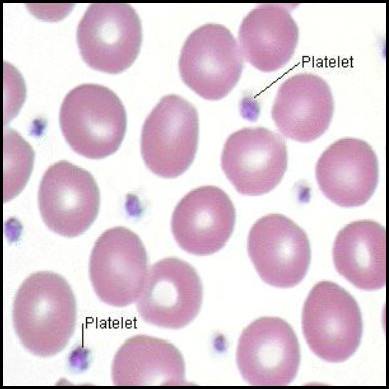
Translation: A deficiency of ALL cellular components in the blood. A patient with pancytopenia has dangerously low levels of red blood cells (anemia), white blood cells (leukopenia), and platelets (thrombocytopenia), usually due to complete bone marrow failure.


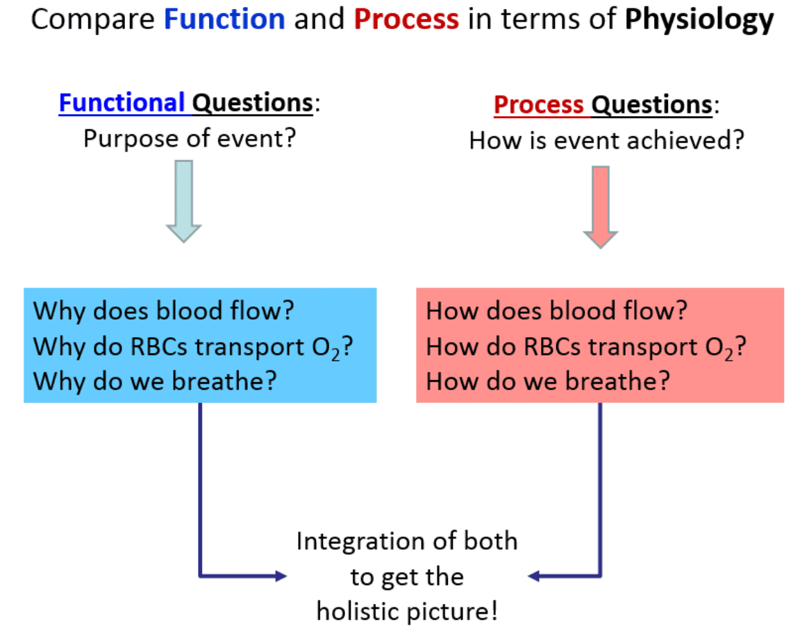
3. Compare Function and Process in Human Physiology

As we look to understand the central themes of physiology, an important concept is learning exactly how to ask questions about what’s occurring in the human body. In general, there are two basic, distinct approaches to physiology: 1) We can ask Functional Questions; and 2) We can ask Process Questions.

1. Functional Questions (The "Why")

These are strictly related to Why something occurs. They seek to understand the ultimate purpose or the evolutionary advantage of a mechanism. For example, what is the survival purpose of the heart beating? These can often be answered from a "big picture" perspective without requiring microscopic detail.

  • Q: Why does blood flow?
    A: To continuously transport vital nutrients, hormones, and gases (oxygen) around the body to sustain tissues, and to carry away toxic metabolic wastes (carbon dioxide, urea) to the excretory organs.
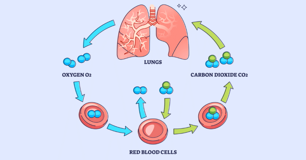
  • Q: Why do Red Blood Cells (RBCs) transport O₂?
    A: To efficiently deliver oxygen to all the body tissues that desperately need it to perform aerobic cellular respiration and generate ATP (energy).
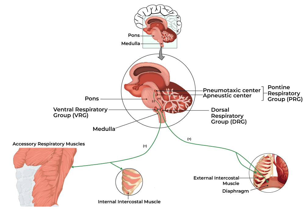
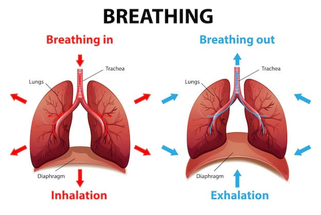
  • Q: Why do we breathe?
    A: To extract essential oxygen (O₂) from the inhaled atmospheric air and to continuously release volatile carbon dioxide (CO₂) back out of the body, thus preventing fatal acid build-up in the blood.

2. Process Questions (The "How")

These are related to How something physically and chemically occurs. For example, how does the heart actually manage to beat? Often these issues must be answered in an extensively detailed, step-by-step, mechanistic manner.

  • Q: How does blood flow?
    A: The muscular ventricles of the heart contract (systole) to generate a massive high-pressure wave. Blood flows strictly down this pressure gradient, moving from the high-pressure aorta through the progressively lower-pressure arteries, capillaries, and veins, finally returning to the heart aided by skeletal muscle pumps and tissue fluid pressures.
  • Q: How do RBCs transport O₂?
    A: Inside the red blood cells, the iron-containing heme portion of the hemoglobin molecule undergoes conformational changes. It exhibits a high chemical affinity for O₂ when the surrounding partial pressure of O₂ is high (such as in the pulmonary capillaries of the lungs, forcing O₂ to bind). Conversely, it exhibits a low affinity for O₂ when the surrounding partial pressure for O₂ is low (such as in metabolically active, oxygen-depleted muscle tissue, forcing the hemoglobin to drop the O₂ where it is needed).
  • Q: How do we breathe?
    A: The diaphragm and external intercostal muscles (skeletal muscles of respiration) contract, pulling the rib cage up and the diaphragm down. This physical action dramatically increases the volume of the thoracic cavity. According to Boyle's Law, an increase in volume causes an inverse decrease in pressure. This creates a negative pressure gradient inside the lungs compared to the outside atmosphere, effectively vacuuming air down its pressure gradient into the lungs. Exhalation is the passive recoil of these same structures.

Things to Notice about Function and Process

Notice that the How part (the process) requires vastly more details and involves a strict ‘pathway’ approach. It is much more like sequential storytelling compared to the broader, less detailed functional aspects. The more arduous and demanding component of physiology is mastering these detailed processes. This is precisely the reason we need to take our time and fully, deeply understand the fundamentals before we delve into intricate, microscopic details.

What most students quickly recognize about physiology is that it is vastly more conceptual than anatomy because there is almost always a chemical or physical process to describe in a logical, step-by-step manner. There are usually two sides to the functions discussed in physiology. This is because at the very center of the human body is balance, which provides the equilibrium necessary to function properly. When we explain the mechanism of how we breathe in, we must simultaneously understand how we breathe out. When we explain how insulin lowers blood sugar, we must understand how glucagon raises it. Often, once you master one side of the physiological story, the other side falls into place much more easily.


4. Basic Functions of a Complex Organism

Holistically, we will examine Human Physiology as it relates to the foundational basics of how a multi-system, trillions-of-cells living organism functions as a single, perfectly coordinated entity. To be considered a living organism, the body must perform several critical basic functions. Below is an excessively detailed breakdown of these functions:

  1. Differentiation: The process by which unspecialized, generic cells (like stem cells) develop into highly specialized cells with distinct structures and functions. Extra Example: A single fertilized egg cell divides and its progeny differentiate into vastly different structures, such as conductive nerve cells, contractile muscle cells, and absorptive intestinal cells.
  2. Responsiveness (Excitability): The body's ability to detect and respond instantly to changes in its internal or external environment. Extra Example: If you touch a hot stove (external stimulus), sensory nerve endings detect the heat and trigger a withdrawal reflex. Internally, if blood calcium levels drop, the parathyroid glands detect this and release hormones to restore balance.
  3. Metabolism: The sum of all the intricate chemical processes that occur in the body. It consists of two phases: Catabolism (the breaking down of complex molecules into simpler ones to release energy, like digesting food) and Anabolism (the building up of complex molecules from simpler ones, requiring energy, like building new muscle protein).
  4. Growth and Repair: Growth refers to an increase in body size, either due to an increase in the size of existing cells (hypertrophy), an increase in the total number of cells (hyperplasia), or an increase in the amount of material surrounding cells. Repair is the ongoing process of replacing dead, damaged, or scraped-off cells (like skin cells constantly replacing themselves).
  5. Movement: Not just walking or running, but motion at every single level. This includes the coordinated action of the entire body, the movement of individual organs (like the stomach churning food or the gallbladder squeezing bile), single cells (like white blood cells physically crawling toward an infection), and even tiny organelles moving inside a cell.
  6. Excretion: The vital process of removing the toxic by-products of digestion and metabolism. If wastes like urea, carbon dioxide, or lactic acid are allowed to accumulate, they rapidly become fatal. The respiratory, digestive, and urinary systems all share this massive burden.
  7. Reproduction: The formation of new cells for tissue growth, repair, or replacement, OR the production of a completely new individual to ensure the continuation of the human species.

What we will find is that all of the diverse systems we will study in this course will contain many, if not all, of these basic functions deeply embedded within them.


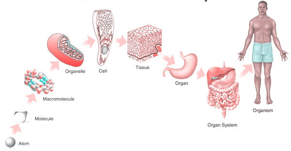
5. Levels of Organization & Body Systems

A body system (also called an organ system) is a highly integrated, complex collection of organs in the body that work seamlessly together to perform a specific, vital function. The truth is that all systems are intimately connected, relying heavily on one another, but it is highly useful for educational purposes to study them separately, even though they are not separate at all. With all of our body systems operating constantly and simultaneously, it is absolutely necessary to have an overarching regulatory system in place to maintain stability, harmony, and equilibrium across all these integrated systems. This unifying, central element in all of physiology is called Homeostasis.

The Six Levels of Structural Organization:

  1. Chemical Level: The most basic level. Includes atoms (like Carbon, Hydrogen, Oxygen, Nitrogen) combining to form essential molecules (like DNA, glucose, water).
  2. Cellular Level: Molecules combine to form cells, the basic structural and functional units of an organism. (e.g., Muscle cells, nerve cells, blood cells).
  3. Tissue Level: Groups of similar cells and their surrounding materials working together to perform a specific function. (The four basic types: Epithelial, Connective, Muscular, Nervous).
  4. Organ Level: Structures composed of two or more different types of tissues. They have specific functions and recognizable shapes. (e.g., The stomach, heart, liver, brain).
  5. System Level: Consists of related organs with a common function. (e.g., The digestive system includes the mouth, esophagus, stomach, intestines, liver, and pancreas working to extract nutrients).
  6. Organismal Level: The highest level. All the parts of the human body functioning together to constitute the total living organism.

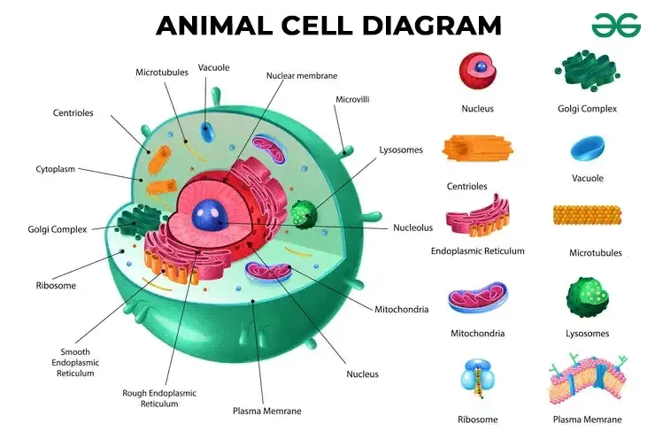
6. The Cell: The Fundamental Unit of Life

The cell is the absolute basic building block of all living things. Every single physiological process begins at the cellular level. To understand how the cell works, we can beautifully compare it to a bustling, highly organized city.

A. The Outer Boundary: The City Wall and Gates

The Cell (Plasma) Membrane

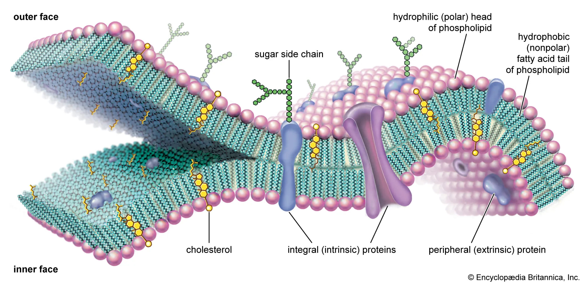
This is the outermost boundary of the cell, an incredibly thin, highly flexible, and selectively permeable (or semipermeable) barrier. It's primarily composed of a phospholipid bilayer, thickly populated with embedded proteins, carbohydrates, and cholesterol molecules.

  • Phospholipid Bilayer: Two opposing layers of phospholipids. Each lipid molecule has a hydrophilic ("water-loving") polar head that faces the watery environments inside and outside the cell, and two hydrophobic ("water-fearing") non-polar fatty acid tails that face inward toward each other, forming the water-repelling core of the membrane.
  • Proteins: These are crucial for nearly all membrane functions. Integral (transmembrane) proteins span the entire width of the membrane, forming open channels, carrier pumps, and signaling receptors. Peripheral proteins are loosely attached to the inner or outer surface, often involved in intracellular signaling cascades or anchoring the cytoskeleton.
  • Cholesterol: Wedged deeply within the hydrophobic core between the fatty acid tails, cholesterol helps stabilize the membrane's fluidity. It prevents the membrane from freezing solid at low temperatures and stops it from becoming too loose and falling apart at high body temperatures.
  • Glycocalyx (Carbohydrates): Chains of complex carbohydrates attached to proteins (forming glycoproteins) or attached to lipids (forming glycolipids) on the outer surface. This forms a unique, fuzzy "sugar coat" or cellular "ID tag." Extra Example: The ABO blood group markers on your red blood cells are actually part of the glycocalyx!

Physiological Functions of the Cell Membrane:

  • Selective Permeability: Strictly controls exactly what enters and leaves the cell, maintaining perfect homeostasis. (Functions like the city's border control).
  • Cell Recognition: The glycocalyx allows immune cells to recognize each other and identify foreign invaders.
  • Communication/Signaling: Receptor proteins bind to chemical messengers like hormones and neurotransmitters, translating outside signals into internal actions.
  • Cell Adhesion: Proteins allow cells to stick tightly together to form robust tissues (like skin).
  • Protection: Provides a flexible physical barrier shielding internal components.
Mnemonic: "People Call Me Protector" for Phospholipids, Cholesterol, Membrane Proteins.

B. The Cell's Internal Environment: The City Hall and Workers

The Cytoplasm

The cytoplasm is literally everything inside the cell membrane but entirely outside the nucleus. It consists of three main elements:

  • Cytosol: The jelly-like, semi-fluid, viscous portion where all organelles are suspended. It's mostly water heavily packed with dissolved solutes (ions, glucose, amino acids, ATP, fatty acids, etc.).
  • Organelles: The highly specialized "little organs" with very specific, indispensable functions (discussed next).
  • Inclusions: Temporary, non-functioning storage bodies. Extra Examples: massive glycogen granules in liver and muscle cells for energy storage, giant lipid droplets in fat cells (adipocytes), and melanin pigment granules in skin cells.

Physiological Functions of the Cytoplasm:

  • Site of Many Metabolic Reactions: Crucial biochemical pathways, such as glycolysis (the first, anaerobic step of glucose breakdown to extract energy), occur entirely free-floating in the cytosol.
  • Suspension of Organelles: Provides the perfect physical and chemical medium for all organelles to exist, interact, and function.

C. The Control Center: The City Hall/Mayor's Office

The Nucleus

Usually the largest and most prominent organelle, the nucleus is securely enclosed by a double-layered membrane called the nuclear envelope, which is punctured by nuclear pores that highly regulate the entry and exit of molecules. Inside, it contains:

  • Chromatin: The relaxed, uncondensed, thread-like form of DNA (our genetic material) carefully wrapped around specialized structural proteins called histones. When the cell prepares to divide, this messy chromatin tightly condenses into distinct, visible structures called chromosomes.
  • Nucleolus: A dense, dark, spherical body deeply embedded within the nucleus. It is the primary site of rapid ribosome synthesis.

Physiological Functions of the Nucleus:

  • Genetic Control: Contains the cell's entire genetic blueprint (DNA), directing absolutely all cell activities by strictly controlling which proteins are synthesized. (The "master architectural plan" for the city).
  • DNA Replication & Transcription: This is where DNA faithfully copies itself before cell division (mitosis) and where DNA's genetic code is safely transcribed into messenger RNA (mRNA), which then leaves the nucleus to give instructions to the factories.
  • Ribosome Production: The nucleolus continuously synthesizes and assembles ribosomal RNA (rRNA) subunits.

D. Protein Synthesis and Processing: The Factories and Delivery Services

Ribosomes

Tiny, granular, non-membranous organelles made of ribosomal RNA (rRNA) and various proteins. They are the ultimate "protein factories" of the cell. They physically read the mRNA code coming from the nucleus to perfectly assemble amino acids into long proteins (a process called translation). They can exist as free ribosomes floating in the cytosol (making proteins for immediate use within the cell) or bound ribosomes securely attached to the Endoplasmic Reticulum (making proteins destined for export out of the cell or for embedding into membranes).

Mnemonic: "Ribosomes Read RNA to make Really good pRotein."

Endoplasmic Reticulum (ER)

An extensive, labyrinth-like network of interconnected membranous tubes and flattened sacs that extends massively throughout the cytoplasm, and is directly continuous with the outer membrane of the nuclear envelope.

  • Rough Endoplasmic Reticulum (RER): Heavily studded with ribosomes, giving it a "rough" or sandpaper-like appearance under a microscope. Its primary function is to synthesize, fold, and modify proteins that are destined for secretion, insertion into cell membranes, or delivery to lysosomes. (e.g., adding sugar chains in a process called glycosylation). Extra Example: Pancreatic cells that produce massive amounts of digestive enzymes have incredibly large, active RERs.
  • Smooth Endoplasmic Reticulum (SER): Completely lacks ribosomes. Its highly specialized functions include massive lipid and steroid hormone synthesis, intense detoxification of harmful drugs and poisons (making it extremely abundant in liver cells), and the storage and release of calcium ions. Extra Example: In skeletal muscle cells, a specialized SER called the sarcoplasmic reticulum stores the calcium absolutely crucial for triggering muscle contraction.
Mnemonic: "Rough ER is Rough on Ribosomes & Really helps Really good pRotein; Smooth ER is Smoothly Synthesizing Steroids & Storing Salcium (calcium) and Speedily Solving Substance Spoilage (detox)."

Golgi Apparatus (Golgi Complex)

A distinct stack of flattened, slightly curved membranous sacs called cisternae. It acts as the ultimate "Post Office" or "Packaging and Shipping Center" of the cell.

Physiological Functions of the Golgi Apparatus:

  • Modification, Sorting, and Packaging: It receives vesicles containing raw proteins and lipids from the ER at its cis face, further modifies them (like adding or trimming sugar tags), sorts them based on their final destination, and packages them into new vesicles leaving from its trans face.
  • Vesicle Formation: It actively forms various types of vesicles, including secretory vesicles (for exocytosis/dumping out of the cell), lysosomes (cellular stomachs), and transport vesicles that seamlessly deliver new structural components to patch the plasma membrane.
Mnemonic: "Golgi Gathers, Grades, and Gets rid of Garbage (or packages good stuff!)."

E. Energy Production: The Power Plant
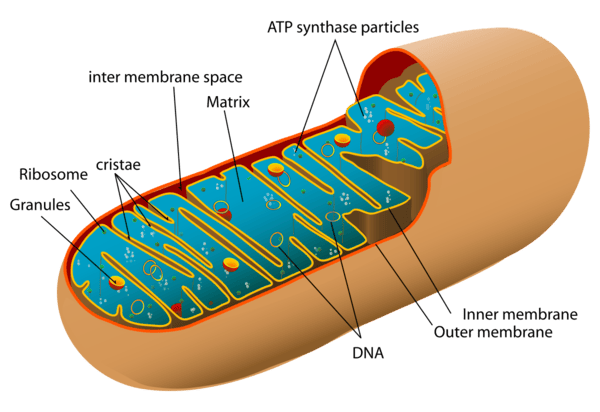

Mitochondria

Oval or bean-shaped organelles distinctly enclosed by a double membrane: a relatively smooth outer membrane and an inner membrane that is highly folded inward to form structures called cristae. These folds drastically increase the surface area available for chemical reactions. The dense, fluid-filled space within the inner membrane is called the matrix. Mitochondria also astonishingly possess their own unique, circular DNA (mtDNA) inherited exclusively from the mother.

Physiological Function (The "Powerhouses of the Cell"):

They are the primary site of aerobic cellular respiration. They utilize oxygen to meticulously break down and convert fuel molecules like glucose and fatty acids into ATP (adenosine triphosphate), the primary, usable energy currency of the entire cell. Extra Example: Cells that require massive, continuous amounts of energy, such as cardiac (heart) muscle cells and swimming sperm cells, have spectacularly high numbers of mitochondria compared to inactive cells.

Mnemonic: "Mighty Mitochondria Make Much More Money (ATP)."

F. Waste Management and Recycling: The Cleaning Crew

Lysosomes

Tiny, spherical membranous sacs packed tightly with incredibly powerful hydrolytic (digestive) enzymes that function best in an acidic environment. They act as the "Recycling Centers" and "Stomachs" of the cell.

  • They aggressively break down ingested foreign substances and bacteria (phagocytosis).
  • They digest worn-out, broken cellular organelles to recycle their raw materials (a process called autophagy).
  • In dying or damaged cells, they can burst open to digest the entire cell (autolysis).
  • Clinical Correlate: Tay-Sachs disease is a fatal genetic disorder where a specific lysosomal enzyme is missing. Toxic lipids build up uncontrollably in the brain because the lysosomes cannot break them down.
Mnemonic: "Lyso-some = "Lysol" – they lyse (break down) stuff."

Peroxisomes

Smaller membranous sacs containing potent oxidative enzymes, most notably catalase and oxidase. They act as the highly specialized "Detoxification Squad".

  • They aggressively neutralize highly reactive, harmful free radicals and detoxify toxins like alcohol (making them very abundant in kidney and liver cells).
  • They heavily assist in the breakdown (beta-oxidation) of very long-chain fatty acids for use in energy production.
  • During their reactions, they produce dangerous hydrogen peroxide (H₂O₂), but their built-in catalase enzyme instantly converts it safely into water and oxygen.
Mnemonic: "Peroxisomes Produce Peroxide to Purify."

G. The Cell's Internal Support and Movement: The Infrastructure
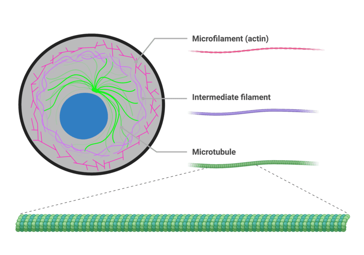

The Cytoskeleton

An intricate, dynamic network of protein filaments extending massively throughout the cytoplasm, providing cell shape, internal support, and physical highways for transport. It consists of three main types of filaments:

  • Microfilaments (Actin): The thinnest fibers; deeply involved in cell movement, gross shape changes, cell division (cleavage furrow), and forming the contractile machinery of muscle cells.
  • Intermediate Filaments: Tough, rope-like proteins that provide immense structural stability and fiercely resist mechanical pulling stress on the cell. Extra Example: Keratin in skin cells is an intermediate filament that makes skin tough and waterproof.
  • Microtubules: The largest elements; hollow tubes made of tubulin. They determine the overall cell shape, form the "railroad tracks" for motor proteins to drag organelles across the cell, and are the primary structural core of cilia, flagella, and the mitotic spindle.
Mnemonic: "Cytoskeleton Supports the Cell Shape and Ships things Swiftly."

Centrosomes, Cilia, and Flagella

  • Centrosomes and Centrioles: Located strategically near the nucleus, the centrosome contains two barrel-shaped centrioles positioned at right angles. It acts as the main Microtubule-Organizing Center (MTOC), generating and organizing the mitotic spindle required to pull chromosomes apart during cell division.
  • Cilia and Flagella: Hair-like, motile cellular projections entirely made of microtubules.
    • Cilia are short and numerous, beating in coordinated waves to move substances across the cell surface. Extra Example: Millions of cilia in your respiratory tract constantly sweep mucus and trapped dust up and out of your lungs.
    • Flagella are much longer and usually singular, acting like a whip to propel the entire cell forward. Extra Example: The only human cell with a flagellum is the male sperm cell.
Mnemonic: "Centrosomes and Centrioles Control Cell Civision Carefully."

Summary Table of Organelles

Organelle Key Physiological Functions
Plasma Membrane Selective barrier, cell recognition, intercellular communication, structural protection.
Nucleus Ultimate genetic control, houses DNA, site of DNA replication and mRNA transcription.
Ribosomes Direct protein synthesis via the translation of mRNA into amino acid chains.
Rough ER (RER) Synthesis, folding, and intense modification of proteins destined for export or membrane insertion.
Smooth ER (SER) Lipid and steroid synthesis, detoxification of drugs/poisons, massive intracellular Ca²⁺ storage.
Golgi Apparatus Modifies, sorts, meticulously packages proteins and lipids into transport/secretory vesicles.
Mitochondria Site of aerobic cellular respiration, massive ATP synthesis (the ultimate powerhouse).
Lysosomes Intracellular digestion of debris, bacteria, and old organelles; primary cellular waste removal.
Peroxisomes Detoxification (especially neutralizing free radicals and alcohol), extensive fatty acid breakdown.
Cytoskeleton Maintains cell shape, structural support, internal transport tracks, enables cell motility.
Centrosomes Organize the microtubule network and generate the mitotic spindle during cell division.
Cilia / Flagella Cilia move external substances across the cell surface; Flagella physically propel the entire cell.

7. Biological Membranes: The Fluid Mosaic Model

Biological membranes are highly dynamic, incredibly fluid structures that sharply define the boundaries of cells (plasma membrane) as well as the internal organelles. They are absolutely essential for maintaining cellular integrity, strictly regulating transport, facilitating communication, and housing vital enzymatic reactions. The most universally accepted scientific model describing membrane structure is the Fluid Mosaic Model.

The Fluid Mosaic Model

Proposed brilliantly by Singer and Nicolson in 1972, this model describes the cell membrane as a highly fluid lipid bilayer where an array of proteins are embedded or attached, looking much like a complex, ever-shifting mosaic artwork.

  • "Fluid": Refers to the constant, rapid movement of individual phospholipid molecules and proteins within the two-dimensional plane of the membrane. Lipids and many proteins are not locked in place; they constantly drift laterally, rotate on their axes, and flex their tails.
  • "Mosaic": Refers to the diverse, scattered "patchwork" of different functional proteins and other structural molecules (like cholesterol and carbohydrates) permanently or temporarily embedded within the vast ocean of the lipid bilayer.

A. Lipids of the Cell Membrane

The central, primary structural framework of the membrane is the fluid lipid bilayer, which is predominantly made of phospholipids and cholesterol.

1. Phospholipids

Phospholipids are by far the most abundant lipids in the membrane. They possess a crucial chemical property: they are amphipathic. This means a single molecule contains both a highly hydrophilic (water-loving) polar head and two highly hydrophobic (water-fearing) non-polar fatty acid tails. In the watery environment of the body, they spontaneously and instantly self-assemble to form a bilayer. The hydrophobic tails hide inward, desperately avoiding water, while the hydrophilic heads proudly face the watery environments inside (intracellular fluid) and outside (extracellular fluid) the cell.

2. Cholesterol

Cholesterol molecules are tough, rigid, ring-shaped lipids inserted tightly between the phospholipids. They act as the ultimate membrane temperature buffer, perfectly regulating fluidity. At normal, warm body temperatures, cholesterol restricts movement, reducing excessive fluidity and making the membrane stronger and less leaky. Conversely, at dangerously low temperatures, cholesterol prevents the phospholipid tails from packing too tightly together, thus increasing fluidity and stopping the cell membrane from freezing solid.

Lipid Functions in the Cell Membrane:

  • Forms the fundamental, self-healing bilayer structure.
  • Provides a robust selectively permeable barrier, naturally allowing only small, uncharged, fat-soluble substances (O₂, CO₂, steroid hormones) to easily pass through directly.
  • Acts as an impenetrable physical barrier for all water-soluble substances (glucose, salts, ions), which absolutely require physical assistance from embedded proteins to cross.

B. Membrane Proteins

While lipids form the barrier, proteins are the hardworking machines of the membrane, performing almost all of its specific, dynamic functions.

1. Integral (Transmembrane) Proteins

These are tightly bound proteins that fully span across the entire thickness of the membrane. They are deeply embedded in the hydrophobic core and can only be removed by completely destroying the bilayer. They primarily function as open channels, carrier transport proteins, active pumps, receptors, and enzymes.

2. Peripheral Proteins

These are loosely, temporarily bound to the membrane's inner or outer surface. They do not penetrate the hydrophobic core and are easily detached without harming the membrane. They often function as localized enzymes, signaling relays, or cytoskeletal anchors holding the membrane shape.

Functions of Membrane Proteins:

  • Transport: Facilitating the precise movement of specific, rejected substances across the barrier (channels, carriers, ATP pumps).
  • Enzymatic Activity: Catalyzing vital metabolic reactions directly at the membrane surface.
  • Signal Transduction: Acting as sophisticated receptors catching chemical messengers (like adrenaline) and transmitting the order inside.
  • Cell-Cell Recognition: Acting as strict identification tags (glycoproteins) so immune cells don't attack the body's own tissues.
  • Intercellular Joining: Forming tight junctions and desmosomes to physically link neighboring cells together.
  • Attachment to Cytoskeleton & ECM: Providing immense structural stability by tethering the cell's skeleton to the outside world.

C. Carbohydrates of the Cell Membrane

Carbohydrates are exclusively found on the external, outward-facing surface of the plasma membrane. They never face the inside. They are permanently attached to lipids (forming glycolipids) or to proteins (forming glycoproteins). This entire "sugar coat" covering the cell is called the glycocalyx, which serves as a highly specific, unique molecular signature for every different cell type in your body.

Functions of Membrane Carbohydrates (Glycocalyx):

  • Cell-Cell Recognition: Crucial for distinguishing "self" tissues from "non-self" invaders (e.g., orchestrating immune responses, dictating ABO blood types, and organ transplant rejection).
  • Cell Adhesion: The sticky sugars help cells firmly bind to one another in tissues.
  • Receptors: Can uniquely act as receptors for specific hormones, or tragically, as hijacking points for bacterial toxins and viruses (like the flu or COVID-19).
  • Protection: Provides a cushioning, protective physical barrier against mechanical damage and harsh enzymes.

Properties of the Cell Membrane

The specific composition and arrangement of lipids, proteins, and carbohydrates endow the cell membrane with its most essential, life-sustaining properties:

  1. Selectively Permeable (Semi-permeable): This is arguably its most important property. The membrane precisely and selfishly regulates exactly which substances can enter or leave the cell. The hydrophobic lipid core acts as the primary absolute barrier. Small, nonpolar molecules (O₂, CO₂) and highly lipid-soluble molecules pass directly and effortlessly. However, charged ions (Na⁺, K⁺) and large polar molecules (glucose) are utterly rejected and require specific, designated transport proteins to grant them entry.
  2. Fluidity: The membrane is absolutely not a rigid, solid shell; its molecular components are in constant, swirling motion. Fluidity is heavily influenced by body temperature, the amount of cholesterol (which acts as a stabilizing buffer), and the degree of saturation of the fatty acid tails (kinked, unsaturated tails increase fluidity). This property is absolutely essential for vesicles to fuse with the membrane, for the cell to divide, and for transport proteins to physically shift their shapes.
  3. Asymmetry: The two faces (the inner leaflet touching the cytoplasm, and the outer leaflet touching the blood/fluid) of the plasma membrane are completely structurally and functionally different. For example, carbohydrates are exclusively on the outer surface (glycocalyx), and specific lipids and signaling proteins are oriented strictly in one direction. This is vital for directional signaling, shape maintenance, and cell recognition.
  4. Self-Sealing Capability: Due to the powerful thermodynamic forces of hydrophobic interactions, if the membrane is punctured or torn, the lipids have a natural, spontaneous tendency to instantly re-seal themselves, completely preventing the fatal leakage of cytoplasmic contents. This is crucial for maintaining cell integrity during physical stress or when a needle enters a cell.

Summary of Key Membrane Functions:

  • Protective Barrier: Encloses the cell's delicate contents, safely separating the intracellular from the chaotic extracellular environment.
  • Selective Transport: Heavily regulates the passage of every single substance entering and exiting the cell.
  • Cell-Cell Communication: Contains specific receptors for circulating hormones, drugs, and neurotransmitters.
  • Cell Recognition & Adhesion: Facilitates cell identification by the immune system and the formation of solid tissues.
  • Enzymatic Activity: Houses membrane-bound enzymes that rapidly catalyze specific biochemical reactions.
  • Maintenance of Cell Shape: Provides profound structural support in conjunction with the inner cytoskeleton.
  • Generates Membrane Potential: Crucial for the electrical firing of nerve and muscle cells.
  • Endocytosis & Exocytosis: Manages the bulk, massive transport of large materials into and out of the cell.

8. Membrane Potential: The Electrical Voltage Across the Membrane

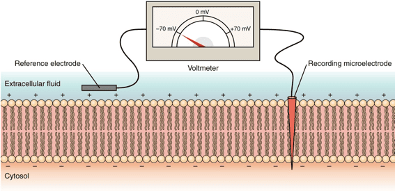
Before looking at exactly how things physically move across the membrane, it's absolutely essential to understand that there is a permanent electrical difference, or voltage, sitting directly across every single cell membrane in your body. This is called the membrane potential.

Membrane Potential is the exact measurable difference in electrical charge (or potential energy) between the inside and the outside of a cell. By strict scientific convention, the inside of the cell is always measured as being negative relative to the outside.

How is the Membrane Potential Established?

It is driven by three distinct, constantly working factors:

  1. Unequal Distribution of Ions: There are radically different concentrations of ions (charged particles) inside versus outside the cell.
    • Outside the cell (Extracellular Fluid - ECF): Features a massively high concentration of Na⁺ (sodium) and Cl⁻ (chloride).
    • Inside the cell (Intracellular Fluid - ICF): Features a massively high concentration of K⁺ (potassium) and huge, negatively charged proteins and phosphates (which are physically too large to ever leave the cell, trapping their negative charge inside).
  2. Selective Permeability of the Membrane: The cell membrane is not equally permeable to all ions. At rest, it is packed with specific "leak channels" that make it vastly more permeable to K⁺ than to Na⁺. Because K⁺ is highly concentrated inside, it constantly leaks out down its concentration gradient. Every time a positive K⁺ leaves, it leaves behind a negative charge, making the inside of the cell significantly more negative.
  3. Sodium-Potassium Pump (Na⁺/K⁺ ATPase): This relentless active transport pump burns massive amounts of ATP to constantly eject exactly 3 Na⁺ ions out of the cell for every 2 K⁺ ions it pumps back in. Because it pumps out more positive charges (3) than it brings in (2), this pump is inherently electrogenic and contributes directly and continuously to the negative charge inside the cell.

Resting Membrane Potential

In a resting (unstimulated) neuron or muscle cell, the steady-state electrical potential established and maintained perfectly by these factors is called the Resting Membrane Potential. It is typically measured at around -70 mV (millivolts), meaning the inside is 70 millivolts more negative than the outside.

Physiological Significance:

The resting membrane potential is absolutely not just a passive, resting state; it is a massive form of stored, coiled potential energy (like a stretched rubber band) crucial for:

  • Excitability: It allows excitable cells (like neurons and heart muscle cells) to instantly open gates and generate rapid, explosive electrical signals (action potentials) for instant communication, thought, and muscle contraction.
  • Secondary Active Transport: The immense energy stored in the steep Na⁺ and K⁺ ion gradients can be brilliantly harnessed by the cell to power the "uphill" transport of other essential substances (like glucose and amino acids) across the membrane.

Clinical Correlate: Hyperkalemia

If a patient's kidneys fail, potassium (K⁺) builds up in the blood outside the cell (a condition called hyperkalemia). This destroys the carefully maintained K⁺ gradient. The resting membrane potential is ruined, the heart muscle cells cannot reset their electrical charge, and the patient suffers a sudden, fatal cardiac arrhythmia (heart attack).


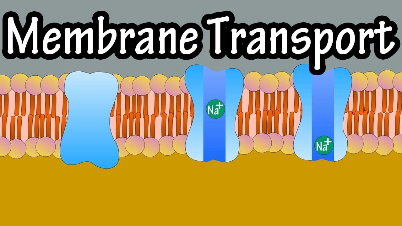
9. Membrane Transport

Membrane transport is the fundamental physiological process that meticulously governs the movement of all substances across biological membranes. It's essential for maintaining cellular homeostasis, acquiring vital nutrients, aggressively expelling toxic waste products, and facilitating cell-to-cell communication. Substances cross the membrane via two general, overarching mechanisms: Passive Transport and Active Transport.

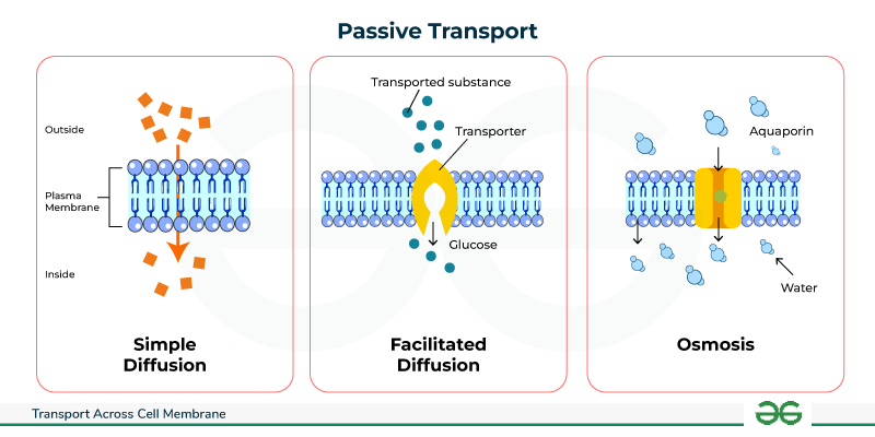
1. Passive Transport: Moving Downhill

Passive transport is the movement of substances across a cell membrane completely without the direct expenditure of cellular metabolic energy (ATP). This movement is always "downhill," meaning down the electrochemical or concentration gradient of the substance. The energy driving this movement comes purely from the inherent, random kinetic energy of the molecules themselves and the potential energy stored in the concentration gradient.

1.1. Simple Diffusion: Through the Lipid Bilayer

In simple diffusion, substances move directly and effortlessly through the lipid bilayer without any help from membrane proteins whatsoever.

  • Highly Permeable: Small, nonpolar (lipophilic/fat-soluble) molecules like O₂, CO₂, and steroid hormones readily dissolve directly into the hydrophobic core and pass completely through to the other side.
  • Moderately Permeable: Small, uncharged polar molecules like water and ethanol can pass to a very limited degree because they are small enough to slip through the lipid gaps.
  • Impermeable: Large polar molecules (like glucose) and absolutely all charged ions (Na⁺, K⁺, Ca²⁺) cannot pass through the lipid core on their own under any circumstances.

The sole driving force is the concentration gradient. The random, chaotic molecular motion (kinetic energy) results in a net movement from an area of highly crowded concentration to an area of lower concentration, continuing unabated until perfect equilibrium is reached across the membrane.

Key Characteristics of Simple Diffusion:

  • Absolutely no membrane proteins are involved.
  • Does not exhibit saturation kinetics: The rate of diffusion increases linearly forever with the concentration gradient; there is no maximum transport rate (Vmax) because there are no proteins to get "full".
  • According to Fick's Law, the rate is directly proportional to the gradient magnitude, lipid solubility, and membrane surface area, and inversely proportional to molecular size and the physical thickness of the membrane.

1.2. Facilitated Diffusion: Protein-Assisted Passage

This process uses specialized integral membrane proteins (channels or carriers) to beautifully facilitate the movement of specific, impermeable substances down their electrochemical gradient. It is still completely passive, as absolutely no ATP is directly consumed.

A. Channel Proteins (Pores)

These proteins form a hollow, water-filled pore directly across the membrane, allowing the incredibly rapid, single-file passage of specific ions or water molecules. Most channels are tightly gated, meaning they act like doors that only open or close in response to specific, required stimuli:

  • Voltage-Gated Channels: Respond instantly to changes in electrical membrane potential (e.g., Na⁺ and K⁺ channels crucial in firing neurons).
  • Ligand-Gated Channels: Respond only when a specific chemical messenger binds to them (e.g., neurotransmitter receptors at muscle synapses).
  • Mechanically-Gated Channels: Respond directly to physical deformation or stretching of the membrane (e.g., touch receptors in your skin, or hearing receptors in the ear).
  • Leak Channels: Are generally always randomly fluttering open and closed, heavily contributing to the resting membrane potential.
  • Extra Examples: Ion channels (Na⁺, K⁺, Cl⁻, Ca²⁺) and aquaporins, which are highly specialized channels dedicated exclusively to letting massive amounts of water through instantly.

B. Carrier Proteins (Transporters)

These proteins work like revolving doors. They physically bind to a specific molecule on one side, undergo a massive conformational (shape) change, and then release the molecule safely on the other side. This physical shifting process makes it much slower than channel-mediated transport.

  • Saturation Kinetics: Because there are a strictly finite number of carrier proteins on a cell, the transport rate has an absolute maximum speed limit (Vmax) that hits when every single carrier is occupied and working as fast as it can.
  • Specificity: Carriers are incredibly highly specific for the exact molecule(s) they are designed to transport.
  • Competition: Structurally similar, "imposter" molecules can compete for the exact same binding site, blocking the real molecule.
  • Extra Examples: Glucose Transporters (GLUT proteins, which insulin commands to surface) and various amino acid transporters.

Key Characteristics of Facilitated Diffusion:

  • Strictly involves specific integral membrane proteins (channels or carriers).
  • Exhibits saturation kinetics (Vmax limit) due to the limited, physical number of transporters.
  • Can be easily subject to chemical competition.
  • Can be heavily regulated by the cell (e.g., by gating channels shut, or inserting/removing carriers from the membrane to control absorption).

1.3. Osmosis: The Grand Movement of Water

Osmosis is the incredibly powerful net movement of water across a selectively permeable membrane, strictly from an area of higher water concentration (which means lower solute concentration) directly to an area of lower water concentration (which means higher, saltier solute concentration). The driving force is the water potential gradient, determined entirely by the difference in non-penetrating solute concentration on either side of the membrane.

Osmotic pressure is the "pulling" or "sucking" force a solution with a higher solute concentration forcefully exerts to draw water toward it. Tonicity refers to the actual physical effect of a surrounding solution on the cell's total volume:

  • Isotonic: The concentration is perfectly equal inside and outside. There is no net water movement; cell volume remains perfectly normal.
  • Hypotonic: The outside fluid is dilute (watery). Water rushes rapidly into the cell, causing it to aggressively swell and potentially burst (a fatal event called lysis).
  • Hypertonic: The outside fluid is extremely concentrated (salty). Water is sucked rapidly out of the cell, causing the cell to drastically shrivel and shrink (a process called crenation).

Clinical Correlate: Intravenous (IV) Fluids

When a patient arrives at the hospital dehydrated, you cannot simply inject pure, hypotonic tap water into their veins. Doing so would cause their red blood cells to rapidly absorb the water, swell, and violently explode (hemolysis), killing the patient. Instead, nurses administer 0.9% Normal Saline, which is perfectly isotonic to human blood plasma, safely rehydrating the body without destroying the cells.

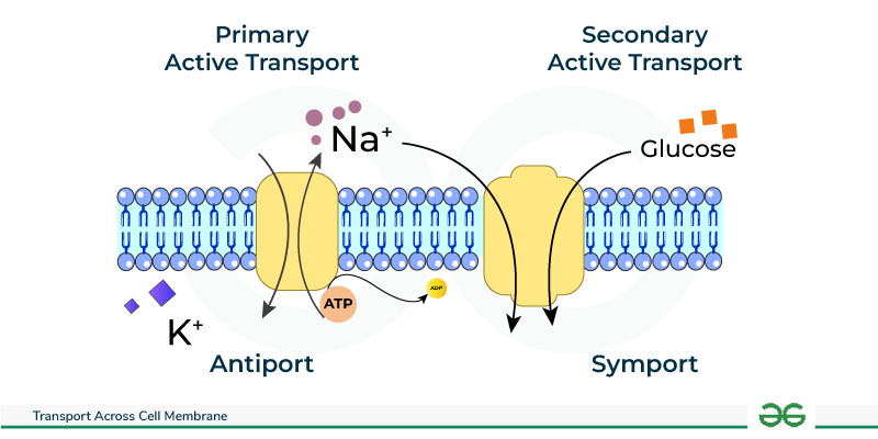
2. Active Transport: Against the Current, with Energy

Active transport is the strenuous, demanding process of actively moving substances across a cell membrane directly against their electrochemical or concentration gradient (i.e., dragging them from a region of lower concentration forcefully into a region of higher concentration). This distinctly "uphill" movement necessitates the direct or indirect expenditure of precious cellular metabolic energy, almost invariably derived from the violent hydrolysis of ATP.

2.1. Primary Active Transport: Direct ATP Expenditure

Primary active transporters are robust integral membrane proteins that function fundamentally as ATPases; they directly bind to and snap the phosphate off of ATP, utilizing that massive burst of released energy to physically power the forced movement of solutes. These heavy-duty transporters are often called "pumps."

  • Na⁺/K⁺ ATPase (Sodium-Potassium Pump): Found aggressively pumping in virtually all animal cells, this absolutely vital, life-sustaining pump forcibly moves 3 Na⁺ ions entirely out of the cell and drags 2 K⁺ ions back into the cell for every single ATP molecule hydrolyzed. It is strictly electrogenic (creates a negative charge imbalance) and is undeniably fundamental for maintaining steep Na⁺/K⁺ gradients, establishing the baseline resting membrane potential, regulating water balance and cell volume, and driving secondary active transport.
  • Ca²⁺ ATPases (e.g., SERCA, PMCA): These relentless pumps maintain the extremely, dangerously low intracellular Ca²⁺ concentration needed for survival. SERCA constantly pumps Ca²⁺ safely into the sarcoplasmic/endoplasmic reticulum for deep storage (which is absolutely crucial for allowing a muscle to relax after a contraction), while PMCA furiously pumps Ca²⁺ completely out of the cell membrane into the blood.
  • H⁺/K⁺ ATPase (Gastric Proton Pump): Located almost exclusively in the specialized parietal cells of the stomach lining, this pump aggressively secretes H⁺ (acid protons) directly into the stomach lumen, creating the highly destructive, acidic environment (pH 1-2) necessary for dissolving food and activating digestion. Extra Example: This exact pump is the sole target of Proton Pump Inhibitor (PPI) drugs, like Omeprazole, which shut the pump down to cure severe stomach ulcers and acid reflux.
  • ABC Transporters (ATP-Binding Cassette): A gigantic, ancient superfamily of transporters that move a vast, diverse array of substrates.
    Extra Example 1: MDR1 (P-glycoprotein) is an ABC transporter notorious in oncology; it causes multidrug resistance in cancer cells by acting as a biological bouncer, actively catching and pumping expensive chemotherapy drugs right back out of the tumor cell before they can work.
    Extra Example 2: The CFTR protein is a specialized Cl⁻ channel; a genetic mutation destroying this exact pump causes the thick, deadly mucus buildup characteristic of the disease Cystic Fibrosis.

2.2. Secondary Active Transport (Co-transport)

Secondary active transport is highly efficient because it does not directly hydrolyze ATP itself. Instead, it ingeniously acts like a waterwheel; it uses the immense potential kinetic energy stored in an existing, steep electrochemical gradient (typically the massive Na⁺ gradient that was previously created and paid for by the Na⁺/K⁺ pump) to power the transport of a completely second substance against its own gradient.

  1. Symporters (Cotransporters): Both the driving ion (e.g., Na⁺ rushing down its gradient) and the "freeloading" transported solute are physically dragged across the membrane in the exact same direction. Extra Example: The Na⁺-Glucose Symporter (SGLT) found heavily in the intestine and kidneys, which utilizes the rushing flow of Sodium to forcefully absorb essential Glucose from your food against a steep gradient, ensuring none is wasted in the feces or urine.
  2. Antiporters (Exchangers): The driving ion rushes in, providing energy to forcefully eject the transported solute in the exact opposite direction. Extra Example: The Na⁺-Ca²⁺ Exchanger (NCX), which is absolutely crucial for aggressively removing toxic excess Ca²⁺ from cardiac (heart) muscle cells after every single heartbeat, allowing the heart to relax. Also, the Na⁺-H⁺ Exchanger (NHE) which pumps acid out of the cell to regulate intracellular pH.

3. Vesicular Transport (Bulk Transport): For the Heavy Lifting

Vesicular transport is the macro-scale mechanism used exclusively for moving massively large molecules, whole macromolecules, and giant particulate matter (even whole bacteria) into or out of the cell. It involves the complex, energy-demanding physical formation, movement, and fusion of large membrane-bound sacs called vesicles. Because it physically reshapes the cell membrane, it absolutely always requires massive amounts of cellular energy (ATP).

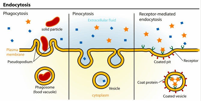
3.1. Endocytosis: Bringing the Outside In

Endocytosis is the active process by which cells internalize large substances. The plasma membrane physically sinks inward (invaginates), wraps entirely around the material, and pinches off, forming a brand-new intracellular vesicle.

  • Phagocytosis ("Cell Eating"): The aggressive, targeted ingestion of massive, solid particles like invading bacteria, dead tissue, or cellular debris by highly specialized immune sentinels (e.g., macrophages and neutrophils). The cell physically extends long "arms" called pseudopods to totally engulf the target, forming a giant internal death-chamber called a phagosome, which then merges with a lysosome for total destruction.
  • Pinocytosis ("Cell Drinking"): The constant, non-specific, routine gulping of tiny droplets of extracellular fluid and whatever dissolved solutes happen to be floating in it. It is how cells sample their environment.
  • Receptor-Mediated Endocytosis: An exquisitely, highly specific, targeted process. Specific extracellular ligands (e.g., LDL-cholesterol particles, iron-carrying transferrin, or even sneaky viruses) must perfectly bind to complementary, matching receptors on the surface. These loaded receptors then rapidly slide together and cluster into specialized depressions called clathrin-coated pits, which fold inward and are cleanly brought into the cell as a customized vesicle.

3.2. Exocytosis: Releasing to the Outside

Exocytosis is the reverse process by which cells actively release large substances. Intracellular, membrane-bound vesicles are driven by motor proteins to the edge of the cell, where they seamlessly fuse with the plasma membrane, violently dumping their contents to the outside world.

  • Constitutive Secretion: A continuous, ongoing, unregulated baseline process operating in all cells. It is used to constantly deliver newly minted lipids and proteins to expand the plasma membrane, and to continuously secrete the structural components of the extracellular matrix (like collagen).
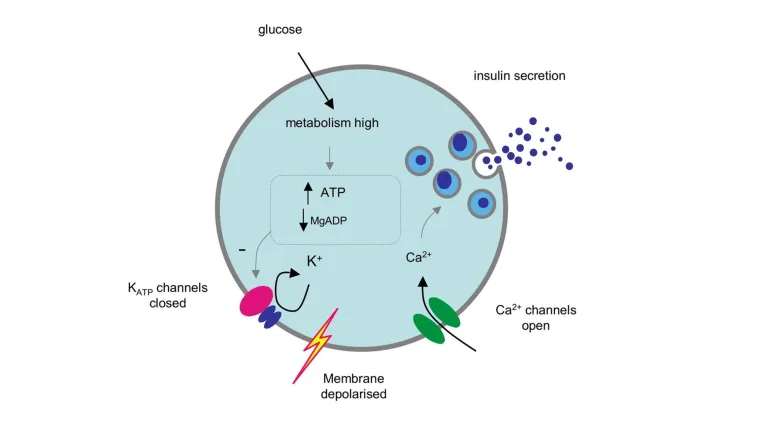
  • Regulated Secretion: A highly controlled process that occurs exclusively in specialized secretory cells (e.g., brain neurons, pancreatic endocrine cells). Secretory vesicles containing highly potent products like neurotransmitters, digestive enzymes, or hormones are stockpiled safely near the membrane. They are strictly held back and only released in a massive burst in response to a very specific, targeted signal (almost always a sudden, sharp rise in intracellular Ca²⁺).

10. The "Why": Importance and Functions of Membrane Transport

The precise, unrelenting control over exactly what enters and exits a cell underlies the success of virtually every single physiological process in the entire human body.

  • Maintenance of Cellular Homeostasis: Strict, life-or-death ion gradients (e.g., maintaining low intracellular Na⁺ and high K⁺) and exact internal pH are aggressively maintained at incredibly great energy cost to absolutely prevent cell death and ensure proper, flawless enzyme function.
  • Nutrient Acquisition: Transport systems enable cells to highly efficiently scavenge, absorb, and aggressively concentrate essential, life-sustaining molecules like glucose, vitamins, and amino acids from the blood.
  • Waste Removal: Active transporters continuously expel harmful, toxic metabolic waste products (like urea, lactic acid, and excess acid protons), totally preventing their lethal accumulation inside the cytoplasm.
  • Generation of Electrical Signals: In complex neurons and muscle cells, the rapid, highly controlled movement of ions surging through gated channels generates action potentials, which are the fundamental electrical basis of every single thought, heartbeat, and physical movement.
  • Cell-to-Cell Communication: Exocytosis violently releases neurotransmitters across synapses and dumps hormones into the blood, while endocytosis helps regulate receptor sensitivity by swallowing up overused receptors from the membrane.
  • Regulation of Cell Volume: Constant ion pumping, most especially the tireless Na⁺/K⁺ ATPase, strictly controls internal intracellular osmolarity, absolutely preventing cells from fatally swelling up and bursting or aggressively shrinking and collapsing.
  • Absorption and Reabsorption: Massive, highly coordinated transport processes in the Gastrointestinal (GI) tract and the nephrons of the kidneys are utterly essential for absorbing nutrients from food and perfectly regulating the entire body's water, total electrolyte, and systemic acid-base balance.
Summary of Membrane Transport Mechanisms
Process Energy Requirement Gradient Direction Transporter Requirement What Specifically Moves? Examples & Clinical Notes
Passive Processes (No ATP Burned)
Simple Diffusion None Downhill (High to Low) None (Straight through lipid) Small, highly lipid-soluble molecules O₂, CO₂, steroid hormones crossing tissues.
Facilitated Diffusion None Downhill (High to Low) Yes (Channel or Carrier protein) Ions, large glucose, amino acids Glucose transporters (GLUT), Na⁺/K⁺ voltage channels.
Osmosis None Downhill (High water to Low water) Yes (Aquaporins heavily aid) Water molecules exclusively Red blood cells expanding or shrinking in different tonic IV solutions.
Active Processes (Requires ATP)
Primary Active Transport Yes (Directly breaks ATP) Uphill (Low to High) Yes (A specialized Pump) Ions against steep gradients Na⁺/K⁺ pump, Ca²⁺ SERCA pump, H⁺ stomach acid pump.
Secondary Active Transport No (Uses kinetic energy of an existing ion gradient) Uphill (Low to High) Yes (Symporter or Antiporter) Ions, glucose, amino acids dragged along Na⁺-glucose co-transporter (SGLT) in the kidney.
Vesicular Transport Yes (Massive ATP used) N/A (Bulk physical movement) No (Uses membrane vesicles) Large particles, whole bacteria, macromolecules, bulk fluids Phagocytosis (macrophages eating bacteria), Exocytosis (releasing insulin).

List of Academic References

  • Hall, J. E., & Hall, M. E. (2020). Guyton and Hall Textbook of Medical Physiology (14th ed.). Elsevier. (Definitive source on membrane potentials, the Na⁺/K⁺ pump, and systemic physiology).
  • Boron, W. F., & Boulpaep, E. L. (2016). Medical Physiology (3rd ed.). Elsevier. (Excellent deep dive into cellular organelles, secondary active transport, and molecular physiology).
  • Alberts, B., Johnson, A., Lewis, J., et al. (2014). Molecular Biology of the Cell (6th ed.). Garland Science. (The absolute gold standard for the Fluid Mosaic Model, vesicular transport, and cytoskeletal structures).
  • Costanzo, L. S. (2017). Physiology (6th ed.). Elsevier. (Highly recommended for clear, mechanistic explanations of functional vs. process physiology and simple diffusion mathematics).
  • Chabner, L. (2020). The Language of Medicine (12th ed.). Saunders. (A premier resource for medical terminology, etymology, roots, and prefixes like pancytopenia).

Quick Quiz

Cell Physiology & Transport Quiz

Physiology - mobile-friendly and focused practice.

Privacy: Your details are used only for quiz tracking and certificates.

Physiology and Cell Physiology Read More »

Want notes in PDF? Join our classes!!

Send us a message on WhatsApp
0726113908

Scroll to Top
Enable Notifications OK No thanks