
Dracunculiasis (Guinea Worm Disease)
Dracunculiasis is a parasitic disease caused by the nematode worm Dracunculus medinensis.
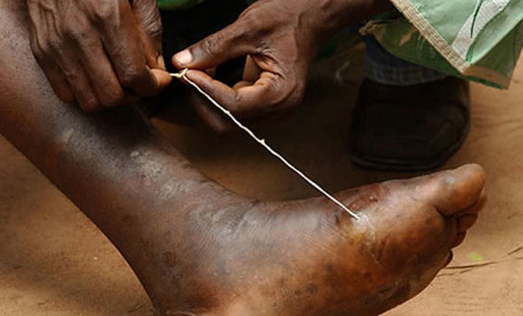
It is characterized by the emergence of a long, thread-like worm from a painful blister on the skin, usually on the legs or feet.
Dracunculiasis is a neglected tropical disease (NTD) that primarily affects poor communities in rural areas with limited access to safe water. It is a debilitating disease that can cause significant pain and disability.
The disease is transmitted through contaminated drinking water.
![]()
Transmission:
Vector: The disease is transmitted by copepods, tiny crustaceans found in stagnant water.
Lifecycle:
- In the Copepod (Vector):
- Copepods ingest the infective larvae of Dracunculus medinensis from contaminated water.
- The larvae develop into infective stage within the copepod.
- In Humans (Host):
- Humans become infected when they drink contaminated water containing the infected copepods.
- In the human stomach, the copepod is digested, releasing the larvae.
- The larvae penetrate the intestinal wall and migrate through the body, typically reaching subcutaneous tissue (beneath the skin).
- Larvae mature into adult worms within one year.
- The female worm migrates to the surface of the skin and emerges from a blister, usually on the legs or feet.
- The worm releases larvae into the water, continuing the cycle.
Routes of Transmission:
- Drinking Contaminated Water: This is the primary route of transmission.
Causes/Aetiology:
- Dracunculus medinensis Worm: The disease is caused by the Dracunculus medinensis nematode.
Clinical Features:
Initial Stage:
- A small, itchy blister appears on the skin, typically on the legs or feet.
- Fever, nausea, and vomiting may occur.
Blister Stage:
- Blister becomes painful, swollen, and filled with fluid.
- The worm may be visible within the blister.
Worm Emergence Stage:
- The female worm emerges from the blister, forming a long, thread-like structure.
- The worm can be several feet long and can cause intense pain as it emerges.
Secondary Complications:
- Infection of the wound: The emergence site can become infected with bacteria.
- Joint pain and stiffness.
- Lymphedema: Fluid buildup in the affected limb.
- Abscess formation: Pus collection around the worm.
Other Symptoms:
- Swelling and tenderness of the lymph nodes.
- Generalized weakness.
- Loss of appetite.
Diagnosis and Investigations:
- Clinical Examination: A typical blister with a visible worm emerging is usually diagnostic.
- Microscopic Examination: Examining the blister fluid or the emerging worm under a microscope can confirm the presence of Dracunculus medinensis.
- Serological Tests: Blood tests to detect antibodies against Dracunculus medinensis.
Prevention:
- Safe Water: Provide access to safe drinking water sources and promote safe water handling practices.
- Water Filtration: Use filters to remove copepods from drinking water.
- Boiling Water: Boiling water for at least 1 minute kills copepods.
- Education: Educate communities about the disease, its transmission, and prevention strategies.
- Environmental Management: Control mosquito breeding sites and improve sanitation in rural areas.

Management:
- There is no known drug treatment for guinea worm
Aims of Management:
- To relieve pain and discomfort.
- To prevent secondary infections.
- To prevent transmission of the disease.
Early Management:
- Wound Care: Clean the affected area with antiseptic solutions and dress the wound.
- Pain Relief: Administer pain relievers as needed.
- Preventing Secondary Infections: Administer antibiotics if secondary bacterial infection is suspected.
- Extraction of the Worm: A healthcare provider can carefully extract the worm from the blister. This process can be slow and painful and may take several days.
Medical Management:
All patients:
- To facilitate removal of the worm, slowly and carefully roll it onto a small stick over a period of days.
- Dress the wound occlusively to prevent the worm passing ova into the water.
- Give analgesics for as long as necessary If there is ulceration and secondary infection give:
- Amoxicillin 500 mg every 8 hours for 5 days
- Child: 250 mg every 8 hours for 5 days
- Or cloxacillin 500 mg every 6 hours for 5 days
- Pain Relief: Over-the-counter pain relievers can help manage pain.
- Antibiotics: Prescribed for any bacterial infections.
- Anti-inflammatory Medications: Can help reduce swelling and inflammation.
Nursing Care:
- Pain Management: Assist patients in managing pain and discomfort.
- Wound Care: Provide wound care and dressing changes as needed.
- Infection Prevention: Monitor for signs of infection and ensure appropriate wound care.
- Education: Teach patients about the disease, treatment, and prevention strategies.
Complications:
- Secondary Infections: The emergence site can become infected with bacteria, leading to cellulitis, abscesses, or sepsis.
- Arthritis: The worm can migrate into joints, causing inflammation and pain.
- Lymphedema: Fluid buildup in the affected limb due to lymphatic obstruction.
- Disability: Chronic pain, joint stiffness, and lymphedema can lead to significant disability.
- Social Stigma: The disease can lead to social isolation and discrimination.