HISTORY, PHYSICAL EXAMINATION AND INVESTIGATIONS IN GYNAECOLOGY

HISTORY, PHYSICAL EXAMINATION AND INVESTIGATIONS IN GYNAECOLOGY

History taking:

Proper history taking is key in determining the proper diagnosis and a guide to the required examination and investigations. The history from the patient should be taken from a private room.

During history taking, which should always start with the patient’s demographics, the following specific information should be captured.

Personal history: This refers to the biographic data of the patient.(NAATRENREM )

  • N Name
  • A Age
  • A Address
  • T Tribe
  • R Religion
  • E Education
  • N Next of kin
  • R Relationship with the next of kin
  • O Occupation
  • M Marital status.

Presenting complaints: The patient should be asked to state her problems. In chronological order. It is important that you first listen to the patient. The patient should describe fully the nature of her problems to enable you to come to a conclusion using the acronym “DOPAPRA.”

  • D Duration
  • O Onset
  • P Progression
  • A Aggravation
  • R Relieving
  • A Association.

NOTE: If the complaint is a disorder of menstruation, take full menstrual history. If about abnormal vaginal discharge, ask about colour, odour, and relationship with menstrual periods. 

History of abnormal vaginal discharge: Vaginal discharge may be normal or abnormal. Normal vaginal discharge is usually off-white and is neither associated with vaginal itching nor presence of a foul smell.

Abnormal vaginal discharge is usually associated with change in quantity, colour, vaginal irritation, smell and sometimes abdominal pain. Conditions commonly associated with abnormal vaginal discharge include trichomoniasis, bacterial vaginosis and vaginal candidiasis.

Cervical cancer may also present with abnormal vaginal discharge especially in the advanced stage of the disease when there is secondary infection.

When taking history, capture the following information:

  • Amount of discharge

  • Duration of discharge

  • Ask about the presence of smell of the discharge

  • Colour change

  • Relationship of the discharge with menstruation cycle

  • Associated lower abdominal pain.

If the presenting complaint is pain, as in abdominal pain, follow the acronym “SOCRRATES.”


S

Site

Where is the pain? Or the maximal site of the pain.

O

Onset

When did the pain start, and was it sudden or gradual? Include also whether it is progressive or regressive.

C

Character

What is the pain like? An ache? Stabbing?

R

Radiation

Does the pain radiate anywhere?

A

Associations

Any other signs or symptoms associated with the pain?

T

Time course

Does the pain follow any pattern?

E

Exacerbating / relieving factors

Does anything change the pain?

S

Severity

How bad is the pain?


Menstrual History:

  • Age of onset of the first period (menarche).
  • Regularity of the cycle.
  • Duration of the period.
  • Length of the cycle.
  • Amount of bleeding—Excess is indicated by the passage of clots or the number of pads used.
  • First day of the last menstrual period (LMP).

The menstrual history can be represented as 13/4/28, indicating that the onset of the period was at the age of 13, bleeding lasts for 4 days, and occurs every 28 days.

Past Gynaecology History: Has she ever had any gynaecological conditions like fibroids, rectal vaginal fistula, vesico-vaginal fistula, perineal tears, and abortions, etc.? Any operations on the cervix or dilation and curettage.

Past Medical History: Relevant medical disorders—systemic, metabolic, or endocrine (diabetes, hypertension, hepatitis) should be enquired. Their presence requires care during operative procedures. Next pertinent point is the interrogation about sexually transmitted diseases.

Find out any chronic conditions that the patient may be suffering from like chronic lung disease and cardiovascular diseases. Find out if the patient is on any medication, hospitalization, and for what reason.

Past Surgical History: This includes general, obstetrical, or gynaecological surgery. The nature of the operation, anaesthetic procedures, bleeding, or clotting complications if any, postoperative convalescence are to be enquired. Any histopathological report or relevant investigation related to the previous surgery is most often helpful.

Past Obstetric History: This entails the number of pregnancies, abortions, type of delivery, history of trauma, prolonged labour, etc., and menstrual cycle i.e. menarche, regularity, duration, and length of cycles, volume of blood loss, etc.

Family History: It is of occasional value. Malignancy of the breast, colon, ovary, or endometrium is often related. Tubercular affection of any family member can give a clue in the diagnosis of pelvic tuberculosis.

Past Social History: Look out for marital status, lifestyle, smoking, alcohol, occupation, etc.

Contraceptive History: Find out which method she uses, any side effects so far experienced, and if not using any, this information is important to rule out the possibility of pregnancy and also to determine whether the present complaint is not a result of the method of conception.

Cervical Cancer Screening: If the woman is over 35 years of age, ask whether she has ever been screened for cervical cancer.

Sexual History: This should be included if appropriate to presenting complaints. Most patients feel embarrassed to talk about their sexual behaviours. Capture information on pain during sexual intercourse or difficulty with coitus. Pain during sexual intercourse (dyspareunia) may be superficial, that is the woman feels pain during the entrance of the penis.

Superficial dyspareunia may be associated with lack of vaginal lubrication or vaginismus. Deep dyspareunia may be due to a scar, endometriosis or a mass. 


EXAMINATION 

After history taking, gynaecological examination is performed to confirm the presence/absence of pathology based on the history obtained.

Before gynaecological examination is carried out, the following should be done:

  • Explain to the patient the need for the examination and its nature.

  • Obtain an informed verbal consent.

  • The male examiner should be accompanied by a chaperone (female who can either be a nurse, fellow clinician or attendant)

  • The examination should be done in a private room respecting the patient’s privacy at all times.

  • The patient should be covered at all times and only relevant parts of her anatomy exposed.

  • Ensure that there is good lighting in the examination room.

  • Instruct the patient to void before the examination.

  • Ensure all the relevant supplies, sundries and equipment is available before beginning the examination.

Note: After the gynaecological examination, explain in a simple language and with the help of learning aids such as drawings or models the findings of the examination.

Physical Examination

The examination includes:

  1. General and systemic examination (General Examination)
  2. Gynaecological examination.
  • Breast examination
  • Abdominal examination
  • Pelvic examination
General Examination

1. Body Composition:

  • Built: Assessment of body weight and distribution.
  • Significance: Endocrinopathy can manifest as obesity or thinness, impacting menstrual patterns.

2. Nutritional Status:

  • Nutrition: Evaluation of dietary habits.
  • Significance: Nutritional factors may influence reproductive health, emphasizing the importance of a balanced diet.

3. Growth and Development:

  • Stature: Monitoring overall height and development of secondary sex characteristics.
  • Significance: Growth abnormalities may indicate hormonal imbalances affecting gynaecological health.

4. Systemic Indicators:

  • Pallor: Examination for paleness, suggesting potential anaemia.
  • Jaundice: Inspection for yellowing of the skin or eyes.
  • Edema of Legs: Assessment for abnormal fluid retention.
  • Significance: Anaemia or liver dysfunction can impact gynaecological well-being.

5. Oral Health:

  • Teeth, Gums, and Tonsils: Inspection for signs of infection or septic foci.
  • Significance: Oral health can contribute to overall systemic health, influencing gynaecological conditions.

6. Neck Examination:

  • Thyroid Gland and Lymph Nodes: Palpation, focusing on left supraclavicular glands.
  • Significance: Thyroid disorders or lymphatic abnormalities may have implications for gynaecological health.

7. Cardiovascular and Respiratory Assessment:

  • Cardiovascular System: Evaluation for any abnormalities.
  • Respiratory System: Inspection for respiratory issues.
  • Significance: Any cardiovascular or respiratory irregularities can impact surgical procedures if intervention is required.

8. Vital Signs Monitoring:

  • Pulse: Measurement of heart rate.
  • Temperature: Assessment of body temperature.
  • Respirations: Monitoring breathing rate.
  • Blood Pressure: Evaluation of systemic blood pressure.
  • Significance: Vital signs provide insights into overall health and may influence gynaecological management.

Gynaecology Examination

A gynaecological examination is done to assess the overall health of the female reproductive system. 

During the exam, the gynaecologist will look at the external and internal reproductive organs, as well as at the breasts, to determine whether there are any problems or conditions present.

Breast examination

This should be a routine, especially in women above the age of 30, to detect any breast pathology, the most important being carcinoma.

This is carried out before pelvic examination to try to allay the woman’s anxiety.

It can be performed while she is sitting on either a chair or an examination coach. Inspect for size, shape, and any other obvious abnormalities. Then the breasts are palpated each at a time to rule out any abnormal lumps and discharges.

It also gives an opportunity to teach the mother self-breast examination.

Gynaecology Examination

Examination of the breasts


  1. Inspection with the arms at her sides.

  2. Inspection with the arms raised above the head.

  3. Inspection with hands at the waist (with contracted pectoral muscle).

  4. Palpation of the axillary nodes.

  5. Palpation of the supraclavicular nodes.

  6. Palpation of the outer half of the breast (a pillow is placed under the patient’s shoulder).

Abdominal examination

Abdominal examination is done when the patient is lying supine with a pillow for headrest.

The arms should be by the sides and the bladder must be emptied. The only exception to the procedure is the presence of a history suggestive of stress incontinence. If history suggests chronic retention of urine, catheterization should be done, taking aseptic precautions, using a sterile simple rubber catheter.

The physician usually prefers to stand on the right side.

Actual steps of abdominal examination

INSPECTION:

Abdominal examination assesses for:

  • Shape of the abdomen

  • Abdominal distension or masses

  • Movement with respiration

  • Presence of scars due to surgery or trauma

  • Distended veins and presence or absence of striae

  • Distribution of the pubic hair

  • Swelling or bulging inguinal orifices (with or without cough impulse)
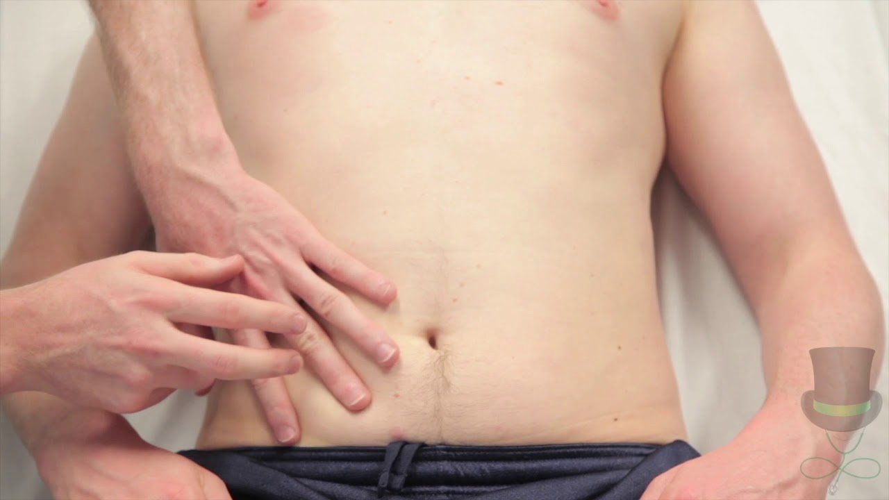
PALPATION

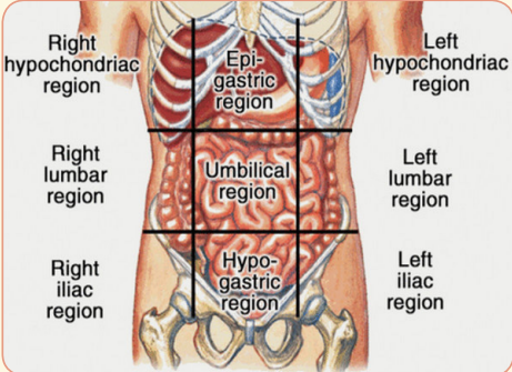
Palpation for tenderness, spleen, liver, kidneys, and for masses. Palpate the abdomen in all the nine regions; right hypochondria (RH), epigastrium (E), Left Hypochondria (LH), Right Lumbar (RL), Umbilical (U), Left Lumbar (LL), Right Iliac (RI), Pubic/Hypogastric (P) and Left Iliac.

  • Palpation should be done with the flat of the hand gently rather than the tips of the fingers.

  •  If a mass is felt in the lower abdomen, its location, size above the symphysis pubis, consistency, feel, surface, mobility from side to side and from above to down, and margins are to be noted. 

PERCUSSION

Percussion is done to assess for presence or absence of gas or fluid in the abdomen. Fluids could be blood or ascites.
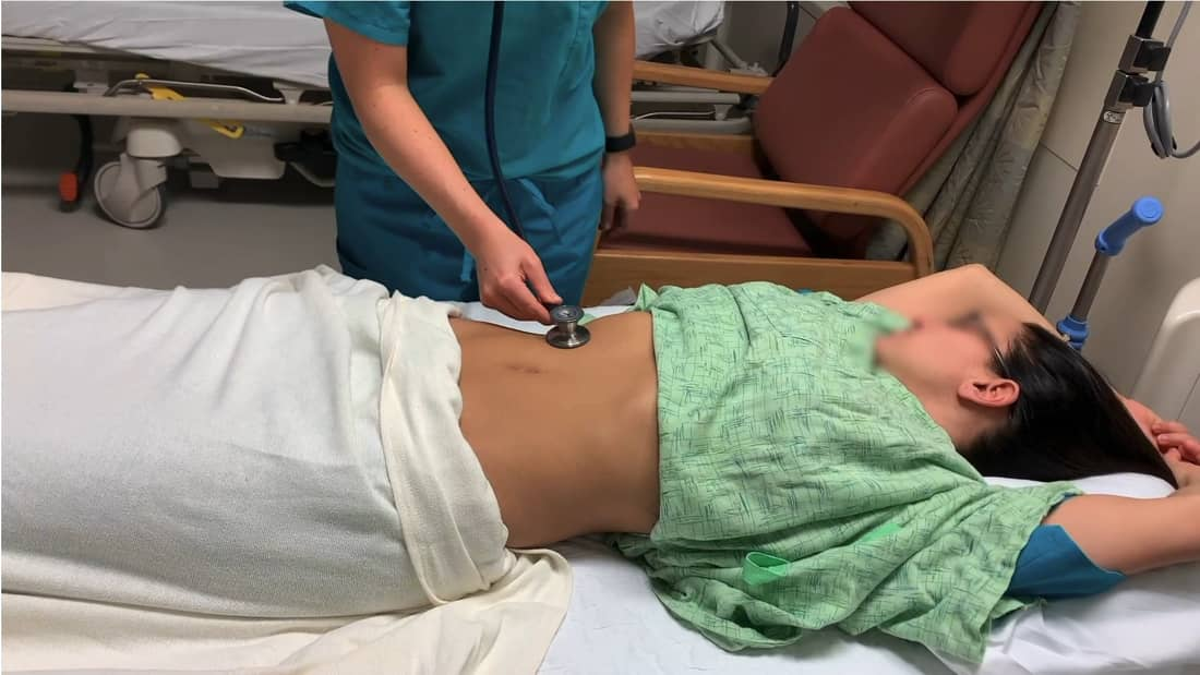
AUSCULTATION

Listen for bowel sounds or for fetal heart sounds and rate in case of pregnancy using a stethoscope or fetal stethoscope (fetoscope) respectively.


PELVIC EXAMINATION
  • The pelvic examination should be done on an examination couch with patient supine, knees and hips flexed, hips abducted and feet together.
  • The examiner stands on the patient’s (right or left) side.
  • A good and adjustable light source is needed for inspection of the vulva and for the speculum examination.
  • Sterile gloves, sterile lubricant (preferably colourless without any antiseptics), speculum, sponge holding forceps and swabs are required.
  • Pelvic examination is usually required when a patient presents with lower abdominal pain, menstrual disorders, abnormal vaginal discharge or to obtain a pap smear.
  • Pelvic examination should never be missed unless the patient is a virgin.
  • To examine a minor or unmarried, a consent from the parent or guardian is required
  • Lower bowel (rectum and pelvic colon) should preferably be empty.

Pelvic examination includes:

1. Inspection of the external genitalia

2. Vaginal examination

  • Inspection of the cervix and vaginal walls
  • Palpation of the vagina and vaginal cervix by digital examination
  • Bimanual examination of the pelvic organs

3. Rectal examination

4. Recto-vaginal examination.

Positions during pelvic examination:

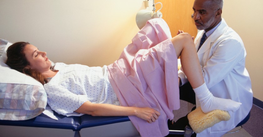
Lithotomy position (patient lying supine with her legs on stirrups) is ideal for examination.

Inspection of the vulva:

  • To note any anatomical abnormality starting from the pubic hair, clitoris, labia, and perineum.
  • To note any palpable pathology over the areas.
  • To note the character of the visible vaginal discharge, if any
  • To separate the labia using fingers of the left hand to note external urethral meatus, visible openings of the Bartholin’s ducts (normally not visible unless inflamed), and character of the hymen.
  • To ask the patient to strain in order to find out Stress incontinence—urine comes out through the urethral meatus or Genital prolapse and the structures involved— anterior vaginal wall, uterus alone or posterior vaginal wall or all the three.
  • Lastly, to look for haemorrhoids, anal fissure, anal fistula, or perineal tear.

Vaginal examination:

We shall look at the following variations of performing vaginal examination:

  • Inspection of the cervix and vaginal walls
  • Palpation of the vagina and vaginal cervix by digital examination
  • Bimanual examination of the pelvic organs

Inspection of the vagina and cervix:

Inspection of the vagina and cervix is done by speculum examination. Two types of speculum are commonly used—Sims’ or Cusco’s bivalve. While in the dorsal position, Cusco is widely used, but in the lateral position, Sims’ variety has advantages.

Introduction of Cusco’s speculum:

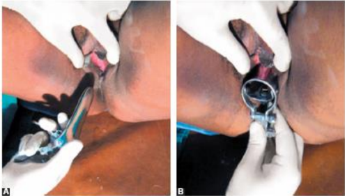
(A) The transverse diameter of the closed blades is placed in the anteroposterior position and inserted slightly obliquely to minimize pressure on the urethra;

(B) Blades are inserted in a downward motion and then rotated. Rotate to 90° and then open up the blades. Inspection is then made using good light. The cervix is best visualized with Cusco’s variety. But while the vaginal fornices are only visualized by Cusco, the anterior vaginal wall is to be visualized by Sims’ variety. Sims’ speculum is advantageous in cases of genital prolapse. Speculum examination should preferably be done prior to bimanual examination.

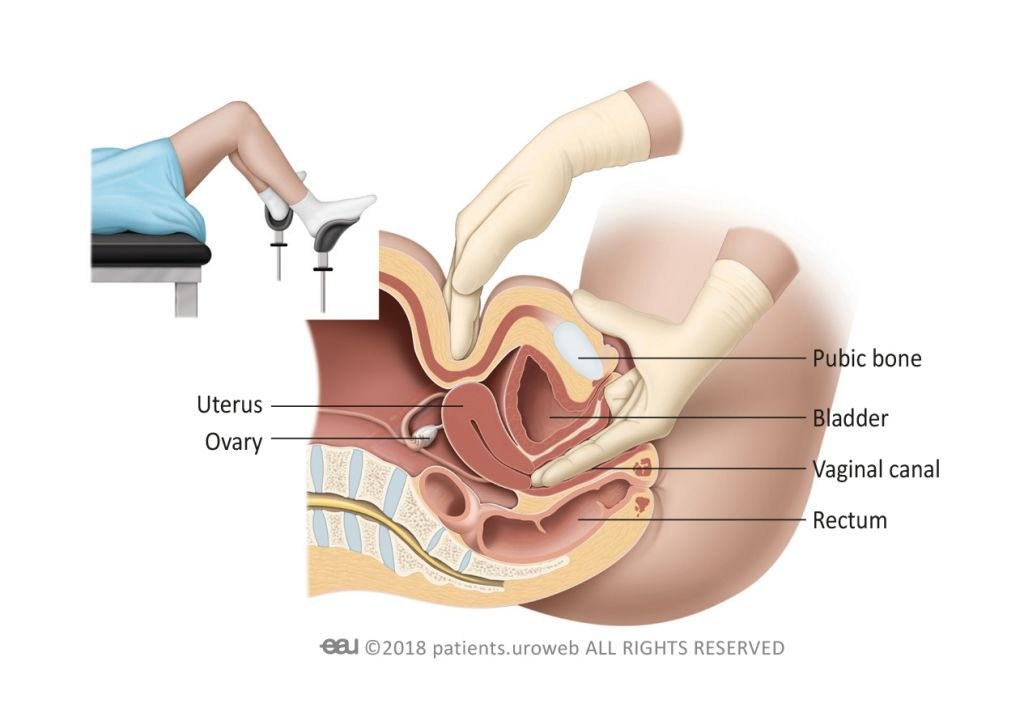
Bimanual Digital examination:

  • Explain every step to the patient and reassure her. Inform her that an internal examination is to be performed.
  • The labia are gently parted with the gloved index finger and thumb of the non-dominant hand.
  • Initially the lubricated index finger of the examiner’s dominant hand is inserted through the introitus into the vaginal canal.
  • If the patient is comfortable with this, the lubricated middle finger of the same hand is also inserted.
  • If not, due to pain, a limited bimanual examination with one finger can be performed.
  • The full length of the finger is introduced, assessing the vaginal walls in transit until the cervix is located.
GYNAECOLOGICAL INVESTIGATIONS

GYNAECOLOGICAL INVESTIGATIONS 

After taking a thorough history and conducting a complete physical examination, some investigations may be required to confirm the diagnosis or rule out differential diagnosis.

1. Blood Values:

  • Full Complete Blood Count (FBC or CBC): Evaluation of haemoglobin levels in cases of excessive bleeding and white blood cell count in infections.
  • VDRL Test for Syphilis: Screening for syphilis infection.
  • Serological Test for HIV Infection: Identification of HIV infection.

2. Urine Examination and Culture:

  • Collection Method: Midstream urine.
  • Purpose: Detection of urinary tract infections or confirmation of pregnancy. Urinalysis for suspected urinary tract infections.

3. Vaginal Swab:

  • Indication: Abnormal vaginal discharge.
  • Tests Conducted: Microscopic examination, culture & sensitivity.
  • Diagnosis: Trichomoniasis, bacterial vaginosis, and vaginal candidiasis.

4. Pap Smear: Routine screening for cervical cancer.

5. Hormonal Assays:

  • Hormones Assessed: FSH, testosterone, progesterone, luteinizing hormone, prolactin, among others.
  • Applications: Diagnosis of menopause, polycystic ovarian syndrome (PCOS), and assessment of ovarian function.

6. Pelvic Ultrasound:

  • Purpose: Evaluation of structural disorders in the genital tract.
  • Diagnostic Significance: Detection of pelvic masses or fluid.

7. Computed Tomography (CT) and Magnetic Resonance Imaging (MRI):

  • Indication: Suspected malignant diseases of the genital organs.
  • Considerations: Reserved for cases with a serious clinical need due to cost implications.

8. Hysterosalpingography:

  • Indication: Assessment of fallopian tube patency in cases of infertility.
  • Procedure: Injection of radio-opaque substance through the cervix.
  • Monitoring: Progress tracked on a screen as the substance fills the uterus and fallopian tubes.

Special Procedures in Gynecology

These are useful to fill gaps which remain after history taking during clinical assessment.

1. Evacuation: Removal of cavity contents, typically performed when a pelvic examination is not feasible. Anaesthesia is utilized, but tenderness signs may be overlooked.

2. Curettage: Scraping the internal organ or body cavity surface using a spoon-shaped instrument (curette).

Purpose:

  • Remove retained products of conception.
  • Obtain diagnostic specimens.

3. Biopsy: Removal of a small living tissue piece for microscopic examination, aiding in disease exclusion.

Sites: Cervix, endometrium, etc.

4. Ultrasound Scan: Utilizes high-frequency sound waves to produce images of pelvic organ structures. Widely used for disease detection and pregnancy monitoring.

5. Hysterosalpingography: X-ray imaging of the uterus and fallopian tubes.

Diagnoses tubal obstruction, adhesions, uterine malformations, small intracavity tumors, and internal os issues causing abortion.

6. Laparoscopy: Examination of abdominal structures using a laparoscope.

Applications:

  • Biopsy.
  • Cyst aspiration.
  • Adhesion division.
  • Ova collection for in vitro fertilization.

Gynaecological Operations

1. Hysterectomy: Surgical removal of the uterus.

Types:

  • Wertheim’s Hysterectomy (Radical).
  • Subtotal Hysterectomy.
  • Total Hysterectomy.
  • Indications: Fibroids, cancers, ruptured uterus.

2. Salpingectomy: Surgical removal of fallopian tubes.

  • Indications: Ruptured ectopic pregnancy, chronic salpingitis.

3. Vesico-vaginal Fistula Repair: Operation to correct abnormal bladder-vagina communication.

4. Oophorectomy: Surgical removal of ovaries.

  • Indications: Ovarian tumors, chronic oophoritis.

5. Myomectomy: Surgical removal of uterine fibroids.

6. Rectal Vaginal Fistula Repair: Operation to correct abnormal rectum-vagina communication.

7. Mastectomy: Surgical removal of the breast.

Types:

  • Radical Mastectomy.
  • Simple Mastectomy.

8. Tubal Ligation: Permanent family planning method involving tying and cutting fallopian tubes.

9. Vulvectomy: Surgical removal of the vulva.

Types:

  • Simple Vulvectomy.
  • Radical Vulvectomy.

10. Dilatation and Curettage: Procedure involving cervical dilation and endometrial scraping for purposes like abortion product removal and biopsy.

11. Perineoplasty: Operation to enlarge the vaginal opening by hymen and perineum incision.

12. Perineorrhaphy: Surgical repair of a damaged perineum resulting from childbirth tears.

Spread the love

1 thought on “HISTORY, PHYSICAL EXAMINATION AND INVESTIGATIONS IN GYNAECOLOGY”

Leave a Comment

Your email address will not be published. Required fields are marked *

Contact us to get permission to Copy

We encourage getting a pen and taking notes,

that way, the website will be useful.

Scroll to Top