
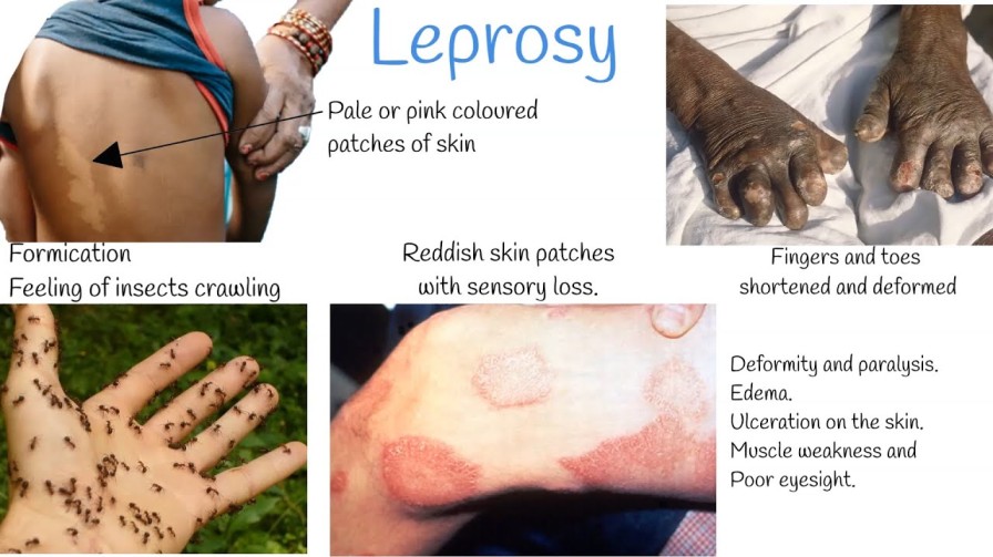
Leprosy, officially known as Hansen’s disease, is a chronic, progressive granulomatous infectious disease caused by the obligate intracellular acid-fast bacillus Mycobacterium leprae (and rarely Mycobacterium lepromatosis).
The organism demonstrates a unique, selective tropism for peripheral Schwann cells and macrophages of the reticuloendothelial system. Consequently, the disease predominantly attacks the peripheral nervous system, the skin, the mucous membranes of the upper respiratory tract, the anterior chamber of the eye, and the testes. Untreated, leprosy leads to irreversible peripheral nerve destruction, sensory anesthesia, motor paralysis, secondary reabsorption of phalanges, severe facial disfigurement, blindness, and profound social exclusion. However, with prompt diagnosis and modern Multidrug Therapy (MDT), leprosy is completely curable.
- Ancient Plague: Leprosy has afflicted human populations for over four millennia, historically regarded with horror and spiritual curse, leading to the forced banishment of sufferers into isolated leper colonies or sanatoria.
- Discovery of the Pathogen: The causative bacterium was identified in 1873 by the Norwegian physician Gerhard-Henrik Armauer Hansen in Bergen, Norway.
- Monumental Scientific Milestone: Mycobacterium leprae was the very first bacterium identified as a causative agent of human disease, antedating Robert Koch’s discovery of Mycobacterium tuberculosis (1882) and Vibrio cholerae (1883).
- Classification: Family Mycobacteriaceae, Order Actinomycetales. Strongly acid-alcohol fast, non-motile, non-spore-forming, straight or slightly curved rod-shaped bacillus (measuring 1–8 μm in length and 0.3–0.5 μm in diameter).
- Staining: Stains bright red against a blue background with the modified Ziehl-Neelsen stain (using 5% sulfuric acid or 1% acid-alcohol for decolorization rather than the standard 20% sulfuric acid, because its mycolic acid cell wall is slightly less acid-fast than M. tuberculosis) or the Fite-Faraco stain in paraffin-embedded histological tissue sections.
- In Vitro Cultivation Impossibility: M. leprae is an obligate intracellular parasite possessing a massively decayed, pseudogene-dense reductive genome. It cannot be cultured on any artificial cell-free bacteriological media or conventional tissue cell cultures. Laboratory propagation is restricted strictly to live animal footpad inoculation in immunocompromised/normal mice or systemic proliferation within the nine-banded armadillo (Dasypus novemcinctus), whose core body temperature is naturally low.
- Microscopic Arrangement: Inside host macrophages (histiocytes), the bacilli produce an abundant extracellular lipid capsule (composed largely of phenolic glycolipid-1 [PGL-1]) that binds them into compact, cigar-shaped parallel bundles and round, dense globular masses termed Globi. Heavily parasitized, lipid-laden macrophages are termed Virchow cells or lepra cells.
- Replication Kinetics: The bacillus has the slowest known doubling time among human bacterial pathogens, dividing once every 12 to 14 days (compared to 20 minutes for E. coli and 20 hours for M. tuberculosis).
- Thermal Preference (Cryotropism): The organism replicates optimally at cooler temperatures between 30°C and 33°C, completely explaining its clinical predilection for superficial, cooler anatomical structures: the dermis, superficial nerve trunks located near bone surfaces (ulnar at the olecranon groove, common peroneal at the fibular head), nasal mucosa, earlobes, and scrotum, while entirely sparing warmer visceral organs such as the brain, lungs, and spinal cord.
- Primary Route of Transmission: Airborne transmission via nasopharyngeal respiratory droplet aerosols expelled during sneezing, coughing, or prolonged conversation by untreated, highly bacilliferous multibacillary (MB) patients. Millions of viable bacilli are shed daily in their nasal secretions.
- Infectivity Requirements: Transmission is not casual. It requires prolonged, close, frequent contact (months to years) within the same household or immediate living environment.
- Innate Human Immunity: Approximately 95% of all human beings possess complete natural, innate cell-mediated immunity against M. leprae and will never develop clinical disease even upon heavy exposure.
- Infectivity Cessation: Within 72 hours to a few days of initiating multidrug therapy (MDT), the bactericidal potency of Rifampicin renders the patient non-infectious, permanently arresting forward transmission.
- Non-Transmission Routes: There is no transplacental transmission (cannot be passed to an unborn fetus), no sexual transmission, and no transmission via casual social contacts (shaking hands, hugging, sharing utensils, sitting together).
- Zoonotic Reservoirs: While humans are the primary reservoir, wild nine-banded armadillos in the southern United States and red squirrels in the United Kingdom harbor endemic M. leprae strains capable of zoonotic transmission to humans via direct handling.
- Due to the exceptionally slow bacterial division rate, the incubation period is prolonged.
- Average Duration: Approximately 5 years.
- Clinical Range: Ranges from 9 months to 20 years or more. Symptoms in Paucibacillary (PB) forms typically appear earlier (2–5 years), whereas Multibacillary (MB) forms average 8–12 years.
- Close Household Contact: Living with an untreated index patient with Multibacillary (MB) disease elevates disease risk up to 5- to 10-fold.
- Geographic Clustering: Residing in hyper-endemic pockets or cross-border corridors (e.g., the West Nile region in Uganda).
- Genetic Predisposition: Host polymorphisms in human leukocyte antigen (HLA-DR2 and HLA-DR3 associated with tuberculoid disease; HLA-DQ1 associated with lepromatous disease) and non-HLA genes (PARK2/PACRG, NOD2, LTA).
- Malnutrition & Extreme Poverty: Food insecurity, overcrowded poor housing, and poor sanitary conditions that weaken baseline mucosal cell-mediated immunity.
Leprosy is fundamentally a neuro-dermatological disease. Mycobacterium leprae is the only human pathogen capable of directly invading the parenchyma of peripheral nerves:
- Attachment: The unique cell wall glycolipid PGL-1 and bacterial adhesins bind specifically to the G-domain of laminin-α2 situated within the basal lamina surrounding peripheral Schwann cells.
- Invasion & Demyelination: This interaction triggers receptor-mediated endocytosis via the α-dystroglycan complex. Once intracellular, the bacillus induces Schwann cell dedifferentiation, downregulating myelin gene expression and upregulating stem-cell-like progenitor traits, initiating spontaneous demyelination and axonal atrophy.
- Immunological Destruction: The subsequent destruction of the peripheral nerve is an immunologically mediated bystander injury. In patients with vigorous cell-mediated immunity (tuberculoid pole), CD4+ T-helper 1 (Th1) lymphocytes infiltrate the endoneurium, releasing interferon-gamma (IFN-γ) and TNF-α, forming compact epithelioid granulomas within the non-distensible perineurial sheath. This generates high intra-neural compartment pressure, localized ischemia, caseous necrosis, and permanent fibrotic nerve trunk strangulation.
- Lepromatous Infiltration: Conversely, in lepromatous leprosy, deficient cellular immunity permits unhindered intracellular bacterial replication within Schwann cells, perineurial macrophages, and vascular endothelial cells, causing painless, gradual, diffuse replacement of nerve fibers by foamy lepra cells and widespread ascending sensorimotor axonal loss.
The clinical, histological, and bacteriological manifestations of leprosy represent a continuous biological spectrum dictated entirely by the potency of the host’s Cell-Mediated Immunity (CMI) against M. leprae:
| Classification | Cell-Mediated Immunity | Bacterial Index (BI) | Skin Lesion Features | Histopathological Hallmarks |
|---|---|---|---|---|
| Tuberculoid (TT) | High / Robust (Th1 dominant: IFN-γ, IL-2) | 0 (Negative) | Single or very few (≤3) large, well-demarcated hypopigmented anesthetic plaques with raised, erythematous, dry, hairless, scaly edges. Marked nerve thickening. | Epithelioid cell granulomas with Langerhans giant cells surrounding dermal nerve twigs; dense cuffing by CD4+ lymphocytes. No demonstrable AFB. |
| Borderline Tuberculoid (BT) | Moderate / Variable | 0 to 1+ | Few lesions (typically 3–10) resembling TT but smaller, less sharply defined margins; multiple satellite lesions; asymmetric nerve enlargement. | Well-formed epithelioid granulomas, few giant cells, moderate lymphocytic infiltration, rare AFB in Schwann cells. |
| Mid-Borderline (BB) | Unstable / Equivocal | 2+ to 3+ | Moderate number of lesions; characteristic "punched-out" or "inverted saucer" plaques (sloping outer margin with a sharply punched-out, anesthetic center). | Diffuse epithelioid granulomas without giant cells, variable lymphocytes, moderate AFB present. |
| Borderline Lepromatous (BL) | Low / Deficient | 3+ to 5+ | Numerous bilateral, asymmetric macules, papules, plaques, and small nodules; sensation mildly reduced or normal in early stages. | Predominantly foamy histiocytes, few lymphocytes, abundant AFB singly and in globi, perineurial onion-skin lamellation. |
| Lepromatous (LL) | Absent / Anergy (Th2 dominant: IL-4, IL-10) | 5+ to 6+ (Extreme) | Innumerable, small, symmetric, shiny, ill-defined erythematous macules, papules, diffuse infiltration, plaques, and nodules. Leonine facies, loss of eyebrows, saddle-nose. | Extensive sheets of foamy Virchow histiocytes packed with thousands of AFB in globi. Uninfiltrated Grenz zone directly below the epidermis. No granulomas. |
The earliest, uncommitted clinical stage of leprosy. Characterized by a single, faint, hypopigmented or erythematous macule with vague borders and only slightly diminished sensation. Histologically shows non-specific perivascular or periadnexal lymphocytic infiltration with rare bacilli located within tiny dermal neurovascular bundles. Depending on the individual's evolving cell-mediated immunity, indeterminate leprosy will either resolve spontaneously or evolve toward one of the Ridley-Jopling poles (TT or LL).
In endemic countries, clinical case confirmation requires the definitive presence of at least ONE of the following three Cardinal Signs:
- Definite Loss of Sensation in a Hypopigmented or Reddish Skin Patch: Tested carefully using a light wisp of cotton wool, a feather, or standardized Semmes-Weinstein monofilaments (0.07g to 2.0g) applied to the patch while the patient's eyes are closed, comparing responses with adjacent normal skin. Thermal and pinprick sensation are lost prior to light touch.
- Thickened, Enlarged Peripheral Nerve Trunk(s): Palpable, tender, or firm peripheral nerves, accompanied by objective sensory impairment in its cutaneous distribution and/or motor weakness of the muscle groups it innervates.
- Microscopic Demonstration of Acid-Fast Bacilli (AFB): Visualized in a stained slit-skin smear or full-thickness skin/nerve biopsy specimen.
Clinicians must routinely palpate specific superficial nerve trunks bilaterally for tenderness, enlargement, irregular beading, and firmness:
- Ulnar Nerve: Palpated in the groove between the olecranon process and medial epicondyle of the humerus.
- Sensory Deficit: Numbness over the hypothenar eminence, medial palm, little finger, and ulnar half of the ring finger.
- Motor Deficit: Weakness of intrinsic hand muscles, interossei wasting, lumbrical paralysis, producing Claw Hand (main-en-griffe) deformity (hyperextension of metacarpophalangeal joints and flexion of interphalangeal joints, most marked in digits 4 and 5).
- Median Nerve: Palpated as it passes deep to the flexor retinaculum at the wrist.
- Sensory Deficit: Numbness over the thenar eminence, palmar aspects of thumb, index, middle finger, and radial half of the ring finger.
- Motor Deficit: Thenar atrophy and paralysis of the abductor pollicis brevis, resulting in loss of thumb opposition (Ape Hand deformity) and total clawing when combined with ulnar paralysis.
- Radial Cutaneous Nerve: Palpated across the anatomical snuffbox over the styloid process of the radius. Motor branch (posterior interosseous) involvement leads to Wrist Drop.
- Common Peroneal (Lateral Popliteal) Nerve: Palpated rolling over the neck of the fibula on the lateral aspect of the knee.
- Motor Deficit: Paralysis of the ankle dorsiflexors and peronei, resulting in Foot Drop with an unnatural high-stepping gait, leading to severe dragging of toes and extensive neuropathic trophic ulcerations.
- Posterior Tibial Nerve: Palpated behind and below the medial malleolus of the ankle.
- Sensory Deficit: Complete anesthesia of the entire sole of the foot.
- Motor Deficit: Paralysis of intrinsic foot muscles leading to Clawed Toes and high peak planter pressures during ambulation, predisposing to chronic, painless neuropathic plantar ulcers.
- Facial Nerve (Cranial Nerve VII): Specifically its zygomatic and temporal branches running over the zygomatic arch.
- Motor Deficit: Paralysis of the orbicularis oculi muscle causing Lagophthalmos (inability to close the eyelid completely), absence of the corneal blink reflex, continuous exposure keratitis, corneal ulceration, secondary bacterial infection, and irreversible blindness.
- Great Auricular Nerve: Palpated crossing diagonally across the superficial surface of the sternocleidomastoid muscle in the lateral neck; frequently visibly enlarged and cord-like in lepromatous and borderline cases.
- Leonine Facies (Facies Leonina): Extensive nodular thickening, deep furrowing of the forehead, glabellar ridges, and earlobes, imparting a lion-like facial appearance.
- Madarosis: Bilateral, symmetrical, permanent loss of eyebrows (beginning characteristically at the outer lateral thirds) and eyelashes.
- Saddle-Nose Deformity: Extensive chronic granulomatous infiltration and ischemic necrosis of the cartilaginous nasal septum, leading to septal perforation, structural collapse, and sunken bridge of the nose.
- Gynecomastia & Testicular Atrophy: Direct bacillary infiltration of the seminiferous tubules and interstitial Leydig cells, causing hypergonadotropic hypogonadism, impotence, and sterility.
A simple, highly specific semi-invasive bedside test performed to detect and quantify viable and non-viable acid-fast bacilli:
- Standard Sampling Sites (Minimum 4 to 6 Sites):
- Both earlobes (lower fleshy margin).
- At least two active, erythematous, indurated skin lesion edges.
- Dorsal aspect of fingers or normal-appearing skin over the forehead or chin.
- Procedure: Pinch the skin firmly between thumb and forefinger to render the tissue ischemic (preventing bleeding, which dilutes the smear with red blood cells). Make a superficial scalpel slit (5 mm long, 2 mm deep) into the dermis. Turn the scalpel blade 90 degrees and scrape the side and bottom of the slit firmly to obtain clear dermal tissue pulp and cellular fluid. Smear onto a clean glass slide, heat fix, and stain with the modified Ziehl-Neelsen method.
- Microscopic Enumeration:
- Bacterial Index (BI - Ridley Scale): Reflects total bacterial load (live and dead):
- 0: No bacilli observed in 100 oil immersion fields.
- 1+: 1 to 10 bacilli per 100 fields.
- 2+: 1 to 10 bacilli per 10 fields.
- 3+: 1 to 10 bacilli per single field.
- 4+: 10 to 100 bacilli per single field.
- 5+: 100 to 1,000 bacilli per single field (clumps/globi).
- 6+: >1,000 bacilli per field (massive globi throughout).
- Morphological Index (MI): The percentage of solid-staining, fully intact, viable bacilli out of 200 counted bacilli. A falling MI is the earliest, most sensitive indicator of bacteriological response to effective MDT.
- Bacterial Index (BI - Ridley Scale): Reflects total bacterial load (live and dead):
Full-thickness punch or incisional biopsy taken from the advancing, elevated border of an active skin lesion including subcutaneous fat, or a superficial sensory nerve twig (e.g., radial cutaneous or sural nerve). Stained with Hematoxylin & Eosin (H&E) and the Fite-Faraco acid-fast stain. Provides absolute confirmation by demonstrating neural architecture, perineurial inflammation, granuloma morphology, and intra-neural bacilli.
Utilizes targeted nucleic acid amplification targeting the repetitive element (RLEP), groEL, or the 16S rRNA genes of M. leprae. Highly sensitive (>90%) and specific, capable of detecting as few as 1 to 10 bacterial genomes in paucibacillary lesions, indeterminate cases, blood, or fine-needle aspirates where smears are completely negative.
- Hypopigmented Macules: Tinea versicolor (pityriasis versicolor—shows superficial scaling, KOH-positive fungal hyphae, completely normal sensation); Vitiligo (total chalky depigmentation, no sensory loss); Pityriasis alba (common in atopic children, cheeks, normal sensation); Post-inflammatory hypopigmentation.
- Infiltrated Plaques & Nodules: Cutaneous Leishmaniasis; Sarcoidosis (non-caseating naked granulomas, no sensory deficit); Cutaneous Tuberculosis (Lupus vulgaris); Secondary and Tertiary Syphilis; Neurofibromatosis type 1; Kaposi Sarcoma; Granuloma annulare.
- Peripheral Neuropathies: Diabetic peripheral sensory-motor polyneuropathy (bilateral, symmetrical, glove-and-stocking without thickened nerve trunks); Alcoholic neuropathy; Nutritional neuropathies (Vitamin B12 deficiency); Hereditary motor-sensory neuropathy (Charcot-Marie-Tooth disease).
To eliminate reliance on laboratory microscopy at primary health centers, the WHO established a pragmatic operational classification based entirely on clinical lesion counting:
- Paucibacillary (PB) Leprosy: 1 to 5 skin lesions with definite sensory loss, affecting no more than one nerve trunk. Slit-skin smears (if performed) are negative. (Corresponds to TT and BT on the Ridley-Jopling spectrum).
- Multibacillary (MB) Leprosy: 6 or more skin lesions, OR involvement of more than one peripheral nerve trunk (regardless of lesion count), OR any positive slit-skin smear finding. (Corresponds to BB, BL, and LL on the Ridley-Jopling spectrum).
MDT blister packs are supplied by the WHO free of charge globally. Treatment combines three core antimycobacterial agents:
Crucial Paradigm Shift — WHO 2018 Guidelines:
Historically, PB leprosy was treated with only 2 drugs (Rifampicin and Dapsone for 6 months), while MB leprosy received 3 drugs (adding Clofazimine for 12 months). The WHO Guidelines for the Diagnosis, Treatment, and Prevention of Leprosy 2018 strongly recommended a Uniform 3-Drug Regimen (Rifampicin, Dapsone, and Clofazimine) for BOTH PB and MB leprosy (6 months for PB, 12 months for MB). This prevents relapse, simplifies drug procurement, and ensures that misclassified MB cases are not undertreated.
*Note on Operational Practice: While Uganda and many national programmes have phased in 3-drug therapy, traditional 2-drug blister packs remain clinically relevant in many field settings. Both regimens are detailed below.
| Drug | Dosage & Frequency | Mode of Administration | Duration |
|---|---|---|---|
| Rifampicin | 600 mg once monthly | Directly Observed Therapy (DOT) by health worker | PB: 6 months MB: 12 months |
| Clofazimine | 300 mg once monthly (supervised DOT) PLUS 50 mg daily | DOT (monthly dose) Self-administered (daily dose) | PB: 6 months (WHO 2018) MB: 12 months |
| Dapsone | 100 mg daily | Self-administered daily | PB: 6 months MB: 12 months |
| Medication | Children Aged 10 to 14 Years | Children <10 Years (or <40 kg Body Weight) |
|---|---|---|
| Rifampicin | 450 mg once monthly (supervised DOT) | 10 mg/kg body weight once monthly (DOT) |
| Clofazimine | 150 mg once monthly (DOT) + 50 mg every other day | 6 mg/kg once monthly (DOT) + 1 mg/kg every other day |
| Dapsone | 50 mg daily | 2 mg/kg body weight daily |
- PB Leprosy: 6 monthly blister packs to be completed within a maximum timeframe of 9 months.
- MB Leprosy: 12 monthly blister packs to be completed within a maximum timeframe of 18 months.
In HIV-positive patients already receiving daily Cotrimoxazole (TMP-SMX) preventive prophylaxis, DAPSONE MUST NEVER BE CO-ADMINISTERED. Both agents are folate-synthesis inhibitors (sulfonamides/sulfones) and share identical pathways of hematological toxicity. Concurrent use precipitates catastrophic, life-threatening bone marrow aplasia, severe agranulocytosis, and severe hemolytic crisis. In such patients, Dapsone must be replaced with Clofazimine or second-line regimens under specialist guidance.
- Rifampicin (Rifadin):
- Mechanism: Binds to the β-subunit of bacterial DNA-dependent RNA polymerase, blocking transcription and RNA synthesis. It is the single most powerful, rapidly bactericidal drug in the regimen, killing 99.9% of viable M. leprae within a single 600 mg dose.
- Adverse Effects: Harmless orange-red discoloration of urine, sweat, saliva, and tears (essential patient counseling point!); transient hepatotoxicity; cutaneous flushing; rare flu-like syndrome with intermittent high-dose therapy.
- Dapsone (4,4’-diaminodiphenylsulfone):
- Mechanism: Competitive antagonist of para-aminobenzoic acid (PABA), inhibiting bacterial dihydropteroate synthase and halting folic acid synthesis. Slow, bacteriostatic agent.
- Adverse Effects: Dose-dependent mild hemolysis and methemoglobinemia; severe, life-threatening acute hemolytic crisis in patients with Glucose-6-Phosphate Dehydrogenase (G6PD) deficiency; agranulocytosis; peripheral neuropathy; drug-induced lupus; and the severe idiosyncratic "Dapsone Hypersensitivity Syndrome" (exfoliative dermatitis, high fever, lymphadenopathy, hepatosplenomegaly, mononucleosis, and hepatitis, occurring 2–6 weeks after starting therapy; requires immediate, permanent discontinuation).
- Clofazimine (Lamprene):
- Mechanism: Lipophilic rhimophenazine dye that binds selectively to bacterial DNA guanine bases, inhibiting replication and generating toxic intracellular reactive oxygen species. Possesses potent anti-inflammatory properties by suppressing lysosomal enzymes and neutrophil chemotaxis, significantly suppressing Type 2 Lepra Reactions (ENL).
- Adverse Effects: Causes reversible brownish-black or purplish hyperpigmentation of the skin, macules, and conjunctivae (occurs in >90% of patients and persists for months after treatment completion—a major driver of psychosocial distress); severe skin xerosis (ichthyosis); gastrointestinal deposition in mesenteric lymph nodes and bowel wall causing intermittent abdominal cramps, diarrhea, and rare paralytic ileus.
- Second-Line & Alternative Agents: Indicated in documented rifampicin resistance, severe Dapsone hypersensitivity, or patient refusal of Clofazimine pigmentation:
- Fluoroquinolones (Ofloxacin 400 mg daily or Moxifloxacin 400 mg daily): Inhibit bacterial DNA gyrase (topoisomerase II). Highly bactericidal.
- Minocycline (100 mg daily): Semisynthetic tetracycline; lipophilic, crosses host cell membranes and inhibits bacterial protein synthesis at the 30S ribosomal subunit.
- Clarithromycin (500 mg daily): Macrolide; binds the 50S ribosomal subunit, suppressing peptide translocation. Highly active against M. leprae.
Lepra reactions are acute, immunologically mediated systemic inflammatory episodes that occur before, during, or years after successful completion of MDT. They do NOT indicate treatment failure or bacterial relapse, but represent a sudden immunological response to dead bacterial antigens. If not treated promptly, reactions cause irreversible, permanent nerve trunk destruction within days.
- Immunological Basis: Delayed-type hypersensitivity (Type IV immune reaction) driven by an abrupt, spontaneous upregulation of Cell-Mediated Immunity (CMI) against M. leprae antigens residing within skin lesions and Schwann cells. Seen almost exclusively in the borderline spectrum (BT, BB, BL).
- Clinical Features:
- Existing skin patches become acutely swollen, erythematous, shiny, hot, and highly tender. New satellite lesions may erupt. Desquamation and skin ulceration can follow.
- Acute Neuritis (Medical Emergency): Peripheral nerves become enlarged, intensely painful, and exquisitely tender to light palpation. Rapid loss of motor and sensory function (e.g., sudden onset of foot drop, complete facial palsy, or rapid loss of hand sensation).
- Systemic fever and constitutional symptoms are usually mild or absent.
- Management Protocol:
- Continue MDT without interruption.
- Systemic Corticosteroids: High-dose Oral Prednisolone is the definitive first-line standard. Initiate at 40 mg daily (or 1 mg/kg/day) taken in the morning. Maintain for 2 weeks, then gradually taper down by 5 mg every 2 weeks over a 12- to 24-week period depending on clinical recovery.
- Limb Splinting: Immobilize and rest the acutely inflamed limb in functional positions using plaster slabs or splints to relieve mechanical tension and prevent contractures.
- Surgical Nerve Decompression: Indicated for unrelenting, excruciating neural pain or progressive motor/sensory paralysis failing to respond to 72 hours of maximal steroid therapy. Involves longitudinal neurolysis and incision of the epineurium.
- Immunological Basis: Systemic immune-complex-mediated vasculitis (Type III hypersensitivity). High bacterial antigen loads combine with circulating polyclonal antibodies, forming antigen-antibody complexes that deposit within post-capillary venule walls, activating the classical complement cascade and recruiting neutrophils. Seen exclusively in multibacillary patients (BL and LL).
- Clinical Features:
- Cutaneous Eruptions: Sudden eruption of crops of bright red, warm, intensely tender, evanescent subcutaneous nodules and plaques situated predominantly on the extensor surfaces of limbs, thighs, and face. Nodules subside in 3 to 7 days, leaving faint brown macules, while new crops continue to erupt. In severe cases, nodules undergo central necrosis and ulcerate (ENL necroticans).
- Severe Systemic Manifestations: High swinging fever (39°C–40°C), profound prostration, generalized arthralgia, synovitis, bone pain (dermo-periostitis), lymphadenopathy, and hepatosplenomegaly.
- Visceral & Organ Complications: Acute bilateral iridocyclitis/uveitis (risk of secondary glaucoma and blindness), acute orchitis/epididymitis (extreme testicular agony, testicular necrosis, secondary sterility), glomerulonephritis, and severe acute polyneuritis.
- Management Protocol:
- Continue MDT without interruption.
- Systemic Corticosteroids: Oral Prednisolone (40–60 mg daily), tapering slowly over several months. Recurrences are exceptionally common when tapering below 15–20 mg.
- Thalidomide: The single most potent, rapidly acting therapeutic agent for severe ENL. Inhibits TNF-α synthesis, alters integrin expression, and suppresses neutrophil extravasation. Dose: 100 to 400 mg orally at bedtime, tapering gradually to 50–100 mg maintenance.
ABSOLUTE CONTRAINDICATION: Thalidomide is devastatingly teratogenic, causing severe phocomelia (seal-like limb deformities) and fetal agenesis. It is strictly prohibited in women of childbearing potential unless absolute contraception (dual barrier methods) is documented under rigid regulatory frameworks.
- High-Dose Clofazimine: Increased to 300 mg daily (divided into 100 mg three times daily) for 1 month, then 200 mg daily for 3 to 6 months, and finally 100 mg daily. Highly effective for controlling steroid-dependent chronic ENL and weaning patients off prolonged steroid regimens.
- Analgesics / NSAIDs: Paracetamol, Indomethacin, or high-dose Ibuprofen for mild ENL manifestations.
- Ophthalmic Care: Topical 1% Atropine drops (to dilate the pupil and prevent posterior synechiae formation) plus topical 0.1% Dexamethasone eye drops for acute leprotic anterior uveitis.
| Nursing Diagnosis | Assessment Findings | Desired Outcomes | Nursing Interventions | Scientific Rationale | Evaluation |
|---|---|---|---|---|---|
| Impaired Skin Integrity related to sensory loss, bacterial infiltration, and pressure necrosis. |
- Painless ulcers on soles/palms - Anesthetic hypopigmented plaques - Fissured, dry skin - Loss of protective pain sensation | Patient will demonstrate intact skin integrity, wound healing of ulcers, and absence of secondary osteomyelitis. |
1. Teach daily foot/hand visual inspection. 2. Implement the "Soak, Scrape, Oil" protocol daily: soak feet for 20 mins, gently scrape hyperkeratotic calluses with a pumice stone, coat immediately with petroleum jelly or mineral oil. 3. Perform sterile wound dressing for open ulcers. 4. Ensure total off-loading of plantar ulcers with plaster boots or custom orthotics. |
1. Loss of pain sensation prevents awareness of microtrauma; visual inspection compensates for absent nociception. 2. Denervated skin loses autonomic sweat function (anhidrosis); hydration and lubrication prevent cracking and secondary infection. 3. Off-loading eliminates shearing forces, allowing epithelial migration. | Goal Met: Trophic plantar ulcers granulated completely; skin margins soft and supple; no new skin breaks. |
| Risk for Peripheral Neurovascular Dysfunction / Injury related to nerve inflammation (lepra reactions) and compression. |
- Acute nerve tenderness - Developing claw hand or foot drop - Inability to hold objects securely - Tingling or sudden numbness | Patient will maintain baseline motor/sensory nerve function without progressive paralysis or irreversible deformity. |
1. Perform voluntary muscle testing (VMT) and sensory testing with monofilaments at every visit. 2. Immediately administer prescribed systemic Prednisolone upon detection of neuritis. 3. Apply resting splints to affected extremities. 4. Instruct patient to use padded handles, insulated cooking utensils, and wear thick leather gloves. |
1. Early detection of nerve function impairment within 6 months enables steroid reversal. 2. Glucocorticoids reduce endoneurial edema and halt ischemic axonal death. 3. Insulation prevents catastrophic thermal burns from hot pans and fire embers. | Goal Met: Nerve tenderness eliminated; ulnar/peroneal motor power preserved; no accidental burn injuries sustained. |
| Disrupted Body Image related to visible facial infiltrations, madarosis, skin discoloration, and physical deformities. |
- Clofazimine reddish-brown skin pigmentation - Leonine facies / saddle nose - Depressed affect, social withdrawal - Avoidance of eye contact | Patient will verbalize realistic acceptance of physical alterations and actively participate in social and self-care activities. |
1. Pre-counsel patient extensively before initiating Clofazimine that skin darkening is temporary and will fade post-treatment. 2. Encourage verbalization of grief and feelings regarding facial/limb changes. 3. Refer for reconstructive surgery (temporalis transfer for lagophthalmos, rhinoplasty, claw hand tendon transfers). 4. Connect with peer survivor networks. |
1. Foreknowledge prevents non-compliance fueled by cosmetic alarm. 2. Therapeutic listening validates self-worth and decreases depressive symptomatology. 3. Corrective reconstructive surgery restores physical function and normalizes anatomical appearance. | Goal Met: Patient verbalizes that drug-induced hyperpigmentation is a sign of effective cure; joins local self-help cooperative. |
| Inadequate health Knowledge regarding transmission, treatment duration, and lepra reactions. |
- Inquiring whether disease is a curse - Fear of infecting family members - Stopping MDT when lesions flatten - Confusing lepra reaction with cure failure | Patient and family will explain the biological cause, mode of transmission, and necessity of finishing the 6- or 12-month MDT course. |
1. Educate patient that leprosy is a bacterial disease, not a curse, witchcraft, or inherited genetic trait. 2. Reassure family that patient is non-infectious within days of beginning MDT. 3. Provide color-coded calendar reminders for monthly clinic blister-pack collections. 4. Teach warning signs of lepra reactions (red, painful swelling of skin or nerves) requiring immediate hospital visit. |
1. Dispels destructive cultural stigma and eliminates domestic isolation. 2. Directly observed therapy (DOT) and structured health literacy prevent premature treatment default. 3. Early presentation for reactions prevents irreversible paralysis. | Goal Met: Caregiver accurately details 3 cardinal signs and commits to uninterrupted 12-month clinic visits. |
To standardize clinical evaluation and programmatic monitoring globally, the WHO developed a simplified three-tier grading system assessing the hands, feet, and eyes:
| Grade | Hands and Feet Criteria | Eyes Criteria |
|---|---|---|
| Grade 0 | No anesthesia (loss of sensation); no visible deformity or damage. | No eye problems due to leprosy; no loss of vision (vision 6/60 or better; can count fingers at 6 meters). |
| Grade 1 | Loss of sensation (anesthesia) present, but NO visible deformity or damage. | Eye problems present due to leprosy (e.g., lagophthalmos, iridocyclitis), but vision is not severely impaired (can count fingers at 6 meters). |
| Grade 2 | Visible deformity or damage PRESENT: Ulcers, claw hand, foot drop, wrist drop, shortened/reabsorbed digits, severe bone absorption. | Severe visual impairment (vision worse than 6/60; inability to count fingers at 6 meters); severe lagophthalmos, corneal opacity, or complete blindness. |
The myth that digits "spontaneously rot and drop off" is completely false. The true sequence of progressive phalangeal shortening involves:
- Severe, unperceived repetitive mechanical trauma or thermal injury to totally anesthetic digits.
- Breakdown of epidermal barrier leading to indolent, painless soft-tissue ulcerations.
- Secondary invasive polymicrobial bacterial infection extending through fascial planes into the periosteum and bone cortex (pyogenic osteomyelitis).
- Development of avascular bone necrosis, microfractures, chronic osteolytic remodeling, and gradual aseptic osteoclast-mediated bone reabsorption by the host tissue, leading to stubbed, shortened, retracted digits.
- Lagophthalmos Care: Paralysis of the seventh cranial nerve prevents protective blinking. Patients must perform active forced eyelid closure exercises, wear wide-brimmed hats and wrap-around sunglasses outdoors to block dust and insects, apply sterile artificial tear drops hourly during the day, and apply thick antibiotic ointment (Tetracycline) at night, taping the eyelids shut with hypoallergenic paper tape.
- Corneal Anesthesia: Sensory denervation of the ophthalmic branch of the trigeminal nerve (Cranial Nerve V1) results in total loss of corneal sensation; small abrasions and foreign bodies produce no pain, predisposing to undetected ulceration and hypopyon.
Standard footwear exerts high localized mechanical shear forces over the metatarsal heads and heel, rapidly inducing blister formation and chronic neuropathic ulcers in anesthetic feet. MCR footwear utilizes specialized microcellular rubber insoles (15° to 20° Shore hardness) that conform dynamically to the plantar arch, distributing weight evenly over the entire sole surface, combined with rigid outer soles and firm heel counters to eliminate shearing.
Single-Dose Rifampicin Post-Exposure Prophylaxis (SDR-PEP) is the global standard for interrupting the transmission chain of leprosy among close contacts.
- All household, neighborhood, and social contacts who have had close association with a newly diagnosed PB or MB index patient within the past 1–2 years.
- Contact must be at least 2 years of age.
- Must undergo thorough clinical screening to rule out active leprosy disease and active tuberculosis prior to administration (giving monotherapy Rifampicin to a patient with undiagnosed active TB precipitates rifampicin-resistant TB!).
- No history of liver disease or jaundice; not pregnant; no known rifampicin allergy; has not received rifampicin for any indication within the preceding 2 years.
| Age / Weight Category | Single Oral Dose of Rifampicin |
|---|---|
| Adults (≥15 years, ≥35 kg) | 600 mg single dose |
| Children aged 10 to 14 years (or 20–34 kg) | 450 mg single dose |
| Children aged 6 to 9 years (or 15–19 kg) | 300 mg single dose |
| Children aged 2 to 5 years (or <15 kg) | 10 to 15 mg/kg body weight single dose |
Administration of the Bacillus Calmette-Guérin (BCG) vaccine at birth (part of national infant immunization schedules) confers significant cross-reactive cell-mediated protective immunity against M. leprae, reducing the overall incidence of clinical leprosy by approximately 50% to 60% and providing even higher protective efficacy against severe multibacillary and lepromatous forms.
The global roadmap is anchored on four fundamental goals: Zero Infection, Zero Disease, Zero Disability, and Zero Stigma and Discrimination:
- Pillar 1: Implement integrated, country-owned zero-leprosy roadmaps in all endemic nations.
- Pillar 2: Scale up leprosy prevention alongside integrated active case detection (shifting from passive waiting to proactive active case searches in communities and schools).
- Pillar 3: Manage leprosy and its complications early, preventing new disabilities through expanded access to reconstructive surgery, assistive devices, and psychological support.
- Pillar 4: Combat discrimination and promote inclusion, ensuring human rights protection and repealing outdated, discriminatory legislation against persons affected by leprosy.
- Current National Burden: In 2024, Uganda reported 275 new leprosy cases, corresponding to an incidence of 6 per 1,000,000 population.
- The Geographic Epicenter (West Nile): Leprosy transmission is heavily geographically clustered. The West Nile sub-region registers the highest disease rates in the nation, ranging between 56 and 280 per 1,000,000 population. Between 2020 and 2024, 49% of all districts in Uganda (72 out of 146) recorded at least one new case, proving that low-level active transmission persists widely.
- High Proportion of Child Cases: A critical 17% of all new cases occur in children (<15 years). This is an alarming epidemiological indicator demonstrating intense, recent, ongoing community transmission.
- Overwhelming Multibacillary Ratio: Approximately 86% of newly notified cases are Multibacillary (MB), carrying severe risks of extensive onward droplet spread and high bacterial loads.
- Alarmingly High Grade 2 Disability (G2D) at Diagnosis: In 2024, 24% of new leprosy patients presented with established Grade 2 visible disabilities at the very time of initial clinical diagnosis, an increase from previous years and dramatically higher than the national elimination target of <5%, underscoring severe, chronic diagnostic delays at primary healthcare levels.
- Treatment Completion Rate: Treatment completion stood at 78% in 2022, improving to 91% in 2024 through intensified tracking, but remaining vulnerable to drug supply imbalances.
| Strategic Objective / Indicator | Baseline (2024/25) | Target by 2029/30 |
|---|---|---|
| SO5: Proportion of child leprosy notifications | 14% – 17% | < 3% |
| SO5.1: Districts reporting zero new autochthonous leprosy cases | 116 districts (79%) | 132 districts (90%) |
| SO5.2: Absolute annual new and relapse cases detected nationally | 273 – 275 cases | Reduce to 30 cases |
| SO5.3.1: Household contact tracing coverage | 67% | ≥ 90% |
| SO5.3.2: Proportion of index contacts screened for signs/symptoms | 74% | 90% |
| SO5.3.3: Eligible contacts receiving Single-Dose Rifampicin (SDR-PEP) | 55% | ≥ 90% |
| SO5.3.4: New leprosy cases presenting with Grade 2 Disability (G2D) | 18% – 24% | < 5% |
| SO5.4: Treatment completion rate (TCR) for leprosy | 91% | ≥ 95% |
| SO5.5: Patients in need provided with custom protective MCR footwear | 72% | ≥ 90% |
- Child Case Audits: Conducting rapid epidemiological audits of every newly identified child leprosy case within 2 weeks of diagnosis, mapping exposure networks across household, school, and neighborhood contacts.
- CAST+ Integration: Embedding targeted leprosy screening modules into high-impact Community Awareness, Screening, and Testing (CAST+) outreach campaigns across high-burden West Nile districts and border areas.
- Designation of Specialized Skin Clinics: Establishing dedicated skin clinics at all Regional Referral Hospitals (RRHs) and Health Centre IVs (HC IVs) equipped with standardized SOPs for cardinal sign examination, slit-skin smears, reaction triage, and referral.
- End-to-End Contact Tracing with SDR-PEP: Enforcing universal, immediate line-listing of all household contacts, offering same-day clinical examination and SDR-PEP delivery within 7 days.
- Decentralized MCR Footwear Production: Fixing last-mile procurement bottlenecks for Microcellular Rubber (MCR) sandals, standardizing foot-mapping at baseline, and scheduling mandatory replacement every 12 to 18 months.
- Digital Surveillance Integration: Fully migrating leprosy case notifications and contact tracking into the electronic Case-Based Surveillance System (eCBSS) and integrating routine reporting into DHIS2 (HMIS 106a).
- Supply Chain Security via TWOS: Incorporating MDT and SDR-PEP rifampicin supplies into the web-based ordering system (TWOS) and National Medical Stores (NMS) distribution schedules to eradicate facility-level stock-outs.
Myth 1: "Leprosy is a hereditary punishment, witchcraft, or divine curse."
Clinical Fact: Leprosy is purely a biological infectious disease caused by a bacterium (M. leprae). It has zero connection to genetics, ancestral spirits, or curses. It is completely treatable and curable with modern antibiotics.
Myth 2: "Leprosy causes body parts and fingers to spontaneously rot and drop off."
Clinical Fact: The bacterium destroys peripheral sensory nerves, eliminating protective pain sensation. Patients unknowingly suffer severe burns, cuts, and pressure injuries. Unperceived secondary bacterial infections and bone infections (osteomyelitis) lead to aseptic bone reabsorption, making digits appear shortened and retracted. With daily self-care and protective footwear, deformities are 100% preventable.
Myth 3: "Leprosy is highly contagious and warrants immediate physical isolation."
Clinical Fact: Over 95% of all humans have complete natural immunity to leprosy. Furthermore, within days of starting MDT, patients can no longer transmit the infection. There is zero medical justification for isolating patients in segregated settlements or barring them from employment, school, or community life.
- Ministry of Health Uganda. National Tuberculosis and Leprosy Strategy 2025/26–2029/30. Kampala: National Tuberculosis and Leprosy Control Programme (NTLP). November 2025.
- Ministry of Health Uganda. Uganda Clinical Guidelines (UCG) 2023: National Guidelines for Common Medical Conditions. Section 2.1.4: Leprosy.
- World Health Organization. WHO Guidelines for the Diagnosis, Treatment and Prevention of Leprosy. Geneva: World Health Organization; 2018.
- World Health Organization. Towards Zero Leprosy: Global Leprosy (Hansen’s Disease) Strategy 2021–2030. New Delhi: WHO Regional Office for South-East Asia; 2021.
- Doctors Revision Uganda. Clinical Guide: Leprosy (Hansen's Disease) Diagnosis, Referral, Management and NTLP Protocols. 2026.
- National Tuberculosis and Leprosy Programme (NTLP). Manual for Health Workers in Uganda. Ministry of Health, Kampala.
Quick Quiz
Leprosy Quiz
Medical Nursing
Privacy: Your details are used only for quiz tracking and certificates.
Leprosy Quiz
Medical Nursing
Preparing questions...
Choose your answer and keep your streak alive.
Great effort.
Here is your quick performance summary.