
Micro-organisms, commonly called microbes, are organisms or infectious agents that are too small to be seen clearly with the unaided eye. They are found in soil, water, air, food, animals, plants, the human body and healthcare environments. Some are useful or harmless; some cause disease when they enter the wrong site, multiply excessively, produce toxins or overcome host defences.
This topic explains what microbes are made of, how they differ structurally and why the structure of a microbe matters in microscopy, staining, treatment, infection prevention and nursing care.
- Define micro-organism, pathogen, commensal and opportunistic pathogen.
- Classify microbes as cellular prokaryotes, cellular eukaryotes or acellular infectious agents.
- Describe the major structures of bacteria, fungi, protozoa and viruses.
- Relate bacterial morphology and cell-wall structure to laboratory identification and patient care.
- Apply microbiology structure knowledge to infection prevention and safe nursing practice.
A micro-organism is a microscopic living organism. In health sciences, the term is commonly used for bacteria, fungi, protozoa and microscopic parasites. Viruses are also studied in microbiology although they are acellular: they are not made of cells and can reproduce only inside a living host cell.
| Term | Meaning | Example / clinical significance |
|---|---|---|
| Microbe | A microscopic organism or infectious agent. | Bacterium, fungus, protozoan or virus. |
| Pathogen | A micro-organism capable of causing disease. | Mycobacterium tuberculosis causes tuberculosis. |
| Commensal / normal flora | A microbe that normally lives on or in the body without causing disease in its usual site. | Some gut bacteria assist normal body processes but may cause infection if introduced into wounds or blood. |
| Opportunistic pathogen | A microbe that causes disease when host defences are reduced or when it reaches an abnormal site. | Candida may overgrow after antibiotics, in diabetes or immunosuppression. |
| Contaminant | A microbe unintentionally introduced into a specimen, item or area. | Skin flora contaminating a poorly collected blood sample may give a misleading result. |
Colonisation means microbes are present and multiplying without tissue invasion or symptoms. Infection means invasion and multiplication in the host; disease occurs when infection produces tissue damage or symptoms. A nurse must collect specimens correctly and interpret results with the patient's clinical condition, not merely the presence of an organism.
| Group | Cell type | Nucleus / organelles | Typical examples | Key point |
|---|---|---|---|---|
| Bacteria | Prokaryotic | No true nucleus; no membrane-bound organelles. | Staphylococcus, Streptococcus, Escherichia coli, Vibrio cholerae | Usually reproduce by binary fission; have 70S ribosomes; most have peptidoglycan cell walls. |
| Archaea | Prokaryotic | No true nucleus; structurally and genetically distinct from bacteria. | Environmental archaea | Important in classification; not a routine major cause of human infection. |
| Fungi | Eukaryotic | True nucleus and membrane-bound organelles. | Candida, Aspergillus, Cryptococcus | Cell wall contains chitin and glucans; can occur as yeasts or moulds. |
| Protozoa | Eukaryotic | True nucleus and organelles. | Plasmodium, Giardia, Entamoeba | Usually single-celled; many have trophozoite and cyst stages. |
| Helminths | Eukaryotic multicellular parasites | True nuclei and organ systems. | Roundworms, tapeworms, flukes | Adult worms are visible, but eggs/larvae are microscopic and are studied in microbiology/parasitology. |
| Microscopic algae | Eukaryotic | True nucleus and chloroplasts in many species. | Mostly environmental species | Usually not central to routine human infection but important in general classification. |
- Viruses: consist of nucleic acid enclosed in a protein coat, sometimes with a lipid envelope. They depend on host cells to replicate.
- Prions: infectious misfolded proteins that contain no nucleic acid. They are rare but important because they are unusually resistant to ordinary decontamination.
- Viroids: small infectious RNA molecules mainly associated with plant disease; they are not a routine cause of human disease.
Microbes differ greatly in size. A typical bacterium is around 1 micrometre (µm), while many viruses are measured in nanometres (nm). Most protozoa and fungal cells are larger than bacteria, but they still require microscopy for detailed examination.
| Approximate scale | What may be seen | Practical meaning |
|---|---|---|
| 10–100 nm | Many viruses | Too small for ordinary light microscopy; special methods are needed. |
| 0.5–5 µm | Many bacteria | Usually visualised after staining by light microscopy. |
| 5–100 µm | Yeasts, protozoa, some fungal elements | May be seen with light microscopy; morphology helps identification. |
| Micrometres to centimetres | Helminth eggs/larvae to adult worms | Eggs/larvae may be found microscopically; adult worms may be visible. |
- 1 millimetre (mm) = 1,000 micrometres (µm).
- 1 micrometre (µm) = 1,000 nanometres (nm).
- Therefore, a 100 nm virus is far smaller than a 1 µm bacterium.
| Feature | Prokaryotic cell: bacteria | Eukaryotic cell: fungi / protozoa / helminths | Why it matters |
|---|---|---|---|
| Nucleus | No membrane-bound nucleus; DNA lies in a nucleoid. | True nucleus enclosed by a nuclear membrane. | Major classification feature. |
| DNA | Usually one circular chromosome; may have plasmids. | Multiple linear chromosomes inside nucleus. | Plasmids can carry antimicrobial-resistance genes. |
| Organelles | No membrane-bound organelles. | Mitochondria, endoplasmic reticulum, Golgi apparatus and other organelles present. | Provides different possible drug targets. |
| Ribosomes | 70S ribosomes. | 80S cytoplasmic ribosomes. | Some antibacterial medicines selectively target bacterial ribosomes. |
| Cell wall | Usually peptidoglycan; absent in Mycoplasma. | Fungal wall contains chitin/glucans; protozoa lack a bacterial-type peptidoglycan wall. | Cell-wall differences influence staining and therapy. |
| Division | Usually binary fission. | Mitosis; some have sexual stages. | Explains growth and life cycles. |
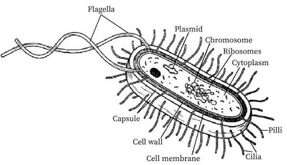
A bacterium is a complete living prokaryotic cell. Although small, it can take in nutrients, make proteins, generate energy, grow, respond to its environment and reproduce independently by binary fission.
| Structure | Description | Main function | Clinical relevance |
|---|---|---|---|
| Capsule / glycocalyx | Outer sticky layer in some bacteria, often polysaccharide. | Adherence, protection from drying and sometimes resistance to phagocytosis. | May increase virulence; can help bacteria form biofilms on catheters and devices. |
| Cell wall | Rigid layer outside the cell membrane; in most bacteria contains peptidoglycan. | Maintains shape and prevents osmotic lysis. | Basis of Gram staining; target of several antibiotics. |
| Cell membrane | Selective phospholipid-protein barrier beneath wall. | Controls entry/exit; involved in energy generation and transport. | Damage to the membrane can kill the cell; essential in every bacterium. |
| Cytoplasm | Water-based internal matrix containing enzymes, nutrients, ribosomes and inclusions. | Site of many metabolic reactions. | Metabolism determines growth requirements and laboratory tests. |
| Nucleoid | Region containing the main circular bacterial chromosome. | Stores essential genetic information. | Genetic changes can alter virulence or drug susceptibility. |
| Plasmids | Small extra-chromosomal circular DNA molecules. | Carry additional traits that can be shared between bacteria. | May carry genes for antimicrobial resistance or toxin production. |
| 70S ribosomes | Small protein-synthesising particles. | Make bacterial proteins. | Target of several antibacterial drug groups. |
| Flagella | Long, whip-like protein filaments. | Motility and chemotaxis. | Motility can assist spread through tissues or fluids. |
| Pili / fimbriae | Short, hair-like surface projections. | Attachment; some sex pili transfer DNA between bacteria. | Adherence supports colonisation; DNA transfer can spread resistance. |
| Endospore | Dormant, highly resistant survival form produced by some bacteria. | Survives heat, drying and chemicals; not a reproductive method. | Bacillus and Clostridium spores require strict decontamination procedures. |
A bacterial endospore is a survival structure formed inside one bacterial cell under unfavourable conditions. One vegetative cell forms one endospore and, when conditions improve, one vegetative cell emerges. It does not increase bacterial numbers. Fungal spores are commonly reproductive structures.
Morphology means the form, shape and structural arrangement of an organism. In the laboratory, shape and arrangement give early clues but do not identify a species by themselves.
| Shape | Description | Typical arrangement / example |
|---|---|---|
| Cocci | Spherical or oval bacteria. | Pairs (diplococci), chains (streptococci), clusters (staphylococci), tetrads or packets. |
| Bacilli | Rod-shaped bacteria. | Single rods, pairs, chains or palisades; Escherichia coli is a common rod. |
| Coccobacilli | Very short rods that may appear oval. | Intermediate appearance between cocci and bacilli. |
| Vibrios | Comma-shaped curved rods. | Vibrio cholerae is the major clinical example. |
| Spirilla | Rigid spiral-shaped cells with external flagella. | Usually environmental organisms. |
| Spirochaetes | Flexible, slender corkscrew-shaped bacteria. | Motile by axial filaments; includes Treponema species. |
| Filamentous / branching bacteria | Long branching filaments that can resemble fungi. | Includes actinomycetes such as Actinomyces. |
The cell envelope includes the cell membrane and structures outside it. Gram staining divides bacteria into Gram-positive and Gram-negative groups according to differences in their cell-wall envelope. This is one of the first and most useful laboratory tests in bacteriology.
| Feature | Gram-positive bacteria | Gram-negative bacteria |
|---|---|---|
| Peptidoglycan | Thick layer. | Thin layer. |
| Outer membrane | Absent. | Present outside the thin peptidoglycan. |
| Stain appearance | Retains crystal violet: purple/blue. | Decolourised then takes counterstain: pink/red. |
| Outer membrane components | No lipopolysaccharide outer membrane. | Contains lipopolysaccharide; lipid A is associated with endotoxin activity. |
| Practical implication | Cell-wall characteristics influence antibiotic selection. | Outer membrane can limit entry of some antimicrobial agents. |
- It is a rapid classification test, not a final species identification.
- Old, damaged or poorly stained cultures may give unreliable results.
- Mycobacterium has a waxy cell wall and is better assessed with acid-fast staining.
- Mycoplasma lacks a cell wall, so it does not Gram stain in the usual way.
Fungi are eukaryotic organisms. Their cell wall contains chitin and glucans, and their cell membrane contains ergosterol rather than cholesterol. These structural differences are important targets for antifungal therapy.
- Yeasts: unicellular fungi, often round or oval; many reproduce by budding. Example: Candida.
- Moulds: multicellular fungi formed by branching filaments called hyphae; a mass of hyphae is called a mycelium. Example: Aspergillus.
- Dimorphic fungi: can change form, often existing as moulds in the environment and yeasts/tissue forms in the host depending on conditions.
- Fungal spores: may be reproductive and dispersal structures; they are different from bacterial endospores.
Protozoa are single-celled eukaryotes. They have a nucleus, cytoplasm, cell membrane and specialised structures for movement, feeding or attachment. Depending on the group, they may move by pseudopodia, flagella or cilia. Many parasitic protozoa have a feeding/multiplying trophozoite stage and a resistant/transmission cyst stage.
- Plasmodium: malaria parasite with stages in mosquitoes and human liver/red cells.
- Entamoeba histolytica: may form cysts and trophozoites; can cause amoebic dysentery.
- Giardia duodenalis: has flagella and an attachment disc; causes diarrhoeal disease.
Helminths are multicellular parasitic worms. Their eggs and larval forms are often examined microscopically in stool, urine, blood or tissue specimens. They include nematodes (roundworms), cestodes (tapeworms) and trematodes (flukes).
A virus is not a cell. The complete infectious particle is called a virion. It contains a genome of either DNA or RNA, never both as its genomic material, enclosed by a protein coat called a capsid. Some viruses also have a lipid envelope derived from host-cell membranes.
| Viral component | Description | Importance |
|---|---|---|
| Genome | DNA or RNA carrying viral genetic instructions. | Directs production of new viral components inside host cells. |
| Capsid | Protein coat surrounding the genome, built from capsomeres. | Protects genome and helps determine viral shape. |
| Envelope | Lipid membrane present in enveloped viruses. | Contains viral glycoprotein spikes; relatively fragile to drying, heat, detergents and some disinfectants. |
| Spikes / attachment proteins | Surface proteins on capsid or envelope. | Bind specific receptors on host cells and influence tissue tropism. |
| Viral enzymes | Present in some viruses. | Assist replication when host-cell enzymes are insufficient. |
- Helical: capsid proteins arranged in a spiral around nucleic acid.
- Icosahedral: roughly symmetrical capsid with 20 triangular faces.
- Complex: more complicated structure, for example bacteriophages that infect bacteria.
- Enveloped or non-enveloped: enveloped viruses have a lipid envelope; non-enveloped viruses have only nucleic acid and capsid.
- Specimen collection: a correct specimen from the correct site, collected with aseptic technique and labelled accurately helps the laboratory distinguish true infection from contamination.
- Hand hygiene: breaks transfer of bacteria, fungi, viruses and parasites between patients, staff, equipment and the environment.
- Device care: capsules and biofilms help some bacteria adhere to catheters, cannulae and other devices; follow aseptic insertion and maintenance bundles.
- Decontamination: bacterial spores are highly resistant; use approved sterilisation/disinfection methods rather than assuming all organisms are equally easy to kill.
- Antimicrobial stewardship: bacterial structures provide drug targets, but unnecessary antibiotics select resistant bacteria. Antibiotics do not treat viral infections.
- Patient education: explain that not every “germ” causes disease, but hygiene, safe water/food, immunisation, correct medicine use and early care-seeking reduce infection risk.
A patient with a urinary catheter develops fever and cloudy urine. The nurse should not simply report “infection.” Consider the device as a potential surface for bacterial adherence and biofilm formation; assess the patient, obtain specimens correctly when ordered, maintain aseptic technique, review catheter need, monitor vital signs and escalate signs of sepsis promptly.
- Writing that viruses are prokaryotes. Viruses are acellular, not prokaryotic or eukaryotic cells.
- Confusing bacterial endospores with fungal reproductive spores.
- Stating that all microbes cause disease. Many are harmless, beneficial or opportunistic only under certain conditions.
- Forgetting that Gram-negative bacteria have an outer membrane and thin peptidoglycan.
- Calling every round bacterium “staphylococcus.” Shape and arrangement are clues, not a full identification.
- Using antibiotics for viral illness without a bacterial indication.
- Classify bacteria, fungi, protozoa, helminths and viruses as prokaryotic, eukaryotic or acellular.
- Draw and label a typical bacterial cell, stating one function of each structure.
- Differentiate a bacterial endospore from a fungal spore.
- Compare Gram-positive and Gram-negative bacterial cell envelopes.
- Describe the structures of an enveloped virus and explain why the envelope matters for infection prevention.
- Explain how microbial structure influences specimen collection, antibiotic use and decontamination.