
Table of Contents
ToggleRuptured spleen is a potentially life-threatening emergency because bleeding from the injured spleen can rapidly fill the abdominal cavity and cause haemorrhagic shock. Recognise the mechanism of injury, assess circulation early, activate emergency and surgical support, and arrange urgent transfer to a facility able to provide blood, imaging and surgery. A patient who is unstable or has peritonitis must not wait for CT imaging before emergency treatment.
- Meaning: A tear or disruption of the splenic capsule or substance that allows blood to escape into the splenic tissue, beneath the capsule, or into the peritoneal cavity.
- Common context: Blunt abdominal trauma such as a road traffic crash, fall, assault or sports injury; penetrating injuries can also damage the spleen.
- Main immediate danger: Internal haemorrhage, which may be severe even when there is no visible external wound.
- Classic clue: Left upper abdominal pain after trauma, sometimes with referred pain to the left shoulder (Kehr’s sign). Its absence does not exclude splenic injury.
- Diagnostic principle: Use the patient’s haemodynamic state to choose investigations. FAST/eFAST can rapidly detect free fluid; contrast-enhanced CT is the preferred anatomical assessment in a stable or stabilised patient.
- Treatment principle: Unstable patients, those with diffuse peritonitis, or those with another injury requiring laparotomy need urgent operative management. Carefully selected stable patients may be managed without an operation only where close monitoring and urgent rescue surgery are available.
By the end of this lesson, the learner should be able to:
- Define splenic rupture and describe the spleen’s relevant anatomy and functions.
- Explain traumatic and less common atraumatic causes, risk factors and the development of haemorrhagic shock.
- Recognise symptoms, signs and danger indicators of splenic injury, including possible delayed deterioration.
- Outline a focused history, primary survey, investigations and differential diagnoses.
- Distinguish the broad roles of observation, splenic artery embolisation and surgery according to stability, associated injuries and available resources.
- Plan safe pre-operative, post-operative and non-operative nursing care and patient education.
1. Introduction and definition
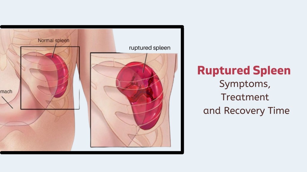
The spleen is a highly vascular organ in the left upper quadrant of the abdomen. Its position beneath the left lower ribs offers partial protection, but the organ can tear when the chest or abdomen is struck, compressed or penetrated. A splenic injury may range from a small capsular tear to a shattered spleen with major vascular damage.
Splenic rupture is disruption of the splenic capsule or tissue with bleeding. Blood may collect inside the splenic parenchyma, beneath the capsule as a haematoma, or spill into the peritoneal cavity as a haemoperitoneum. Bleeding can be immediate or, less commonly, delayed when a contained haematoma or vascular injury later gives way.
Treat suspected splenic rupture as an emergency. The amount of blood lost may be difficult to estimate from the abdomen’s appearance, and an initially alert patient can deteriorate quickly.
2. Relevant anatomy and functions of the spleen
The spleen lies in the left hypochondrium, mainly under ribs 9–11, close to the diaphragm, stomach, tail of the pancreas, left kidney and splenic flexure of the colon. It is covered by a fibrous capsule and has a rich arterial supply from the splenic artery, a branch of the coeliac trunk. Venous blood drains through the splenic vein toward the portal circulation.
The spleen is soft, friable and richly perfused. Its location near the ribs and diaphragm explains why blunt force to the left lower chest or upper abdomen can injure it and why blood irritating the diaphragm may cause referred shoulder pain.
- Blood filtration: Removes aged or damaged red blood cells and helps clear cellular debris from circulation.
- Immune defence: Supports antibody production and removal of certain organisms from the blood.
- Blood-cell storage: Holds a portion of platelets and blood cells and can contribute to blood-cell regulation.
- Clinical importance: Preserving splenic function is desirable when safe, but stopping life-threatening haemorrhage takes priority. Removal of the spleen increases lifelong risk of severe infection and requires planned preventive care.
3. Causes and risk factors
A. Traumatic splenic injury
- Blunt trauma: Road traffic collisions, motorcycle crashes, falls from height, assault, sports injuries, crush injuries and direct blows to the left lower chest or upper abdomen.
- Rapid deceleration: Sudden movement can stretch or tear the spleen and its supporting attachments, even when external bruising is slight.
- Penetrating trauma: Stab wounds, gunshot wounds or other penetrating objects may directly lacerate the spleen. These injuries can coexist with damage to the stomach, bowel, diaphragm, kidney, pancreas or major blood vessels.
- Rib or lower chest injuries: Fractures overlying the spleen may indicate substantial force and should prompt a careful search for internal injury.
B. Atraumatic or pathological rupture
Spontaneous rupture without a clear injury is uncommon. It may occur when the spleen is enlarged, infiltrated or weakened by an underlying disorder. Examples include infectious illnesses, haematological disease, malignancy, inflammatory conditions, pregnancy-related conditions or a pre-existing splenic lesion. Anticoagulant use or a bleeding disorder may increase the severity of bleeding. Atraumatic rupture still requires urgent assessment and should not be assumed to be harmless because the patient reports no injury.
Factors that may increase concern
- High-energy mechanism or direct blow to the left upper abdomen.
- Known splenomegaly, blood dyscrasia, malignancy or recent infection.
- Use of anticoagulants or antiplatelet medicines, or a known coagulation disorder.
- Older age, major associated injuries, persistent tachycardia or worsening pain.
- Pregnancy or possible pregnancy, which changes assessment and imaging considerations.
- Delayed presentation, syncope, increasing abdominal distension or unexplained falling haemoglobin after trauma.
4. Classification and severity
Clinicians describe anatomical injury using a recognised splenic injury scale, commonly the American Association for the Surgery of Trauma (AAST) Organ Injury Scale. The grade is determined from imaging or operative findings and is not something a nurse should try to assign from symptoms alone.
| Broad severity | Typical anatomical pattern | Clinical meaning |
|---|---|---|
| Low grade | Small haematoma or shallow laceration with limited tissue disruption. | May be suitable for monitored non-operative care if the patient is stable and there are no other indications for surgery. |
| Moderate grade | Larger or deeper laceration, expanding haematoma, or more substantial parenchymal injury. | Requires careful observation; vascular imaging or embolisation may be considered in selected stable patients. |
| High grade | Extensive laceration, major vascular injury, substantial devascularisation or a shattered spleen. | High risk of complications. Management depends on physiology, bleeding, associated injuries, specialist capability and rapid access to intervention. |
The World Society of Emergency Surgery (WSES) approach combines anatomical grade with haemodynamic status. This matters because a patient with a low-grade anatomical injury can still be critically unstable, while some stable patients with higher-grade injuries can be managed without immediate splenectomy in a suitably equipped centre. The patient’s physiology and associated injuries guide urgent decisions; the scan grade alone does not.
5. Pathophysiology
- Force damages splenic tissue or vessels. A direct blow, compression, deceleration or penetrating object tears the capsule, parenchyma or splenic vessels.
- Blood collects or escapes. Bleeding may be contained beneath the capsule at first or may enter the peritoneal cavity. A contained haematoma can enlarge or rupture later.
- Circulating volume falls. Ongoing internal blood loss reduces venous return, cardiac output and oxygen delivery to organs.
- Compensation may mask early shock. The body initially increases heart rate and peripheral vasoconstriction. Blood pressure can remain near normal for a time, especially in children, so a normal single reading does not rule out serious haemorrhage.
- Decompensated haemorrhagic shock develops. Continued blood loss causes hypotension, confusion, weak pulses, reduced urine output, lactic acidosis, organ dysfunction and cardiac arrest if untreated.
- Delayed complications may occur. Rebleeding, pseudoaneurysm rupture, infection or complications of splenectomy can arise later and require education and follow-up.
6. Clinical manifestations
A. Symptoms
- Pain or tenderness in the left upper quadrant, left lower chest or upper abdomen.
- Pain that may spread across the abdomen or become generalised if blood irritates the peritoneum.
- Referred left shoulder pain (Kehr’s sign), especially when the diaphragm is irritated by blood. It is a clue, not a required feature.
- Dizziness, weakness, fainting, restlessness, thirst, nausea or a feeling of impending collapse.
- Shortness of breath or chest discomfort, particularly with associated rib or thoracic injury.
- Increasing pain, abdominal fullness or a new episode of collapse after an initially stable period, suggesting possible ongoing or delayed bleeding.
B. Signs
- Left upper abdominal tenderness, guarding or rigidity; bruising may or may not be visible.
- Abdominal distension or signs of peritoneal irritation.
- Tachycardia, weak peripheral pulses, cool clammy skin, pallor, delayed capillary refill and sweating.
- Falling blood pressure, narrowed pulse pressure, altered mental status or reduced level of consciousness. These may be late signs of shock.
- Reduced urine output, increasing respiratory rate, low oxygen saturation or worsening peripheral perfusion.
- Associated injuries such as lower rib fractures, abdominal wall bruising, pelvic injury or signs of other internal bleeding.
- Hypotension, collapse, altered consciousness or rapidly worsening vital signs.
- Persistent tachycardia or signs of poor perfusion despite initial resuscitation.
- Generalised guarding, rigidity or diffuse peritonitis.
- Positive FAST/eFAST with instability or other evidence of ongoing intra-abdominal bleeding.
- Rapidly increasing abdominal pain or distension, recurrent syncope, or a falling haemoglobin with clinical deterioration.
Do not wait for the complete classic picture. Early splenic injury can present with subtle symptoms, and a patient may deteriorate before abdominal bruising or marked hypotension appears.
7. Assessment of a patient with suspected splenic rupture
A. Primary survey: ABCDE
| Step | Assessment and action | Reason |
|---|---|---|
| A | Assess airway patency, voice, airway protection and cervical-spine risk. Call for skilled airway support if the patient cannot maintain the airway. | Trauma may cause airway obstruction or associated head, neck and facial injuries. |
| B | Assess respiratory rate, chest movement, breath sounds and oxygen saturation; expose the chest enough to identify injury while preserving warmth. Give oxygen when indicated by hypoxaemia, respiratory distress or local emergency protocol. | Chest trauma can coexist with splenic injury; oxygenation and ventilation must be supported. |
| C | Look for shock and external bleeding. Apply direct pressure to accessible external bleeding, obtain large-bore IV access or IO access if needed, send blood samples, request cross-match, activate the local major haemorrhage pathway when indicated and prepare blood products. Keep the patient warm. | Internal haemorrhage is the immediate threat; early resuscitation and blood availability support perfusion while definitive bleeding control is arranged. |
| D | Check level of consciousness, pupils and glucose if indicated; reassess for deterioration. | Confusion may reflect shock, head injury, hypoxia or another cause that changes immediate care. |
| E | Expose enough to examine the abdomen, back, flanks and limbs for injury, then cover promptly with warm blankets and prevent heat loss. | A full trauma assessment can identify hidden injuries, while hypothermia worsens trauma-associated coagulopathy. |
Activate the trauma, emergency and surgical teams early. Arrange rapid transfer to a facility with blood, theatre and appropriate imaging or interventional capability if these are not available where the patient first presents. Stabilisation and referral should follow local protocols and must not be delayed by non-essential procedures.
B. Focused history
- Mechanism and timing: Ask what happened, when it happened, the direction and force of impact, any fall or collision, penetrating object, loss of consciousness and whether pain is worsening.
- Symptoms: Ask about left upper abdominal or shoulder pain, dizziness, fainting, weakness, nausea, vomiting, shortness of breath and urine output.
- Medicines and bleeding risks: Ask about anticoagulants, antiplatelet medicines, bleeding disorders, previous transfusion or allergies when feasible.
- Medical background: Ask about splenomegaly, blood disease, malignancy, recent infection, liver disease, pregnancy possibility and previous abdominal surgery.
- Use a structured approach: Obtain SAMPLE history when the immediate resuscitation allows; keep gathering information without interrupting life-saving care.
C. Focused examination and monitoring
- Record baseline and repeated airway, breathing, circulation, neurological status, temperature and pain observations.
- Monitor pulse, blood pressure, respiratory rate, oxygen saturation, skin perfusion, mental status and response to resuscitation at the frequency set by acuity and protocol.
- Inspect and gently assess the abdomen for tenderness, guarding, rigidity, distension, bruising and wounds. Avoid repeated forceful palpation.
- Examine the chest, ribs, pelvis, back and limbs for associated injuries while maintaining spinal precautions when indicated.
- Measure urine output when clinically appropriate and safe; report oliguria promptly as a possible sign of poor perfusion.
- Document trends, times, findings, interventions, medicines, fluid or blood given, calls made and the patient’s response.
8. Investigations
| Investigation | Purpose and interpretation | Important limitation |
|---|---|---|
| FAST/eFAST ultrasound | Rapid bedside assessment for free fluid in the abdomen or pericardium; useful during the initial trauma survey when the patient is unstable or too unwell to leave resuscitation. | A negative examination does not exclude splenic injury or early bleeding. Repeat assessment and the clinical picture matter. |
| Contrast-enhanced CT abdomen | Preferred detailed assessment for a haemodynamically stable or stabilised patient; identifies injury pattern, haemoperitoneum, active contrast extravasation, pseudoaneurysm and associated injuries. | Do not send an unstable patient or a patient needing immediate laparotomy away from resuscitation for CT. |
| Full blood count | Provides baseline haemoglobin, haematocrit and platelet count; serial measurements may help identify ongoing blood loss. | Early haemoglobin may not reflect the full amount of acute blood loss. Interpret with vital signs and repeated clinical assessment. |
| Blood group, screen and cross-match | Prepares for transfusion and supports emergency blood provision. | Follow the facility’s emergency-release blood protocol when life-threatening bleeding does not allow time for a full cross-match. |
| Coagulation tests and fibrinogen | Assess coagulopathy and guide blood-component treatment when available. | Results must not delay haemorrhage control in a critically unstable patient. |
| Lactate, blood gas or base deficit | May help assess tissue hypoperfusion and response to resuscitation. | Use together with clinical observations; a single result does not determine the need for surgery. |
| Renal function and electrolytes | Provide a baseline and help plan contrast imaging, fluid therapy and medication use. | Do not delay urgent life-saving care while awaiting non-critical results. |
| Pregnancy test when relevant | Helps guide imaging and management for a patient who could be pregnant. | Urgent treatment of life-threatening trauma remains the priority; involve the appropriate team. |
Diagnostic choices depend on the patient’s haemodynamic status. WSES guidance identifies E-FAST as a rapid test for free fluid and contrast-enhanced CT as the gold-standard anatomical assessment in stable or stabilised trauma patients. EAST guidance likewise recommends CT with intravenous contrast in a stable patient without peritonitis. A negative bedside scan must not override clinical deterioration.
9. Differential diagnoses
Consider associated or alternative causes of abdominal pain, bleeding or shock, including:
- Ruptured liver, kidney, mesenteric or other solid-organ injury.
- Hollow-viscus injury or perforation with peritonitis.
- Pelvic fracture with retroperitoneal bleeding.
- Rib fracture, haemothorax, pneumothorax or cardiac injury.
- Ruptured ectopic pregnancy or another gynaecological emergency where relevant.
- Acute pancreatitis, renal injury, peptic ulcer perforation or other acute abdominal condition.
- In atraumatic cases, disease-related splenic enlargement, infection, haematological malignancy or coagulopathy.
More than one injury may be present. A diagnosis of splenic injury should not stop a complete trauma assessment.
10. Emergency and definitive management
Management is determined by the patient’s physiology, associated injuries, imaging, response to resuscitation and the resources available. Haemorrhage control and adequate perfusion take priority. The following outline is for nursing education; follow the current local trauma pathway and instructions of the responsible clinical team.
A. Immediate priorities for all patients
- Recognise and escalate: Treat suspected splenic rupture as major trauma. Call senior emergency, anaesthesia and surgical staff early; communicate deterioration using the local escalation or SBAR process.
- Support ABCDE: Maintain airway and oxygenation, minimise movement when spinal injury is possible, assess perfusion frequently, establish access and control external bleeding.
- Resuscitate and prepare blood: Take blood samples promptly, request cross-match, activate major haemorrhage procedures when indicated and administer fluids, blood components, tranexamic acid or other medicines only as prescribed and in line with the local trauma protocol.
- Prevent avoidable heat loss: Use warm blankets and warmed fluids or blood when available and appropriate. Record temperature.
- Keep the patient prepared for intervention: Maintain nil by mouth when urgent anaesthesia or surgery is possible, obtain consent through the responsible team when feasible, and prepare for urgent transfer or theatre without delaying resuscitation.
- Reassess continuously: Repeat observations, abdominal assessment and response-to-treatment checks. Escalate a new change immediately.
B. Stable patient: selected non-operative management
Non-operative management (NOM) may be appropriate for a haemodynamically stable patient without peritonitis or another injury requiring surgery. NOM is active hospital care, not “doing nothing.” It requires skilled observation, serial clinical assessment, appropriate laboratory testing, access to blood and an operating room for urgent laparotomy. Where these capabilities are absent, early referral or transfer to a capable centre is essential.
- Admit to the level of monitoring indicated by injury severity, comorbidities, trends and local protocol.
- Perform serial vital signs and abdominal examinations; report increasing pain, tachycardia, hypotension, abdominal distension, reduced urine output, falling haemoglobin or new peritoneal signs.
- Follow the prescribed schedule for repeat blood tests and imaging. Follow-up imaging is guided by the injury grade, clinical findings, resources and specialist plan.
- Provide analgesia and prescribed supportive care while continuing to reassess alertness, respiratory status and abdominal findings.
- Observe activity and oral-intake orders; advance only when the treating team considers it safe.
- Escalate promptly if signs suggest ongoing bleeding or failure of non-operative care.
C. Splenic artery angiography and embolisation
In an appropriately resourced centre, splenic artery angiography with embolisation can control selected arterial bleeding while preserving the spleen. WSES consensus guidance suggests splenic artery embolisation for a haemodynamically stable adult with an arterial blush on CT, with decisions made by the trauma and interventional radiology teams. It does not replace surgery for persistent instability, diffuse peritonitis or another urgent operative indication.
- Prepare the patient, confirm identity and required checks, review allergies and relevant results, maintain prescribed fasting and explain the procedure within the nurse’s role.
- Maintain monitoring and IV access during transfer; hand over the mechanism, observations, resuscitation, medicines, allergies and investigation results.
- After the procedure, monitor vital signs, abdominal pain, access site, distal circulation and signs of recurrent bleeding according to the interventional radiology protocol.
- Report worsening pain, persistent fever, hypotension, access-site bleeding or new signs of infection promptly.
D. Unstable patient or patient with peritonitis: urgent operative management
Persistent haemodynamic instability despite resuscitation, diffuse peritonitis, ongoing intraperitoneal haemorrhage or an associated injury requiring exploration calls for urgent surgical management. EAST recommends urgent laparotomy for patients with diffuse peritonitis or haemodynamic instability after blunt abdominal trauma. Do not delay operative control in order to obtain a CT scan when the patient’s condition or surgical indication makes it unsafe.
At laparotomy, the surgeon controls bleeding and assesses the spleen and other abdominal organs. Depending on the injury and the patient’s condition, treatment may include splenectomy or a spleen-preserving approach. The priority in a critically bleeding patient is rapid control of haemorrhage and restoration of perfusion.
E. Care after splenectomy
- Continue post-operative monitoring for bleeding, shock, respiratory complications, ileus, wound infection and other complications.
- Explain that removal of the spleen increases the risk of severe infection. Arrange vaccination and preventive antibiotic advice in accordance with current national guidance, local availability and the treating clinician’s plan.
- Teach the patient to seek urgent medical assessment for fever, chills or sudden deterioration, and to inform every healthcare provider that they have had a splenectomy.
- Encourage the patient to keep written medical information or an alert card and attend follow-up appointments.
11. Nursing management
Nursing priorities are early recognition of deterioration, support of oxygenation and circulation, preparation for definitive haemorrhage control, safe transfer and clear communication. A nurse should not wait for a physician’s routine round when a trauma patient develops shock signs.
| No. | Nursing action | Rationale |
|---|---|---|
| 1 | Receive the patient in a prepared resuscitation area; call the trauma team and allocate roles using the local emergency system. | Early coordinated response shortens delays to resuscitation, imaging and bleeding control. |
| 2 | Assess airway, breathing, circulation, disability and exposure systematically; repeat the assessment after every intervention or change in condition. | Trauma physiology can change rapidly, and repeated assessment identifies deterioration early. |
| 3 | Measure and document vital signs and perfusion trends at the frequency set by the patient’s acuity; use continuous monitoring where indicated. | Trends in pulse, blood pressure, respiratory rate, oxygenation, mental state and skin perfusion may reveal haemorrhage before a single value becomes extreme. |
| 4 | Establish and maintain the prescribed IV or IO access; collect and label blood specimens correctly; request cross-match promptly. | Reliable access and prepared blood support resuscitation and urgent surgery. |
| 5 | Administer prescribed oxygen, fluids, blood components, analgesia and other medicines; check product identity and monitor for transfusion reactions. | These interventions support oxygen delivery and comfort while treating bleeding and shock under the clinical protocol. |
| 6 | Keep the patient warm, limit unnecessary exposure and use warming measures as available. | Hypothermia contributes to trauma-associated coagulopathy and can worsen bleeding. |
| 7 | Maintain nil by mouth when surgery or anaesthesia is possible; explain the reason and provide mouth care. | Urgent surgery may be required, and fasting reduces aspiration risk during anaesthesia. |
| 8 | Assess the abdomen gently and consistently; note pain location, guarding, distension and changes; avoid repeated vigorous palpation. | Serial findings help detect progression while avoiding unnecessary discomfort and interference with the assessment. |
| 9 | Prepare promptly for FAST, CT, angiography, theatre or transfer as ordered; check that lines, monitoring, documentation and handover accompany the patient. | Safe movement preserves monitoring and continuity of care during time-critical diagnostics or intervention. |
| 10 | Maintain accurate intake/output records and monitor urine output when indicated and safe. | Low output may signal poor renal perfusion or worsening shock. |
| 11 | Provide calm explanations, privacy and emotional support; involve family when appropriate without delaying care. | Trauma is frightening; clear communication can reduce distress and improve cooperation. |
| 12 | Document injury history, assessment findings, serial observations, times, calls, instructions, medicines, fluids, blood products and response. | Accurate records support clinical decisions, handover, audit and legal accountability. |
12. Pre-operative, post-operative and recovery nursing care
A. Pre-operative care
- Continue ABCDE reassessment, prescribed resuscitation and preparation for urgent theatre.
- Maintain nil by mouth, secure IV access, confirm blood availability and ensure the patient is appropriately monitored during transfer.
- Check allergies, current medicines, anticoagulant use, relevant blood results and consent status; report missing information to the team.
- Explain procedures in clear language within the nurse’s role. Reassure the patient and family while preparing for rapid intervention.
- Use infection-prevention practices, preserve privacy and document all care.
B. Post-operative care
- Airway and breathing: Assess respiratory rate, work of breathing, oxygen saturation and lung sounds; support prescribed oxygen, coughing and deep breathing as appropriate.
- Circulation and bleeding: Monitor observations, skin perfusion, wound and drain output if present, urine output and blood results as ordered. Report any evidence of recurrent bleeding or shock.
- Pain and comfort: Assess pain, administer prescribed analgesia, evaluate its effect and observe for adverse effects.
- Wound and infection prevention: Inspect the incision and lines, use aseptic technique and monitor temperature and other signs of infection.
- Gastrointestinal recovery: Assess nausea, bowel sounds, abdominal distension and tolerance of oral intake; follow the surgical team’s plan.
- Mobility and thromboembolism prevention: Encourage safe mobilisation and prescribed leg exercises; provide ordered mechanical or pharmacological prophylaxis after bleeding risk has been reviewed.
- Psychological support: Discuss recovery and body changes sensitively and give the patient time to ask questions.
C. Patient education after splenectomy
- Explain the increased lifelong risk of serious infection and the importance of following a clinician’s vaccination and preventive antibiotic plan.
- Advise urgent medical review for fever, chills or sudden illness. The patient should tell healthcare workers about the splenectomy at every encounter.
- Provide written follow-up instructions, clinic contact details and a medical alert card or equivalent record where available.
- Discuss activity restrictions, wound care, return to work or school and follow-up imaging based on the injury and the surgeon’s instructions.
- Encourage road safety, protective equipment during sports and avoidance of risky activities until the treating team confirms healing.
13. Nursing care plan
| Nursing problem | Expected outcome | Key nursing interventions |
|---|---|---|
| Deficient fluid volume related to internal haemorrhage | Perfusion is supported, observations are trended, and definitive bleeding control is not delayed. | Perform repeated ABCDE assessment; report deterioration immediately; maintain IV/IO access; prepare blood and prescribed resuscitation; monitor response and urine output; prepare for urgent intervention. |
| Acute pain related to tissue injury | Pain is assessed and reduced to a tolerable level without loss of surveillance for deterioration. | Assess location, intensity and change in pain; administer prescribed analgesia; reassess effect; report new, worsening or referred shoulder pain; use supportive positioning when safe. |
| Anxiety related to trauma and emergency treatment | The patient receives clear information and is supported during urgent care. | Use a calm voice, briefly explain actions, provide reassurance and privacy, involve family when appropriate, and remain attentive to questions while prioritising resuscitation. |
| Risk of infection after splenectomy | The patient and family understand infection prevention and when to seek urgent care. | Teach fever precautions; coordinate vaccine and prophylaxis planning with the clinical team; reinforce follow-up; encourage the patient to disclose splenectomy to future providers. |
| Knowledge deficit regarding injury, treatment or recovery | The patient can describe the plan, warning signs and follow-up instructions before discharge. | Assess what the patient already understands; explain care in plain language; use teach-back; provide written information and contact instructions; include the caregiver when the patient agrees. |
Nursing diagnoses and goals must be individualised to the patient’s assessment, condition, age, associated injuries and facility protocol.
14. Complications
Early complications
- Massive haemorrhage and haemorrhagic shock.
- Delayed rupture of a subcapsular haematoma or vascular lesion.
- Rebleeding or failure of non-operative management.
- Associated injury to the bowel, stomach, diaphragm, pancreas, kidney, liver or chest.
- Coagulopathy, hypothermia, acidosis, acute kidney injury or cardiac arrest.
- Complications of transfusion, anaesthesia, surgery or vascular access.
Later complications
- Splenic pseudoaneurysm, arteriovenous fistula or delayed bleeding.
- Intra-abdominal infection, abscess, wound infection or persistent pain.
- Thromboembolism or complications related to prolonged immobility.
- Overwhelming infection after splenectomy, particularly from encapsulated organisms.
- Psychological distress, delayed functional recovery or complications of associated injuries.
15. Prevention and health education
- Promote seat-belt and helmet use, road safety and appropriate child restraints.
- Use protective equipment and follow safety rules in contact sports and high-risk work.
- Seek assessment after significant left lower chest or upper abdominal trauma, even if symptoms initially appear mild.
- Patients with known splenomegaly or splenic disease should receive advice from their clinician about injury precautions and when to seek care.
- After splenectomy, follow the clinician’s vaccination, antibiotic, follow-up and fever-management plan.
- For a patient managed without surgery, explain delayed bleeding warning signs and the required activity restrictions and follow-up plan before discharge.
16. Key points for revision
- Splenic rupture causes concealed internal bleeding and may progress to haemorrhagic shock.
- Suspect it after blunt or penetrating trauma, especially with left upper abdominal pain, tenderness, shock or referred left shoulder pain.
- Kehr’s sign is a clue and may be absent; no single symptom or normal early blood pressure safely excludes injury.
- Choose investigations according to stability: FAST/eFAST can rapidly detect free fluid; contrast-enhanced CT assesses anatomy in a stable or stabilised patient.
- Unstable patients, diffuse peritonitis or another urgent operative indication require urgent surgical management; do not delay for CT.
- Non-operative care requires a stable patient and a facility able to provide close observation, serial reassessment, blood and urgent surgery.
- Angiography and embolisation may control arterial bleeding in selected stable patients where expertise is available.
- After splenectomy, infection-prevention education and follow-up are essential.
17. Common examination questions
- Define splenic rupture and state the most serious immediate complication.
- List five causes or mechanisms of traumatic splenic injury.
- State four symptoms or signs that may suggest splenic rupture.
- What is Kehr’s sign, and why can it occur?
- State two important limitations of FAST/eFAST in suspected splenic injury.
- When is contrast-enhanced CT generally used in the assessment of splenic trauma?
- Give four nursing priorities during the initial management of a patient with suspected splenic rupture.
- State two situations in which non-operative management is not appropriate without urgent surgical assessment.
- List four items of health education for a patient after splenectomy.
- Splenic rupture is disruption of the splenic capsule or tissue with bleeding; the immediate life-threatening complication is haemorrhagic shock from internal blood loss.
- Examples include road traffic collision, fall, assault, sports injury, crush injury, rapid deceleration and penetrating injury.
- Examples include left upper abdominal pain or tenderness, tachycardia, pallor, cool clammy skin, dizziness or syncope, abdominal distension, hypotension or altered consciousness.
- Kehr’s sign is referred pain to the left shoulder from diaphragmatic irritation by blood; it is not present in every patient.
- A negative scan does not exclude injury or early bleeding, and FAST/eFAST does not provide the same anatomical detail as CT.
- For a haemodynamically stable or stabilised patient when detailed assessment of the spleen and associated injuries is appropriate.
- Examples: ABCDE assessment, urgent escalation, repeated vital signs, IV/IO access and cross-match, prescribed resuscitation, warmth, preparation for transfer or theatre, accurate documentation.
- Persistent haemodynamic instability and diffuse peritonitis are examples; other injuries requiring laparotomy are also operative indications.
- Follow vaccination and antibiotic advice, seek urgent care for fever, disclose splenectomy to healthcare providers, carry medical information, attend follow-up and observe activity guidance.
18. References and further reading
- World Society of Emergency Surgery. Splenic trauma: WSES classification and guidelines for adult and pediatric patients. World Journal of Emergency Surgery. 2017.
- World Society of Emergency Surgery. Follow-up strategies for patients with splenic trauma managed non-operatively: the 2022 WSES consensus document. World Journal of Emergency Surgery. 2022.
- Eastern Association for the Surgery of Trauma. Splenic Injury, Blunt: Selective Nonoperative Management of. Practice Management Guideline. 2012.
- World Health Organization and International Committee of the Red Cross. Basic Emergency Care: Approach to the Acutely Ill and Injured. 2018; trauma training materials updated by WHO as indicated on the resource page.
These notes support learning and do not replace individual clinical assessment, the decisions of a qualified clinician, or current local emergency and trauma protocols. The patient’s condition and available resources determine care.
Return to the Diploma in Nursing Direct Curriculum
- Peritonitis: Causes, Symptoms, Treatment and Nursing Care
- Perforated Peptic Ulcer: Emergency Management and Nursing Care
- Intestinal Obstruction: Causes, Symptoms, Diagnosis, Treatment and Nursing Care
- Chronic Appendicitis: Recurrent Pain, Diagnosis and Nursing Care
- Appendicitis: Symptoms, Diagnosis, Treatment and Nursing Care