
Table of Contents
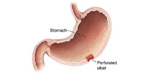
TogglePerforated peptic ulcer is a full-thickness break in the wall of the stomach or duodenum caused by an ulcer. Air and gastric or intestinal contents may escape into the peritoneal cavity, causing chemical irritation, bacterial contamination, peritonitis and sepsis. Suspected perforation is a time-critical surgical emergency.
These nursing lecture notes explain how perforation develops, its risk factors and clinical features, urgent assessment and investigations, treatment principles, perioperative nursing care, complications, prevention and examination points. Management must be led by the surgical and anaesthetic teams and adapted to the patient and available local services.
Emergency reminder: sudden severe upper abdominal pain, a rigid or very tender abdomen, collapse, rapid breathing, a weak rapid pulse, low blood pressure or altered consciousness requires urgent escalation and surgical review. Do not wait for routine investigations before raising concern.
By the end of this lesson, the learner should be able to:
- Define peptic ulcer perforation and distinguish it from ulcer bleeding and gastric outlet obstruction.
- Describe common causes, risk factors and the pathophysiological sequence leading to peritonitis and shock.
- Recognise typical and atypical symptoms, signs and danger features.
- Outline the initial assessment, investigations and emergency treatment principles.
- Plan preoperative, postoperative and ongoing nursing care with clear rationales.
- Explain prevention of recurrence and follow-up after recovery.
Definition and important distinctions
A peptic ulcer is a break in the mucosal lining of the stomach or proximal duodenum that extends through the muscularis mucosae. A perforated peptic ulcer (PPU) has penetrated the entire wall, creating communication between the gastrointestinal lumen and the peritoneal cavity. Perforation most often involves a gastric or duodenal ulcer; the exact site is determined during imaging or surgery.
| Complication | What happens | Typical concern |
|---|---|---|
| Perforation | Full-thickness defect allows air and luminal contents to leak outside the stomach or duodenum. | Sudden abdominal pain, peritoneal irritation, sepsis and shock. |
| Bleeding | An ulcer erodes into a blood vessel. | Haematemesis, coffee-ground vomit, melaena, dizziness or haemodynamic instability. |
| Gastric outlet obstruction | Inflammation or scarring narrows the pylorus or proximal duodenum. | Early satiety, persistent vomiting, abdominal fullness and dehydration. |
| Penetration | An ulcer extends into an adjacent organ without freely opening into the peritoneal cavity. | Persistent or referred pain, depending on the organ involved. |
One patient may have more than one complication. A patient with signs of perforation needs emergency assessment even if there is no history of previously diagnosed ulcer disease.
Causes and risk factors
Peptic ulcers form when mucosal injury and impaired mucosal protection allow acid and pepsin to damage the stomach or duodenal lining. Perforation can occur when an ulcer deepens through the full thickness of the wall.
- Helicobacter pylori infection: a common cause of chronic gastritis and peptic ulcer disease. Infection should be tested for and treated using an appropriate clinician-selected regimen; eradication should be confirmed after treatment.
- Non-steroidal anti-inflammatory drugs (NSAIDs): medicines such as ibuprofen, diclofenac and aspirin can impair mucosal protection, especially with prolonged use, higher doses, older age or a previous ulcer.
- Combination of risk medicines: NSAIDs taken with corticosteroids, anticoagulants or some antiplatelet medicines may increase the risk of ulcer complications or bleeding. Medication decisions require clinical review.
- Previous peptic ulcer or ulcer complication: recurrence risk may be higher if the cause has not been addressed.
- Smoking: associated with poorer ulcer healing and increased recurrence.
- Severe physiological illness: critical illness can cause stress-related mucosal injury in some hospitalised patients; this is different from ordinary emotional stress.
- Other contributors: older age, serious comorbidities and delayed presentation can increase the chance of severe illness or a poor outcome.
Spicy foods and ordinary emotional stress do not by themselves explain most peptic ulcers. They may affect symptoms for some people, but clinicians should look for underlying causes such as H. pylori or ulcerogenic medicines.
Pathophysiology
- Mucosal injury: H. pylori, NSAIDs and other factors weaken mucosal defence or promote injury.
- Ulcer deepening: acid and pepsin continue to damage the ulcer base, which may extend through the muscular wall.
- Full-thickness perforation: a hole forms and air, acid, bile, food particles and microorganisms may escape into the peritoneal cavity.
- Chemical peritonitis: gastric and duodenal contents irritate the peritoneum, producing severe pain and involuntary guarding.
- Bacterial contamination and inflammation: contamination can progress from local to generalised peritonitis. The omentum may sometimes partly seal the defect, temporarily limiting the leak.
- Fluid shifts and sepsis: inflammation increases capillary permeability; fluid moves into the peritoneal space, while vomiting and poor intake reduce circulating volume. Infection and hypoperfusion can progress to acute kidney injury, shock and multi-organ dysfunction.
A contained or sealed perforation may have less obvious signs than a free leak. An initially reassuring examination, absence of fever or a normal early laboratory result does not safely exclude perforation when the history and clinical course are concerning.
Clinical features and danger signs
Symptoms
- Sudden, severe epigastric pain: may begin abruptly and spread across the abdomen. Some patients describe it as sharp, piercing or “like a knife.”
- Pain on movement: coughing, deep breathing, walking or bumps during transport may make pain worse. The patient may lie still with knees flexed.
- Referred shoulder-tip pain: diaphragmatic irritation may cause pain felt at the shoulder, often on either side.
- Nausea and vomiting: may occur and can worsen fluid loss.
- Abdominal distension or fullness: may develop as inflammation and ileus progress.
- Thirst, weakness, dizziness or faintness: may reflect pain, dehydration, bleeding or poor circulation.
Signs
- Marked abdominal tenderness, involuntary guarding or a rigid “board-like” abdomen, especially with free perforation.
- Reduced abdominal movement with breathing and reduced bowel sounds; bowel sounds alone cannot confirm or exclude perforation.
- Tachycardia, pallor, sweating, cool extremities, delayed capillary refill, tachypnoea or shallow breathing.
- Fever may appear later; severe illness may instead cause hypothermia.
- Hypotension, confusion, reduced urine output, collapse or mottled skin may indicate shock or organ dysfunction.
- Generalised severe pain, rapidly worsening pain, involuntary guarding or abdominal rigidity.
- Falling blood pressure, a rapid weak pulse, fast breathing, fainting or altered mental state.
- Cool clammy skin, reduced urine output, increasing abdominal distension or persistent vomiting.
- Any concern for sepsis, shock, an acute abdomen or a deteriorating patient.
Urgent assessment
Suspected perforation is an emergency. Use the facility's emergency and sepsis pathways and obtain urgent surgical review. Resuscitation and referral should proceed in parallel; do not delay escalation while completing a lengthy history or waiting for every test.
Focused history
- Clarify the time and suddenness of onset, site, severity, spread and progression of pain; ask whether movement or coughing makes it worse.
- Ask about nausea, vomiting, appetite, bowel motions, passage of flatus, fever, thirst, faintness and last oral intake.
- Ask about previous ulcer, H. pylori testing or treatment, upper gastrointestinal bleeding and prior abdominal surgery.
- Review prescribed and non-prescribed medicines, particularly NSAIDs, aspirin, steroids, antiplatelets and anticoagulants; document allergies and the last dose.
- Identify important conditions such as kidney, liver, heart or lung disease, diabetes, frailty and pregnancy possibility when relevant.
Primary survey and examination
- Airway and breathing: assess airway patency, work of breathing, respiratory rate and oxygen saturation; provide oxygen if indicated under local protocol.
- Circulation: measure pulse, blood pressure, capillary refill, skin temperature and perfusion; look for shock and establish intravenous access promptly.
- Disability and exposure: check mental status, pain, temperature and hydration while preserving dignity and warmth.
- Abdominal assessment: a trained clinician assesses distension, scars, tenderness, involuntary guarding and rigidity. Avoid repeated painful manoeuvres that do not change management.
- Trend and communicate: record observations with the time, repeat them according to acuity and promptly report deterioration using SBAR.
Investigations
Investigations support diagnosis and operative planning. Choice depends on the patient's stability, age, pregnancy status, renal function and available services. In an unstable patient, imaging must not delay an urgent decision by the surgical team.
| Investigation | Purpose | Important limitation or nursing point |
|---|---|---|
| Full blood count | May show leukocytosis, anaemia or other changes. | Early results may be non-specific; interpret with the examination and trend. |
| Electrolytes, urea and creatinine | Assess dehydration, renal function and electrolyte disturbance. | Useful for resuscitation and procedural planning; report significant abnormalities. |
| Blood gas and lactate when indicated | Help assess acid-base disturbance and tissue perfusion in a seriously ill patient. | A normal early value does not exclude serious disease. |
| Group and save / crossmatch | Supports timely blood availability if transfusion is needed. | Use correct patient identification and local transfusion procedures. |
| Liver tests, lipase and other tests as indicated | Help assess alternative diagnoses or associated disease. | Tests do not replace clinical assessment or surgical review. |
| Blood cultures when sepsis is suspected | May help identify bloodstream infection and guide later antimicrobial review. | Collect promptly before antibiotics only if this causes no unsafe delay. |
| CT of the abdomen | Preferred imaging when available and the patient can safely undergo it; may show free air, leakage or the source. | Consider renal function, contrast risks, pregnancy, radiation and local protocol. A scan should not delay urgent surgery when the patient is unstable. |
| Upright chest or abdominal X-ray | May show free subdiaphragmatic air when CT is not promptly available. | A negative X-ray does not rule out perforation. |
| Water-soluble contrast study | May be considered by the treating team when suspicion persists despite no visible free air. | Use only as directed; it should not delay urgent care. |
Endoscopy is not the first diagnostic step in a suspected free perforation. Once the patient has recovered, upper gastrointestinal evaluation and ulcer follow-up may be arranged by the specialist team, especially for a gastric ulcer where malignancy needs to be excluded.
Differential diagnosis
Several conditions can cause sudden epigastric or generalised abdominal pain. The team may consider pancreatitis, acute cholecystitis, appendicitis, intestinal obstruction, mesenteric ischaemia, ruptured abdominal aortic aneurysm, myocardial ischaemia, lower-lobe pneumonia, ectopic pregnancy or other causes according to the patient and context. Do not assume that pain is “just an ulcer” based on history alone.
Emergency and definitive management
Key aims are to stabilise the patient, reduce ongoing contamination, treat infection, control the perforation and support organ function. Management is individualised and directed by the surgical, anaesthetic and critical-care teams.
1. Immediate actions and resuscitation
- Activate urgent medical and surgical assessment; use the local emergency or sepsis pathway when indicated.
- Keep the patient nil by mouth while urgent surgery or anaesthesia is possible. Explain why and record the time of last intake.
- Establish reliable IV access. Administer prescribed isotonic fluids with frequent reassessment of blood pressure, perfusion, urine output and signs of overload.
- Provide prescribed analgesia and antiemetics; assess and document the response. Pain relief should not be withheld solely to preserve examination findings.
- Give timely prescribed broad-spectrum antimicrobial therapy for suspected perforation and peritoneal contamination, following the local guideline, allergy history, renal function and resistance patterns.
- Collect ordered laboratory tests, group and crossmatch and cultures; do not delay antimicrobials or source control for samples that cannot be obtained promptly.
- Give acid suppression and other medicines as prescribed. A nasogastric tube, urinary catheter or oxygen may be used when clinically indicated and ordered.
- Prepare for theatre, transfer or higher-level care. Use structured handover and communicate deterioration immediately.
2. Operative source control
For significant pneumoperitoneum, contrast leakage or clinical peritonitis, operative treatment is generally recommended. Surgery should be arranged as soon as possible, particularly when presentation is delayed or the patient is older. The exact procedure depends on the site and size of the perforation, contamination, physiological stability, surgical expertise and concern for an underlying tumour.
- Primary closure with or without an omental patch: commonly used for many small gastroduodenal perforations.
- Laparoscopic repair: may be appropriate for selected stable patients when expertise and equipment are available.
- Open surgery: may be needed when the patient is unstable, the defect is large or complex, or laparoscopy is unsuitable or unavailable.
- Additional procedures: a large gastric ulcer, suspicious lesion, non-viable tissue or another abdominal finding may require biopsy, resection, drainage or a different operation.
At operation, the team may collect peritoneal fluid for culture and assess the extent of contamination. Antibiotic choice and duration are reviewed according to the source, cultures, clinical response and local antimicrobial guidance.
3. Non-operative management is exceptional
Non-operative care is not routine for a perforated peptic ulcer. A specialist may consider it only in a carefully selected, stable patient when investigations confirm a sealed perforation and there is no ongoing contrast leak, generalised peritonitis or sepsis. It requires close repeated assessment, laboratory monitoring, supportive treatment and immediate access to surgery if the patient worsens. If deterioration occurs or sealing is uncertain, the patient needs urgent reassessment for operative source control.
4. Ulcer treatment and recurrence prevention
- After emergency stabilisation, review for H. pylori and arrange treatment if infection is confirmed, using current local guidance and a regimen chosen by a qualified prescriber.
- Plan a test to confirm eradication after treatment, at the interval and with medicine holds advised by the treating clinician.
- Review whether NSAIDs or other ulcerogenic medicines can be stopped, replaced or protected against; do not independently discontinue prescribed antiplatelet or anticoagulant treatment.
- Provide smoking-cessation support and explain follow-up, adherence and symptoms that require urgent review.
- Arrange appropriate follow-up of gastric ulcers and histology where indicated to ensure healing and exclude malignancy.
Nursing management
Nurses play a central role in early recognition, continuous reassessment, preparation for source control, safe medicines, fluid monitoring and recovery. Follow prescribed orders and local protocols; communicate promptly when the patient's condition changes.
A. Preoperative and emergency nursing care
| No. | Nursing action | Rationale |
|---|---|---|
| 1 | Recognise suspected perforation and escalate immediately to the clinician and surgical team; activate the emergency response when indicated. | Peritoneal contamination and shock can progress rapidly, and timely source control matters. |
| 2 | Assess and trend airway, breathing, pulse, blood pressure, temperature, oxygen saturation, pain, mental status, perfusion and urine output as appropriate. | Trends may reveal deterioration earlier than one set of observations. |
| 3 | Keep the patient nil by mouth when ordered; document last intake and explain the reason in calm, clear language. | Urgent anaesthesia or an operation may be needed. |
| 4 | Maintain IV access; administer prescribed fluids, analgesia, antiemetics and antimicrobials promptly; check allergies and document response. | Supports circulation, comfort and timely treatment while reducing medication risk. |
| 5 | Measure intake and output; report reduced urine output, hypotension, increasing tachycardia or signs of fluid overload. | Third spacing, vomiting and resuscitation can alter circulating volume and renal function. |
| 6 | Collect ordered blood samples and cultures with correct identification and aseptic technique; avoid delaying urgent treatment. | Reliable results support diagnosis, transfusion planning and later antimicrobial review. |
| 7 | Prepare for imaging, theatre or transfer; confirm identity, allergies, relevant results, documentation, consent status and handover details according to policy. | Structured preparation reduces preventable delays and errors. |
| 8 | Provide warmth, privacy, reassurance and emotional support; involve a chosen family member when the patient agrees. | Severe pain and unexpected surgery can cause fear and distress. |
| 9 | Document findings, times, communications, treatment and response; use SBAR for urgent handover. | Clear records support continuity and accountability. |
B. Postoperative nursing care
- Airway, breathing and circulation: receive a structured handover, assess consciousness and observations, and escalate concerning trends promptly.
- Sepsis and perfusion: monitor temperature, pulse, blood pressure, respiratory rate, skin, mental status, fluid balance and urine output at the prescribed frequency.
- Pain and nausea: assess location and severity, give prescribed treatment and reassess. New or increasing pain may indicate a complication and requires review.
- Abdomen and bowel function: observe distension, tenderness, vomiting, bowel activity and passage of flatus. Advance oral intake only according to the surgical plan.
- Wound, drains and tubes: use aseptic technique; inspect the wound and record drain or nasogastric output, colour and patency. Report unexpected blood, bile, faeculent material or sudden changes.
- Fluid and renal care: maintain accurate intake/output records; monitor ordered renal tests and electrolytes; report oliguria or overload.
- Infection prevention: administer prescribed antimicrobials on time, monitor for adverse reactions and support review when culture results are available.
- Respiratory care and mobilisation: assist breathing exercises, coughing with wound support and progressive mobilisation as appropriate; follow VTE-prevention orders and falls precautions.
- Nutrition and recovery: follow the prescribed feeding plan and seek nutritional support if recovery is prolonged or intake remains poor.
- Education and discharge: explain wound care, medicines, follow-up and danger signs. Use teach-back to check understanding.
C. Example nursing care plan
| Nursing problem | Expected outcome | Key interventions |
|---|---|---|
| Acute pain related to peritoneal irritation or surgery | Pain is reduced to a tolerable level and any change is assessed promptly. | Assess pain pattern; position for comfort; give prescribed analgesia; reassess; escalate new or worsening pain. |
| Risk of deficient fluid volume or impaired perfusion related to third spacing and vomiting | Observations, perfusion and urine output remain within the target set by the clinical team. | Trend vital signs and fluid balance; administer prescribed fluids; assess response and overload; report oliguria, hypotension or confusion. |
| Risk of sepsis and organ dysfunction related to intra-abdominal contamination | Deterioration is recognised and communicated without avoidable delay. | Monitor observations and mental state; give prescribed antimicrobials; collect ordered samples without unsafe delay; escalate red flags. |
| Anxiety related to sudden illness and possible surgery | The patient can describe the immediate plan and feels supported. | Explain care in short steps; invite questions; protect privacy; avoid false reassurance; involve chosen support with consent. |
Complications and prognosis
- Generalised peritonitis, intra-abdominal abscess or persistent leakage.
- Sepsis, septic shock, acute kidney injury and multi-organ dysfunction.
- Ongoing haemorrhage, anaemia or need for transfusion.
- Postoperative wound infection, leak, fistula, ileus or bowel obstruction.
- Pulmonary complications, venous thromboembolism, poor nutrition or delayed wound healing.
- Recurrent ulcer or re-perforation if the underlying cause is not treated.
Outcome depends on the patient's physiological condition, the site and size of the perforation, the degree of contamination, comorbidities and how quickly effective treatment is started. Older adults and patients presenting late or in shock may have a higher risk of complications.
Prevention and patient education
- Take prescribed ulcer medicines as directed and complete H. pylori treatment; attend the planned test to confirm eradication.
- Do not start or continue NSAIDs without discussing risks with a health professional, especially after an ulcer complication. Ask before stopping aspirin, antiplatelet or anticoagulant medicines prescribed for another condition.
- Stop smoking with support where available. Limit alcohol if it worsens symptoms or conflicts with medicines and recovery advice.
- Attend follow-up, including any endoscopy or biopsy arranged for a gastric ulcer.
- Seek urgent care for sudden severe abdominal pain, a rigid or swollen abdomen, repeated vomiting, fainting, black stools, blood in vomit, fever with worsening pain or marked weakness.
Exam revision
- Perforation is a full-thickness ulcer defect that allows gastrointestinal contents to enter the peritoneal cavity.
- Classic presentation is sudden severe epigastric pain followed by peritoneal signs; an atypical or sealed perforation can be less obvious.
- CT is preferred when available; upright chest or abdominal X-ray may be used when CT is not promptly available. A negative X-ray does not exclude perforation.
- Significant free air, contrast leakage or peritonitis generally indicates operative source control.
- Non-operative management is reserved for a carefully selected, confirmed sealed perforation with close monitoring and immediate surgical backup.
- Nursing priorities are urgent escalation, serial observations, nil by mouth as ordered, IV access and prescribed resuscitation, timely antimicrobials, accurate fluid balance and theatre preparation.
- Define a perforated peptic ulcer and explain how it differs from ulcer bleeding.
- List five risk factors for peptic ulcer disease or perforation.
- Explain how a full-thickness ulcer can lead to peritonitis, shock and acute kidney injury.
- Describe four symptoms and four signs of perforation.
- What immediate findings should be reported to the surgical team?
- State the role and limitations of CT and plain radiography in suspected perforation.
- Outline the initial treatment priorities before definitive source control.
- Why is non-operative management not routine, and what features are required for exceptional consideration?
- Outline five preoperative and five postoperative nursing responsibilities.
- Give four measures that may reduce recurrent ulcer disease after recovery.
References and related notes
- Tarasconi A, Coccolini F, Biffl WL, et al. Perforated and bleeding peptic ulcer: WSES guidelines. World Journal of Emergency Surgery. 2020;15:3. doi:10.1186/s13017-019-0283-9.
- Chey WD, Howden CW, Moss SF, et al. ACG Clinical Guideline: Treatment of Helicobacter pylori Infection. American College of Gastroenterology, 2024.
- Republic of Uganda, Ministry of Health. Uganda Clinical Guidelines, 2023. Follow current Ministry of Health guidance and facility protocols.
- Peritonitis: causes, symptoms, treatment and nursing care
- Cholecystitis lecture notes
- Intestinal obstruction lecture notes
- Appendicitis lecture notes
Return to the Diploma in Nursing Direct Curriculum
Last updated: October 2026. These notes support learning and do not replace individual clinical assessment, a qualified prescriber's decisions or current local guidance.