
Table of Contents
ToggleChronic cholecystitis is long-standing or recurrent inflammation of the gallbladder, most often associated with repeated irritation from gallstones. A person may describe recurring right upper abdominal or epigastric pain, especially after meals, with nausea but without fever or jaundice between uncomplicated attacks. The diagnosis often overlaps clinically with symptomatic gallstones (cholelithiasis); ultrasound, the full clinical picture and sometimes examination of the removed gallbladder help clarify the condition. For suitable patients with symptomatic gallbladder stones, laparoscopic cholecystectomy is the usual definitive treatment. Nurses contribute through recognition of complications, safe perioperative preparation, recovery monitoring and practical discharge education.
- Chronic cholecystitis: Recurrent or persistent gallbladder inflammation, usually related to gallstones and repeated episodes of cystic duct obstruction.
- Typical symptom: Recurrent biliary-type pain in the right upper quadrant or epigastrium, sometimes radiating to the back or right shoulder, with nausea.
- Stable chronic disease: Usually has no fever, jaundice or systemic toxicity between attacks. New fever, persistent pain or jaundice requires urgent assessment.
- First-line imaging: Abdominal ultrasound to identify gallstones and assess the gallbladder and bile ducts. Blood tests are selected according to symptoms and suspected complications.
- Definitive treatment: Offer laparoscopic cholecystectomy to suitable people with symptomatic gallbladder stones. Open surgery or conversion may be needed when this is safer.
- After gallbladder removal: Bile continues to be made by the liver and flows to the intestine. Most people return to a usual balanced diet as tolerated.
- Urgent red flags: Severe continuous pain, fever or chills, jaundice, dark urine, pale stool, persistent vomiting, confusion, fainting or shock.
By the end of this lesson, the learner should be able to:
- Define chronic cholecystitis and distinguish it from biliary colic, acute cholecystitis and common bile duct stones.
- Describe gallbladder anatomy and explain how repeated stone obstruction can cause inflammation, fibrosis and recurrent pain.
- Recognise typical symptoms, important examination findings and complications that require urgent escalation.
- Outline the assessment and investigations used for suspected chronic gallbladder disease.
- Explain indications for cholecystectomy and the broad differences between laparoscopic and open approaches.
- Plan nursing care before, during and after cholecystectomy, including patient teaching and discharge precautions.
1. Introduction and definition
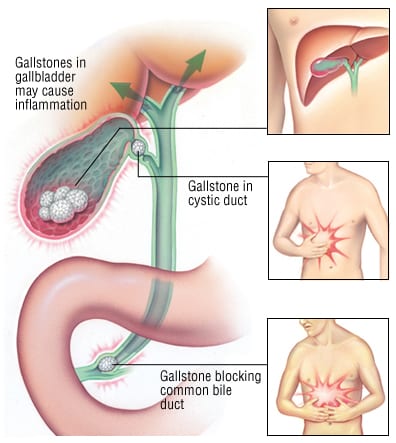
The gallbladder stores and concentrates bile made by the liver. Chronic cholecystitis describes repeated or persistent inflammation and structural change in the gallbladder. In most cases, gallstones intermittently obstruct the gallbladder outlet or cystic duct. Repeated obstruction and irritation may cause a thickened, scarred or contracted gallbladder.
In practice, chronic cholecystitis may be recorded alongside symptomatic cholelithiasis because the symptoms, ultrasound findings and tissue changes do not always correspond perfectly. Some people have gallstones with typical attacks but little visible inflammation on imaging; histology after surgery may show chronic inflammatory change. Others have gallstones that never cause symptoms. Always interpret the history, examination, investigations and clinical course together.
- Cholelithiasis: Gallstones in the gallbladder.
- Biliary colic: An episode of biliary-type pain, commonly when a stone temporarily obstructs the cystic duct. Despite the word “colic,” the pain may feel steady during an attack. Uncomplicated biliary colic does not by itself prove infection.
- Chronic cholecystitis: Recurrent or prolonged gallbladder inflammation, usually related to repeated gallstone irritation.
- Acute cholecystitis: A current episode of gallbladder inflammation, often with persistent right upper quadrant pain and local or systemic inflammatory findings.
- Choledocholithiasis: A stone in the common bile duct. This can obstruct bile flow and cause jaundice, cholangitis or pancreatitis.
- Cholecystectomy: Surgical removal of the gallbladder.
Clinical distinction: Recurrent brief attacks without fever are different from a patient who now has persistent severe pain, fever, jaundice or systemic illness. The latter may have acute cholecystitis, a common bile duct stone, cholangitis or pancreatitis and needs urgent hospital assessment.
2. Relevant anatomy and normal function
The liver continuously produces bile, which travels through the right and left hepatic ducts. These join to form the common hepatic duct. The gallbladder connects to this drainage system through the cystic duct. The common hepatic duct and cystic duct join to form the common bile duct, which carries bile toward the duodenum. The common bile duct usually joins the pancreatic duct near the ampulla before opening into the small intestine.
When a person eats, hormones and nerve signals stimulate the gallbladder to contract. Bile passes through the cystic duct and common bile duct into the duodenum, where it helps emulsify dietary fat. If a stone blocks the cystic duct, the gallbladder contracts against an obstruction. The resulting pressure and wall stretch can produce pain. If obstruction and irritation persist or recur, inflammation and scarring may develop.
- The cystic duct and cystic artery must be identified safely during surgery because the bile ducts and hepatic vessels are close by and anatomical variation occurs.
- The hepatocystic triangle is an important operative landmark. The surgeon confirms anatomy before dividing structures; the team uses established safety practices such as the critical view of safety.
- Stones can remain in or move into the common bile duct. These duct stones need assessment and may require endoscopic or surgical treatment in addition to gallbladder removal.
- After cholecystectomy, the liver still makes bile. Bile drains directly into the intestine rather than being stored in a gallbladder.
3. Causes and risk factors
A. Main cause
Most chronic calculous cholecystitis is associated with gallstones and repeated episodes of partial or temporary obstruction near the gallbladder neck or cystic duct. The gallbladder wall is repeatedly distended and irritated. Over time, it may become thickened, fibrotic, less distensible or contracted. Chronic gallbladder inflammation can also occur less commonly without visible stones; the cause and significance need clinician assessment.
B. Factors associated with gallstones
Gallstones develop when substances in bile form crystals and grow. A person may have more than one risk factor, and many people with risk factors never develop symptomatic stones.
- Age and family history: Risk tends to rise with age, and family or genetic factors can contribute.
- Hormonal factors: Pregnancy and oestrogen exposure can affect bile composition and gallbladder emptying.
- Higher body weight or metabolic risk: Obesity, diabetes and some lipid disorders are associated with gallstone formation.
- Rapid weight loss or prolonged fasting: Rapid changes in weight and reduced gallbladder emptying can increase risk in some people.
- Haemolytic disorders: Conditions that increase red cell breakdown can increase pigment gallstone risk.
- Terminal ileal disease or resection: Reduced bile salt recycling may alter bile composition.
- Some medicines or medical conditions: Assess these in the patient’s full clinical history rather than assuming they are the cause of an individual case.
C. Factors that may increase operative difficulty
Chronic inflammation may make laparoscopic dissection more difficult because of adhesions, fibrosis or altered anatomy. Other factors include previous upper abdominal surgery, obesity, cirrhosis, acute inflammation, a large or inflamed liver, duct stones and anatomical variation. These factors help the surgical team plan the operation; they do not automatically mean that open surgery is required.
4. Pathophysiology and tissue changes
- A gallstone intermittently blocks the gallbladder outlet. The obstruction may resolve when the stone moves, allowing pain to settle.
- The gallbladder contracts against resistance. Increased pressure and stretching of the wall stimulate visceral pain, commonly felt in the right upper abdomen or epigastrium.
- Repeated irritation triggers inflammation. The lining can become inflamed and the gallbladder may not empty normally.
- Healing occurs with fibrosis and structural change. Repeated inflammation can thicken or scar the wall and may reduce the gallbladder’s capacity to distend and empty.
- Complications can develop if obstruction persists or a stone migrates. Ongoing inflammation may become acute or progress to infection, empyema, gangrene or perforation. A migrating stone can obstruct the common bile duct or ampulla and cause jaundice, cholangitis or pancreatitis.
Histological descriptions can include chronic inflammatory cells, fibrosis and mucosal changes. These findings are usually confirmed from the removed gallbladder rather than used alone to diagnose the patient before surgery.
5. Clinical manifestations
A. Typical symptoms
- Recurrent right upper quadrant or epigastric pain: Attacks may follow meals, particularly a fatty meal, but meal timing is not diagnostic and attacks can occur at other times.
- Steady or cramping pain: The pain may build over minutes and last from about half an hour to several hours. It may spread to the back or below the right shoulder blade.
- Nausea, sometimes vomiting: These can accompany pain. Persistent vomiting increases concern for dehydration or another complication.
- Symptom-free intervals: Pain may settle when temporary obstruction resolves. A symptom-free interval does not remove the risk of another attack.
- Indigestion, bloating or food intolerance: These symptoms are common in the general population and are not specific to gallbladder disease. Investigate other causes when the pain pattern is atypical.
B. Findings between uncomplicated attacks
Between attacks, a person may look well and have a normal abdominal examination. There is usually no fever or jaundice in uncomplicated biliary colic. Murphy’s sign is an examination finding that may support acute gallbladder inflammation, but it is not a reliable stand-alone test and may be absent, especially after analgesia or in some older or immunocompromised patients.
C. Red flags for an acute complication
- Severe pain that persists, worsens or lasts longer than the patient’s usual attacks.
- Fever, chills, increasing tenderness or a generally unwell appearance.
- Yellow skin or eyes, dark urine, pale stool or intense itching, which may indicate impaired bile drainage.
- Repeated vomiting, inability to keep fluids down, reduced urine output or signs of dehydration.
- Severe epigastric pain radiating to the back, especially with vomiting, which may suggest pancreatitis.
- Confusion, fainting, weak pulses, low blood pressure, rapid breathing or other signs of sepsis or shock.
Escalate urgently: Do not label persistent pain with fever or jaundice as “chronic” and leave the patient waiting for routine surgery. Arrange urgent clinical review because acute cholecystitis, cholangitis, duct obstruction or pancreatitis may need immediate treatment.
6. Complications
| Complication | What happens | Clinical clue or concern |
|---|---|---|
| Acute cholecystitis | Persistent obstruction and inflammation of the gallbladder. | Continuous right upper quadrant pain, tenderness, fever or inflammatory findings. |
| Empyema, gangrene or perforation | Severe infection, tissue injury or rupture of the gallbladder. | Systemic illness, worsening pain, sepsis or signs of peritonitis; emergency surgical assessment is needed. |
| Choledocholithiasis | A stone enters and obstructs the common bile duct. | Jaundice, dark urine, pale stool or abnormal liver tests; can occur with or without gallbladder pain. |
| Acute cholangitis | Infection above an obstructed bile duct. | Fever or chills, jaundice and upper abdominal pain; confusion or hypotension signals severe illness. |
| Gallstone pancreatitis | A stone obstructs near the shared outlet of the bile and pancreatic ducts. | Persistent severe epigastric pain, often radiating to the back, with nausea and vomiting. |
| Gallstone ileus | A large stone passes through an abnormal connection into the bowel and causes obstruction. | Abdominal distension, vomiting and failure to pass stool or flatus, especially in an older patient. |
| Hydrops or mucocele | Prolonged cystic duct obstruction leads to distension of the gallbladder with fluid or mucus. | May cause persistent discomfort or a palpable mass; assess for infection and other complications. |
7. Assessment of suspected chronic gallbladder disease
A. Focused history
- Ask the patient to describe the pain location, onset, duration, character, severity, radiation and frequency.
- Ask whether attacks follow meals, occur at night, interrupt activity or are becoming more frequent or prolonged.
- Ask about fever, chills, jaundice, dark urine, pale stool, itching, nausea, vomiting and weight change.
- Clarify previous gallstones, episodes of acute cholecystitis, pancreatitis, jaundice, ERCP or abdominal surgery.
- Review medical conditions, medicines, allergies, pregnancy possibility where relevant, alcohol use and risk factors for anaesthesia and surgery.
- Ask how symptoms affect eating, work, sleep and daily life. Determine whether the symptoms are typical enough to be explained by gallstones.
B. Examination
Assess general appearance, temperature, hydration, pulse, blood pressure and pain. Examine the abdomen gently for right upper quadrant tenderness, guarding, distension or a mass. Look for jaundice and signs of systemic illness. A normal examination between attacks does not exclude symptomatic gallstones. If the patient appears unstable, has peritonism or is septic, prioritise emergency assessment and escalation.
C. Nursing assessment priorities
- Record symptom onset and trends, observations, pain score, intake, vomiting and urine output as indicated.
- Identify and report features suggesting acute inflammation, bile duct obstruction, cholangitis, pancreatitis or sepsis.
- Check that the patient understands investigations and planned care; answer within the nursing role and refer clinical decisions to the responsible practitioner.
- Assess nutrition, anxiety, previous anaesthetic or surgical problems and support needs before a planned procedure.
8. Investigations
A. Abdominal ultrasound
Ultrasound is commonly the first imaging test when gallstone disease is suspected. It can identify stones, assess gallbladder wall and surrounding fluid, and measure the bile ducts. A thickened or contracted gallbladder may be seen in chronic disease, but wall thickening has other causes and should not be interpreted in isolation. Ultrasound may miss small common bile duct stones.
B. Blood tests
- Liver tests: Bilirubin, alkaline phosphatase and transaminases help assess possible bile duct obstruction or liver involvement.
- Full blood count and inflammatory markers: Consider when fever, persistent pain or infection is suspected. These may be normal in uncomplicated chronic disease.
- Renal function and electrolytes: Help assess hydration and prepare for treatment, particularly with vomiting or acute illness.
- Lipase (or amylase, according to local practice): Consider if pancreatitis is suspected from the pain pattern or clinical findings.
- Pregnancy test or other tests: Use when clinically indicated and according to local policy.
C. Further imaging or procedures
- MRCP: Non-invasive magnetic resonance imaging of the bile and pancreatic ducts may be used when liver tests or ultrasound suggest a common bile duct stone.
- Endoscopic ultrasound: May detect small duct stones when other imaging is inconclusive and the result will change management.
- ERCP: An endoscopic procedure used mainly to treat selected common bile duct stones or obstruction. It has risks and is not a routine test for uncomplicated gallbladder stones.
- Hepatobiliary scintigraphy (HIDA) or other imaging: A clinician may request additional tests when the diagnosis remains unclear or gallbladder function is being evaluated.
- Histopathology: The removed gallbladder may be examined after surgery. Tissue findings can confirm chronic inflammation and assess for unexpected pathology.
A normal blood test does not exclude symptomatic gallstones. Conversely, abnormal liver tests need interpretation alongside symptoms and imaging because they can have causes other than a duct stone.
9. Differential diagnosis
| Condition | How it may resemble gallbladder pain | Clues that guide assessment |
|---|---|---|
| Peptic ulcer disease or gastritis | Epigastric pain, nausea or symptoms related to meals. | Burning or gnawing quality, medication history and gastrointestinal bleeding symptoms; investigate as clinically indicated. |
| Gastro-oesophageal reflux | Upper abdominal or lower chest discomfort. | Heartburn, acid regurgitation or relation to lying down; chest symptoms still require appropriate cardiac assessment. |
| Acute pancreatitis | Epigastric pain, often with nausea and vomiting. | Persistent severe pain radiating to the back; assess lipase and other findings. |
| Hepatitis or liver disease | Right upper abdominal discomfort and nausea. | Jaundice, malaise, exposure history, liver enlargement or abnormal liver tests. |
| Renal or urinary disease | Flank or abdominal pain that can be felt near the right upper abdomen. | Urinary symptoms, haematuria, flank tenderness or renal imaging findings. |
| Cardiac or pulmonary disease | Chest, epigastric or upper abdominal discomfort. | Breathlessness, exertional symptoms, cardiovascular risk or abnormal chest findings; assess urgently when indicated. |
| Functional dyspepsia or irritable bowel syndrome | Bloating, nausea, abdominal discomfort or food intolerance. | Symptoms may be diffuse or related to bowel habit. Finding gallstones does not prove they explain every symptom. |
10. Medical and definitive management
A. Stable chronic symptoms while awaiting definitive review
Management depends on symptom pattern, ultrasound findings, co-existing conditions, operative risk and the patient’s preferences. A clinician may advise planned review or surgery for recurrent, typical symptoms. While awaiting assessment, patients may find that smaller meals or reducing foods that reliably trigger symptoms improves comfort, but dietary change does not remove gallstones or prevent every complication.
- Use prescribed analgesia and antiemetics according to the clinical plan and the person’s contraindications.
- Encourage a balanced diet and adequate fluids as tolerated. Avoid extreme fasting, crash diets or unverified “gallbladder flushes.”
- Antibiotics are not a routine cure for uncomplicated chronic gallstone disease. They are prescribed by clinicians when infection or acute inflammation is suspected or confirmed.
- Arrange urgent assessment if the pain becomes persistent or is accompanied by fever, jaundice, repeated vomiting or systemic illness.
B. When cholecystectomy is considered
Laparoscopic cholecystectomy is generally offered to a suitable patient with symptomatic gallbladder stones because it removes the gallbladder source of recurrent biliary attacks. Surgery is also considered after some gallstone complications, with timing guided by the acute episode and local surgical expertise. The decision is individual: the team weighs expected symptom benefit against anaesthetic and operative risks, investigates atypical symptoms and discusses alternatives and patient preferences.
- Common reason: Recurrent typical biliary pain with gallbladder stones.
- After complications: Cholecystectomy may be recommended after acute cholecystitis or gallstone pancreatitis when the patient is suitable; timing is determined by the treating team.
- Asymptomatic stones: Many people with incidentally found gallbladder stones do not need surgery unless symptoms or a specific risk condition develops.
- Possible common bile duct stone: The duct may need further imaging and stone clearance by ERCP or another approach before, during or after gallbladder surgery.
- High operative risk: The team may consider observation, medical optimisation or a drainage procedure in selected acute cases. These decisions require specialist review.
C. When urgent hospital management takes priority
Persistent severe pain, fever, jaundice, suspected cholangitis, pancreatitis, sepsis or peritonitis changes the plan from routine elective review to urgent assessment. The team may provide monitoring, IV fluids, prescribed pain relief, investigations and antibiotics when infection is suspected, and arrange early surgical or endoscopic treatment as appropriate. Exact timing depends on severity, patient stability and available expertise.
11. Cholecystectomy: procedure overview
A. Laparoscopic cholecystectomy
Laparoscopic cholecystectomy is performed under general anaesthesia through several small abdominal incisions. A camera and instruments allow the surgical team to view and remove the gallbladder. Carbon dioxide is commonly used to create working space in the abdomen. The surgeon identifies the relevant anatomy and follows safe dissection principles before dividing the cystic duct and artery. The gallbladder is separated from the liver bed, removed and the incisions are closed.
The operation may be completed as day surgery or with a short admission, depending on the patient, clinical indication and facility protocol. The surgeon may use imaging of the bile ducts when indicated. If the anatomy is unsafe or the operation becomes difficult, the surgeon may change the plan, including conversion to an open operation, subtotal cholecystectomy or another safety approach.
B. Open cholecystectomy
Open cholecystectomy removes the gallbladder through a larger abdominal incision. It may be planned for selected patients or used when laparoscopic surgery is unsuitable or cannot be completed safely. Recovery often involves a larger wound and may take longer, but the safest approach is determined by the operative findings and the surgical team.
C. Expected effect on digestion
Removing the gallbladder does not stop the liver from making bile. Bile flows directly into the small intestine. Most people gradually return to their usual diet. Some experience temporary loose stool, bloating or difficulty tolerating very fatty meals; persistent or severe symptoms should be reviewed rather than assumed to be normal.
12. Preoperative nursing care
Preoperative nursing care reduces avoidable risk, prepares the patient and caregiver, and ensures that the team has the information and equipment needed. Follow the facility’s perioperative policy and the anaesthetist’s and surgeon’s instructions.
| Nursing action | Rationale |
|---|---|
| Confirm identity, planned procedure, site documentation, allergies and responsible clinician. | Correct identification and verification prevent wrong-patient, wrong-procedure and avoidable medication or material errors. |
| Check that consent has been completed by the authorised clinician and the patient has had an opportunity to ask questions. | Consent is a process of informed decision-making. The nurse verifies documentation and raises unanswered questions without signing on behalf of the patient. |
| Record baseline observations, pain, temperature, hydration, mobility and relevant abdominal findings. | Baseline data help the team recognise deterioration or postoperative change. |
| Review the health history, previous anaesthetic or surgical problems, comorbidities, pregnancy possibility where relevant, and current medicines. | These factors can affect anaesthetic planning, bleeding risk, infection risk and postoperative monitoring. |
| Clarify allergies and document the substance and reaction; communicate latex, medication or antiseptic concerns. | Accurate allergy information supports safe selection of perioperative products and medicines. |
| Follow the prescribed fasting plan and document last food and fluid intake. | Fasting instructions are set by the anaesthetic team and local policy to reduce aspiration risk. Do not invent or extend fasting periods without an order. |
| Check requested investigations and results, such as ultrasound, blood count, liver tests, renal function, coagulation studies or blood group when indicated. | Available results help confirm the plan and identify issues requiring review before anaesthesia. |
| Confirm prescribed medicines, IV access and preoperative treatments; administer only as ordered. | Safe preparation supports analgesia, hydration and timely perioperative care. |
| Prepare the skin and remove or secure jewellery, dentures, contact lenses or other items according to local policy. | Preparation supports infection prevention, equipment safety and protection of the patient’s belongings. |
| Explain the broad pathway: anaesthesia, recovery area, likely incisions, pain control, breathing exercises, early movement and discharge plan. | Clear explanations can reduce anxiety and help the patient take part in recovery. |
| Use the surgical safety checklist and complete handover with the theatre team. | Structured checks strengthen communication, confirm essential information and support safer surgery. |
- Explain that the gallbladder is being removed to prevent further gallbladder-related attacks; the patient should ask the surgeon about individual risks and expected benefits.
- Teach deep breathing, supported coughing and early mobilisation as appropriate to the procedure and the patient’s condition.
- Explain how to use the prescribed pain scale and how to request help for uncontrolled pain or nausea.
- Discuss practical matters such as transport home, an adult caregiver where required, wound care instructions and follow-up.
- Invite questions and use teach-back to check understanding. Provide an interpreter or communication support when needed.
13. Intraoperative nursing responsibilities
The scrub and circulating nurse work with the surgeon, anaesthetist and theatre team. Duties follow local scope of practice, theatre policy and the WHO Surgical Safety Checklist.
- Participate in the sign-in, time-out and sign-out checks; confirm identity, procedure, allergies, antibiotic plan if prescribed, anticipated risks and completion of counts.
- Maintain aseptic technique, prepare and check instruments and equipment, and report any fault or missing item promptly.
- Assist with safe positioning and pressure-area protection; maintain dignity, warmth and appropriate skin protection.
- Complete instrument, needle and swab counts according to policy and communicate discrepancies immediately.
- Label, document and send specimens correctly when the surgeon requests histopathology.
- Record key events, implants or devices if used, fluids, counts and handover information accurately.
- Support team communication if the surgical plan changes, such as conversion to open surgery or use of a drain.
14. Postoperative nursing care
A. Immediate recovery
- Airway and breathing: Assess airway patency, respiratory rate, work of breathing and oxygen saturation. Provide oxygen or airway support as prescribed and monitor recovery from anaesthesia.
- Circulation: Monitor pulse, blood pressure, skin perfusion, temperature and wound or drain loss. Report unexpected bleeding, hypotension or tachycardia promptly.
- Consciousness and comfort: Assess alertness, pain, nausea, shivering and response to prescribed treatment.
- Wounds and abdomen: Inspect laparoscopic port sites or the open incision for bleeding, increasing swelling or leakage. Assess abdominal pain and distension.
- Fluids and urine: Monitor intake, IV therapy and urine output as indicated by the patient’s condition and local protocol.
B. Ongoing ward care
- Continue observations at a frequency appropriate to the patient’s condition and escalate any deterioration.
- Give prescribed analgesics and antiemetics; assess effect and adverse reactions. Encourage supported coughing and movement to reduce respiratory complications.
- Support early mobilisation when safe and ordered. Assist with gradual return to oral fluids and food according to recovery, nausea and the surgical plan.
- Maintain wound care using local policy; assess for redness, warmth, discharge, separation or increasing pain.
- Monitor for fever, jaundice, persistent vomiting, increasing abdominal pain, distension or signs of bile leak or duct obstruction.
- Provide emotional support and include the patient in decisions about pain relief, activity and discharge teaching.
C. Discharge education
- Follow the surgeon’s instructions about wound care, bathing, activity, driving, lifting, medicines and follow-up. Advice depends on the operation and the patient’s recovery.
- Resume a balanced diet as tolerated. Smaller or lower-fat meals may be more comfortable during early recovery, but a strict long-term low-fat diet is not required for everyone.
- Explain that mild incision discomfort, fatigue or temporary changes in bowel habit may occur. The patient should seek review if symptoms are severe, worsening or persistent.
- Seek urgent care for fever or chills, worsening abdominal pain, persistent vomiting, increasing abdominal swelling, jaundice, dark urine, pale stool, wound pus or spreading redness, breathing difficulty, fainting or uncontrolled bleeding.
- Attend the follow-up appointment and ask where to obtain help if symptoms recur or the wound changes.
15. Possible complications of cholecystectomy
| Complication | What the nurse may notice | Priority response |
|---|---|---|
| Bleeding | Increasing wound or drain blood, pallor, tachycardia, hypotension, dizziness or deteriorating perfusion. | Escalate immediately, repeat observations, maintain access and follow the emergency response plan. |
| Wound or intra-abdominal infection | Fever, worsening pain, redness, warmth, pus or systemic illness. | Assess, document and promptly notify the surgical team; follow prescribed investigations and treatment. |
| Bile leak or bile duct injury | Persistent or increasing abdominal pain, fever, abdominal distension, bile-stained drainage, jaundice or sepsis. | Urgent surgical review is needed. Do not dismiss these findings as routine postoperative discomfort. |
| Retained common bile duct stone | Jaundice, dark urine, pale stool, itching, abnormal liver tests or recurrent biliary pain. | Report promptly for assessment and possible imaging or duct clearance. |
| Postoperative nausea, vomiting or ileus | Poor oral intake, repeated vomiting, distension or failure to pass flatus when expected. | Assess hydration and abdomen, maintain prescribed restrictions and inform the responsible clinician. |
| Respiratory or thromboembolic complication | Breathlessness, chest pain, low oxygen saturation, unilateral leg swelling or sudden deterioration. | Activate urgent assessment according to the facility emergency pathway. |
| Persistent digestive symptoms | Ongoing diarrhoea, pain, bloating or food intolerance after recovery. | Arrange clinical review to assess retained stones, another diagnosis or post-cholecystectomy symptoms. |
16. Nursing care plan
| Nursing problem | Expected outcome | Key nursing interventions |
|---|---|---|
| Acute pain related to biliary obstruction or surgical incision | The patient reports acceptable pain relief and can breathe, rest and mobilise as appropriate. | Assess location, intensity and pattern; distinguish recurrent familiar pain from new persistent pain; administer prescribed analgesia; reassess effect; support comfortable positioning and splinting during cough or movement; escalate unexpected pain or deterioration. |
| Nausea or risk of deficient fluid volume | The patient maintains hydration and progressively tolerates oral intake as permitted. | Record vomiting and intake; assess hydration and urine output as indicated; administer prescribed antiemetics and fluids; provide mouth care; follow fasting and diet instructions; report persistent vomiting or reduced urine output. |
| Anxiety related to recurrent pain or surgery | The patient describes the care plan and identifies whom to ask for help. | Listen to concerns; explain procedures in simple language within the nursing role; provide updates; invite questions; include a support person with consent; arrange interpreter support when needed. |
| Risk of infection related to inflammation or surgical wounds | The patient remains free of preventable infection and the wound shows expected healing. | Use hand hygiene and aseptic technique; monitor temperature and wound appearance; administer prescribed antibiotics only when ordered; teach wound care and infection warning signs. |
| Knowledge gap regarding recovery and red flags | The patient explains the home plan, follow-up and symptoms requiring urgent review. | Teach medication, activity, wound and diet instructions; explain jaundice, fever, persistent pain, vomiting and wound changes as warning signs; use teach-back; provide written information and contact details. |
Nursing diagnoses and goals should be individualised to the patient’s symptoms, age, comorbidities, procedure, preferences and local policy.
17. Prevention, self-care and patient education
- Gallstones cannot always be prevented. Encourage healthy, sustainable eating and activity rather than rapid weight-loss plans or prolonged fasting.
- People with known gallstones should understand which symptoms have been attributed to them and what follow-up was agreed with their clinician.
- Do not recommend unproven gallbladder cleanses, herbal products or laxatives as a way to dissolve or pass stones; they can delay assessment and may cause harm.
- After cholecystectomy, encourage gradual return to usual activities as directed and a balanced diet adjusted to the person’s tolerance.
- Teach urgent warning symptoms before discharge and confirm that the patient knows where and how to seek help.
18. Key points for revision
- Chronic cholecystitis is recurrent or persistent gallbladder inflammation, most often associated with repeated irritation from stones.
- Typical symptoms are recurrent right upper quadrant or epigastric biliary-type pain, sometimes radiating to the right shoulder or back, with nausea.
- Uncomplicated biliary colic and chronic gallbladder symptoms usually do not cause fever or jaundice between attacks.
- Persistent pain, fever, jaundice, dark urine, pale stool or severe vomiting suggests a complication and needs urgent assessment.
- Ultrasound is commonly first-line imaging. Blood tests support assessment of inflammation, obstruction, hydration or pancreatitis but do not independently diagnose chronic cholecystitis.
- Symptomatic gallbladder stones are commonly treated with laparoscopic cholecystectomy when the patient is suitable; asymptomatic stones often need no treatment.
- Common bile duct stones may need MRCP, ERCP or another duct-clearance approach in addition to gallbladder removal.
- Safe perioperative nursing includes patient verification, allergy and medicines review, fasting checks, checklist use, observation, pain control, mobilisation and clear discharge teaching.
- Most people digest normally without a gallbladder, although some have temporary loose stool or difficulty tolerating very fatty meals.
- Report possible bleeding, infection, bile leak, duct injury, retained stones, jaundice or postoperative deterioration promptly.
19. Common examination questions
- Define chronic cholecystitis and state its most common underlying cause.
- Differentiate biliary colic from acute cholecystitis using symptoms and signs.
- Describe the route of bile from the liver and gallbladder to the duodenum.
- List five red flags in a patient with gallstones that require urgent assessment.
- What is the usual first-line imaging investigation for suspected gallbladder stones?
- State four reasons a patient may be offered cholecystectomy.
- Outline six preoperative nursing responsibilities for a patient scheduled for laparoscopic cholecystectomy.
- List five postoperative complications or warning signs the nurse should monitor for.
- Explain why jaundice after cholecystectomy should be reported promptly.
- What advice should a patient receive about diet and seeking help after discharge?
- Chronic cholecystitis is recurrent or persistent inflammation of the gallbladder, usually due to repeated irritation and intermittent obstruction by gallstones.
- Biliary colic is generally episodic pain that settles when temporary obstruction resolves, usually without fever or systemic illness. Acute cholecystitis more often causes persistent pain and local or systemic inflammatory findings such as tenderness and fever.
- The liver produces bile; it drains through the hepatic ducts. The gallbladder stores and concentrates bile and empties through the cystic duct into the common bile duct, which carries bile to the duodenum.
- Examples include persistent severe pain, fever or chills, jaundice, dark urine or pale stool, repeated vomiting, confusion, fainting, hypotension or signs of sepsis.
- Abdominal ultrasound is commonly first-line to identify gallstones and assess the gallbladder and bile ducts.
- Examples include recurrent typical biliary pain with gallbladder stones, selected patients after gallstone complications, or other specialist indications. Individual suitability and patient preferences are considered.
- Verify identity and consent documentation; review allergies, medicines and history; record baseline observations; follow fasting orders; confirm requested results; prepare IV access and skin as ordered; explain recovery; and complete the surgical safety checklist and handover.
- Examples include bleeding, wound infection, bile leak or duct injury, retained duct stone, postoperative vomiting or ileus, respiratory complication, thromboembolism or persistent digestive symptoms.
- Jaundice may indicate retained common bile duct stone, bile duct injury or another obstruction; prompt assessment can prevent worsening obstruction, infection or pancreatitis.
- Follow the individual wound, activity, medicine and follow-up plan; resume a balanced diet as tolerated; seek urgent care for fever, worsening pain, persistent vomiting, jaundice, dark urine, pale stool, wound infection, breathlessness or bleeding.
20. References and further reading
- National Institute for Health and Care Excellence (NICE). Gallstone disease: diagnosis and management (CG188). Recommendations on investigation, symptomatic gallbladder stones and cholecystectomy.
- SAGES and partner societies. Safe Cholecystectomy Multi-Society Practice Guideline. Strategies to reduce bile duct injury and support safe operative decisions.
- Society of American Gastrointestinal and Endoscopic Surgeons (SAGES). Guidelines for the Clinical Application of Laparoscopic Biliary Tract Surgery.
- World Health Organization. WHO Surgical Safety Checklist: tools and resources.
- World Health Organization. WHO Guidelines for Safe Surgery: Safe Surgery Saves Lives.
- Tokyo Guidelines 2018. Diagnostic criteria and severity grading of acute cholecystitis. Use for distinguishing and assessing an acute episode; clinical decisions follow current local protocols.
These notes support nursing education and do not replace individual clinical assessment, a surgeon’s or anaesthetist’s advice, or current facility and national protocols. Operative suitability, investigations, timing and recovery plans vary by patient and available resources.
Return to the Diploma in Nursing Direct Curriculum
- Cholecystitis: Lecture Notes — overview of gallbladder inflammation and acute disease.
- Peri-operative Care — general preparation and recovery principles.
- Peritonitis: Causes, Symptoms, Treatment and Nursing Care.
- Intussusception: Emergency Management and Nursing Care.