
Table of Contents
ToggleLiver trauma is injury to the liver caused by blunt force or a penetrating object. Because the liver is highly vascular, a severe injury may cause rapid internal bleeding and haemorrhagic shock. Early recognition, resuscitation and timely surgical or interventional care can be lifesaving.
Emergency reminder: Suspected significant abdominal trauma is an emergency. Activate the local emergency response, arrange urgent transfer to a facility able to assess and treat trauma, and follow the facility’s trauma protocol. This lesson is for nursing education; it does not replace bedside assessment, senior clinical decisions or local guidelines.
Learning objectives
After studying this lesson, the learner should be able to:
- Explain the anatomy and functions of the liver that make liver injury clinically important.
- Describe common mechanisms, patterns and classifications of hepatic trauma.
- Recognise the signs of internal haemorrhage, associated injury and delayed complications.
- Outline the initial assessment, investigations and multidisciplinary management of a patient with suspected liver trauma.
- Explain nursing responsibilities during resuscitation, observation, surgery, recovery and discharge planning.
- Use clear documentation, escalation and patient education to support safe care.
1. Definition and overview
Hepatic trauma means damage to liver tissue or its blood vessels and bile ducts following injury. It may occur alone or alongside injuries to the chest, ribs, diaphragm, bowel, spleen, kidneys, pancreas, spine or major blood vessels. The term includes a small capsular bruise as well as a deep laceration, vascular disruption or extensive destruction of liver tissue.
The immediate danger is usually bleeding into the abdominal cavity or around the liver. A patient may initially appear alert and relatively well while blood loss is continuing. A normal early haemoglobin result or a single reassuring set of observations does not safely exclude serious haemorrhage. Repeated assessment and attention to trends are essential.
Care depends on the patient’s physiology, the pattern and severity of injury, associated injuries and the resources available. Many haemodynamically stable patients can be managed without an operation, but this means active hospital observation with the ability to intervene quickly if the patient deteriorates. It does not mean sending an injured patient home to wait.
2. Relevant anatomy and functions of the liver
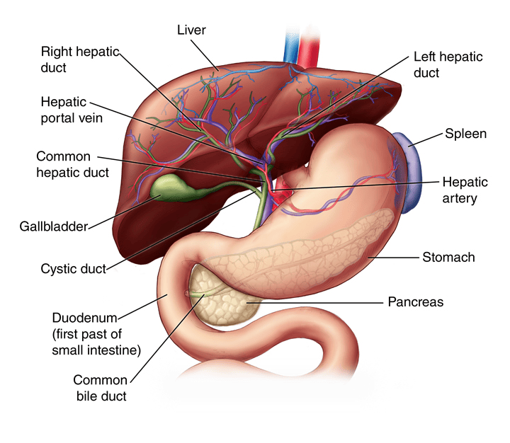
The liver is a large solid organ in the right upper part of the abdomen, immediately below the diaphragm and protected partly by the lower ribs. A portion extends across the upper abdomen. Its close relation to the right chest explains why a lower chest wound or fractured lower rib may also injure the liver, diaphragm or lung.
The liver receives blood from two major sources: the hepatic artery and the portal vein. Blood drains through the hepatic veins into the inferior vena cava. The liver’s large blood supply makes haemorrhage possible when these vessels or the surrounding tissue are disrupted. Bile produced by liver cells drains through small intrahepatic ducts into the larger bile ducts.
Important functions include metabolism of nutrients and medicines, production of bile and plasma proteins, storage of glycogen and selected vitamins, and processing of substances absorbed from the gut. After trauma, liver injury can therefore be associated with bleeding, bile leakage, infection, impaired perfusion and, in extensive injury or pre-existing disease, reduced liver function.
3. Causes and mechanisms of liver injury
Blunt abdominal trauma
Blunt trauma does not break the skin. The liver may be compressed between the abdominal wall and the spine, torn by rapid deceleration, or injured by a direct blow. Common mechanisms include:
- Road traffic collisions, including impact against a steering wheel, dashboard, seat belt or motorcycle handlebar.
- Falls from a height or onto a hard surface.
- Assault, kicks, blows or sports injuries.
- Crush injuries at work or during other accidents.
- Fractures of the lower right ribs associated with an abdominal impact.
Penetrating abdominal trauma
A knife, sharp object, bullet or other penetrating force may pass through the abdominal wall and cut liver tissue or major vessels. The external wound may be small while the internal injury is extensive. The direction and depth of a wound cannot be judged from skin appearance alone.
Iatrogenic or procedure-related injury
Rarely, liver injury may occur during abdominal procedures or interventions. The cause and pattern are assessed by the treating team, and the same priorities apply: identify haemorrhage or bile leakage, monitor the patient and provide appropriate source control.
4. Pathophysiology
Trauma may produce a subcapsular haematoma, a tear through the liver tissue, disruption of a vessel, or injury to a bile duct. Blood may accumulate within the liver, beneath its capsule or freely in the peritoneal cavity. A contained haematoma can enlarge or rupture; a vascular injury can cause ongoing or delayed bleeding.
As circulating blood volume falls, venous return and cardiac output may decrease. The body initially compensates by increasing the heart rate and constricting peripheral blood vessels. The patient may become anxious, restless, pale, cool or sweaty before blood pressure falls. Continued blood loss reduces oxygen delivery to tissues and can progress to altered consciousness, weak pulses, oliguria, metabolic acidosis, coagulopathy, hypothermia, organ failure and death.
Damage to the biliary system can allow bile to collect in the abdomen or within a local cavity, forming a biloma or biliary fistula. Tissue devitalisation, collections and invasive procedures may increase the risk of infection or abscess. These complications may appear after the first emergency has passed, so discharge teaching and follow-up matter.
5. Classification and severity
The American Association for the Surgery of Trauma (AAST) organ injury scale describes anatomic severity from lower-grade injury through extensive vascular or parenchymal damage. The World Society of Emergency Surgery (WSES) approach adds haemodynamic status to anatomic grading. This distinction is clinically important: a patient with a less extensive injury can still be critically unwell if bleeding causes instability, while a higher-grade injury may sometimes be managed without surgery in a stable patient in a suitably equipped setting.
| Clinical group | General description | Implication for care |
|---|---|---|
| Minor injury | Lower-grade anatomic injury in a haemodynamically stable patient. | May be suitable for non-operative hospital management after assessment. |
| Moderate or severe injury in a stable patient | More extensive anatomic injury, but circulation remains stable after assessment. | Requires specialist review, close monitoring and access to urgent intervention; selected cases may be managed non-operatively. |
| Unstable or non-responding patient | Persistent or recurrent evidence of shock despite resuscitation, regardless of the anatomic grade. | Needs immediate trauma-team escalation and haemorrhage control; imaging must not delay definitive treatment. |
This table is an educational summary, not a grading tool. Only the responsible trauma and surgical teams should assign injury grades and decide treatment. The WSES 2020 liver trauma guideline explains the combined classification and treatment approach.
6. Clinical features and warning signs
Presentation varies with the amount and speed of bleeding, the injury mechanism, associated injuries, age, pregnancy and pre-existing illness. Some patients have abdominal pain but little visible bruising; others may have significant internal bleeding with only subtle early signs.
Possible local findings
- Pain or tenderness in the right upper abdomen, epigastrium or across the abdomen.
- Abdominal guarding, distension or increasing discomfort.
- Pain referred to the right shoulder from irritation beneath the diaphragm.
- Bruising, abrasions, a seat-belt mark, lower rib tenderness or a penetrating wound.
- Reduced bowel sounds, which may occur but are not specific to liver injury.
Features of blood loss and shock
- Increasing pulse rate, weak peripheral pulses or a narrowing pulse pressure.
- Pale, cool, clammy skin; delayed capillary refill; sweating or thirst.
- Restlessness, anxiety, confusion, drowsiness or reduced responsiveness.
- Rapid or shallow breathing, dizziness, fainting or collapse.
- Falling blood pressure, although hypotension may be a late sign, especially in children and young adults.
- Reduced urine output, worsening lactate or other evidence of poor tissue perfusion.
Escalate immediately for deteriorating observations, new confusion, increasing abdominal distension or tenderness, repeated fainting, ongoing bleeding, a falling haemoglobin with clinical change, or any return of shock after initial improvement. A transient response to fluids does not mean the bleeding has stopped.
7. Immediate first aid and pre-hospital priorities
In a suspected serious abdominal injury, first aid should minimise delay and further harm while emergency transport and trained help are arranged:
- Ensure scene safety and call the local emergency response or arrange urgent transfer to an appropriate facility.
- Do not move the patient unnecessarily if a spinal or major limb injury is possible, unless movement is needed to protect life or maintain an airway.
- Check responsiveness, airway and breathing. Give basic life support within training and local protocol if required.
- If there is external bleeding, apply direct pressure around the wound using a clean dressing when safe. Do not remove an embedded object or push exposed tissue back into the abdomen.
- Keep the patient warm, reassure them and monitor breathing and responsiveness while awaiting transfer.
- Do not give food, drink or oral medicines before urgent clinical assessment. Do not delay transfer to apply traditional substances or unproven treatments.
- Share the injury mechanism, time of injury, observed changes and any treatment already given with the receiving team.
These measures do not replace rapid transfer or professional resuscitation. A patient who looks stable after an accident still requires urgent assessment when a significant abdominal injury is suspected.
8. Emergency assessment: the ABCDE approach
Care is delivered by a multidisciplinary trauma team. Nurses should work within their scope, local protocol and delegated responsibilities, while escalating concerns early. The primary survey identifies and treats immediate threats in order; treatment and reassessment occur together.
| Survey step | What the team assesses | Nursing priorities and rationale |
|---|---|---|
| A — Airway with cervical-spine protection | Airway patency, speech, obstruction, facial injury and possible cervical injury. | Call for airway support when needed, maintain indicated spinal precautions, prepare suction and prescribed airway equipment, and report any change in voice or consciousness. An obstructed airway can rapidly compromise oxygen delivery. |
| B — Breathing | Respiratory rate and effort, chest movement, oxygenation and associated chest injury. | Observe continuously, attach pulse oximetry when available, administer oxygen as prescribed or per protocol, and alert the team to respiratory distress or asymmetry. A lower chest injury may coexist with hepatic injury. |
| C — Circulation and haemorrhage control | Pulse, blood pressure, skin perfusion, external bleeding, likely internal bleeding and response to resuscitation. | Obtain help early, prepare appropriate intravenous access and blood sampling as directed, control visible bleeding, warm the patient, and record response after each intervention. Do not wait for hypotension if other signs suggest shock. |
| D — Disability | Level of consciousness, pupils, glucose if indicated and neurological deficit. | Record a recognised consciousness score and changes, check point-of-care glucose when ordered, and consider head injury or hypoxia as well as shock. A deteriorating mental state may be an early sign of poor perfusion. |
| E — Exposure and environment | Full examination for injury while preventing heat loss and preserving dignity. | Expose only as needed to identify injury, inspect the back during a safe log-roll when indicated, document wounds, then cover with warm blankets. Hypothermia worsens trauma-related coagulopathy. |
After immediate threats are addressed, the team repeats the primary survey and continues monitoring. A trauma handover should be structured and concise, for example using the local SBAR or trauma handover format.
9. Focused history and secondary survey
Once immediate threats are being managed, obtain a focused history without delaying resuscitation. Use a structured approach such as AMPLE when appropriate:
- A — Allergies: medicines, latex or other relevant allergies.
- M — Medicines: anticoagulants, antiplatelets, prescribed medicines, traditional medicines or substances that may affect bleeding or assessment.
- P — Past history: liver disease, bleeding disorders, previous surgery, pregnancy possibility and important comorbidities.
- L — Last oral intake: time and type of the last food or drink.
- E — Events: mechanism, direction and force of injury; time; loss of consciousness; symptoms and any first aid or treatment.
During the secondary survey, examine the patient systematically for other injuries. Inspect the chest, abdomen, pelvis, back and limbs as indicated. Document the location and appearance of wounds, bruising and tenderness without repeatedly or forcefully palpating an unstable abdomen. Record who was informed, when escalation occurred, and the patient’s response.
10. Investigations
Investigations are selected according to the patient’s haemodynamic status and the resources available. They should support, not delay, urgent haemorrhage control.
Bedside assessment
- Extended focused assessment with sonography for trauma (E-FAST): may rapidly detect free fluid or associated chest complications. A negative scan does not exclude all liver or abdominal injuries, particularly early in the course or when views are limited.
- Serial observations and examination: repeated assessment of pulse, blood pressure, respiratory rate, oxygen saturation, temperature, mental state, abdominal findings and urine output helps detect deterioration.
- Point-of-care tests: glucose, blood gas or lactate may be used according to the clinical setting and local availability.
Laboratory investigations
- Full blood count with serial haemoglobin and haematocrit when indicated.
- Blood group, antibody screen and crossmatch if transfusion may be required.
- Coagulation profile, platelet count, electrolytes, renal function and liver enzymes as directed.
- Blood gas, lactate or base deficit to assess perfusion and response to resuscitation when available.
- Pregnancy testing when relevant and when it does not delay emergency care.
An early haemoglobin can be misleading in acute blood loss. Interpret results alongside the mechanism, examination, vital-sign trends and treatment response. A rise in liver enzymes may support concern for tissue injury but does not by itself determine severity or exclude haemorrhage.
Imaging and other procedures
- Contrast-enhanced CT of the abdomen: the preferred detailed imaging test for a haemodynamically stable or stabilised patient when available. It can identify injury pattern, active bleeding and associated abdominal injury.
- Ultrasound: may be useful when CT is unavailable or for selected follow-up, but a normal study should not overrule concerning clinical findings.
- Diagnostic peritoneal lavage: may be considered by an appropriately trained team in selected settings when imaging is unavailable or inconclusive. It is invasive and is not a routine nursing procedure.
- Chest or pelvic radiographs: may be requested to assess associated injuries as part of the trauma work-up.
Do not send a persistently unstable patient to CT if doing so delays a needed operation or other definitive haemorrhage control. The diagnostic pathway is determined by the trauma team.
11. Initial medical management and resuscitation
The immediate goal is to restore oxygen delivery while controlling the source of bleeding and preventing further physiological deterioration. The trauma, emergency, anaesthesia, surgical, radiology and transfusion teams coordinate treatment according to patient needs and local resources.
- Maintain airway and breathing support; provide oxygen or assisted ventilation when clinically indicated.
- Establish appropriate vascular access, take ordered samples and begin resuscitation as directed.
- Activate the facility’s major haemorrhage or massive transfusion protocol when criteria are met. Use blood components and warming measures according to protocol.
- Prevent heat loss during examination, transport, infusion and procedures.
- Use intravenous fluids and blood products judiciously under clinical direction; avoid allowing repeated fluid boluses to delay definitive haemorrhage control.
- Provide prescribed analgesia and antiemetics, and reassess the patient’s response. Pain relief should not replace observation for deterioration.
- Assess tetanus protection and antimicrobial needs for open wounds or surgery according to the injury, prescription and local protocol.
- Consider reversal of anticoagulation only after senior review of bleeding risk, indication and available reversal options.
Medication doses and transfusion decisions depend on age, weight, injury, laboratory results, available products and local protocols. Nurses should verify prescriptions, patient identity, blood-product checks and monitoring requirements before administration.
12. Non-operative management
Many patients with blunt liver injury do not require an operation if they remain haemodynamically stable, have no other injury requiring surgery, and can be monitored in a setting that can respond promptly to deterioration. This is called non-operative management or selective observation. It is an active treatment plan led by the surgical or trauma team.
Depending on the injury and available services, the plan may include monitored admission, serial clinical examinations, repeat blood tests, pain control, careful fluid balance and repeat imaging when the patient’s course suggests it is needed. Patients with more severe injury, ongoing transfusion needs or other risks may require a higher-acuity area. There is no single observation schedule suitable for every patient; follow the prescribed plan.
For selected haemodynamically stable patients with active arterial bleeding on CT, angiography with embolisation may control bleeding without open surgery where the service is available. This requires careful patient selection and continued monitoring. A patient who becomes unstable, develops peritonitis or has another operative injury may need urgent surgery even if initial observation was planned.
In a setting with limited imaging or interventional capacity, clinicians must weigh the patient’s stability, serial examination findings, blood-test trends, associated injuries, staffing, theatre availability and transfer options. A plan for observation is unsafe if required monitoring or rapid rescue cannot be provided.
13. Operative and interventional management
Patients with persistent haemodynamic instability, ongoing uncontrolled haemorrhage, peritonitis or another injury requiring laparotomy may need urgent operative management. The operating surgeon decides the approach according to the injury and overall condition.
Surgical priorities
- Rapidly identify and control life-threatening haemorrhage.
- Control bile leakage when possible and assess for associated bowel, vascular or other abdominal injuries.
- Use damage-control principles in a severely unstable patient: abbreviated haemorrhage control and resuscitation may be followed by planned reassessment and definitive repair.
- Perihepatic packing and other specialist techniques may be used to control bleeding. Complex vascular or parenchymal procedures are performed by appropriately trained surgical teams.
- Major liver resection is generally avoided as an initial damage-control step and is considered selectively by experienced teams.
Interventional radiology and endoscopic care
Angiography and hepatic artery embolisation may help control selected arterial bleeding in a stable patient or persistent bleeding after surgery. Percutaneous drainage, endoscopic procedures such as ERCP and biliary stenting, or repeat surgery may be used for selected delayed bile or infectious complications. Their availability differs between facilities; arrange referral or transfer when clinically required and feasible.
Nursing care around surgery includes preparation for theatre, safe transfer, documentation, blood-product coordination, consent processes led by the responsible clinician, and support for the patient and family. After surgery, monitor for recurrent bleeding, respiratory complications, infection, bile leakage, pain and organ dysfunction.
14. Nursing management
Nursing management is continuous from the first contact to recovery. It combines close observation, rapid escalation, safe delivery of prescribed care, clear documentation and compassionate communication.
| Nursing action | Rationale | What to document or escalate |
|---|---|---|
| Use a structured trauma handover and identify the mechanism and time of injury. | Mechanism and timing guide the search for associated injuries and help the receiving team interpret changes. | Mechanism, symptoms, pre-hospital care, time of arrival and team members notified. |
| Repeat and trend vital signs at the frequency ordered; increase observation and call for review if the patient changes. | Trends may show blood loss or shock before one isolated reading becomes abnormal. | Pulse, blood pressure, respiratory rate, oxygen saturation, temperature, consciousness and response to interventions. |
| Observe perfusion, mental status, skin colour, abdominal distension and pain. | Internal bleeding can present with subtle changes and no visible external blood. | New or worsening findings, time observed, escalation time and response. |
| Maintain accurate intake and output; measure urine output as prescribed. | Urine output contributes to assessment of renal perfusion and response to resuscitation. | Urine volume and time interval, catheter output if present, and any significant decline. |
| Prepare and assist with ordered blood tests, crossmatch and imaging. | Timely results support resuscitation, diagnosis and treatment planning. | Specimen times, labelled samples, blood-bank communication and any delay or result requiring review. |
| Use warming measures and minimise unnecessary exposure. | Preventing hypothermia supports coagulation and reduces avoidable complications. | Temperature trends, warming interventions and response. |
| Administer prescribed fluids, blood products, analgesia and other treatment using safety checks. | Correct, timely therapy supports perfusion and comfort while reducing preventable medication and transfusion errors. | Product or medication details, checks, start and finish times, observations and reactions. |
| Maintain nil-by-mouth status when ordered or while urgent surgical assessment is ongoing. | A procedure or anaesthesia may be needed; oral intake can create additional risk. | Last oral intake, status, mouth care and any exception authorised by the team. |
| Support non-operative observation with reliable serial assessment. | Observation is safe only when deterioration is recognised and acted on quickly. | Assessment findings, prescribed frequency, missed observations and immediate escalation. |
| Provide privacy, explanation and emotional support to the patient and family. | Trauma is frightening; clear communication helps preserve dignity and cooperation. | Information given, questions raised, interpreter or support needs and referrals offered. |
Nursing priorities during resuscitation
- Recognise the possibility of internal bleeding from the mechanism and patient presentation.
- Call the appropriate emergency, surgical or trauma team early; do not wait for every test result before escalating shock.
- Assist with airway, oxygenation, vascular access, ordered blood sampling and resuscitation within scope and protocol.
- Prepare equipment and documentation for transfer, imaging, theatre or higher-acuity care as directed.
- Reassess after each intervention and communicate changes using a structured handover.
Nursing care during observation
For a patient being managed without surgery, carry out serial assessments exactly as prescribed and record them promptly. Check for increasing pain, abdominal distension, dizziness, pallor, sweating, fever, reduced urine output or a change in consciousness. Keep emergency call systems accessible and ensure the patient can be observed at the planned level. Report a missed observation, a deteriorating trend or difficulty obtaining reliable readings.
Administer analgesia as prescribed and reassess comfort, alertness and respiratory status. Avoid assuming that reduced pain means the injury has resolved. Encourage mobility, feeding and other recovery activities only when the treating team confirms they are appropriate for the patient’s stability and injury.
15. Post-operative and recovery care
After laparotomy, embolisation or another procedure, care depends on the intervention and the patient’s physiological status. Follow the postoperative plan and local recovery-unit standards.
- Assess airway, breathing, circulation, oxygenation, level of consciousness, pain and temperature at the prescribed frequency.
- Inspect the abdominal dressing, drains, wound sites and access site for fresh bleeding, leakage, swelling or infection.
- Monitor ordered haemoglobin, coagulation, renal function, liver tests and other results; escalate concerning trends.
- Maintain fluid balance and observe urine output. Report a significant decline or unexpectedly dark or bloody urine.
- Assess for abdominal distension, worsening pain, persistent vomiting, fever, jaundice or signs of bile leakage.
- Prevent pressure injury, support respiratory exercises and early mobility as allowed, and provide thrombosis prevention prescribed by the team.
- Explain procedures and progress in language the patient understands, and include family or a chosen support person when appropriate.
A patient may need intensive monitoring or transfer to a higher-level facility. Handover should include the injury mechanism and grade if known, interventions, blood products given, latest observations and results, drains, access devices, allergies and outstanding concerns.
16. Complications
| Complication | Possible clues | Nursing response |
|---|---|---|
| Ongoing or delayed haemorrhage | Increasing pulse, pallor, dizziness, falling blood pressure, abdominal expansion, new confusion or falling haemoglobin with clinical change. | Escalate urgently, repeat ABCDE assessment, maintain monitoring and prepare for the team’s plan for resuscitation or haemorrhage control. |
| Bile leak, biloma or biliary fistula | Persistent or increasing abdominal pain, fever, abdominal collection, bile-stained drainage or jaundice. | Report findings promptly; monitor drains, observations and ordered investigations. |
| Hepatic artery pseudoaneurysm or delayed arterial bleeding | New abdominal pain, gastrointestinal bleeding, unexplained anaemia or recurrent instability after initial improvement. | Urgent medical or surgical review; do not reassure solely because the original observation period was stable. |
| Infection or liver abscess | Fever, rigors, increasing pain, raised inflammatory markers or an unwell appearance. | Escalate, collect ordered specimens and administer prescribed treatment safely. |
| Necrosis, biliary obstruction or liver dysfunction | Worsening jaundice, abnormal liver tests, persistent fever or other organ dysfunction. | Report trends and support specialist assessment and follow-up. |
| Trauma-related coagulopathy, hypothermia or organ failure | Oozing, abnormal coagulation tests, low temperature, reduced urine output, worsening perfusion or altered mental state. | Continue warming and prescribed monitoring; urgently communicate findings to the team. |
| Associated chest, bowel, spleen, renal or pancreatic injury | Breathing difficulty, peritonism, blood in urine, new symptoms or deterioration not explained by the known injury. | Repeat a complete assessment and escalate; the liver injury may not be the only injury. |
Some complications occur days or weeks after the original injury. New severe abdominal pain, fainting, blood in vomit or stool, fever, jaundice, increasing abdominal swelling or sudden weakness after discharge requires urgent clinical assessment.
17. Special considerations
Children
Children can compensate for blood loss and maintain blood pressure until they suddenly deteriorate. Use age-appropriate observation ranges, equipment and escalation pathways. Involve paediatric and trauma expertise early. A negative E-FAST may not exclude intra-abdominal injury in a child with concerning symptoms or mechanism.
Pregnancy
Trauma assessment should prioritise maternal stabilisation while involving obstetric and trauma teams according to the facility pathway. Pregnancy may alter normal vital signs and creates additional maternal-fetal assessment needs. Do not delay urgent life-saving evaluation or treatment because the patient is pregnant.
Older adults and patients taking anticoagulants
Older adults, people with frailty, liver disease or bleeding disorders, and patients using anticoagulant or antiplatelet medicines may have atypical signs or a greater risk of serious bleeding. Obtain a careful medicine history, monitor trends closely and escalate early.
Limited-resource settings
Where CT, interventional radiology, blood products or specialist surgery are not immediately available, early recognition, stabilisation within capability, senior consultation and timely referral are essential. Non-operative observation should be chosen only when the required monitoring and rescue capability are realistically available. Arrange transfer before the patient deteriorates whenever a higher level of care is needed and transfer is feasible.
18. Health education and discharge planning
Discharge is planned by the treating team after the patient is clinically suitable. Explain the diagnosis and treatment in simple language, check understanding and provide written instructions when available. The patient should know who to contact and where to return for urgent care.
- Take medicines only as prescribed and clarify how to manage medicines that were stopped or adjusted during admission.
- Attend the surgical or trauma follow-up appointment and complete any planned tests or imaging.
- Resume work, sport, heavy lifting and other strenuous activity only according to the treating team’s advice; restrictions depend on injury grade, symptoms and recovery.
- Keep wounds and drain sites clean and follow the specific wound-care instructions provided.
- Seek urgent help for worsening abdominal or shoulder pain, fainting, weakness, fever, vomiting, jaundice, abdominal swelling, wound discharge, shortness of breath or any new bleeding.
- Discuss transport, costs, caregiver support, safe return to home and how to reach the facility if symptoms recur.
Before discharge, assess whether the patient can explain the warning signs in their own words. Provide an interpreter or other communication support when needed.
19. Sample nursing care plan
The following examples are a learning guide. Individualise nursing diagnoses, goals and interventions to the patient’s assessment, facility policy and current orders.
| Problem or nursing diagnosis | Expected outcome | Key nursing interventions |
|---|---|---|
| Risk of shock related to suspected internal haemorrhage. | Patient maintains or improves perfusion while definitive management is arranged. | Trend observations and mental state; assess perfusion and abdomen; maintain prescribed access and monitoring; prepare ordered blood samples or products; keep warm; escalate any deterioration immediately. |
| Acute pain related to abdominal tissue injury or procedure. | Patient reports acceptable pain relief while remaining appropriately alert and stable. | Assess pain using an appropriate scale; administer prescribed analgesia; reassess pain, sedation and breathing; position for comfort without delaying assessment; report sudden change in pain. |
| Anxiety related to trauma, emergency procedures and uncertainty. | Patient and family demonstrate improved understanding of immediate care and know whom to contact. | Give brief, clear explanations; stay calm; facilitate communication with the team; protect privacy; include a chosen support person when appropriate. |
| Risk of infection related to open injury, surgery, drains or invasive devices. | Patient remains free of preventable wound or device infection. | Use aseptic technique; inspect wounds and devices; perform ordered wound care; monitor temperature and clinical signs; administer prescribed prophylaxis or treatment. |
| Knowledge need related to recovery and warning signs after liver injury. | Before discharge, patient describes follow-up, activity advice and symptoms that require urgent review. | Teach using plain language and teach-back; provide written instructions; address access barriers; document education and referrals. |
20. Revision questions and answers
Question 1
Why can a patient with liver trauma initially have a normal blood pressure?
The body can compensate for early blood loss by increasing heart rate and constricting peripheral blood vessels. Hypotension may develop later, so repeated assessment of the full clinical picture is essential.
Question 2
Does a negative E-FAST rule out liver injury?
No. E-FAST can rapidly detect free fluid, but a negative result does not exclude every liver injury or early bleeding. Further assessment depends on the patient’s stability, examination and clinical suspicion.
Question 3
Which patient may be considered for non-operative management?
A carefully selected patient who is haemodynamically stable, has no other injury requiring surgery, and can receive serial clinical assessment and timely intervention if the condition changes. The treating trauma team makes this decision.
Question 4
What is the nurse’s priority when an observed patient develops a rising pulse, pallor and increasing abdominal distension?
Recognise possible ongoing haemorrhage, repeat the immediate assessment, escalate urgently to the responsible team, maintain monitoring and prepare for the team’s resuscitation and haemorrhage-control plan.
Question 5
Name three complications that may occur after the initial injury.
Examples include delayed haemorrhage, bile leak or biloma, hepatic artery pseudoaneurysm, liver abscess, infection, tissue necrosis and associated-organ complications.
21. Key points to remember
Think LIVER in suspected hepatic trauma
- L — Look for the mechanism, local injury and signs of internal bleeding.
- I — Identify airway, breathing and circulation threats using a repeated ABCDE assessment.
- V — Verify trends in vital signs, mental state, urine output and ordered investigations.
- E — Escalate early for shock, deterioration, peritonitis or delayed warning signs.
- R — Reassess after every intervention and hand over clearly.
Liver trauma can be life-threatening even when external injuries look minor. The safest care combines early recognition, repeated assessment, prompt escalation, appropriate resuscitation, and definitive haemorrhage control when required. Stable patients may avoid surgery only when careful monitoring and rescue treatment are available.
22. References and further reading
- Coccolini F, et al. Liver trauma: WSES 2020 guidelines. World Journal of Emergency Surgery. 2020;15:24.
- Eastern Association for the Surgery of Trauma (EAST). Hepatic Injury, Blunt, Selective Nonoperative Management of. Practice Management Guideline. 2012.
- Coccolini F, et al. WSES classification and guidelines for liver trauma. World Journal of Emergency Surgery. 2016;11:50.
- Ministry of Health, Uganda. Uganda Clinical Guidelines 2023.
Use current facility and Ministry of Health protocols for emergency assessment, blood transfusion, referral, surgery and follow-up. These notes are intended for nursing revision and should be taught alongside supervised clinical practice.