
A systematic, respectful, evidence-informed approach to clinical assessment. Prepared for clinical teaching and skills practice, incorporating essential nursing rationale and detailed anatomical context.
- Define the scope of a genitourinary (GU) examination and explain its clinical purpose in evaluating renal, urinary, and reproductive health.
- Prepare the patient, environment, and equipment while strictly protecting the patient's dignity, modesty, and privacy.
- Take a focused history covering urinary, renal, and reproductive symptoms.
- Perform a structured examination of the abdomen, flanks, external genitalia, and—when indicated and appropriately trained—the rectum or pelvis.
- Recognize abnormal findings, document them clearly and legally, and identify urgent Red Flags requiring emergency intervention.
The Genitourinary (GU) system comprises two intricately linked systems: the urinary tract (kidneys, ureters, bladder, and urethra) and the reproductive organs (testes, epididymis, prostate, penis in males; vulva, vagina, uterus, ovaries in females).
- Tailored Assessment: Choose examination components according to the patient's symptoms, age, anatomy, clinical indications, and personal preference. You do not need to examine the genitalia of a patient presenting with simple isolated flank pain unless complications are suspected.
- Informed Consent: Explain each and every step before you do it. Obtain explicit informed consent.
- Chaperoning: Always offer and strongly encourage a chaperone for intimate examinations, according to institutional policy. Document the chaperone's name and presence.
- Trauma-Informed Care: Use a trauma-informed, non-judgmental approach. Be highly sensitive to the fact that many patients may have a history of sexual trauma or abuse. If the patient withdraws consent or experiences significant distress, stop the examination immediately.
- Holistic Evaluation: A GU examination complements—but does not replace—a thorough clinical history, urinalysis, laboratory testing (renal function), and imaging (ultrasound/CT).
In many African settings, discussing genitourinary and sexual health is highly taboo. As a nurse, you must build immense trust. Additionally, keep local endemic diseases in mind: a patient presenting with terminal haematuria (blood at the end of passing urine) in Uganda must be evaluated for Schistosomiasis (Bilharzia), especially if they reside near freshwater lakes.
Intimate examinations provoke severe anxiety in patients. Your preparation sets the tone for a professional, safe encounter.
- The Environment: Provide a completely private, warm, and well-lit room. Ensure the door/curtain is securely closed. Only strictly necessary medical staff should be present.
- Communication & Consent: Introduce yourself clearly, confirm the patient's identity, explain the purpose and exact sequence of the exam, and obtain verbal consent before any physical contact.
- Chaperone: Offer a chaperone (preferably of the same gender as the patient). You must legally document the offer, and whether the patient accepted or declined it.
- Standard Precautions: Perform rigorous hand hygiene. Wear non-sterile gloves and other Personal Protective Equipment (PPE) as indicated (e.g., aprons).
- Gather Equipment: Ensure you have drapes/gowns, a good light source (goose-neck lamp), a stethoscope, and a measuring tape if needed. For intimate exams, prepare appropriate swabs, lubricating jelly, and a speculum only when clinically indicated and if you are trained to use them.
- Patient Comfort: Ask about current pain, mobility issues, allergies (especially to latex gloves or lubricants), and recent surgical procedures. Crucial step: Ask whether the patient needs to empty their bladder before the abdominal exam (a full bladder is incredibly uncomfortable to palpate and can mimic a pelvic mass).
A comprehensive history often reveals the diagnosis before you even touch the patient. Use the SOCRATES acronym for pain and explore the following:
| Symptom Category | Specifics to Ask & Clinical Medical Terms |
|---|---|
| Pain | Onset, site, severity, and radiation (e.g., classic renal colic pain radiates from the flank/loin down to the groin/testicles). Aggravating or relieving factors. |
| Voiding (Storage) Symptoms | Frequency: Needing to pass urine often. Urgency: Sudden, compelling need to pass urine. Nocturia: Waking up multiple times at night to urinate. Dysuria: Pain or a burning sensation when passing urine (classic UTI sign). |
| Voiding (Emptying) Symptoms | Hesitancy: Difficulty starting the urine stream. Weak stream: Poor flow force. Intermittency: Flow stops and starts. Incomplete emptying: Feeling like the bladder is still full. (These are classic signs of Benign Prostatic Hyperplasia - BPH in older men). |
| Urine Changes | Haematuria: Visible (macroscopic) blood in urine. Cloudiness or unusual foul odour (infection). Reduced output (Oliguria: <400ml/day; Anuria: <100ml/day). Frothiness (indicates heavy proteinuria/nephrotic syndrome). Incontinence: Unintentional leaking of urine (stress, urge, or overflow). |
| Associated Systemic Symptoms | Fever, chills/rigors, nausea/vomiting, severe flank pain, peripheral oedema (swelling of legs/face), severe fatigue, or acute confusion (especially in the elderly with UTIs). |
| Past Medical History | Previous UTIs, kidney stones, chronic kidney disease (CKD), diabetes, hypertension (major causes of renal failure), recent catheterization, surgery, and a full medication review. |
Also ask about: Daily fluid intake, the possibility of pregnancy (when relevant), sexual health, and STI exposure. Handle STI questions sensitively and in strict privacy.
- Clarify the concern: Use the patient's own words. Ask permission before diving into sensitive sexual health questions.
- General genital symptoms: Ask about genital pain, swelling, sores/ulcers, abnormal discharge, intense itching (pruritus), lumps, bleeding, or changes in sexual function.
- For patients with testes (Male): Enquire about scrotal pain or swelling, history of trauma to the groin, testicular lumps (cancer risk), and abnormal urinary/urethral discharge (STI risk).
- For patients with a vulva/vagina (Female): Enquire about deep pelvic pain, abnormal vaginal discharge (colour, smell, consistency), vulval symptoms, menstrual history (LMP - Last Menstrual Period, regularity), and abnormal/post-coital bleeding.
- Sexual History: Ask about sexual practices and partners only as clinically relevant. Use inclusive, non-assumptive language (e.g., "Do you have sex with men, women, or both?").
- Wider context: Explore prior pregnancies (gravidity/parity), current contraception, menopause status, fertility concerns, past STI history, and importantly, safeguarding needs (signs of intimate partner violence or sexual assault) when appropriate.
Before examining the specific system, step back and perform a general "end-of-bed" inspection.
- General Appearance: Assess comfort level, hydration status (dry mucous membranes, sunken eyes), marked pallor (anaemia from chronic kidney disease), signs of fever, distress, and their level of consciousness.
- Vital Signs: Measure temperature (fever = infection/pyelonephritis), pulse (tachycardia = pain or sepsis), respiratory rate, blood pressure (hypertension is deeply linked to renal disease), and oxygen saturation when indicated.
- Fluid Status & Oedema: Look for peripheral oedema (pitting swelling in the ankles/sacrum) or facial/periorbital oedema (classic in nephrotic syndrome and renal failure). Assess for systemic illness.
- Weight and Growth: Assess weight and growth trajectories in children when relevant. Consider blood pressure and strict fluid balance monitoring in patients with known renal disease.
- Mobility and Posture: Observe how the patient moves.
- Colicky pain: A patient with a kidney stone (renal colic) cannot sit still and constantly writhes in agony.
- Peritonitic pain: A patient with localized peritonitis lies absolutely still, as any movement hurts.
- Scrotal pathology: May walk with a wide-based, waddling gait to avoid friction.
- Urine Output: Record exact urine output when clinically necessary, especially in acutely unwell or hospitalized patients (using a fluid balance chart or urinary catheter bag).
- Positioning: Position the patient supine (lying flat on their back) with arms by their sides and knees slightly flexed to relax the abdominal muscles. Expose only the abdomen (from the xiphisternum to the symphysis pubis) and maintain draping over the chest and legs.
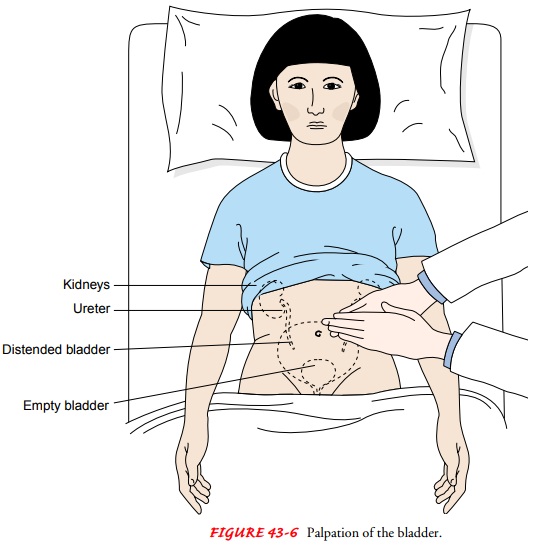
- Inspection: Look for distension (a hugely swollen bladder can reach the umbilicus), surgical scars (e.g., previous nephrectomy, C-section, appendicectomy), visible masses, hernias (inguinal/umbilical), or abnormal skin changes.
- Auscultation: While not usually central to a GU assessment, it may be included in a full abdominal exam. You may listen for renal artery bruits (a whooshing sound) just above and lateral to the umbilicus, indicating renal artery stenosis.
- Palpation: Palpate gently, starting away from the area of reported pain. Feel for tenderness, involuntary guarding (muscle rigidity), abdominal masses, and specifically a distended, firm, globular bladder arising from the pelvis.
- Percussion: Percuss the suprapubic area. A full, distended bladder will sound dull to percussion. Interpret these physical findings alongside the patient's symptoms and use a bedside bladder ultrasound scanner if available.
- Documentation: Accurately document the site, severity of tenderness, presence of any masses, guarding, and whether the bladder was palpable.
- Inspection: Ask the patient to sit up. Inspect the flanks and lower back for severe bruising (e.g., Grey Turner's sign indicating retroperitoneal haemorrhage), surgical scars, swelling, or obvious asymmetry.
- Palpation (Ballotting the Kidneys): Attempted by trained clinicians. Place one hand behind the patient's flank (in the renal angle) and the other hand anteriorly on the abdomen. Push your hands together. Normal kidneys are often not palpable in healthy adults, though the lower pole of the right kidney may occasionally be felt in thin individuals. Enlarged kidneys (e.g., polycystic kidney disease, tumours) will be distinctly palpable.
- Costovertebral Angle (CVA) Tenderness: Assess for CVA tenderness (also known as Murphy's punch sign). Place the palm of your non-dominant hand flat over the angle between the 12th rib and the spine. Strike the back of your hand gently with the ulnar border of your other fist.
Significant CVA tenderness strongly suggests acute pyelonephritis (kidney infection) or renal calculi (stones). However, it is not purely diagnostic on its own. Avoid forceful percussion, particularly if trauma, rib fractures, or severe pain is already suspected. Always correlate CVA tenderness with fever, urinary symptoms, urinalysis, renal function tests, and imaging.
- Strict Indication: Perform only when explicitly indicated by symptoms or clinical need, with explicit consent and a chaperone offered.
- Communication: Explain the examination step-by-step. Ask the patient to report any pain immediately and assure them they hold the power to pause or stop the exam at any second.
- Inspection: Inspect systematically for skin changes, lesions, ulcers (e.g., syphilis chancre, herpes blisters), swelling, unusual discharge, trauma, scars, or masses.
- Infection Control: Wear non-sterile gloves and employ appropriate infection-prevention measures. Avoid unnecessary or prolonged contact and exposure.
- Palpation: Should be exceptionally gentle, highly targeted, and clinically justified. Explain what you are about to feel before every single manoeuvre.
- Limitations: If the clinical findings are outside your competence level, if the patient declines at any point, or if the pain is severe, stop the examination immediately and arrange for an appropriate specialist review.
- Inspection of Shaft and Glans: Inspect the penile shaft, glans penis, and surrounding groin skin for lesions, rashes, sexually transmitted ulcers, swelling, bruising, or anatomical deformity (e.g., Peyronie's disease).
- Meatus: Inspect the urethral meatus (opening). Note its position (hypospadias if on the underside, epispadias if on top), look for acute inflammation, purulent/watery discharge, or visible blood at the meatus.
- Foreskin Management: If the patient is uncircumcised, ask them to retract the foreskin, or retract it yourself very gently only if it moves easily. CRITICAL RULE: Always return the foreskin to its normal forward position immediately afterward!
- History Correlate: Concurrently ask about pain, dysuria, discharge, sores, erectile concerns, or recent pelvic/groin trauma.
- Specimens: Collect urethral swabs or first-catch urine samples only when indicated for STI screening, according to local laboratory protocols.
Phimosis: Inability to retract a tight foreskin over the glans. Do NOT force it, as it will tear and scar.
Paraphimosis: A deeply retracted, tight foreskin becomes trapped behind the glans and cannot be pulled forward. It acts like a tight tourniquet, cutting off venous return. The glans swells massively and becomes painful and necrotic. This requires urgent emergency medical care.
- Inspection: Ask the patient to stand if possible. Inspect the scrotal sac for asymmetry, massive swelling (hydrocele, hernia), erythema (redness/cellulitis), skin lesions, surgical scars, and abnormal testicular position.
- Palpation: Using warm hands, palpate each testis gently between your thumb and first two fingers. Note the size, consistency (should be firm and smooth), tenderness, and any discrete hard lumps. Palpate the epididymis (soft, nodular structure on the posterior aspect) and trace the spermatic cord up toward the inguinal canal.
- Lumps: A new, painless, hard, irregular intratesticular lump is highly suspicious for testicular cancer and requires prompt urgent ultrasound assessment.
- Documentation: Clearly document laterality (left/right), onset of pain, nature of swelling, precise location of tenderness, and any associated systemic symptoms.
- Preparation: Explain the steps and obtain consent. Position the patient comfortably on the examination couch (often in a modified lithotomy or "frog-leg" position) with adequate draping to cover the upper body and thighs. Ensure a chaperone is present.
- Inspection: Under good lighting, inspect the external genitalia (labia majora, labia minora, clitoris) for skin changes, ulcers, warty lesions, swelling, abnormal discharge, fresh bleeding, or traumatic injury.
- Urethra and Vagina: Note the urethral opening and vaginal introitus (opening) only as needed to address the patient's presenting clinical concern.
- Perineum: Assess the perineal skin between the vagina and anus for unhealed episiotomy wounds, scars, signs of pelvic organ prolapse (e.g., cystocele, rectocele visible on straining), or signs of spreading infection.
- Scope of Practice: Avoid conducting an internal (bimanual pelvic) examination unless explicitly indicated, fully consented to, and performed by a suitably trained and credentialed clinician (e.g., a doctor or specialized midwife).
- Urgent Pathways: If there is severe acute pelvic pain, heavy uncontrollable bleeding, pregnancy-related complications (e.g., suspected ectopic), or suspected sexual abuse, stop the exam and follow urgent local referral pathways.
These are highly targeted, advanced clinical examinations—they are not routine steps for every general GU assessment.
- Pelvic/Speculum Examination: May be indicated for selected cases of abnormal vaginal bleeding, taking high vaginal swabs for persistent discharge, investigating deep pelvic pain, performing cervical cancer screening (Pap smear), or assessing suspected pelvic organ prolapse.
- Digital Rectal Examination (DRE): May be considered for specific anorectal complaints, checking anal sphincter tone in neurological disease (cauda equina), or prostate-related indications.
- Prostate Assessment: A DRE to feel the posterior surface of the prostate gland requires a detailed explanation, written or clear verbal consent, appropriate chaperoning, and a highly trained examiner. Follow local clinical protocols.
- Clinical Reasoning: Always consider absolute and relative contraindications, patient comfort, trauma history, and whether a less invasive test (like an ultrasound or urine test) can sufficiently answer the clinical question first.
- Golden Rule: Refer to a specialist or seek senior supervision when the procedure is outside your legal scope of nursing practice. Never perform any intimate examination without clear, ongoing consent.
Connecting what you find on physical examination to potential underlying pathology is key to clinical reasoning.
| Clinical Finding | Possible Clinical Significance / Interpretation |
|---|---|
| Suprapubic fullness / tenderness | May heavily suggest acute urinary retention (distended bladder), severe cystitis (bladder infection), or another space-occupying pelvic condition (like a large fibroid or ovarian cyst). |
| Flank / CVA tenderness WITH fever | Raises high concern for an upper urinary tract infection (Acute Pyelonephritis). Always assess the patient rapidly for signs of systemic sepsis. |
| Haematuria (Visible Blood) | Can arise from severe infection, kidney/bladder stones, blunt trauma, urological malignancy (bladder/renal cancer), Schistosomiasis, or underlying renal disease. Needs comprehensive evaluation. |
| Urethral or genital discharge / ulcers | Strongly consider infection, primarily Sexually Transmitted Infections (STIs) such as Gonorrhoea, Chlamydia, Syphilis, or Herpes. Test according to national public health guidance. |
| Scrotal swelling or Testicular mass | Painless mass = High suspicion for Testicular Cancer. Acute, severe pain = Medical emergency (Torsion or Epididymo-orchitis). Requires immediate prompt assessment. |
| Peripheral Oedema and Hypertension | Often accompany chronic renal disease (kidneys failing to excrete sodium/water). Must correlate with renal function blood tests (Creatinine) and urine protein testing. |
- Urinalysis (Dipstick): The most basic, vital test. Check for blood (stones/tumours), protein (kidney damage), leukocytes and nitrites (bacterial UTI), glucose and ketones (Diabetes/DKA), and specific gravity (hydration status) as relevant.
- Urine Microscopy, Culture, and Sensitivity (MC&S): Send to the lab when a severe or recurrent infection is suspected, or when indicated by local antimicrobial stewardship guidance, to determine exactly which antibiotics will work.
- Blood Tests: Draw blood for Creatinine/eGFR (to measure exact kidney function), Urea, Electrolytes (Potassium is critical in renal failure), Full Blood Count (FBC for infection/anaemia), and inflammatory markers (CRP) when clinically indicated.
- Pregnancy Testing (hCG): Perform a urine or blood pregnancy test on women of childbearing age when pregnancy is biologically possible, especially if the result critically affects your assessment (e.g., ectopic pregnancy) or management (e.g., prescribing teratogenic drugs or ordering X-rays).
- STI Screening: Collect Nucleic Acid Amplification Tests (NAATs) via urine or specific genital swabs based on symptoms, exposure history, and patient consent.
- Imaging: Utilize a bedside bladder scan (to measure retained urine volume), renal/pelvic ultrasound, or CT KUB (Kidneys, Ureters, Bladder) when indicated. Select imaging tests strictly according to your clinical assessment findings.
These findings threaten life or limb. Immediate escalation is mandatory to ensure clinical safety.
- Sudden severe testicular pain: Strongly points to suspected testicular torsion (twisting of the spermatic cord, cutting off blood supply to the testicle). It is a time-critical surgical emergency.
- Acute Urinary Retention: Inability to pass any urine combined with extreme pain and severe bladder distension. Requires urgent catheterization to prevent bladder rupture or kidney damage.
- Urosepsis: Fever, rigors (uncontrollable shivering), flank pain, accompanied by hypotension (low BP), confusion, tachycardia, or other systemic signs of sepsis. Requires urgent IV antibiotics and fluids.
- Gross Haematuria with Clots: Visible blood containing thick clots, or inability to urinate due to clot blockage. Indicates significant bleeding, potential haemodynamic instability, or severe trauma/cancer.
- Trauma & Obstruction: Severe abdominal/flank pain immediately after trauma, a suspected obstructed infected kidney (e.g., a stone blocking a ureter with concurrent infection above it), or rapidly deteriorating renal blood function.
- Ectopic Pregnancy Suspicions: A patient who is pregnant (or potentially pregnant) presenting with severe unilateral pelvic pain, sudden collapse, shoulder-tip pain (diaphragmatic irritation from internal bleeding), or heavy vaginal bleeding.
- Cauda Equina Syndrome: A massive neuro-surgical emergency. Look for new "saddle numbness" (numbness in the buttocks/perineum where you would sit on a horse saddle), severe leg weakness, or sudden loss of bladder/bowel control. Indicates spinal cord compression.
- Comprehensive Records: Record the precise clinical indication for the exam, the relevant focused history, that consent was obtained, the chaperone offer/outcome, and the exact examination components you performed.
- Objective Terminology: Use completely objective, professional, and respectful language. Describe the exact anatomical site, laterality (left/right), measured size, physical appearance, degree of tenderness, and relevant negatives (e.g., "No palpable lumps, no CVA tenderness").
- Justify Omissions: Explicitly document what was not examined and the reason why, including patient preference ("patient declined rectal exam") or clinical contraindication.
- Next Steps: Record all investigations ordered, specimens successfully collected, the findings communicated to the patient, and the clear follow-up or referral plan.
- Escalation (SBAR): Escalate abnormal or urgent Red Flag findings promptly to a doctor. Use a structured handover tool such as SBAR (Situation, Background, Assessment, Recommendation) for clear, rapid communication.
- Legal & Ethical: Maintain strict medical confidentiality and rigidly follow your local institutional documentation and safeguarding reporting policies.
- Hand hygiene, ensuring privacy, step-by-step explanation, obtaining consent, and offering a chaperone.
- Taking a focused urinary, renal, reproductive, and clinically relevant sexual history.
- Assessing general appearance, recording vital signs, and evaluating hydration/oedema status.
- Performing abdominal inspection, palpation, and targeted suprapubic percussion.
- Performing flank inspection and gentle CVA tenderness assessment when indicated.
- Conducting a targeted external genital examination only when strictly indicated and consented to.
- Considering appropriate complementary urine/blood tests, bladder scan, or imaging.
- Documenting findings accurately, explaining the next steps to the patient, and rapidly escalating any red flags.
The Scenario: A 24-year-old male presents with sudden, extremely severe right-sided testicular pain and nausea that began exactly one hour ago.
- What focused history questions would you ask?
- What exactly was he doing when the pain started? (e.g., exercising, sleeping, recent trauma).
- Has he ever had this exact pain before? (Previous intermittent torsion).
- Does he have any dysuria, frequency, or urethral discharge? (To rule out epididymo-orchitis).
- Any associated fever? (Points more toward infection than torsion).
- Which examination findings might support testicular torsion?
- A swollen, exquisitely tender, unilaterally enlarged testicle.
- A high-riding testis (pulled up higher in the scrotum than normal due to the twisted cord shortening).
- Loss of the normal cremasteric reflex on that side.
- The epididymis may be palpable in an abnormal, anterior position.
- What is the immediate priority, and should investigations delay referral?
- Immediate Priority: The absolute priority is recognizing this as a time-critical surgical emergency. The testis will die from ischemia (lack of blood flow) within 4 to 6 hours.
- Should investigations delay referral? NO. Do not wait for urine tests, blood tests, or a scrotal ultrasound if torsion is strongly suspected clinically. Immediate emergency surgical/urological assessment and exploration is required to untwist the cord and save the testicle.
- Hutchison, R. & Glynn, M. Hutchison's Clinical Methods: An Integrated Approach to Clinical Practice. Elsevier. (A core foundation text for systematic clinical examination methodologies).
- Bickley, L. S. Bates’ Guide to Physical Examination and History Taking. Wolters Kluwer. Use the latest available edition.
- National Institute for Health and Care Excellence (NICE). Guidance on urinary tract infection, acute kidney injury and suspected cancer. https://www.nice.org.uk/
- European Association of Urology (EAU). Guidelines on urological infections and acute scrotum/testicular cancer. https://uroweb.org/guidelines
- Centers for Disease Control and Prevention (CDC). Sexually Transmitted Infections Treatment Guidelines. https://www.cdc.gov/sti/hcp/treatment-guidelines/
- Note: Local hospital policies, Ugandan Ministry of Health clinical guidelines, and institutional protocols should ultimately guide examination, investigations, chaperoning, and referral pathways.
Quick Quiz
Genitourinary Examination Quiz
Health Assessment
Privacy: Your details are used only for quiz tracking and certificates.
Genitourinary Examination Quiz
Health Assessment
Preparing questions...
Choose your answer and keep your streak alive.
Great effort.
Here is your quick performance summary.