
Pathology of Aneurysms, Aortic Dissection, and Vascular Disorders
I. Introduction to Aneurysms
In vascular pathology, an aneurysm is defined as a localized, abnormal dilation of a blood vessel or the wall of the heart. These dilations can be congenital (present from birth) or acquired (developing later in life due to underlying vascular disease). To understand the clinical risk, we must first anatomically classify them based on the integrity of the vessel wall.
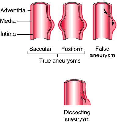
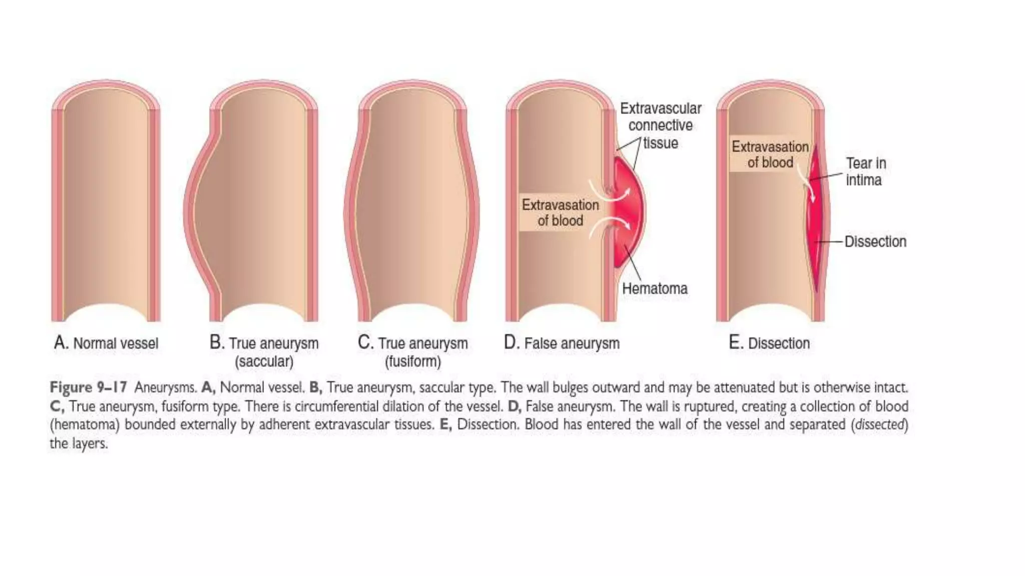
A true aneurysm involves all three layers of an intact arterial wall (intima, media, and adventitia) or the thinned ventricular wall of the heart.
- Vascular Examples: Atherosclerotic aneurysms, syphilitic aneurysms, and congenital vascular aneurysms.
- Cardiac Example: Ventricular aneurysms that follow a transmural myocardial infarction (the dead heart muscle heals into a thin, bulging fibrotic scar that contains all wall layers).
A false aneurysm is a breach or defect in the vascular wall leading to an extravascular hematoma. The blood freely communicates with the intravascular space, creating a "pulsating hematoma," but it is contained only by the outer adventitia or surrounding extravascular connective tissue.
- Examples: Ventricular free wall rupture after a massive myocardial infarction (contained by the pericardium), or an arterial leak at the site of a vascular graft anastomosis.
Macroscopic (Morphological) Classification
Aneurysms are visually categorized by their macroscopic shape and size:
- Saccular Aneurysms: Essentially spherical outpouchings that involve only a portion of the vessel wall circumference. They look like a small "berry" or balloon attached to the side of the vessel (e.g., Berry aneurysms in the Circle of Willis).
- Fusiform Aneurysms: Involve diffuse, circumferential dilation of a long vascular segment. They are spindle-shaped and vary greatly in diameter (5 to 10 cm) and length. They frequently involve extensive portions of the aortic arch, abdominal aorta, or even the iliac arteries.
- Cylindrical Aneurysms: Display a continuous, parallel dilation along a segment of the vessel.
- Serpentine or Varicose: A tortuous, winding dilation of the vessel.
- Racemose or Circoid: A tangled mass of intercommunicating, dilated small arteries and veins (often resembling a cluster of grapes).
II. Pathogenesis of Aneurysms
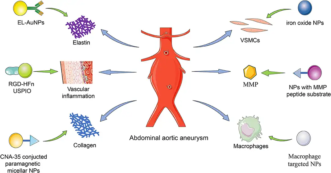
Why do blood vessels balloon out? The structural integrity of a normal blood vessel depends entirely on the connective tissue (collagen and elastin) within the tunica media. Aneurysms occur when the structure or function of this connective tissue is severely compromised. This pathobiology applies to both heritable genetic syndromes and common, sporadic forms of aneurysms.
The vascular wall can be weakened by four primary pathophysiological mechanisms:
Genetic mutations can result in structurally weak collagen or elastin. A classic example is the vascular form of Ehlers-Danlos Syndrome. Patients with this syndrome have defective synthesis of Type III Collagen (the crucial scaffolding collagen in blood vessels and the bowel), leading to incredibly weak vessel walls that spontaneously dilate and rupture.
TGF-β heavily regulates smooth muscle survival and extracellular matrix (ECM) production. Excessive TGF-β activity alters vascular wall remodeling (primarily in the ascending aorta), diminishing ECM integrity and leading to dilation.
- Marfan Syndrome: Caused by a mutation in the FBN1 gene, resulting in defective synthesis of the scaffolding protein fibrillin-1. Normally, fibrillin sequesters (hides) TGF-β. Without functional fibrillin, free TGF-β runs rampant in the aorta, destroying elastic fibers.
- Loeys-Dietz Syndrome: Caused by direct mutations in the TGF-β receptors or its downstream intracellular signaling molecule (SMAD3). This causes aggressive, increased TGF-β activity. Clinical Note: Aneurysms in Loeys-Dietz syndrome follow an highly aggressive course and can rupture at very small sizes.
Inflammation brings macrophages into the vessel wall. These inflammatory cells release destructive proteases (like Matrix Metalloproteinases, or MMPs) which chew up the elastin and collagen faster than the body can synthesize it. This is a primary driver in atherosclerotic aneurysms.
The tunica media relies on SMCs to secrete collagen and elastin. If SMCs undergo apoptosis due to ischemia (e.g., atherosclerosis blocking nutrient diffusion) or inflammation, the wall thins out. Ischemia leads to the inappropriate synthesis of non-collagenous or non-elastic ECM (scar tissue), which cannot withstand arterial blood pressure.
All of the destructive processes above lead to a nonspecific histopathologic change called medial degeneration (historically termed cystic medial necrosis). Under the microscope, you see marked elastin fragmentation, loss of smooth muscle cells, and the formation of cleft-like spaces (cysts) that are actually filled with amorphous, mucoid proteoglycans. Note: If you see striking lymphoplasmacytic inflammation surrounding the vasa vasorum on a biopsy, it strongly suggests Tertiary Syphilis!
III. Specific Aneurysm Syndromes
1. Abdominal Aortic Aneurysm (AAA)
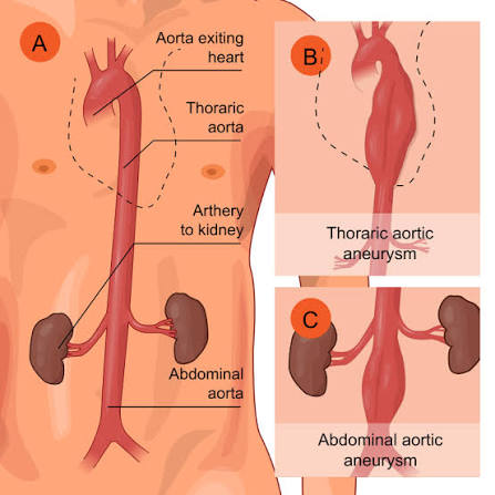
AAAs occur almost exclusively as a direct consequence of severe atherosclerosis. Atherosclerotic plaques thicken the intima so much that nutrients and oxygen cannot diffuse from the aortic lumen into the tunica media. The underlying media starves, undergoes ischemic necrosis, loses its elastic fibers, and balloons outward.
- Epidemiology & Risk Factors: Atherosclerosis is the greatest risk factor. However, because < 5% of men over 60 develop AAAs despite having universal atherosclerosis, genetic and environmental factors are also at play. AAAs occur far more frequently in men, in smokers, and rarely develop before age 50.
- Morphology & Location: The classic AAA is located in the infrarenal aorta (between the renal arteries and the bifurcation of the common iliac arteries). Why? This segment of the aorta lacks its own extensive vasa vasorum blood supply, making it highly susceptible to ischemic damage. They are usually fusiform or saccular, measuring > 3 cm (often > 5.5 cm) in diameter and up to 25 cm in length.
- Internal Appearance: The wall is destroyed and severely thinned. Because the dilated sac creates stagnant blood flow, the aneurysm frequently contains a bland, poorly organized, laminated mural thrombus.
- Clinical Features: Most cases are completely asymptomatic "silent time bombs" discovered incidentally as a pulsating abdominal mass during a physical exam or ultrasound.
- Rupture: The most feared complication. Rupture into the peritoneal cavity or retroperitoneal tissues causes massive, instantly fatal hemorrhage. (Risk of rupture directly correlates with size; > 5.5 cm warrants surgical repair).
- Obstruction: Can occlude vessel ostia (renal, superior/inferior mesenteric arteries) resulting in downstream tissue ischemic injury.
- Embolism: Pieces of the atheroma or the massive mural thrombus can break off and embolize to the legs ("trash foot").
- Impingement: The massive sac can compress adjacent structures, such as compressing a ureter (causing hydronephrosis) or eroding into the lumbar vertebrae (causing severe back pain).
2. Thoracic Aortic Aneurysm (TAA)
Unlike AAAs, Thoracic Aortic Aneurysms are most commonly associated with Hypertension. Other major causes include connective tissue disorders (Marfan syndrome, Loeys-Dietz syndrome) and inflammatory conditions (Tertiary Syphilis - syphilitic aortitis).
Because the thorax is a tight anatomical space, TAAs produce specific compressive symptoms before they dissect or rupture:
- Chest Pain: From the massive aneurysm encroaching on or eroding into the ribs/vertebral bone.
- Myocardial Ischemia: Aneurysmal dilation at the aortic root can stretch and compress the coronary artery ostia, cutting off blood to the heart. Aortic valve insufficiency/regurgitation is also common due to root dilation.
- Difficulty Swallowing (Dysphagia): Due to physical compression of the esophagus.
- Hoarseness: Due to stretching, irritation, or pressure on the left recurrent laryngeal nerve as it loops under the aortic arch (Clinically known as Ortner's Syndrome).
- Respiratory Complications: Chronic cough or dyspnea from compression of the trachea or primary bronchi.
IV. Aortic Dissection
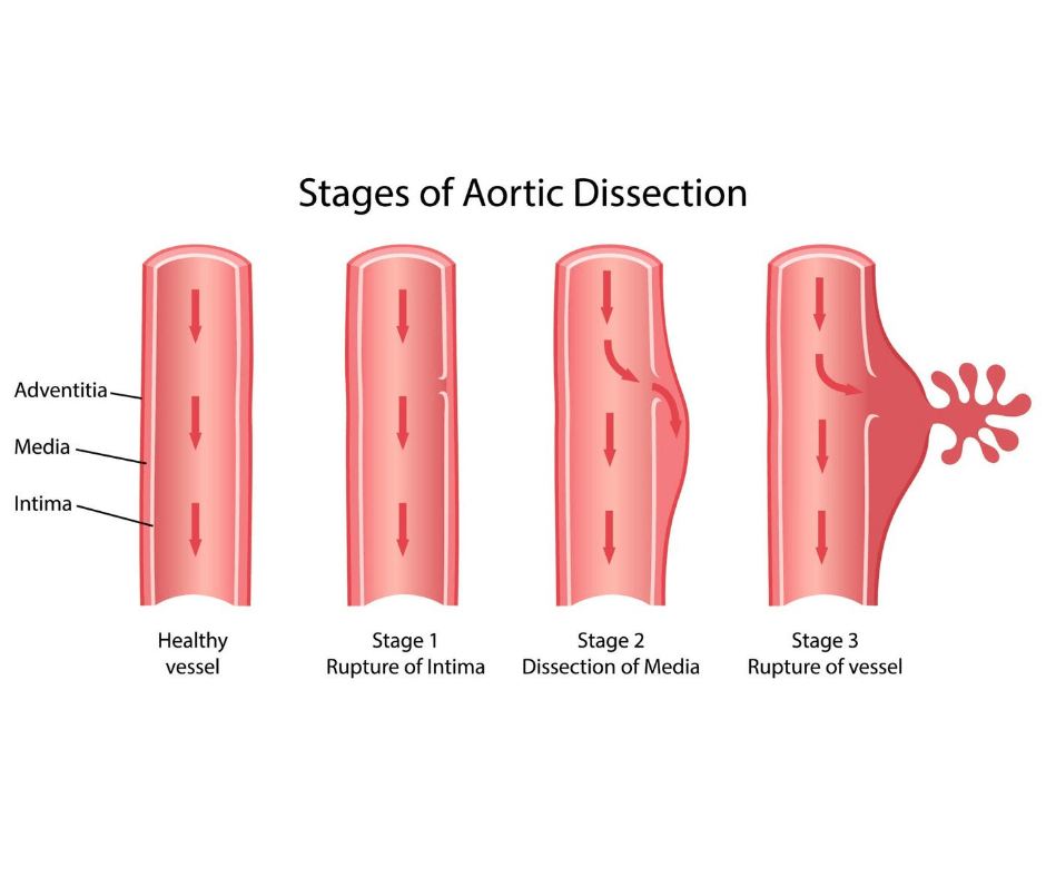
A dissection is vastly different from an aneurysm, though they can occur together. Dissection arises when blood forcefully enters a defect/tear in the arterial wall and tunnels (cleaves) through the laminar planes of the tunica media or between the media and adventitia, creating a blood-filled channel within the aortic wall itself.
Epidemiology & Pathogenesis
Aortic dissection occurs principally in two distinct demographic groups:
- Men aged 40 to 60 years: Almost exclusively associated with antecedent, poorly controlled Hypertension. The high pressure causes medial degenerative changes (SMC loss and altered ECM content) over decades.
- Younger Patients: Those with syndromic connective tissue diseases affecting the aorta, such as Marfan syndrome or Ehlers-Danlos, where the wall is genetically defective.
Other Causes: Can be iatrogenic (physician-caused) following arterial cannulation during diagnostic catheterization or cardiopulmonary bypass. It can also famously occur during the third trimester of pregnancy (due to pregnancy-induced hypertension and hormone-induced connective tissue remodeling).
The Trigger: Regardless of the etiology (HTN or Marfan's), the ultimate trigger for the initial intimal tear is often unknown. However, once the tear occurs, systemic blood pressure forces blood into the tear, violently dissecting the media. Therefore, aggressive pressure-reducing therapy (e.g., IV Beta-blockers) is highly effective in limiting an evolving dissection!
Morphology of Dissection
- The Tear: Initiates with an intimal tear. In the vast majority of spontaneous cases, the tear occurs in the ascending aorta (usually within 10 cm of the aortic valve). The tears are typically transverse, with sharp, jagged edges, measuring 1 to 5 cm in length.
- Propagation: The dissecting hematoma spreads between the lamellar units of the outer third of the media. It can extend retrograde (backward toward the heart) or distally (down into the abdomen, sometimes as far as the iliac and femoral arteries).
- Double-Barreled Aorta: Occasionally, the dissecting hematoma tunnels down and ruptures back into the true aortic lumen through a second, distal intimal tear. This creates a false vascular channel that averts fatal hemorrhage. Over time, this false channel can endothelialize, becoming a chronic dissection.
- Traumatic Tears: Severe deceleration injuries (e.g., motor vehicle accidents) or extremely vigorous CPR can cause intimal tears. These distinctly originate just distal to the great vessels at the ligamentum arteriosum. The connective tissue tethers the aorta here; sudden extreme movement of the heart shears the vessel at this fixed point.
Clinical Features & Classification
The morbidity and mortality of a dissection depend entirely on where the tear is. The most catastrophic complications occur with dissections between the aortic valve and the distal arch.
| Classification | Definition | Clinical Severity & Treatment |
|---|---|---|
| Type A Dissections (Stanford Type A / DeBakey I & II) | Proximal lesions. Involves the ascending aorta (either just the ascending, or both ascending and descending). | Extremely dangerous. High risk of retrograde dissection into the pericardium. Requires immediate surgical plication and intensive antihypertensive therapy (saves 65-85% of patients). |
| Type B Dissections (Stanford Type B / DeBakey III) | Distal lesions. Involves the descending aorta only, usually beginning distal to the takeoff of the left subclavian artery. | Less acutely lethal. Often managed medically with aggressive blood pressure control (beta-blockers, vasodilators) unless complications arise. |
The classic clinical symptom is the sudden onset of excruciating, "tearing" pain, usually beginning in the anterior chest, radiating directly to the back between the scapulae, and moving downward as the dissection violently progresses. This is frequently misdiagnosed as a massive Myocardial Infarction.
Cause of Death: The dissecting blood eventually ruptures through the thin adventitia. If it ruptures backward, it floods the pericardial sac, causing fatal Cardiac Tamponade (the heart is crushed by blood and cannot beat). It can also rupture into the pleural or peritoneal cavities.
V. Venous Disorders
Thrombophlebitis and Phlebothrombosis (DVT)
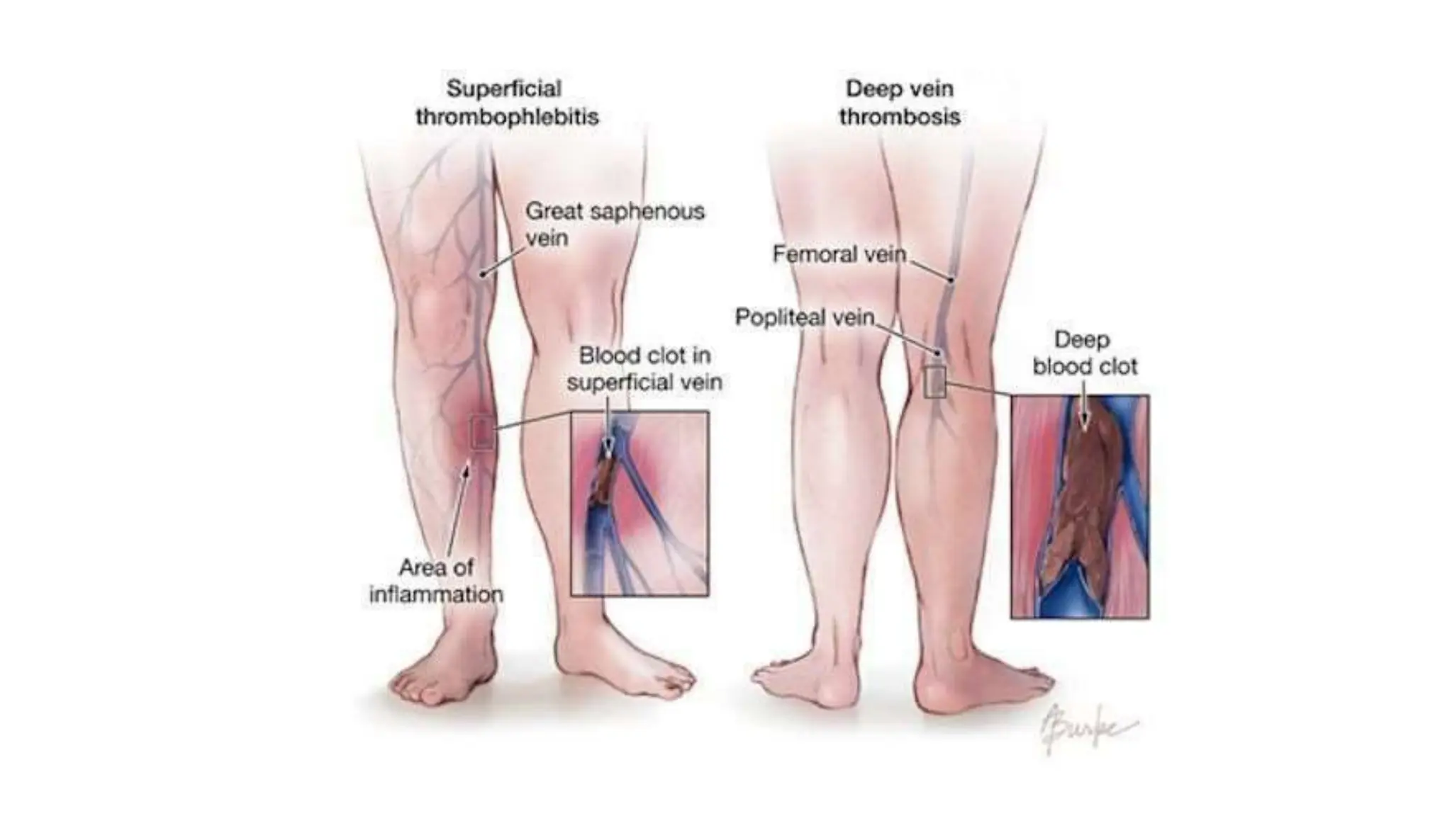
These terms refer to venous thrombosis (clotting) coupled with inflammation of the vein wall. Over 90% of cases occur in the deep veins of the legs (Deep Vein Thrombosis - DVT).
Special Note: Portal vein thrombosis can occur secondary to peritoneal infections (appendicitis, peritonitis) or thrombophilic conditions associated with platelet hyperactivity.
Thrombosis is driven by three risk factors:
- Stasis of blood flow: Prolonged immobilization / bed rest, Congestive Heart Failure (sluggish venous return), Obesity.
- Hypercoagulability: Pregnancy, Oral Contraceptive Pill (OCP) use, systemic Malignancy (Trousseau syndrome), and genetic hypercoagulability syndromes (Factor V Leiden).
- Endothelial injury: Trauma or surgery.
Symptoms are often highly unreliable. DVT can present with:
- Vein dilation, distal edema, cyanosis, heat, and erythema.
- Homan Sign: Pain elicited by pressure over the affected veins, squeezing the calf muscles, or forced dorsiflexion of the foot.
Warning: Pain and physical signs may be entirely absent. The absence of these findings absolutely does not exclude a diagnosis of DVT. The main danger is embolization to the lungs (Pulmonary Embolism).
VI. Lymphatic Disorders
1. Lymphangitis
Lymphangitis represents the acute inflammation of the lymphatic channels, almost always caused by the spread of severe bacterial infections (most commonly Group A beta-hemolytic Streptococcus).
- Pathology: The affected lymphatics become acutely dilated and filled with an intense exudate of neutrophils and monocytes. This aggressive infiltrate can burst through the vessel wall, producing widespread cellulitis or focal tissue abscesses.
- Clinical Presentation: Highly characteristic red, painful, subcutaneous streaks tracking up the arm or leg, tracing the path of the inflamed lymphatic vessels, accompanied by painful enlargement of the draining regional lymph nodes (lymphadenitis).
2. Lymphedema
Lymphedema is the accumulation of interstitial fluid due to compromised lymphatic drainage. It is categorized into two types:
- Primary Lymphedema: Occurs as an isolated congenital defect.
- Simple congenital lymphedema.
- Familial Milroy Disease: A heredofamilial congenital lymphedema resulting in complete lymphatic agenesis (failure to form) or severe hypoplasia.
- Secondary (Obstructive) Lymphedema: Stems from the physical blockage or destruction of previously normal lymphatics. Common causes include:
- Malignancy: Tumors directly obstructing lymphatic channels or invading regional lymph nodes.
- Surgical Procedures: Severing lymphatic connections (e.g., axillary lymph node dissection and resection during a radical mastectomy for breast cancer, causing massive arm edema).
- Post-radiation Fibrosis: Radiation therapy burns and scars the lymphatic channels.
- Filariasis: A parasitic infection (e.g., Wuchereria bancrofti) where the worms physically block the inguinal lymphatics, causing massive "Elephantiasis."
- Post-inflammatory thrombosis and scarring.
References & Further Reading
- Kumar, V., Abbas, A. K., & Aster, J. C. (2021). Robbins & Cotran Pathologic Basis of Disease (10th ed.). Elsevier. (Definitive pathology text on medial degeneration, Aneurysms, and Dissections).
- Dietz, H. C. (2010). New therapeutic approaches to mendelian aneurysmal disease. Cardiology in Review. (Deep dive into TGF-β signaling in Marfan and Loeys-Dietz syndromes).
- Hiratzka, L. F., et al. (2010). ACCF/AHA/AATS/ACR/ASA/SCA/SCAI/SIR/STS/SVM Guidelines for the diagnosis and management of patients with thoracic aortic disease. Circulation. (Clinical guidelines on Stanford A/B dissections).