
Pathology of Arteriosclerosis & Atherosclerosis
I. Arteriosclerosis: The Umbrella Term
Arteriosclerosis literally means "hardening of the arteries." It is a generic, umbrella term for arterial wall thickening and loss of elasticity. Pathology recognizes four distinct general patterns of arteriosclerosis, each with entirely different clinical and pathologic consequences:
- Arteriolosclerosis: Affects small arteries and arterioles. It has two anatomic variants: Hyaline and Hyperplastic, both deeply related to systemic hypertension. It causes downstream ischemic injury (e.g., nephrosclerosis).
- Mönckeberg Medial Sclerosis: Characterized by calcifications specifically in the media (middle muscular wall) of muscular arteries, typically starting along the internal elastic membrane. It most frequently affects adults older than 50 years. Crucial Distinction: These calcifications do not encroach on the vessel lumen. Therefore, it does not restrict blood flow and is usually clinically silent.
- Fibromuscular Intimal Hyperplasia: Occurs in muscular arteries larger than arterioles. It is driven by inflammation or mechanical injury (such as stent placement or balloon angioplasty) and can be considered a healing process, though the resultant scar tissue can cause stenosis.
- Atherosclerosis: The most frequent and clinically devastating pattern, which is the focus of the remainder of this masterclass.
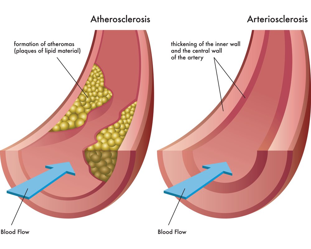
II. Atherosclerosis: Introduction & Risk Factors
Atherosclerosis comes from the Greek root words for "gruel" (athero) and "hardening" (sclerosis). It is the primary underlying pathogenesis of coronary, cerebral, and peripheral vascular disease. It causes more morbidity and mortality in the western world than any other disorder.
The likelihood of developing atherosclerosis is determined by a combination of acquired, inherited, and gender-age-associated risk factors. Acting in concert, they cause intimal lesions called atheromas (atherosclerotic plaques).
- Non-modifiable (Constitutional): Genetic abnormalities, a strong family history, increasing age (40-60 years), and Male gender (estrogen is protective in pre-menopausal females).
- Modifiable: Hyperlipidemia (High LDL), Hypertension, Cigarette smoking, Diabetes Mellitus, and Chronic Inflammation.
III. Pathogenesis: The Response to Injury Hypothesis
Modern pathology views atherosclerosis not merely as "fat clogging a pipe," but as a chronic inflammatory and healing response of the arterial wall to endothelial injury. Lesion progression occurs through the complex interaction of modified lipoproteins, macrophages, T lymphocytes, Endothelial Cells (Ecs), and Smooth Muscle Cells (SMCs) of the arterial wall.
1. The Healthy Endothelium vs. Endothelial Dysfunction
A healthy endothelium produces Prostacyclin (PGI2) and Nitric Oxide (NO), which maintain an anti-coagulant, anti-thrombotic surface and act as vasodilators.
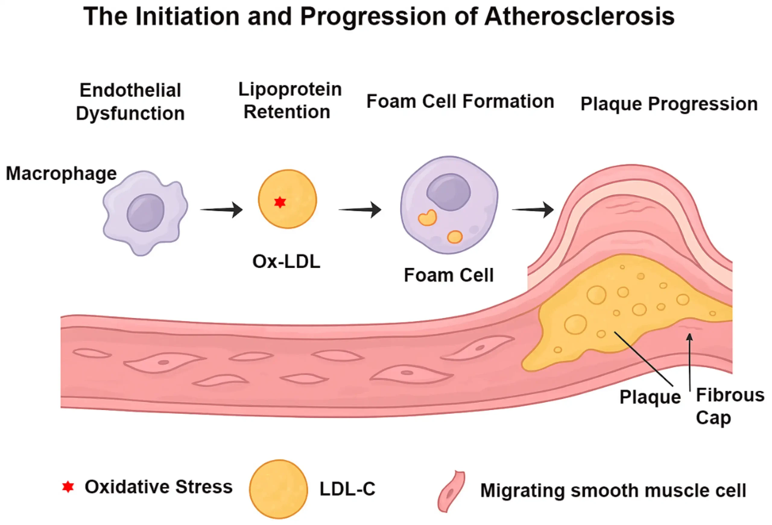
Endothelial Dysfunction is the very first step in atherosclerosis. The endothelium is injured, leading to impaired endothelial-dependent vasodilation, increased vascular permeability, and leukocyte adhesion. The endothelium shifts to a pro-coagulant surface, secreting pro-inflammatory molecules like MCP-1 (Monocyte Chemoattractant Protein-1) and adhesion molecules like VCAM-1. This dysfunction is caused by:
Plaques do not form randomly; they form at the ostia of exiting vessels, branch points, and the posterior abdominal aorta. Why? Because flow here is disturbed and turbulent.
- Laminar Flow increases the production of transcription factors, specifically Krüppel-like factor-2 (KLF2), which turns ON atheroprotective genes (like NO) and turns OFF inflammatory genes.
- Turbulent Flow drives a repertoire of genetic transcription that makes those specific sites atheroprone.
Dyslipoproteinemias include increased LDL cholesterol, decreased HDL cholesterol, and increased levels of abnormal Lp(a).
Causes include genetic mutations in apoproteins or lipoprotein receptors, or secondary disorders like diabetes mellitus, nephrotic syndrome, alcoholism, and hypothyroidism.
Chronic inflammation triggers and drives progression. It is believed that the accumulation of cholesterol crystals and free fatty acids in macrophages triggers cytosolic innate immune receptors that form the Inflammasome.
The inflammasome responds by producing massive amounts of Interleukin-1 (IL-1), an incredibly potent inflammatory cytokine.
2. The Sequence of Plaque Formation
Once the endothelium is injured, a deadly, sequential cascade begins:
- Endothelial Injury & Dysfunction: Causes increased vascular permeability, leukocyte adhesion, and thrombosis.
- Accumulation of Lipoproteins: LDL cholesterol infiltrates the subendothelial space (the intima) of the vessel wall.
- Oxidation of LDL (oxLDL): Free radicals (oxidants) attack and damage the target molecules. Specifically, Polyunsaturated Fatty Acids (PUFAs) in the LDL are oxidized, triggering the oxidation of the apoB100 protein. The LDL becomes highly toxic.
- Monocyte Adhesion & Migration: Monocytes stick to the VCAM-1 on the endothelium, migrate into the intima, and transform into macrophages.
- Foam Cell Formation: The macrophages attempt to clean up the toxic oxLDL by engulfing it. They become bloated with fat and die, becoming Foam Cells.
- Platelet Adhesion & SMC Recruitment: Activated platelets, macrophages, and vascular wall cells release growth factors. This induces Smooth Muscle Cell (SMC) recruitment from the media into the intima.
- SMC Proliferation & Matrix Production: The SMCs proliferate and produce massive amounts of Extracellular Matrix (ECM), including collagen, to create a fibrous cap over the fat.
- Lipid Accumulation & Calcification: Extracellular lipids pool from dead macrophages to form a necrotic core. Late in the pathogenesis, the ECM and necrotic debris undergo dystrophic calcification.
IV. Morphology of Atherosclerosis
1. The Fatty Streak (The Earliest Lesion)
The fatty streak is composed primarily of lipid-filled foamy macrophages. Beginning as small, flat yellow macules, these can eventually coalesce into elongated streaks 1cm long or longer. Crucially: The lesions are not particularly raised and do not cause any significant flow disturbance. (They are present in the aortas of almost all children).
2. The Atherosclerotic Plaque (The Advanced Lesion)
Atheromatous plaques are yellow-tan and are raised above the surrounding vessel wall. If a thrombus superimposes over an ulcerated plaque, it will be red-brown. Plaques vary in size but can coalesce to form larger masses.
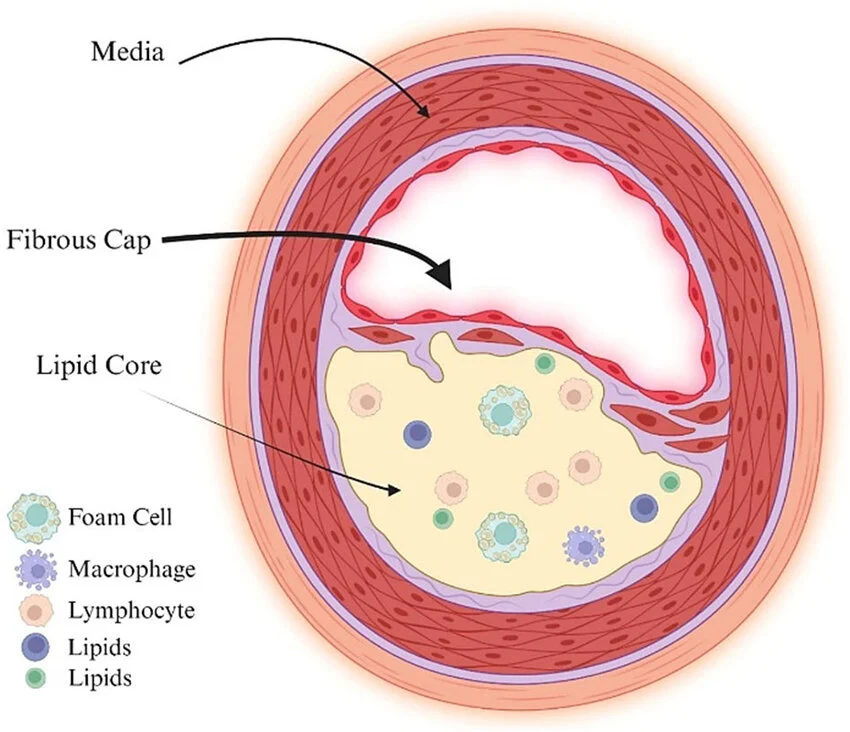
- Focality & Eccentricity: Atherosclerotic lesions are patchy and rarely circumferential. They usually involve only a portion of any given arterial wall; on cross-section, the lesions therefore appear eccentric. This focality—despite the uniform exposure of vessel walls to toxins, LDL, and hyperglycemia—is entirely attributable to the vagaries of vascular hemodynamics (turbulent flow).
- Vessel Distribution: In descending order, the most extensively involved vessels are: The lower abdominal aorta → Iliac arteries → Coronary arteries → Popliteal arteries → Internal carotid arteries → Vessels of the circle of Willis.
The 4 Principal Components of a Plaque:
- Cells: Variable numbers of Smooth Muscle Cells, macrophages, and T lymphocytes.
- Extracellular Matrix (ECM): Collagen, elastic fibers, and proteoglycans (forming the fibrous cap).
- Lipids: Intracellular (within foam cells) and Extracellular (forming the necrotic core).
- Calcifications: Present in later-stage plaques.
V. Plaque Changes & Clinical Consequences
A plaque may sit silently for decades. It becomes lethal when it undergoes acute morphological changes:
- Rupture, Ulceration, or Erosion: The surface of the atheromatous plaque degrades. This exposes the bloodstream to highly thrombogenic substances inside the necrotic core. This instantly leads to thrombosis, which can partially or completely occlude the vessel lumen, causing sudden cardiac death or myocardial infarction.
- Hemorrhage into a Plaque: Rupture of the overlying fibrous cap, or rupture of the fragile, thin-walled neovascular microvessels within the plaque itself, can cause intraplaque hemorrhage. The contained hematoma rapidly expands the size of the plaque, potentially occluding the artery or inducing plaque rupture.
- Atheroembolism: Plaque rupture can discharge actual atherosclerotic debris (cholesterol crystals) into the bloodstream, producing microemboli that lodge in distal capillary beds (e.g., causing "blue toe syndrome" or stroke).
- Aneurysm Formation: The massive plaque creates pressure and induces ischemic atrophy of the underlying media (the muscular layer of the artery that provides structural support). With the loss of elastic tissue, the vessel wall weakens immensely, dilates, and faces potential fatal rupture (e.g., Abdominal Aortic Aneurysm).
References & Further Reading
- Kumar, V., Abbas, A. K., & Aster, J. C. (2021). Robbins & Cotran Pathologic Basis of Disease (10th ed.). Elsevier. (Vascular Pathology and the Response to Injury Hypothesis).
- Ross, R. (1999). Atherosclerosis—an inflammatory disease. New England Journal of Medicine, 340(2), 115-126.