
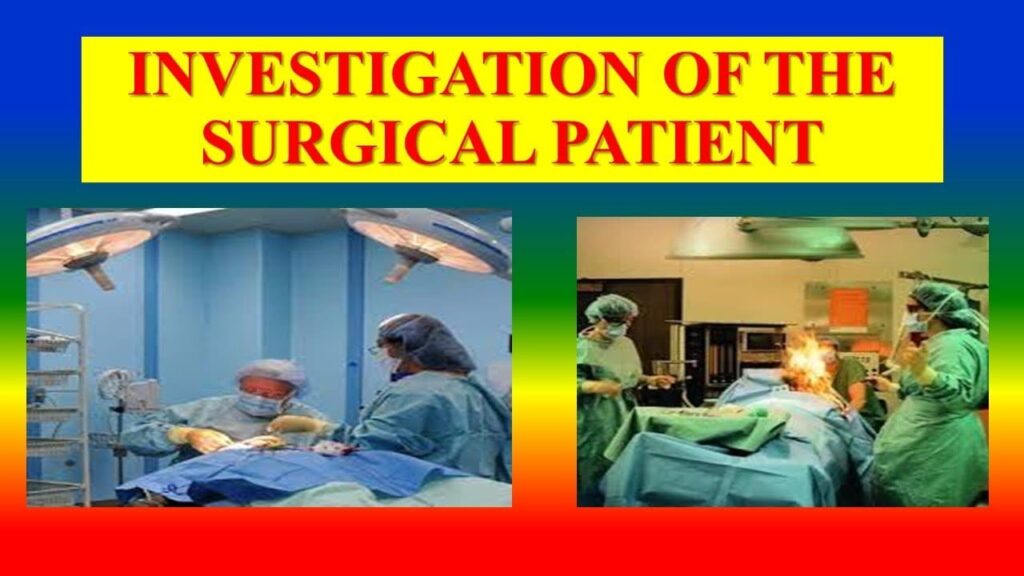
Topic 3.7: Aseptic Technique & Special Investigations
Sub-topic 3.7.3: Aseptic Technique / Surgical Asepsis
Introduction to Surgical Asepsis
- It is defined as the absence of micro-organisms that can cause disease.
- Surgical asepsis promotes tissue healing by determining pathogens from coming into contact with the surgical wound.
- Practices that suppress, reduce and inhibit injection processes are known as aseptic technique.
- Surgical asepsis prevents contamination of surgical wounds.
- All members of the operating theatre (OR) team are responsible for strict adherence to aseptic techniques.
- It is essential that OR nurses acquire a surgical conscience – vigilant adherence to aseptic technique throughout the entire peri-operative period.
Operating Theatre Environment and Asepsis
The purpose of maintaining asepsis in the operating theatre is paramount. The theatre environment should have the following:
- Air conditioned ventilation.
- Charnel enclosure for orthopedic work.
- Easily cleanable fabric.
- A one way traffic circulation from clean area to dirty area.
- Adequate shower facilities for medical staff after finishing a day’s operation.
Basic Rules of Surgical Asepsis in the OR
-
Scrubbed persons function within sterile field
Scrubbed personnel wear gloves and gowns at the surgical field. The gown of scrubbed team member is considered sterile in front, from the chest to the level of the sterile field and the sleeves are sterile front two inches above the elbow to the stockinette cuff. The non-sterile areas of the gowns include; stockinet cuff, neckline, shoulder, axillary region and back. Dressing in OR attire proceeds from head to toe.
-
Sterile drapes are used to create a sterile field
Sterile drapes are placed on the patient equipment and furniture used within the sterile field. Draped tables are sterile only at the table level; items extending over the table edge are contaminated. Handling of the drapes should be minimized.
-
All items used in the sterile field are sterile
If the sterility of an item is questioned, it must be considered unsterile. Packaging materials must guarantee that items will remain sterile until removed.
-
Supplies introduced into the sterile field
Are delivered in a manner that ensures the sterility of the item and maintains the integrity of the sterile field. The nurse opens a sterile package from the far side first and near side last and holds the wrapper tails when the item is presented to the sterile field. The nurse pours solutions carefully to avoid splashing liquids on to the field. After opening a bottle of a sterile solution, the nurse must present the entire contents to the sterile field or discard it.
-
Maintenance and monitoring of sterile field
The possibility for contamination increases with time, therefore the sterile field should be established as close to the time of use as possible. Un-attended sterile field is considered contaminated.
-
The integrity of the sterile field must be maintained by individuals moving within or around the sterile field
Only scrubbed personnel touch and reach over sterile areas. Sterile persons remain close to the sterile field and never turn their backs to it. Sterile individuals change positions by passing back to back or face to face. Un-scrubbed personnel only touch and reach over non-sterile areas, do not walk between sterile fields and approach sterile fields by facing them.
SURGICAL ASEPSIS
- It is defined as the absence of micro-organisms that can cause disease.
- Surgical asepsis promotes tissue healing by determining pathogens from coming into contact with the surgical wound.
- Practices that suppress, reduce and inhibit injection processes are known as aseptic technique.
- Surgical asepsis prevents contamination of surgical wounds.
- All members of the operating theatre (OR) team are responsible for strict adherence to aseptic techniques.
- It is essential that OR nurses acquire a surgical conscience – vigilant adherence to aseptic technique throughout the entire peri-operative period.
The purpose of maintaining asepsis, operating theatre. They should have the following;
- Air conditioned ventilation.
- Charnel enclosure for orthopedic work.
- Easily cleanable fabric.
- A one way traffic circulation from clean area to dirty area.
- Adequate shower facilities for medical staff after finishing a day’s operation.
Client Preparation for Surgery
Although much preparation have taken place prior to clients transfer to the surgical department additional activities such as shaving and positioning may be performed.
Skin preparation
The goal of skin preparation is to reduce the risk of post-operative wound infection by;
- Removing transient microbes from the skin
- Reducing the resident microbes count to sub-pathogenic amounts
- Inhibiting rapid rebound growth of microbes
The skin is prepared by mechanically scrubbing or cleaning around the surgical site with anti-microbial agents. If the patient is very hairy or if the hair will interfere with the surgical procedure, the nurse removes it; usually either wet shaving, clippers or use of depilatory agent. The area is then scrubbed in a circular motion. The principal of scrubbing from the clean area (site of incision) to the dirty area (periphery) is observed at all times. A liberal area is cleansed to allow added protection and unexpected occurrences during the procedure. After preparation of the skin, the sterile members of the surgical team drape the area. Only the site to be incised is left exposed.
Positioning the patient
- It is a critical part of every procedure and usually follows administration of the anesthesia.
- Anesthetist will indicate when to begin the positioning.
- The circulating nurse ensures optimal positioning and continually assess the client.
- The position of the patient should allow accessibility to the operative site, administration and monitoring of anesthetic agents and maintenance of the patient’s airway.
- Improper positioning would potentially result into muscle strain, joint damage and other unwanted effects.
- It is a nurse’s responsibility to secure the extremities provide adequate padding and support and obtain sufficient physical or mechanical help to avoid unnecessary straining of self or patient frequently.
Positions used frequently include;
- The surprise position: it is used for many abdominal surgeries, thoracic surgeries and some surgeries on the extremities
- The semi-sitting up position: it is used for surgeries on the thyroid and neck areas
- The prone position: it is used for spinal fusions and removal of hemorrhoids
- The lateral chest position: it is used for gynecological, perinea or rectal surgeries
- The jackknife: it is used for proctologic and some spinal surgeries
- The Trendelenburg position: it is used for examinations and for performing abdominal surgeries
- Lateral position: it is used for surgeries of the anal area
NB: see positions in medical surgical nursing (patient centered collaborative care 8th edition)
Anesthesia
The term anesthesia is derived from the word anesthesis meaning “no sensation” therefore anesthesia is limited or total loss of feeling (normal sensation) with or without loss of consciousness. There are two broad classifications of anesthesia; general and local anesthesia.
General Anesthesia
Involves unconsciousness, complete insensitivity to pain, amnesia, motionless and muscle relaxation. It involves four overlapping stages i.e. induction (going to sleep), maintenance, emergence (waking up) and recovery.
- Induction time period starting with pre-operative medication, initiation of appropriate IV access, application of monitors, initiation of sequence of medication that render the patient unconscious, securing airway, drugs used include; benzodiazepines, narcotics, hypnotics and volatile gases.
- Maintenance-time period during which the surgical procedures is performed, patient remains in an unconscious state with appropriate measures to ensure safety of the airway. Drugs are the same as above.
- Emergence-time – it is a period during which the surgical procedure is completed. Patient is prepared for return to return to consciousness and removal of airway assist devices. Drugs used; reversal agents – anticholinergic, sympathometics, narcotic, antagonists, benzodiazepines antagonist.
- Recovery-time / period during which the patient regains his/her clear thinking ability. This often takes longer with some residual thinking difficulty persisting for several days or even weeks. Many anesthetic drugs are metabolized slowly. The speed of metabolism depends on amount of drug given, the length of surgery and how deeply the patient is breathing.
Local Anesthesia
Allows operative procedures to be performed on a particular part of the body without loss of consciousness or sedation. The duration of action of the local anesthetic frequently carries into the post-operative period providing continued analgesia.
The disadvantages
- Inadvertent IV administration producing hypotension and potential seizures
- Inability to precisely match the duration of action of the agents administered to the duration of surgical procedure
- Technique difficulty and discomfort that may be associated with infections
Methods of administration
- Topical application – application of the agent directly to the skin, mucous membranes or open surface
- Local infiltration – injection of the agent into the tissues through which the surgical incision will pass
- Regional nerve block – injection of the agent into or around a specific nerve or group of nerves. Examples of spinal anesthesia (injection of the agent into CSF found in the subarachnoid space, usually below L2 ) and epidural black (injection of agent epidural space via either a thoracic or lumber approach)
Conscious sedation
A minimally depressed level of consciousness with maintenance of patient’s protective airway, reflexes. Its primary goal is to reduce the patient’s anxiety and discomfort and to facilitate cooperation. Often a combination of sedative. The anesthetist determines the choice and method of administering the anesthesia according to;
- Patient’s preferences, age, physical status and emotional status
- Type and length of the surgical procedure
- Patient’s positioning during surgery
- Co-existing disease
NB: operating theatre nurses do not administer anesthetic agents but they must understand the various anesthetics used in surgery and the potential side effects and complications (check pharmacology). This knowledge enables the nurse to plan intra-operative nursing care to assist the anesthesia team.
Sub-topic 3.7.4: Special Investigations in Surgical Nursing
Special investigations are diagnostic procedures used to confirm or rule out a surgical condition, determine the extent of disease, and plan for surgery. The nurse plays a vital role in patient preparation, education, and post-procedure care.
X-ray & Contrast Studies
The X-ray has been called one of the most significant advances in medical history. Routine X-rays involve exposing a body part to a small dose of radiation to produce an image of an internal organ. It is a fast and easy procedure. Patients will experience no discomfort or side effects from their examination and are allowed to leave immediately following their X-ray test.
General Preparation of Patients for X-rays
- Explain to the patient what is going to happen. This is especially necessary for x-rays which are done in a darkened room e.g. barium meal.
- Remove jewellery e.g. necklaces for a chest X-rays.
- Take the patient to the X-ray room, in a chair, or on a stretcher, or walking as ordered by the doctor, and bring with you the patient’s chart and previous x-rays, if any.
- On arrival, remove the patient’s clothing and put on an X-rays gown.
Contrast Studies
- Esophagram (Barium Swallow): An examination of the pharynx and oesophagus using still and fluoroscopic X-ray images, after the patient drinks a barium solution.
- Upper GI Series: A series of X-rays of the oesophagus, stomach, and small intestine taken after the patient drinks a barium solution.
- Small Bowel or Small Intestine Series: A series of X-rays of the part of the digestive tract that extends from the stomach to the large intestine.
- Barium Enema / Lower GI Series: A series of X-rays of the lower intestine (colon) and rectum taken after the patient is given an enema with a barium solution.
- Intravenous Pyelogram (IVP): An X-ray examination of the kidneys, their drainage to the bladder, and the bladder, using an injected contrast dye.
- Hysterosalpingogram: X-ray of the uterus and Fallopian tubes; usually done in diagnosing infertility.
- Arthrogram: X-ray of a joint after the injection of a contrast medium.
Advanced Imaging Techniques
MRI (Magnetic Resonance Imaging)
MRI is a method of obtaining detailed pictures of internal body structures without the use of radiation. It uses a magnetic field and radio waves. The patient will hear a repeated drum-like knocking sound as the scans are recorded. High quality images require the patient to lie still.
How to Prepare For the MRI Exam
- Patient wears loose, comfortable clothing without metal snaps or zippers.
- Patient goes with a referral form from the doctor.
- If the patient is having an MRI of the abdomen performed, advise the patient not to eat or drink anything after midnight the night before your procedure.
CT (Computed Tomography)
CT scanning is a rapid, painless diagnostic examination that combines X-rays and computers to see the location, nature, and extent of many different diseases or abnormalities.
HOW to Prepare For the CT Exam
- The meal prior to your CT examination should consist of CLEAR liquids ONLY.
- If oral contrast (barium drink) is required, specific instructions on when to drink it will be given (e.g., TWO HOURS BEFORE and ONE HOUR BEFORE the appointment).
Nuclear Imaging
This provides information about both structure and function by using safe and painless techniques to image the body and treat disease. It involves introducing a small amount of a radioactive chemical (radionuclide or radiotracer) into the body.
- PET/CT: Combines Positron Emission Tomography (PET) with CT to identify areas of abnormal metabolic activity, often used in cancer diagnosis and staging.
- SPECT/CT: Combines Single-Photon Emission Computed Tomography (SPECT) with CT for similar purposes.
Common Nuclear Scans
- Bone Scan: A radionuclide collects in areas of high bone activity (fractures, infection, cancer), seen as 'hot spots'.
- Cardiac Scan: Assesses blood flow to the heart muscle.
- Renal Scan, Hepatobiliary Scan, etc.
Preparation for Nuclear Medicine Exams
Preparation varies. Some scans require no prep (Bone Scan), while others require fasting (Cardiac Scan, PET/CT). Patients must inform staff if they are diabetic or pregnant.
Endoscopy
Endoscopy means looking inside and typically refers to looking inside the body for medical reasons using an endoscope, an instrument used to examine the interior of a hollow organ or cavity of the body. Unlike most other medical imaging devices, endoscopes are inserted directly into the organ.
Components of an Endoscope:
- A rigid or flexible tube
- A light delivery system
- A lens system
- An eyepiece
- An additional channel to allow entry of medical instruments or manipulators
Uses (Examples by Body System):
- GI Tract: Esophagogastroduodenoscopy (EGD), Colonoscopy, ERCP.
- Respiratory Tract: Rhinoscopy, Bronchoscopy.
- Urinary Tract: Cystoscopy.
- Joints: Arthroscopy.
Preparation and Risks
Preparation usually involves fasting to ensure the organ is empty. Risks, though infrequent, include infection, perforation (a tear) of the organ lining, and bleeding.
Advanced Imaging Techniques
MRI (Magnetic Resonance Imaging)
Magnetic Resonance Imaging (MRI) is a method of obtaining detailed pictures of internal body structures without the use of radiation or radioactive substances of any kind.
This is accomplished by placing the patient in a magnetic field while radio waves are turned on and off.
This causes the body to emit its own weak radio signals which vary according to tissue characteristics.
These signals are then picked up by a sensitive antenna and fed to a computer which produces detailed images of the body for interpretation by trained radiologists.
During the examination the patient will not feel anything unusual. He/she will, however, hear a repeated drum-like knocking sound as the scans are recorded. The patient is free to bring a favourite CD or cassette tape to listen to during the scan to make her/himself comfortable. Hearing protection are provided to those patients who do not wish to listen to music.
To produce high quality images, the patient has to lie still during the examination while breathing normally. The average scan takes 5 to 15 minutes—the complete examination about 30 to 45 minutes—during which time several dozen images will be produced.
How to Prepare For the MRI Exam
- Patient wears loose, comfortable clothing without metal snaps or zippers.
- Patient goes with a referral form from the doctor.
- If the patient is having an MRI of the abdomen performed, advise the patient not to eat or drink anything after midnight the night before your procedure.
CT (Computed Tomography)
Computed Tomography (CT) scanning is a rapid, painless diagnostic examination that combines X-rays and computers.
A CT scan allows the radiologist to see the location, nature, and extent of many different diseases or abnormalities inside your body.
HOW to Prepare For the CT Exam
The meal prior to your CT examination should consist of CLEAR liquids ONLY. (You may have coffee/tea WITHOUT milk; broth; soda; and grape, cranberry or apple juice.)
If you are having an out patient, provide the barium drink to the patient to take home. The patient SHOULD NOT REFRIGERATE the barium drink.
TWO (2) HOURS BEFORE THE SCHEDULED APPOINTMENT
- The patient removes cap and drinks the liquid within 30 minutes to the first designated line on the container.
ONE (1) HOUR BEFORE THE SCHEDULED APPOINTMENT
- Drink the liquid within 30 minutes to the 2nd designated line on the container.
REMAINDER OF LIQUID
- THE patient brings the remainder of the liquid to the hospital.
- The patient will finish drinking the liquid when the study begins.
- Prescription medications may be taken as usual.
- EXCEPTION: Do not take Glucophage.
Nuclear Imaging
Nuclear Medicine provides doctors with information about both structure and function by using safe and painless techniques to image the body and treat disease. It is a superior way to gather medical information that would otherwise be unavailable or require surgery.
Nuclear Imaging now offers two of the most advanced nuclear imaging modalities for the early detection of disease: PET/CT and SPECT/CT.
PET/CT
PET/CT is a state-of-the-art technique that combines Positron Emission Tomography (PET) with Computed Tomography (CT) to image tissue and organ function. This scan is designed to accurately identify even small areas of abnormal metabolic activity, which are associated with several disease processes. PET/CT’s major clinical impact to date is in cancer diagnosis and staging; however, PET/CT is also a useful modality for imaging the heart and brain. PET/CT can show more than just where tumours are located. PET/CT can reveal whether lesions are benign or malignant and can assess the effectiveness of treatment, whether surgery, chemotherapy, or radiation therapy.
When the patient arrives at the Nuclear Imaging Suite, a technologist will discuss the PET/CT procedure with him/her and ask if s/he has any questions. When the patient is ready for the PET/CT scan, s/he will have the blood sugar tested. Next, most patients will receive an oral contrast (barium drink). An IV will then be started, and s/he will receive an injection of a small amount of safe, radioactive sugar (radiotracer). The patient will then be asked to wait very quietly in a seated area. Any activity, even talking or gum chewing, may affect the results of the test. Prior to the scan, the patient will be asked to empty his/her bladder.
The patient will lie on a bed that passes slowly through the scanner. For scanning purposes, it is important that the patient lies quietly and remain still on the bed during the scan. The length of time between scans can vary depending on the body areas being studied, typically between 30 to 60 minutes. The patient should plan to spend approximately three hours total time at the Nuclear Imaging Suite for the entire PET/CT procedure.
How to Prepare For the PET/CT Exam
- Refrain from eating for at least six hours prior to the exam since the results of the test are affected by the blood sugar level.
- It is important to be well hydrated for the test, so please make sure that the patient drinks plenty of water beginning the day before the exam up to the appointment time.
- Do not perform any heavy lifting or exercising the day before or the day of the PET/CT scan.
- If the patient is diabetic, please notify the technologist so that s/he may administer special instructions to you as necessary prior to the PET/CT scan.
- It is also recommended that the patient wears comfortable clothing.
SPECT/CT
SPECT/CT is an advanced medical imaging technology that combines Single-Photon Emission Computed Tomography (SPECT) with Computed Tomography (CT) to enable physicians to detect heart disease, cancer and other diseases earlier and target treatments with greater precision.
SPECT, like Positron Emission Tomography (PET), is a nuclear medicine exam that allows direct visualization of tissues, tumours and organs, such as the heart. SPECT/CT system allows physicians to obtain more detailed information and increased image clarity in a single, non-invasive procedure than is possible through separate procedures. The system detects changes in patients’ molecular activity – before structural changes become visible – and combines this information with precise anatomical detail obtained through CT technology to pinpoint the location of abnormal tissue.
When the patient arrives at the Nuclear Imaging Suite, a technologist will discuss the SPECT/CT procedure with him/her and ask ifs/he has any questions. Then a small amount of radiopharmaceuticals will be introduced into the body by injection, swallowing or inhalation. The radiopharmaceuticals are attracted to specific organs, bones or tissues. The amount of radiopharmaceuticals used for the patient’s exam will be carefully determined to provide the least amount of radiation exposure and to ensure an accurate test.
The scanner then creates images of the area being examined and identifies “hot spots” that indicate the location and extent of disease, such as the increased metabolic activity characteristic of cancer. The combination of high-resolution CT through the SPECT/CT allows physicians to accurately localize these hot spots and make a definitive diagnosis.
How to Prepare For the Nuclear Medicine Exam
- Bone Scan
- The patient may eat and drink prior to his/her scan.
- Please do not schedule an X-ray barium study on the same day as the patient’s Bone Scan.
- You may schedule a CT exam on the day of the patient’s Bone Scan.
- If the patient had a Barium Enema (BE) a day or two before the scheduled appointment time, an X-ray may be taken to make sure that the barium is all out of the system.
- Cardiac Scan
- Please do not eat or drink after midnight, the day before the Cardiac Scan.
- At the time of scheduling your exam, the patient will be told whether or not s/he will receive Persantine during the exam. If the patient will be receiving Persantine, let him/her not ingest caffeine for 24 hours prior to the exam.
- The doctor will advise the patient of which medications s/he may and may not take the morning of exam.
- Hepatobiliary: Please do not eat or drink after midnight, the day before the scan.
- Gastric Emptying: Please do not eat or drink after midnight, the day before your scan.
- Brain: There is no preparation for this exam. The doctor will advise the patient of which medications s/he may and may not take the morning of exam.
- Parathyroid: There is no preparation for this exam.
- Renal Scan: There is no preparation for this exam.
Ultrasound
Ultrasound uses sound waves to obtain a medical image or picture of various organs and tissues in the body. It is a painless and safe procedure. Ultrasound produces very precise images of the soft tissues (heart, blood vessels, uterus, bladder, etc.) and reveals internal motion such as heart beat and blood flow. It can detect diseased or damaged tissues, locate abnormal growths and identify a wide variety of changing conditions, which enable the doctor to make a quick and accurate diagnosis.
What will the exam be like?
A technologist will assist the patient onto the examination table. At this time, a water-based transmission gel will be applied to the area of the body that will be examined. A transducer will be moved slowly over the body part being imaged. The transducer sends a signal to an on-board computer which processes the data and produces the ultrasound image. It is from this image that the diagnosis is made.
The patient won’t feel a thing except for the slight pressure and movement of the transducer over the part of the body being imaged. It is important that the patient remains still and relaxed during the procedure. The ultrasound images will appear on a monitor similar to a TV screen and will be recorded either on paper or film for a detailed study.
How to prepare for The Ultrasound exam of the pelvis
Eat meals - DO NOT FAST! Drink 32 ounces of clear liquids (no soda) one hour and 15 minutes prior to the time of the appointment. (All of the liquid is to be in your system one hour before the appointment so that the bladder will be full.) DO NOT EMPTY the bladder until the study has been completed or the patient has spoken with a technologist.
How to prepare for The Ultrasound exam for pregnancy, kidneys, and bladder
- Eat meals - DO NOT FAST! Drink 20 ounces of water one hour and 15 minutes prior to the time of the appointment.
- Continue as above
How to Prepare For the Ultrasound Exam of the Abdomen
- Do not eat or drink anything after midnight the night before the procedure.
Bone Density (DEXA)
Bone Densitometry is a fast, safe and painless test that uses advanced technology called DEXA (Dual Energy X-Ray Absorptiometry) to measure symptoms of osteoporosis -- such as low density and mineral content of bone -- that may have developed unnoticed over many years. Because osteoporosis can result in bone fractures that can cause chronic pain, disability and loss of independence, it is important to begin treating osteoporosis at an early stage. Bone densitometry can detect the early signs of osteoporosis so that patients can begin treating it before a debilitating fracture occurs.
During a comprehensive DEXA bone evaluation, a patient lies comfortably on a padded table while the DEXA unit scans one or more areas of his/her body, usually the spine or hip because they are particularly prone to fracturing.
When the exam is complete, the patient’s images are sent to a computer and analyzed. They are then given to a radiologist, a physician who specializes in the diagnostic interpretation of medical images. After the study has been reviewed, the doctor will receive a report of the findings. This report will include patient’s bone mineral density (BMD), along with the FRAX (Fracture Risk Assessment Tool) results. The radiologist will use the FRAX assessment tool, developed by the World Health Organization, to obtain two results, expressed as percentages. These numbers are a ten-year probability of hip fracture and ten-year probability of a major osteoporotic fracture (clinical spine, forearm, hip or shoulder fracture).
Digital Mammography
A mammogram is a safe low-dose X-ray procedure that takes pictures of the internal tissues of the breasts. This simple exam is performed as a screening or diagnostic study, to determine the possibility of irregularities within the breast. It can reveal areas too small or deep to feel, which may or may not require further investigation. Digital Mammography is the most advanced diagnostic technology available for the early detection of breast cancer.
What are the benefits of Digital Mammography?
There are numerous benefits to digital mammography. For the patient, digital mammograms are faster. The test is "filmless," so nothing has to be developed. Images are read on a monitor and stored electronically in the PACS (Picture Archiving and Communications System). For the radiologist, digital mammograms provide more comprehensive visibility. Calcifications can be enhanced or magnified on the screen to aid the radiologist in interpreting whether or not the calcifications are suspicious. That is good news for younger women and those who have dense breasts. Digital mammography units are also able to accommodate women with larger breasts. This means fewer images and less radiation for these patients.
Radionuclide (Isotope) Scan
A radionuclide scan is a way of imaging bones, organs and other parts of the body by using a small dose of a radioactive chemical. A radionuclide (sometimes called a radioisotope or isotope) is a chemical which emits a type of radioactivity called gamma rays. A tiny amount of radionuclide is put into the body, usually by an injection into a vein. (Sometimes it is breathed in, or swallowed, depending on the test.)
Gamma rays are detected by a device called a gamma camera. The computer builds a picture by converting the differing intensities of radioactivity emitted into different colours or shades of grey. Areas of the target organ or tissue which emit lots of gamma rays may be shown as red spots ('hot spots'). Areas which emit low levels of gamma rays may be shown as blue ('cold spots').
Are there any risks with radioisotope scans?
The term 'radioactivity' may sound alarming. But, the radioactive chemicals used in radionuclide scans are considered to be safe, and they leave the body quickly in the urine. The dose of radiation that your body receives is very small. However:
- As with any other types of radiation (such as X-ray), there is a small risk that the gamma rays may affect an unborn child. So, tell your doctor if you are pregnant or if you may be pregnant.
- Rarely, some people have an allergic reaction to the injected chemical. Tell your doctor if you are allergic to iodine.
Endoscopy
Endoscopy means looking inside and typically refers to looking inside the body for medical reasons using an endoscope, an instrument used to examine the interior of a hollow organ or cavity of the body. Unlike most other medical imaging devices, endoscopes are inserted directly into the organ.
Risks
- Infection
- Punctured organs
- Over-sedation
The main risks are perforation, or a tear, of the stomach or oesophagus lining and bleeding. Although perforation generally requires surgery, certain cases may be treated with antibiotics and intravenous fluids. Bleeding may occur at the site of a biopsy or polyp removal. Seldom does surgery become necessary.
After the Endoscopy
After the procedure the patient will be observed and monitored by a qualified nurse in the endoscopy room or a recovery area until a significant portion of the medication has worn off. Occasionally the patient is left with a mild sore throat, which may respond to saline gargles, or chamomile tea. The patient may have a feeling of distention from the insufflate air that was used during the procedure. Both problems are mild and fleeting. When fully recovered, the patient will be instructed when to resume their usual diet.