Nurses Revision

Microbiology

Rickettsiae, Spotted Fevers & Typhus

Rickettsiae, Spotted Fevers & Typhus

Rickettsiae, Spotted Fevers, & Typhus

Module Learning Objectives

By the conclusion of this highly detailed master guide, you will be deeply conversant with:

  • The unique microbiological identity and evolutionary biology of the Rickettsiaceae family.
  • The profound pathophysiological mechanisms of endothelial infection, explaining the classic triad of fever, headache, and rash.
  • The exhaustive classification and geographic distribution of the Spotted Fever Group, Typhus Group, and related organisms (Ehrlichia/Anaplasma).
  • The definitive clinical timeline, deadly complications, and diagnostic pitfalls of Rocky Mountain Spotted Fever (RMSF).
  • The precise pharmacological interventions, including pediatric exceptions and alternative therapies, alongside vector control strategies.

I. Introduction to Rickettsiae & Historical Context

Rickettsiae represent a highly specialized, unique group of bacteria that conceptually bridge the evolutionary gap between classic, free-living bacteria and obligate intracellular viruses. Named after Dr. Howard Taylor Ricketts (who tragically died of typhus in 1910 while studying the very organisms that bear his name), they are historically classified as the devastating causative agents of epidemic typhus and severe spotted fevers.

Despite being among the oldest recognized infectious agents in modern medicine—responsible for altering the course of human history and wars through massive typhus outbreaks—rickettsial diseases remain a massive, ongoing cause of undifferentiated febrile illnesses (fevers of unknown origin) worldwide, particularly in resource-limited, tropical, and heavily forested regions.

Core Biological Identity:

  • Obligate Intracellular Parasites: They are fundamentally incapable of surviving, metabolizing, or multiplying outside of a living host cell. They must hijack the host's cellular machinery to live.
  • Gram-Negative Architecture: Structurally, they possess an outer membrane, a thin peptidoglycan layer, and an inner membrane, classifying them biologically as Gram-negative.
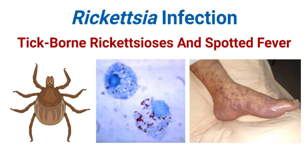
  • Zoonotic Transmission: They are primarily arthropod-borne. They rely heavily on vectors such as hard and soft ticks, fleas, human body lice, and mites (chiggers) to move from host to host.
  • Mammalian Reservoirs: The primary reservoir (where the bacteria naturally live, multiply, and maintain their life cycle in the wild) consists of various mammals, including rodents, dogs, and humans.
  • Pathological Target (The Endothelium): Once introduced into the human bloodstream, they exhibit a profound tropism (specific target affinity) for the vascular endothelium—the single layer of squamous cells lining the interior of blood vessels. This leads directly to severe systemic vasculitis, microscopic hemorrhages, and characteristic petechial rashes.

II. General Characteristics & Microbiology

Because they are obligate intracellular organisms, investigating them in a clinical or research laboratory requires highly specialized techniques that differ drastically from standard bacteriological protocols.
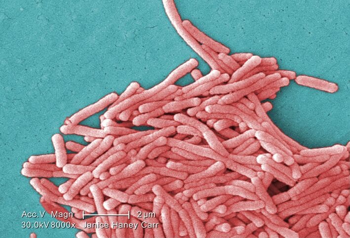
Morphology & Size:

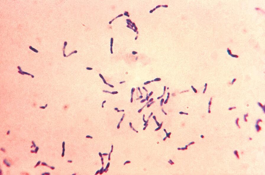
  • They are extremely small, pleomorphic coccobacilli (meaning they can alter their shape, appearing somewhere between a sphere and a tiny rod).
  • Their microscopic size ranges from 0.3 to 0.5 μm in width to 0.8 to 2.0 μm in length, making them barely visible under standard light microscopy.

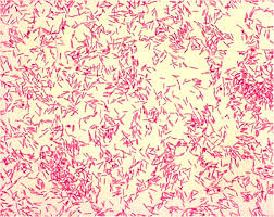
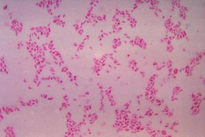
Staining & Culturing (High-Yield Board Concept):

  • The Gram Stain Failure: Although they possess a Gram-negative cell wall architecture, they stain incredibly poorly with a standard Gram stain. This is due to their minuscule size, the lipid-rich nature of their outer membrane, and the fact that they hide deep within the cytoplasm of host cells.
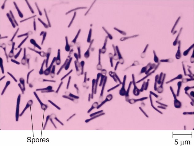
  • Specialized Stains Required: To visualize them microscopically, pathologists must utilize specialized tissue stains such as the Giemsa stain, Gimenez stain, or Machiavello stain.
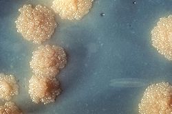
  • Culturing Limitations: Rickettsiae cannot grow on artificial, cell-free agar media (such as standard Blood agar, Chocolate agar, or MacConkey agar). To culture them, the reference laboratory must utilize living tissue cultures (e.g., Vero cells), embryonated chicken eggs (yolk sac inoculation), or susceptible live laboratory animals (like guinea pigs).

Replication & Evolutionary Genetics:

  • They divide strictly by binary fission, but this division occurs entirely within the nutrient-rich cytoplasm (and sometimes the nucleus) of the host cell. They have a very slow generation time (8 to 12 hours).
  • Reductive Evolution: Rickettsiae have an incredibly small genome (ranging from 1.1 to 1.6 Mb) that is heavily loaded with "pseudogenes" (broken, fragmented, non-functional genes). Because they have lived safely inside host cells for millions of years, they underwent reductive evolution—they literally threw away their own genes for synthesizing amino acids, nucleotides, and energy.
  • The ATP/ADP Translocase: Instead of making their own energy, they possess a unique membrane transport protein (ATP/ADP translocase) that allows them to parasitize the host by physically stealing ATP directly from the host cell's cytoplasm, trading it for depleted ADP!
Pathophysiology Expansion

Vascular Leakage & Virchow's Triad

Why do Rickettsial infections cause such deadly drops in blood pressure, distinct rashes, and organ failure? It comes down to the destruction of the blood vessel lining.

When the bacteria aggressively infect the endothelial cells lining the capillaries and arterioles, the host cells eventually undergo severe necrosis (death) and detach from the blood vessel wall. This physical destruction creates microscopic "holes" in the capillaries.

  • Red Blood Cell Leakage: RBCs leak out of the ruptured vessels into the dermal skin layers, causing a non-blanching, petechial/purpuric rash (a rash that does not turn white when you press on it, because the blood is trapped outside the vessel).
  • Plasma Leakage: Fluid leaks massively into the interstitial tissues. This causes severe total-body edema, profound hypovolemia (low intravascular blood volume), and ultimately, hypotensive shock.
  • Microvascular Thrombosis: The exposed, damaged sub-endothelial collagen powerfully activates the body's clotting cascade. Micro-clots form throughout the body's capillary beds (consumptive coagulopathy/DIC), leading to ischemic microinfarcts in the brain, kidneys, and digits. This systemic vasculitis is the primary mechanism of multi-organ failure and death.

III. Exhaustive Classification of Rickettsial Diseases

Rickettsial diseases are notoriously confusing to categorize. They are strictly and historically grouped based on their clinical presentation, their genetic relatedness, and the specific arthropod vector that transmits them to humans.

1. The Spotted Fever Group (SFG)

Characterized by ticks and mites, causing severe fevers and classic spreading rashes.

  • Rocky Mountain Spotted Fever (RMSF): Caused by Rickettsia rickettsii. Transmitted by hard ticks (Dermacentor species). Carries a devastating 5-10% mortality rate if untreated. Found primarily in the Americas.
  • Mediterranean Spotted Fever (Boutonneuse fever): Caused by R. conorii. Transmitted by the brown dog tick (Rhipicephalus sanguineus).
    Clinical Hallmark: Frequently presents with a "Tache Noire" (a black, necrotic eschar/scab at the exact site of the tick bite).
  • African Tick Bite Fever: Caused by R. africae. Transmitted by Amblyomma ticks. Common in travelers returning from sub-Saharan African safaris. Also presents with a prominent inoculation eschar and regional swollen lymph nodes.
  • Rickettsialpox: Caused by R. akari. The Exception: This is the notable exception in the spotted fever group because it is transmitted by a MOUSE MITE (Liponyssoides sanguineus), not a tick. Presents with a vesicular rash resembling chickenpox.
  • Emerging Spotted Fevers: R. parkeri (US), R. 364D (Pacific Coast tick fever), R. japonica (Japanese spotted fever).
2. The Typhus Group

Characterized by lice and fleas, notorious for explosive, devastating historical outbreaks in crowded populations.

  • Epidemic Typhus: Caused by Rickettsia prowazekii. Transmitted by the human body louse (Pediculus humanus corporis). Highly contagious in crowded, freezing, unsanitary conditions (e.g., refugee camps, wartime trenches). Anne Frank famously died of epidemic typhus in the Bergen-Belsen concentration camp.
    Clinical Phenomenon: The bacteria can lay dormant in the reticuloendothelial system (lymph nodes/macrophages) for decades and reactivate later in life when the patient's immunity wanes. This milder, reactivated form is known as Brill-Zinsser disease.
  • Murine Typhus (Endemic Typhus): Caused by Rickettsia typhi. Transmitted by rat fleas (Xenopsylla cheopis). Often seen in urban areas with high rodent populations.
3. The Scrub Typhus Group

A distinct geographical entity.

  • Scrub Typhus: Caused by Orientia tsutsugamushi (formerly classified as a Rickettsia, but genetically reclassified). Transmitted by the bite of the chigger (the microscopic larval stage of the Leptotrombidium mite).
  • Geography: Highly prevalent in the "Tsutsugamushi Triangle" (the Asia-Pacific region, including Japan, India, Australia, and the Pacific Islands). Patients frequently present with a blackened eschar, deafness, and tinnitus alongside the fever.
4. Ehrlichiosis and Anaplasmosis

Closely related intracellular bacteria that target leukocytes (white blood cells) rather than endothelial cells.

  • Human Monocytic Ehrlichiosis (HME): Caused by Ehrlichia chaffeensis. Transmitted by the Lone Star tick (Amblyomma americanum). Targets monocytes.
  • Human Granulocytic Anaplasmosis (HGA): Caused by Anaplasma phagocytophilum. Transmitted by Ixodes ticks (the exact same tick that transmits Lyme disease). Targets neutrophils.
  • Sennetsu Fever: Caused by Neorickettsia sennetsu. Uniquely transmitted to humans by ingesting a trematode (fluke) found in raw, undercooked fish in Japan.
Mnemonic Masterclass

Vectors of the Rickettsial World

To easily keep the bugs and their specific vectors straight for board exams:

  • R. rickettsii = Ticks (RMSF).
  • R. typhi = Fleas (Murine/Endemic Typhus) - "Typhi flies on Fleas."
  • R. prowazekii = Lice (Epidemic Typhus) - "Pro wars cause Epidemics (and body lice)."
  • O. tsutsugamushi = Chiggers (Scrub typhus).
  • R. akari = Mites (Rickettsialpox).

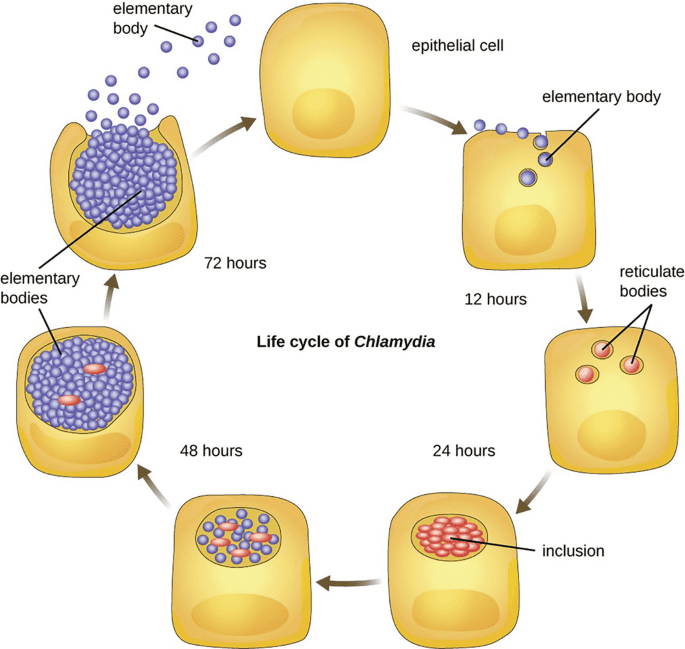
IV. Cellular Pathogenesis: The Hijacking Mechanism

How do these tiny bacteria cause such massive damage at the cellular level? The process is a highly choreographed molecular invasion.

  • Invasion & Cellular Adherence: Rickettsiae utilize outer membrane proteins, specifically OmpA and OmpB, to firmly adhere to specific receptors (like Ku70) on the host endothelial cell surface. Surface Cell Antigen (Sca) proteins further facilitate the actual endocytosis (swallowing) into the cell.
  • Phagosomal Escape: Once swallowed, the bacteria find themselves trapped in a phagosome (a cellular stomach). To avoid being digested by lysosomes, the bacteria immediately secrete phospholipase D and hemolysin C, enzymes that rapidly dissolve the phagosome membrane. The bacteria escape directly into the safety of the host's cytoplasm to multiply freely.
  • Actin-Based Motility (The Comet Tail): Some species (most notably R. rickettsii) do not wait to lyse and destroy the cell to escape. Instead, they express a surface protein called RickA, which hijacks the host cell's Arp2/3 complex. This forces the host to rapidly polymerize actin filaments directly behind the bacteria, creating a powerful "actin comet tail." This tail physically rockets the bacteria through the cytoplasm and propels it directly through the cell membrane into the adjacent neighboring cell.
    Clinical Significance: This allows the bacteria to spread from cell to cell rapidly without ever entering the extracellular blood space, completely hiding from the host's circulating antibodies! (Note: Listeria monocytogenes and Shigella use this exact same actin-rocket mechanism).

V. Rocky Mountain Spotted Fever (RMSF)

RMSF is the most common, most severe, and most frequently fatal tick-borne disease in the United States and the Americas. It requires immediate, aggressive clinical intervention based purely on clinical suspicion.

Epidemiology & Vector Dynamics:

  • Vectors: Transmitted primarily by Dermacentor variabilis (the American dog tick, found heavily in the Eastern and South-Central US, notably North Carolina, Oklahoma, and Arkansas) and D. andersoni (the Rocky Mountain wood tick, found in the Western US).
  • Seasonality: Highly seasonal. Over 90% of cases peak between April and September when ticks are in their nymph and adult stages, actively seeking mammalian blood meals.
  • Transmission Time: The tick must generally be attached and feeding for 6 to 24 hours to successfully reactivate and transmit the dormant bacteria into the human bloodstream.

Clinical Features & The Deadly Timeline:

RMSF is a rapidly progressing disease. Understanding the day-by-day evolution is critical for survival.

  • Incubation Period: 2 to 14 days (Average: 7 days) following the tick bite. Many patients never even recall being bitten by a tick.
  • Early Phase (Days 1-3): Sudden, abrupt onset of extremely high fever (often 103-104°F), severe unrelenting frontal headache, profound myalgia (muscle pain), and nausea/vomiting.
    Crucial Clinical Note: There is absolutely NO RASH during the first three days! Because of this, patients are tragically misdiagnosed with a severe viral illness or a migraine and sent home without antibiotics.
  • The Rash Phase (Days 3-5): Begins as a faint, macular, blanching rash (pink spots that turn white when pressed).
    • Centripetal Spread Pattern: It characteristically starts on the extreme periphery—the wrists, forearms, and ankles—and then spreads centripetally (inward) toward the trunk and chest over the next 24 hours.
    • Palmar/Plantar Involvement: It heavily and famously involves the palms of the hands and the soles of the feet.
    • Petechial Evolution: By day 5 or 6, as the vasculitis deeply worsens and red blood cells leak out, the rash becomes petechial or purpuric (dark red/purple hemorrhagic dots that do NOT blanch when pressed).
  • Late Phase (Severe Complications, Days 7+): The widespread, uncontrolled vascular leakage leads to massive systemic collapse:
    • Neurological: Altered mental status, confusion, seizures, and meningoencephalitis.
    • Pulmonary: Non-cardiogenic pulmonary edema leading to Acute Respiratory Distress Syndrome (ARDS).
    • Renal: Acute Kidney Injury (AKI) driven by hypovolemia (pre-renal) and micro-clots (intra-renal).
    • Digital Necrosis: The profound lack of blood flow causes fingers and toes to turn black and gangrenous, frequently requiring surgical amputation.
    • Mortality: Untreated mortality historically approaches 20-25%. Even with treatment, modern mortality is 5-10% if antibiotics are delayed past day 5.

The "Spotless" Exception

Approximately 10-15% of RMSF patients NEVER develop a rash, or the rash is highly atypical. This condition is termed "Spotless RMSF". Older patients and darker-skinned individuals are particularly at risk for missing the rash. These patients paradoxically have a significantly higher mortality rate simply because their diagnosis is severely delayed, as physicians wait for a rash that never comes before starting antibiotics.

Mnemonic Masterclass

Rashes on the Palms and Soles

Very few infectious diseases in all of medicine cause a distinct rash on the palms of the hands and the soles of the feet. When you observe this clinical sign, immediately think of driving CARS:

  • Coxsackievirus A (Hand, Foot, and Mouth Disease)
  • A (Ignore - used for phonetic flow, though some use 'A' for atypical measles/syndromes)
  • Rocky Mountain Spotted Fever
  • Syphilis (Specifically Secondary Syphilis)

VI. Laboratory Diagnosis

Rickettsial diseases progress incredibly fast. Clinical diagnosis must drive treatment. Do NOT wait for laboratory confirmation to start antibiotics, or the patient may suffer irreversible damage or death.

  1. Serology (IFA) - The Gold Standard:
    • Detects specific antibodies formed against rickettsial antigens via the Indirect Immunofluorescence Assay (IFA).
    • Definitive diagnosis requires paired sera showing a fourfold rise in antibody titers between the acute phase (week 1) and the convalescent phase (week 3-4).
    • The Limitation: Diagnostic IgG and IgM antibodies typically do not appear in detectable quantities until 7 to 14 days after the onset of illness. Therefore, serology is essentially useless for making the initial life-saving decision in the emergency department. It is used retrospectively to confirm what you already treated. A single titer of ≥1:64 with a compatible clinical illness confirms the diagnosis.
  2. Direct Detection:
    • Skin biopsy of the petechial rash evaluated with immunohistochemistry (IHC) or direct immunofluorescence. This is the fastest way to confirm RMSF in the acute phase, but it has low sensitivity (the bacteria are patchy).
    • PCR (Polymerase Chain Reaction) of the blood or skin biopsy is highly specific but suffers from variable sensitivity because the bacterial load in the circulating blood is actually quite low.
  3. Culture:
    • Extremely dangerous to laboratory staff due to the risk of aerosolization. Culturing is restricted strictly to Reference Laboratories requiring high-security BSL-3 (Biosafety Level 3) containment. Utilizes the shell vial technique with cell monolayers.
  4. The Weil-Felix Test (Historical Context):
    • An obsolete, historical agglutination test you may encounter in older literature. It relied on the phenomenon of immunological cross-reactivity: antibodies produced against Rickettsia will accidentally agglutinate (clump) the O-antigens of certain innocuous strains of Proteus bacteria (OX-19, OX-2, OX-K strains).
    • It possesses terrible sensitivity and specificity, yielding numerous false positives and false negatives, and has been completely abandoned in modern, evidence-based medicine.

VII. Treatment Pharmacology

A delay in treatment is the single largest cause of mortality in Rickettsial diseases. Empirical therapy must be initiated immediately based on clinical suspicion, often before the rash even fully develops.

1. Doxycycline (The Undisputed Drug of Choice):

  • Mechanism: A broad-spectrum tetracycline antibiotic. It is bacteriostatic, entering the host cell and binding reversibly to the 30S ribosomal subunit of the rickettsial bacteria, totally shutting down their protein synthesis.
  • Dosage: 100 mg BID (twice daily) intravenously or orally for 5 to 7 days, or strictly until the patient has been completely afebrile (fever-free) and clinically improving for at least 3 consecutive days.

❓ Applied Clinical Question: Pediatric Pharmacology Exception

Case: A 6-year-old boy presents to the Emergency Department with a 103°F fever, severe headache, and a petechial rash spreading from his wrists to his chest. His mother recently pulled an engorged tick off his neck after a camping trip in North Carolina. The attending physician orders Doxycycline IV. The new pediatric nurse questions the order, stating, "Doxycycline is strictly contraindicated in children under 8 years of age due to the risk of permanent enamel hypoplasia and severe grey/yellow tooth discoloration." How should the charge nurse respond?

Answer: The charge nurse must firmly explain that while Doxycycline is indeed normally contraindicated in young children for routine infections, Rocky Mountain Spotted Fever is a rapidly fatal, life-threatening emergency. The CDC and the American Academy of Pediatrics specifically dictate that Doxycycline is the absolute first-line treatment for RMSF for patients of ALL ages, including infants and young children. The risk of neurological devastation, limb amputation, and death from the disease vastly, undeniably outweighs the minimal, often merely cosmetic risk of dental staining from a short 5-to-7-day course of the drug. There is no comparable second-line drug.

2. Alternative Agents:

  • Chloramphenicol: The classic historical alternative, particularly used in pregnant women. However, it is highly toxic, binding to the 50S ribosomal subunit. It is heavily associated with severe, unpredictable bone marrow toxicity (idiosyncratic, fatal aplastic anemia) and Grey Baby Syndrome in neonates (due to the baby's lack of liver UDP-glucuronyl transferase to metabolize the drug, leading to cyanosis, cardiovascular collapse, and an ashen grey skin color). It is rarely used in the US today.
  • Azithromycin / Fluoroquinolones (Ciprofloxacin): Can be used with varied success for very mild rickettsial diseases (like Mediterranean Spotted fever), but are NOT recommended for severe RMSF.
  • Rifampin: Highly effective specifically for severe, doxycycline-resistant cases of Scrub typhus in Southeast Asia.

VIII. Ehrlichiosis and Anaplasmosis

These diseases are closely related to Rickettsiae genetically, present with similar vague febrile symptoms, and are treated with the exact same drugs, but they target completely different cells in the human immune system.

Pathological Target Cells (The Leukocytes):

  • Ehrlichia chaffeensis: Actively infects Monocytes and Macrophages.
  • Anaplasma phagocytophilum: Actively infects Neutrophils (Granulocytes).
Microbiology Feature

The "Morulae" Phenomenon

Once inside the white blood cells, these bacteria avoid lysosomal destruction and multiply rapidly inside host vacuoles. They pack tightly together into massive, distinct, membrane-bound, mulberry-like clusters called Morulae (from the Latin word for mulberry) inside the cytoplasm of the WBC. Finding these morulae on a peripheral blood smear is diagnostic.

Clinical Presentation & Co-Infection Risks:

  • Symptoms: Non-specific flu-like illness: high fever, profound headache, severe myalgia, and malaise.
  • The Rash: Unlike RMSF, a rash is UNCOMMON in these diseases (occurring in <30% of Ehrlichiosis cases and <10% of Anaplasmosis cases). This heavily differentiates it clinically from RMSF.
  • Laboratory Findings: Because they actively destroy white blood cells and platelets while inflaming the liver, patients present with a classic triad of profound Leukopenia (low WBCs), Thrombocytopenia (low platelets), and elevated hepatic transaminases (AST/ALT).
  • Co-Infection Alert: Anaplasma is transmitted by the Ixodes scapularis tick in the Northeast and Midwest US. This is the exact same tick that transmits Lyme disease (Borrelia burgdorferi) and Babesiosis (Babesia microti). A patient bitten by one tick can be simultaneously infected with all three diseases, presenting with a highly complex, confusing, and severe clinical picture!

Diagnosis and Treatment:

  • Diagnosis: Examining a peripheral blood smear under a microscope to physically see the morulae inside the WBCs (fast, but low sensitivity). NAAT (PCR of the blood) and Serology (IFA) are definitive.
  • Treatment: First-line is strictly Doxycycline for all ages. Rifampin is utilized as an alternative exclusively for patients with true, severe, anaphylactic allergies to tetracyclines or for pregnant women.

IX. Prevention & Environmental Control Strategies

Because the pathophysiology of these diseases is so destructive, and because there are currently NO commercially available vaccines for any Rickettsial diseases, Ehrlichiosis, or Anaplasmosis, prevention relies entirely on rigid environmental and vector control mechanisms.

1. Tick Avoidance & Chemical Repellents:

  • Clothing: Wear light-colored protective clothing (makes tiny ticks easier to spot), with long pants tucked securely into high socks to prevent ticks from crawling up the legs from tall grass.
  • DEET (N,N-Diethyl-meta-toluamide): A highly effective chemical repellent applied directly to exposed skin. It disrupts the tick's olfactory receptors, masking the human's scent.
  • Permethrin: A synthetic neurotoxin specifically lethal to arthropods but generally safe for mammals. It must be applied exclusively to clothing, boots, and camping gear, never directly to the skin. Ticks that crawl across permethrin-treated fabric die rapidly.
  • Tick Checks: Conduct thorough full-body tick checks (especially in hair, groin, and axilla) immediately after outdoor activity in endemic areas.

2. Proper Tick Removal Technique:

Removing a tick incorrectly can be deadly. If you squeeze the tick's swollen abdomen, burn it with a hot match, or try to suffocate it with petroleum jelly, the stressed tick will aggressively regurgitate its infected salivary and gut contents directly into the patient's bloodstream, guaranteeing infection.

  • The Correct Method: Use fine-tipped tweezers. Grasp the tick as close to the skin's surface (the mouthparts) as physically possible. Apply a steady, even, upward pull without twisting or jerking. Disinfect the bite site immediately after removal.

3. Louse Control (For Epidemic Typhus):

Body lice (unlike head lice) live and lay their eggs in the seams of clothing, only moving to the skin to feed. They thrive exclusively in unhygienic, impoverished, crowded conditions. Control focuses on improving basic sanitation, boiling clothes in hot water (killing both lice and nits), and utilizing systemic chemical delousing procedures (e.g., mass dusting with safe insecticidal powders in refugee situations).


References

  • Centers for Disease Control and Prevention (CDC): Diagnosis and Management of Tickborne Rickettsial Diseases: Rocky Mountain Spotted Fever and Other Spotted Fever Group Rickettsioses, Ehrlichioses, and Anaplasmosis (MMWR Recommendations and Reports).
  • Mandell, Douglas, and Bennett's: Principles and Practice of Infectious Diseases (9th Edition) - Section on Rickettsiaceae.
  • Harrison's Principles of Internal Medicine: (21st Edition) - Chapters detailing tick-borne illnesses and systemic vasculitis.
  • American Academy of Pediatrics (AAP): Red Book: Report of the Committee on Infectious Diseases - Guidelines regarding the pediatric administration of Doxycycline.
  • World Health Organization (WHO): Guidelines on the management and vector control of Epidemic and Scrub Typhus in developing nations.

Quick Quiz

Bacteriology Intro Quiz

Microbiology - mobile-friendly and focused practice.

Privacy: Your details are used only for quiz tracking and certificates.

Rickettsiae, Spotted Fevers & Typhus Read More »

Chlamydia and Chlamydophila

Chlamydia and Chlamydophila

Chlamydia and Chlamydophila

Module Learning Objectives

By the conclusion of this exhaustive master guide, you will be deeply conversant with:

  • The highly unique, obligate intracellular nature of Chlamydiaceae and their biphasic life cycle.
  • The specific serovars of Chlamydia trachomatis and their distinct clinical manifestations (from blinding Trachoma to Lymphogranuloma Venereum).
  • The complex virulence factors, including the Type III Secretion System and Hsp60-induced immunopathology.
  • Comprehensive clinical presentations, complications (e.g., Fitz-Hugh-Curtis syndrome, Reactive Arthritis), and the gold-standard diagnostic protocols (NAAT).
  • Detailed pharmacological treatment protocols and global prevention strategies (like the WHO S.A.F.E. Strategy).

I. Introduction to Chlamydiae

The family Chlamydiaceae consists of highly specialized, obligate intracellular bacteria. They are unique pathogens that have evolved to replicate exclusively within the protective confines of cytoplasmic vacuoles (inclusions) inside eukaryotic host cells. They are responsible for a massive, global burden of human disease, affecting millions across both developed and developing nations.

Clinical & Epidemiological Significance:

  • The Global STI Epidemic: C. trachomatis is unequivocally the most common sexually transmitted bacterial infection globally. The World Health Organization (WHO) estimates there are over 129 million new cases annually. Because the vast majority of cases are asymptomatic, it heavily drives the "silent epidemic" of infertility.
  • The Trachoma Burden: It is the leading cause of preventable infectious blindness worldwide, predominantly ravaging pediatric populations in resource-limited settings across Sub-Saharan Africa and parts of Asia.

Historical Misclassification: The "Viral" Myth

Historically, up until the mid-20th century, Chlamydiae were completely misclassified by scientists as viruses. This fundamental misunderstanding was due to two factors:

  1. Incredibly Small Size: Their infectious particles (Elementary Bodies) are so small that they could pass through porcelain filters designed to trap bacteria, a hallmark trait originally thought to belong exclusively to viruses.
  2. Obligate Intracellular Nature: They absolutely could not be grown on standard artificial agar plates (like blood agar or MacConkey agar), behaving exactly like viruses that require living host cells to multiply.

The Modern Taxonomic Correction: We now definitively know they are true bacteria. Unlike viruses, Chlamydiae possess both DNA and RNA simultaneously, feature a distinct bacterial cell wall structure, synthesize their own proteins using bacterial ribosomes (70S, comprised of 30S and 50S subunits), divide by bacterial binary fission, and—crucially for clinical practice—are highly susceptible to broad-spectrum antibacterial antibiotics (like macrolides and tetracyclines).


II. General Characteristics & The "Energy Parasite" Phenomenon

Understanding the structure and metabolic limitations of Chlamydiae is key to understanding how they evade the immune system and cause disease.

  • Morphology: They are extraordinarily small, coccoid-shaped bacteria, measuring between 0.2 and 1.5 micrometers depending on their life cycle stage.
  • Staining & The "Chlamydial Anomaly": They possess an envelope structure that resembles a Gram-negative cell wall (containing an outer membrane and lipopolysaccharide), but they stain very poorly with a standard Gram stain.
    Pathophysiology Expansion: For decades, scientists could not detect a rigid peptidoglycan layer, calling it the "chlamydial anomaly." We now know they rely heavily on extensively cross-linked Major Outer Membrane Proteins (MOMPs) and Cysteine-Rich Proteins (CRPs) for their structural integrity, which act like a chain-mail corset to protect them from osmotic pressure.
  • Cultivation: They cannot be cultured on artificial, cell-free media. They strictly require living eukaryotic cells for in vitro cultivation. In the laboratory, they are typically grown in specific cell lines, most notably McCoy cells or HeLa cells.
Microbiology Deep Dive

Energy Parasitism: Stealing the Host's Life Force

Chlamydiae are famously known as "energy parasites." They lack the necessary enzymatic machinery to synthesize their own Adenosine Triphosphate (ATP), meaning they cannot generate their own metabolic energy. They depend entirely on the host cell to survive. To accomplish this, they utilize a highly specialized, parasitic enzyme called an ATP/ADP translocase. This molecular pump sits on the bacterial membrane and literally steals high-energy ATP molecules from the host cell's cytoplasm, swapping them out for depleted bacterial ADP.


III. The Unique Biphasic Chlamydial Developmental Cycle

Chlamydiae possess a highly unique, biphasic (two-stage) developmental cycle that alternates between two distinct morphological forms: the Elementary Body (EB) and the Reticulate Body (RB). This entire cycle takes approximately 48 to 72 hours to complete.

1. Elementary Body (EB)

The Infectious Form.

  • Role: Think of it like a tough bacterial spore. It is the extracellular survival form.
  • Metabolism: It is highly condensed, extremely rigid (due to cross-linked MOMPs), and metabolically inactive (dormant).
  • Function: Its sole job is to survive harsh environmental conditions outside the host, travel to a new host, and initiate infection by attaching to and entering a new eukaryotic cell.
  • Mnemonic: "E" is for Extracellular, Enters the cell, and Elementary.
2. Reticulate Body (RB)

The Replicative Form.

  • Role: This is the fragile, intracellular form.
  • Metabolism: It is metabolically highly active, aggressively synthesizing RNA, proteins, and stealing host ATP.
  • Function: Once the EB is safely inside the host cell, it transforms into the RB. The RB cannot survive outside the host cell. Its sole job is to divide rapidly by binary fission.
  • Mnemonic: "R" is for Replicative, Remains inside, and Reticulate.

The Step-by-Step Sequence of Infection:

  1. Attachment: The infectious EB attaches to specific host cell receptors (e.g., heparan sulfate proteoglycans) via the OmcB protein on its surface.
  2. Entry: The EB enters the host cell via receptor-mediated endocytosis or induced phagocytosis. Crucially, it remains safely within a membrane-bound vesicle known as an inclusion. Through specialized virulence factors, it actively evades lysosomal destruction (preventing the host from digesting it).
  3. Differentiation (Hours 0-8): Once safely inside, the tough EB sheds its protective cross-links, absorbs water, swells, and differentiates into the metabolically active RB.
  4. Replication (Hours 8-24): The RBs begin to divide exponentially by binary fission within the expanding inclusion vacuole, forming massive microcolonies that push against the host cell nucleus.
  5. Redifferentiation (Hours 24-48): After sufficient replication depletes the host's nutrients, the RBs receive an environmental signal to stop dividing. They condense, form rigid cross-links, and transform back into infectious EBs.
  6. Release (Hours 48-72): The exhausted host cell undergoes either explosive lysis or orderly exocytosis, releasing thousands of newly minted, infectious EBs into the surrounding tissue to infect adjacent, healthy cells.

Pathology Expansion: The Persistent State (Aberrant Bodies)

Under highly unfavorable or stressful conditions—such as when the patient takes a low dose of penicillin, or when the host's immune system floods the area with Interferon-gamma (IFN-γ), or when amino acids (like tryptophan) are depleted—the Chlamydia halts its normal life cycle. The RBs stop dividing and swell into massive, bizarre, non-replicative forms known as Aberrant Bodies.

In this "Persistent State," the bacteria can hide in the tissue for months or years. They are temporarily immune to antibiotics that require actively dividing cells. This smoldering, chronic presence is a massive clinical problem, as it continuously provokes the host immune system, contributing heavily to the chronic inflammatory scarring seen in Pelvic Inflammatory Disease (PID) and blinding Trachoma.


IV. Chlamydia trachomatis: Serovars & Virulence Factors

Chlamydia trachomatis is not a single, uniform pathogen. It is intricately divided into distinct serovars (serological variants) based on structural differences in their Major Outer Membrane Protein (MOMP). These serovars have strict "tissue tropism," meaning specific serovars only attack specific organs.

Serovars Target Tissue / Disease Association Clinical Memory Hack
A, B, Ba, C Endemic Trachoma (chronic follicular keratoconjunctivitis). It strictly targets the conjunctival epithelium of the eye, leading to severe corneal scarring and irreversible blindness. A, B, C = Africa, Blindness, Children.
D, Da, E, F, G, H, I, Ia, J, K Genitourinary infections (urethritis, cervicitis, PID, epididymitis), adult inclusion conjunctivitis, and infant pneumonia/ophthalmia neonatorum acquired during vaginal birth. D through K = "Dick" to "Koochie" (Targets the Genital Tract).
L1, L2, L2a, L3 Lymphogranuloma venereum (LGV). Unlike the others that stay on the surface epithelium, the LGV biovars are highly invasive, penetrating the mucosa to aggressively attack and destroy regional macrophages and lymphatic tissue. L = Lymph nodes.

The Arsenal: Chlamydial Virulence Factors

Chlamydiae are master manipulators of the host cell. They utilize a massive arsenal of weapons to survive intracellularly:

  • Type III Secretion System (T3SS): This acts as a microscopic molecular "syringe." The EB uses the T3SS to puncture the host cell membrane and inject toxic effector proteins directly into the host cytoplasm. These injected proteins instantly paralyze the host's actin cytoskeleton, forcing the cell to swallow the bacteria.
  • Inclusion Membrane Proteins (Incs): Once inside the vacuole, Chlamydia inserts Inc proteins into the vacuole's membrane. These act as traffic controllers, actively blocking the host cell's lysosomes from fusing with the vacuole, completely stopping the host from digesting the bacteria.
  • Chlamydial Protease-Like Activity Factor (CPAF): This enzyme is secreted heavily into the host cytoplasm to degrade host transcription factors and cytoskeletal proteins, effectively blinding and paralyzing the host cell's internal alarm systems.
  • Heat Shock Protein 60 (Hsp60): Highly Clinical: This protein is produced when the bacteria are stressed. It is extremely immunogenic. The human body recognizes Hsp60 and mounts a massive, aggressive inflammatory response against it. Tragically, human cells also possess a similar Hsp60. This molecular mimicry causes the immune system to attack the body's own tissues, leading to the severe immunopathology, dense fibrotic scarring, fallopian tube strictures, and ectopic pregnancies seen in chronic infections.
  • Major Outer Membrane Protein (MOMP): The most abundant surface protein, making up 40% of the outer membrane weight. It functions as a porin channel for nutrients and determines the specific serotype.
  • Lipopolysaccharide (LPS): Genus-specific and highly cross-reactive. Notably, chlamydial LPS has very poor endotoxin activity compared to classic Gram-negative bacteria (like Neisseria meningitidis or E. coli), which is why patients with Chlamydia rarely present with septic shock.

V. Clinical Manifestations of C. trachomatis

1. Urogenital Infections in Women

  • Primary Presentation: Often presents as mucopurulent cervicitis (a yellow/green discharge from the cervix) and urethritis (dysuria or painful urination). However, up to 80% of female infections are completely asymptomatic, earning it the title of the "silent epidemic."
  • Ascending Complications: If left untreated, the bacteria ascend through the uterus into the fallopian tubes, causing Pelvic Inflammatory Disease (PID). The severe Hsp60-driven inflammation destroys the delicate ciliated lining of the fallopian tubes. This scarring blocks the path of fertilized eggs, resulting in chronic pelvic pain, life-threatening ectopic pregnancy, and irreversible tubal factor infertility.
  • Clinical Expansion: Fitz-Hugh-Curtis Syndrome In severe or neglected cases of PID, the chlamydial infection escapes the fallopian tubes and tracks up the peritoneal cavity to the liver capsule (perihepatitis). It causes intense inflammation and the formation of distinct "violin-string" fibrinous adhesions between the liver and the anterior abdominal wall. Patients present with severe Right Upper Quadrant (RUQ) pain that perfectly mimics acute cholecystitis (gallbladder attack), referred pain to the right shoulder, and a friction rub heard on auscultation.

2. Urogenital Infections in Men

  • It is the absolute most common cause of Non-Gonococcal Urethritis (NGU). Patients present with dysuria and a watery, mucoid, or clear urethral discharge (as opposed to the thick, purulent, bloody discharge typical of Gonorrhea).
  • Ascending infections can cause severe unilateral testicular pain and swelling (Epididymitis) and inflammation of the prostate (Prostatitis).

3. Lymphogranuloma Venereum (LGV - Serovars L1, L2, L3)

This is a highly invasive, aggressive systemic sexually transmitted disease.

  • Primary Stage: Begins as a small, painless, transient genital papule or ulcer that heals rapidly, often going completely unnoticed by the patient.
  • Secondary Stage: Weeks later, the infection tracks to regional draining lymph nodes (usually inguinal nodes). It causes massive, severely painful, fluctuant lymphadenopathy known as buboes. The inflamed nodes can mat together and adhere to the skin above and deep fascia below, creating a distinct indentation known as the "Groove Sign" (separated by the inguinal ligament). If untreated, these buboes can rupture through the skin, forming chronic draining fistulas.
  • Anorectal Syndrome: In cases of receptive anal intercourse, LGV can cause extreme proctocolitis, mimicking inflammatory bowel disease, leading to severe rectal strictures and even lymphatic obstruction causing genital elephantiasis.

4. Ocular Infections

  • Endemic Trachoma (Serovars A-C): A chronic, repeated infection of the tarsal conjunctiva transmitted by eye-seeking flies (Musca sorbens), dirty hands, and shared fomites (towels). Chronic inflammation leads to dense subepithelial scarring. As the scar tissue contracts, it pulls the eyelid margins inward (entropion). The tough eyelashes then scrape relentlessly against the cornea every time the patient blinks (trichiasis), causing agonizing corneal abrasions, dense corneal opacification, and permanent blindness.
  • Adult Inclusion Conjunctivitis (Serovars D-K): Typically affects sexually active young adults. Spread via autoinoculation (touching infected genital secretions and rubbing the eyes) or direct oral-genital contact. Presents with a foreign-body sensation, tearing, and distinct cobblestone-like follicles and papillary hypertrophy on the lower conjunctiva.

5. Infant Complications (Vertical Transmission)

If a pregnant mother has an untreated cervical infection, the infant has a 50% chance of acquiring the bacteria while passing through the birth canal.

  • Ophthalmia Neonatorum: Inclusion conjunctivitis developing 5 to 14 days postpartum (later than Gonococcal conjunctivitis, which appears in 2-5 days). It is characterized by severe eyelid swelling and purulent discharge.
  • Infant Pneumonia: Onset is typically 4-12 weeks postpartum. Hallmark Clinical Signs: The infant presents with a distinctive, repetitive staccato cough (short, machine-gun-like bursts), tachypnea (rapid breathing), hyperinflation on chest X-ray, and notably, the infant is remarkably afebrile (no fever).

6. Reactive Arthritis (Reiter Syndrome)

This is a severe, systemic autoimmune complication that occurs weeks after the initial genitourinary chlamydial infection has cleared. The immune system, primed by the bacterial antigens, mistakenly attacks the body's own joints and tissues. It is heavily associated with patients who carry the HLA-B27 genetic marker.

Classic Triad Mnemonic

Reactive Arthritis (Reiter's Syndrome)

Remember the classic triad of symptoms triggered 1 to 4 weeks after a Chlamydia infection: "Can't see, can't pee, can't climb a tree."

  • Can't see: Conjunctivitis or Anterior Uveitis (eye inflammation).
  • Can't pee: Non-gonococcal Urethritis (painful urination).
  • Can't climb a tree: Arthritis (typically asymmetric, severe oligoarthritis of the large weight-bearing joints of the lower limbs, like knees and ankles).

Extra detail: Patients may also present with unique dermatological findings such as Keratoderma blennorrhagicum (crusted, hyperkeratotic lesions on the palms and soles) and Balanitis circinata (painless ulcers on the glans penis).


VI. Laboratory Diagnosis

Because Chlamydiae are obligate intracellular organisms, classic microbiological swabs and agar plating are completely useless. Diagnosis requires specialized molecular or cellular techniques.

1. Nucleic Acid Amplification Test (NAAT)

The Undisputed Gold Standard. NAATs (like PCR, Transcription-Mediated Amplification, or Strand Displacement Amplification) are incredibly sensitive and specific. They detect minute traces of chlamydial DNA or RNA.

Clinical Advantage: Because it only looks for nucleic acids, the bacteria do not need to be alive. This eliminates cold-chain transport issues. It can be run on non-invasive samples: a first-catch urine sample (not mid-stream, you want the bacteria washed out of the urethra), self-collected vaginal swabs, or rectal/oropharyngeal swabs.

2. Cell Culture

Historically the gold standard, but highly technically demanding, expensive, and slow (takes 3-7 days). It requires living McCoy cell lines.

Laboratory Trick: The host cells are pre-treated with cycloheximide (a toxin that halts host cell protein synthesis). This suppresses the host cell and allows the Chlamydia to steal all the ATP unopposed, growing massive inclusions that are then stained with iodine or fluorescent antibodies.
Current Use: Restricted mostly to medical-legal applications (e.g., suspected child sexual abuse cases) because it offers absolute 100% specificity.

3. Antigen Detection & Serology

Antigen Detection: Direct fluorescent antibody (DFA) staining and Enzyme Immunoassay (EIA). These are faster but have significantly lower sensitivity than NAAT and are largely being phased out.

Serology: Complement fixation (CF) and microimmunofluorescence (MIF). Serology has very limited utility for active, routine genital infections because antibodies persist for years, making it impossible to distinguish past from current infections. However, looking for a massive four-fold rise in paired antibody titers is highly useful for diagnosing invasive LGV or infant pneumonia.


VII. Pharmacological Treatment Protocols

Because Chlamydia lives exclusively inside host cells, antibiotics must be lipophilic enough to penetrate both the host cell membrane and the inclusion vacuole membrane. Beta-lactams (like Penicillin) are generally ineffective because Chlamydiae lack classical peptidoglycan targets and can enter a persistent state. The drugs of choice are protein synthesis inhibitors (targeting the 30S or 50S bacterial ribosomes).

  • Uncomplicated Genital Infection (Cervicitis/Urethritis):
    • Doxycycline 100 mg orally twice a day (BID) for 7 days. (A tetracycline; targets the 30S ribosome. Requires strict patient adherence).
    • Azithromycin 1 gram orally as a single dose. (A macrolide; targets the 50S ribosome. Highly favored due to observed therapy and perfect compliance, as its extremely long tissue half-life allows for single-dose curative efficacy).
  • Lymphogranuloma Venereum (LGV): The invasive nature requires extended, aggressive therapy: Doxycycline 100 mg BID for a full 21 days.
  • Trachoma: Annual mass drug administration of Azithromycin 20 mg/kg as a single dose to entire communities, OR continuous application of topical 1% tetracycline eye ointment for 6 weeks.
  • Pregnancy Modifications: Doxycycline is absolutely contraindicated in pregnancy (it stains fetal teeth and retards bone growth). Pregnant women are treated with Azithromycin (1g single dose) or Amoxicillin (500mg TID for 7 days).
  • Partner Treatment (Expedited Partner Therapy): It is absolutely essential to test and concurrently treat all sexual partners from the past 60 days to prevent immediate "ping-pong" reinfection.
  • Test of Cure (TOC): A follow-up NAAT is not routinely needed for non-pregnant patients treated with first-line regimens because treatment failure is exceedingly rare. However, a TOC is strictly mandated for pregnant women (at 3 to 4 weeks post-treatment) to absolutely ensure clearance and prevent devastating neonatal transmission.

VIII. Other Chlamydiaceae (C. pneumoniae & C. psittaci)

While C. trachomatis dominates sexual health and ophthalmology, two other distinct species within the family are formidable respiratory pathogens.

1. Chlamydophila pneumoniae

  • Epidemiology & Pathology: Transmitted via respiratory droplets from human to human. It is a major cause of "Atypical" Community-Acquired Pneumonia (CAP), bronchitis, and sinusitis. It accounts for approximately 10% of all CAP cases globally. It is commonly referred to as "walking pneumonia" because patients often look remarkably well, exhibiting mild, gradual-onset symptoms (sore throat, hoarseness, low-grade fever) despite having nasty, patchy interstitial infiltrates on a chest X-ray.
  • Clinical Associations: It is strongly associated with acute asthma exacerbations in both children and adults. (Note: Decades of research have attempted to link persistent C. pneumoniae infection within macrophage foam cells to coronary atherosclerosis and Alzheimer's disease, but these links remain controversial and largely unproven therapeutically).
  • Diagnosis & Treatment: Microimmunofluorescence (MIF) serology is the standard (looking for a fourfold rise in IgG titer or IgM ≥ 1:16). Culturing on specialized HL or Hep-2 cell lines is technically brutal. Multiplex PCR panels are the best modern tool but are expensive. Treatment mirrors standard atypical pneumonia protocols: Doxycycline, Macrolides (Azithromycin), or respiratory Fluoroquinolones (Levofloxacin).

2. Chlamydophila psittaci

  • Epidemiology & Transmission: Causes a severe zoonotic infection known as Psittacosis (or Ornithosis). It is strictly acquired from inhaling the aerosolized, dried feces, urine, or respiratory secretions of infected birds (specifically parrots, parakeets, pigeons, turkeys, and ducks). High-risk populations include pet shop owners, veterinarians, poultry processing workers, and exotic bird smugglers.
  • Clinical Presentation: An abrupt-onset, severe atypical pneumonia accompanied by a splitting, severe headache, photophobia, and a non-productive cough.
    Classic Exam Clues: Patients exhibit Relative Bradycardia (pulse is noticeably slower than what is physiologically expected for their high degree of fever), palpable hepatosplenomegaly (enlarged liver and spleen), and the presence of Horder spots (a rare, faint pink blanching maculopapular rash resembling the rose spots of typhoid fever).
  • Severe Complications: If unchecked, it disseminates rapidly, causing fatal encephalitis, myocarditis, and severe hepatitis.
  • Diagnosis & Treatment: Culturing this organism is highly discouraged and requires maximum Biosafety Level 3 (BSL-3) containment due to the extreme, lethal inhalation risk to lab workers! Diagnosis relies on Serology (CF, MIF) or targeted PCR. Treatment is primarily 10-14 days of Doxycycline (macrolides are used as second-line for children/pregnant women).

IX. Global Prevention Strategies

Because the consequences of untreated chlamydial infections—blindness and infertility—are so profound, massive public health initiatives are in place globally.

  • Vaccines: Despite decades of intense research, there is currently NO viable vaccine available for any human chlamydial disease. The complex intracellular life cycle and the risk of a vaccine actually triggering the Hsp60 autoimmune reaction have stymied development.
  • Widespread Screening: Because genital infections are notoriously asymptomatic, the US Preventive Services Task Force (USPSTF) universally mandates routine, annual C. trachomatis NAAT screening for all sexually active women under the age of 25, older women with new or multiple partners, and all pregnant women at their first prenatal visit.
  • Safe Sex Practices: Consistent and correct use of latex or polyurethane condoms provides a highly effective physical barrier against transmission. Expedited partner therapy (providing prescriptions for partners without requiring a clinic visit) is highly encouraged to break transmission chains.
  • Psittacosis Prevention: Strict, mandatory 30-day quarantine and prophylactic antibiotic treatment (chlortetracycline-laced bird feed) of all legally imported exotic birds; use of heavy N95 respiratory protection by poultry workers and veterinarians when cleaning cages.
Global Health Initiative

The S.A.F.E. Strategy for Trachoma Eradication

The World Health Organization (WHO) has spearheaded a massive, international, multi-faceted initiative aiming to completely eliminate Trachoma as a public health problem. It relies on four interlocking pillars:

  • S - Surgery: Free, mobile surgical camps to correct advanced stages of the disease (rotating the eyelids outward to fix trichiasis/entropion, saving the cornea from further destruction).
  • A - Antibiotics: Massive, community-wide drug administration (MDA) of single-dose oral Azithromycin to instantly clear the chlamydial reservoir from the entire village population.
  • F - Facial cleanliness: Extensive community education programs promoting daily face washing. This removes the infectious ocular and nasal discharge that attracts the Musca sorbens flies, effectively cutting the vector transmission route from child to child.
  • E - Environmental improvement: Long-term infrastructure investments building latrines, separating human dwellings from animal pens, and establishing permanent access to clean, running water to suppress fly breeding grounds.

❓ End of Module Review Question

Case: A 20-year-old female presents to the university health clinic with vague, dull lower abdominal pain, a new-onset dull backache, and a slight yellowish vaginal discharge. A first-catch urine NAAT test returns positive for C. trachomatis. The physician prescribes a 7-day course of oral Doxycycline. Before leaving, she asks the nurse why her long-term boyfriend needs to seek treatment if he "feels completely fine and has no burning or dripping." What is your precise biological and clinical explanation to secure her compliance?

Answer: First, you must explain that Chlamydia is notoriously asymptomatic—upwards of 50% of infected males and 80% of infected females exhibit absolutely no symptoms. He is almost certainly infected and shedding the bacteria despite feeling perfectly fine. Second, because Chlamydia evades the immune system by entering a persistent, intracellular state inside the mucosal cells, if he is not concurrently treated with antibiotics, he will harbor the bacteria indefinitely. Consequently, he will immediately re-infect her the very next time they have unprotected intercourse. This "ping-pong" transmission cycle will subject her fallopian tubes to repeated waves of severe Chlamydial Hsp60-driven immune inflammation, inevitably causing massive fibrotic tubal scarring. This scarring leads directly to Pelvic Inflammatory Disease (PID), a drastically increased risk of a ruptured ectopic pregnancy, and permanent, irreversible tubal infertility.


X. List of References & Evidence-Based Guidelines

  • Centers for Disease Control and Prevention (CDC): Sexually Transmitted Infections Treatment Guidelines. Detailed protocols on the screening, NAAT diagnostics, and pharmacological management of C. trachomatis and LGV.
  • World Health Organization (WHO): Trachoma: Fact Sheets and S.A.F.E. Strategy Guidelines. Comprehensive global health directives on the epidemiology and mass drug administration efforts for endemic blinding trachoma.
  • Mandell, Douglas, and Bennett's: Principles and Practice of Infectious Diseases. In-depth microbiological analysis of the Chlamydiaceae family, including the intricate details of the biphasic life cycle, T3SS, and ATP parasitism.
  • Robbins & Cotran: Pathologic Basis of Disease. Extensive detailing on the immunopathology of Chlamydia infections, specifically focusing on Hsp60 cross-reactivity, tubal stricture formation, and Reiter's Syndrome pathophysiology.
  • U.S. Preventive Services Task Force (USPSTF): Chlamydia and Gonorrhea: Screening. Clinical recommendations establishing the necessity of annual screening for high-risk demographics to prevent PID and infertility.

Quick Quiz

Bacteriology Intro Quiz

Microbiology - mobile-friendly and focused practice.

Privacy: Your details are used only for quiz tracking and certificates.

Chlamydia and Chlamydophila Read More »

Legionella pneumophila

Legionella pneumophila

Legionella pneumophila


(Legionnaires' Disease & Pontiac Fever)

Module Learning Objectives

By the conclusion of this exhaustive master guide, you will be deeply conversant with:

  • The fascinating historical context and epidemiology of Legionnaires' disease.
  • The unique taxonomic, morphological, and highly fastidious metabolic profiles of Legionella species.
  • The complex molecular pathogenesis, specifically the Dot/Icm Type IV Secretion System used to hijack human macrophages.
  • The profound clinical differentiation between severe Legionnaires' pneumonia and the benign Pontiac Fever.
  • The limitations of standard diagnostics and the crucial reliance on Urinary Antigen Testing and BCYE culture.
  • The targeted pharmacological rationale for utilizing intracellularly concentrating antibiotics while strictly avoiding beta-lactams.

I. Introduction & Historical Context

Legionella pneumophila is a highly specialized, intracellular bacterial pathogen responsible for two entirely distinct clinical syndromes: Legionnaires' disease (a severe, rapidly progressive, and potentially fatal form of atypical pneumonia) and Pontiac fever (a milder, self-limiting, acute flu-like illness).

The Historical Discovery: The 1976 Outbreak

This organism remained completely unknown to medical science until July 1976. During the United States Bicentennial, a massive, highly publicized, and mysterious outbreak of severe pneumonia swept through attendees of an American Legion convention stationed at the Bellevue-Stratford Hotel in Philadelphia. Out of more than 2,000 attendees, 221 became critically ill, and 34 died of acute respiratory failure.

The Centers for Disease Control and Prevention (CDC) launched an unprecedented epidemiological investigation. Months later, they isolated a previously unidentifiable, fastidious Gram-negative bacterium. They found it circulating and aerosolizing in the hotel's air conditioning cooling tower. This watershed moment birthed the name "Legionnaires' disease" and permanently established environmental water systems as major, deadly vectors for respiratory outbreaks.

Clinical Significance

Legionella species are ubiquitous in natural aquatic environments (lakes, streams). Because they naturally colonize artificial, man-made water systems (like complex hospital plumbing, cooling towers, and fountains), they are recognized today as a premier cause of both Community-Acquired Pneumonia (CAP) and highly lethal Nosocomial (Hospital-Acquired) Pneumonia (HAP). In the ICU setting, Legionella ranks among the top three causes of severe CAP globally.


II. Taxonomic Classification

While the genus Legionella is vast, clinical medicine focuses intensely on a highly specific subset of species and serogroups that drive human pathology.

  • Species Distribution: The genus contains over 60 distinct species and more than 70 serogroups.
  • The Primary Pathogen: Legionella pneumophila is the undisputed primary pathogen, responsible for approximately 90% of all documented human disease.
  • Serogroup 1: Within L. pneumophila, Serogroup 1 is the specific serotype responsible for the vast majority (over 80%) of severe clinical infections. This is a crucial fact for diagnostic testing, as rapid tests target this specific serogroup.

Other Pathogenic Species (The "Non-Pneumophila" Legionellae):

While rare, other species occasionally cause severe disease. These opportunistic infections typically occur in severely immunocompromised patients (e.g., organ transplant recipients or those on high-dose corticosteroids):

  • L. micdadei: Originally known as the Pittsburgh pneumonia agent.
  • L. bozemanii & L. dumoffii: Known to cause atypical pneumonia in immunosuppressed hosts.
  • L. longbeachae:
    Clinical Pearl: Unlike all the other species which are strictly water-borne, L. longbeachae is famously associated with gardening and inhaling contaminated potting soil or compost. It is a major cause of Legionellosis in Australia and New Zealand.

III. General Characteristics & Cellular Anatomy

Understanding the microscopic structure and metabolic demands of Legionella explains precisely why it is notoriously difficult to identify using standard hospital laboratory protocols.

Morphology & Staining

They are slender, pleomorphic (highly variable in shape, ranging from short coccobacilli to long filaments) Gram-negative rods, measuring 0.3-0.9 × 2-20 micrometers.

The Staining Problem: They stain incredibly poorly with a standard Gram stain. While technically Gram-negative, their unique cell wall lipid content (branched-chain fatty acids) causes them to take up the red counterstain (safranin) very weakly. They often appear as faint, barely visible "ghost" coccobacilli rather than clear rods. In clinical pathology, a Dieterle silver stain or modifying the Gram stain by extending the counterstain time or using basic fuchsin is often required to visualize them in tissue biopsies.

They are non-spore-forming but are highly motile, utilizing a single polar or subpolar flagellum to swim through aquatic environments and host mucus.

Metabolic Profile

Obligate Aerobe: Legionella absolutely requires oxygen to generate ATP and survive. This physiological requirement perfectly explains its distinct pathological affinity for the highly oxygenated human lungs (alveoli).

They are Catalase-positive, weakly Oxidase-positive, and notoriously biochemically unreactive (they do not ferment carbohydrates like most Gram-negative rods).

Pathophysiology Expansion

The Fastidious Trojan Horse

Legionella is incredibly fastidious (a "picky eater"). It strictly requires L-cysteine (an amino acid) and iron salts to grow. It will absolutely not grow on standard Blood agar, Chocolate agar, or MacConkey agar.

It is a highly adapted facultative intracellular pathogen. In the natural aquatic environment, it survives and replicates inside free-living water amoebae (such as Acanthamoeba and Vermamoeba). In the human body, it utilizes this exact same evolutionary tactic to hijack and replicate inside human alveolar macrophages.

Alveolar macrophages are the "garbage collectors" of the lungs, designed to engulf and destroy invading bacteria. However, because Legionella spent millions of years evolving to survive inside amoebae, it views the human macrophage as a comfortable home rather than a threat. The macrophage eats the bacteria via a unique mechanism called coiling phagocytosis, but the bacteria chemically prevent the macrophage from digesting it. It replicates massively inside the macrophage until the cell ruptures, releasing thousands of new bacteria deep into the delicate lung parenchyma.


IV. Growth Requirements and Specialized Culture

Because standard culture media will yield a false-negative result, the clinical nurse or physician must specifically communicate with the lab and order a "Legionella culture" if this disease is suspected.

The BCYE Agar (The Gold Standard):

The organism requires Buffered Charcoal-Yeast Extract (BCYE) agar. Every ingredient serves a highly specific biochemical purpose:

  • L-cysteine & Ferric Pyrophosphate (Iron): Absolute nutritional requirements for the bacteria to synthesize energy and enzymes.
  • Activated Charcoal: Acts as a chemical sponge to absorb toxic oxygen radicals, hydrogen peroxide, and metabolic byproducts (like toxic fatty acids) present in the agar that would otherwise kill these highly sensitive bacteria.
  • ACES Buffer (Alpha-ketoglutarate): Serves as an essential carbon source and stabilizes the pH strictly around 6.9.

Culture Environment & Colony Appearance:

  • To prevent the slow-growing Legionella from being overgrown by the patient's normal respiratory flora, BCYE is typically supplemented with antibiotics (forming BCYE-alpha, GPVC, or MWY selective media using polymyxin B, anisomycin, and cefamandole).
  • Growth is remarkably slow, requiring 3 to 7 days of incubation at 35-37°C in a humidified environment with 2.5-5.0% CO2.
  • Colony Morphology: Gray-white to blue-green, convex, and glistening. When examined under a dissecting stereomicroscope, the colonies exhibit a distinct iridescence and a highly characteristic "cut-glass" appearance (speckled or mosaic pattern).
Mnemonic

Culture Requirements for Legionella

To remember exactly what Legionella needs to grow on a board exam, think of a French Legionnaire:

  • A Legionnaire uses his Iron sword.
  • He cooks his food over Charcoal.
  • He is accompanied by his "Sister" (Cysteine).

Translation: Needs Iron, Charcoal, and L-cysteine (BCYE Agar).


V. Virulence Factors & Molecular Pathogenesis

Legionella pneumophila is armed with highly sophisticated molecular weaponry that allows it to dominate and completely rewrite the host's immune system from the inside out.

  1. Type IV Secretion System (Dot/Icm):

    This is the absolute master key to Legionella's survival. "Dot/Icm" stands for Defect in Organelle Trafficking/Intracellular Multiplication. It acts as a microscopic molecular "syringe" that physically injects over 300 highly specialized effector proteins directly from the bacteria into the host macrophage's cytoplasm.

    Mechanism: These effector proteins completely hijack host cell signaling and vesicle trafficking. Most importantly, they prevent phagolysosomal fusion. Normally, a macrophage merges the bacteria-containing vacuole with a lysosome full of deadly acid to melt the bacteria. The Dot/Icm system halts this fusion. Furthermore, it forces the macrophage's rough endoplasmic reticulum (RER) and ribosomes to physically wrap around the vacuole. This creates a safe, highly camouflaged bubble called the Legionella-Containing Vacuole (LCV), tricking the cell into thinking the bacteria is just a normal host organelle, allowing free and uninhibited multiplication.

  2. Tissue Destruction Enzymes:

    Once the macrophage bursts, the bacteria release enzymes to destroy the surrounding lung architecture:

    • Legionella collagenase: Breaks down collagen in the pulmonary septa, promoting deep tissue invasion, alveolar damage, and hemorrhage.
    • Phospholipase A and C: Destroys the phospholipid bilayers of host cell membranes, causing massive cellular necrosis and the hallmark purulent pulmonary exudate.
    • Zinc metalloprotease (MIP - Macrophage Infectivity Promoter): Facilitates initial binding to the macrophage and guarantees intracellular survival.
  3. Structural Virulence:
    • Lipopolysaccharide (LPS): The major outer membrane component providing Serogroup specificity. Interestingly, it is far less endotoxic than the LPS found in Enterobacteriaceae (like E. coli). This lower toxicity may help the bacteria quietly establish a massive infection before triggering systemic septic shock.
    • Flagella: Absolutely required for initial invasion and swimming through the thick pulmonary mucus. Brilliantly, the bacteria rapidly repress flagellar gene expression once they are safely inside the macrophage, ensuring the host's intracellular immune sensors cannot detect the highly immunogenic flagellar proteins.

VI. Epidemiology & Transmission Dynamics

Unlike most respiratory pathogens (such as Influenza, COVID-19, or Tuberculosis), Legionella does not require strict droplet or airborne isolation precautions, because it is not contagious from human to human.

Reservoir & Amplification:

  • Reservoir: Aquatic environments. This includes natural waters (lakes, rivers) where they exist in low numbers, and man-made systems where they thrive: cooling towers, hospital hot water plumbing systems, whirlpool spas/Jacuzzis, decorative fountains, grocery store produce misters, and respiratory therapy equipment (like unsterilized CPAP/BiPAP machines or nebulizers filled with tap water instead of sterile water).
  • Amplification: The bacteria multiply exponentially inside free-living amoebae within plumbing biofilms. Warm water (25-42°C / 77-108°F) aggressively promotes this growth and biofilm formation.

Transmission & Risk Factors:

  • Transmission: Strictly via aerosol inhalation or micro-aspiration of contaminated water. (CRITICAL NURSING FACT: There is NO person-to-person transmission. You cannot catch Legionnaires' disease from a coughing patient or by sharing a room with them).
  • High-Risk Demographics: Advanced age (greater than 50 years), male sex, heavy smoking (which destroys the mucociliary escalator of the respiratory tract), chronic lung disease (COPD, emphysema), profound immunosuppression (high-dose corticosteroids, organ transplant recipients, TNF-alpha inhibitors, HIV/AIDS), diabetes mellitus, and recent travel (hotel/cruise ship exposure).

❓ Applied Clinical Question: Infection Control

Case: A 65-year-old male with a history of COPD is admitted to the ICU with confirmed Legionnaires' disease. The bedside nurse is preparing the room. Which type of isolation precautions should the nurse implement?

Answer: Standard Precautions only. Legionella is contracted purely by inhaling aerosolized contaminated water from an environmental source. It is never transmitted from human to human. Therefore, contact, droplet, or airborne isolation protocols are completely unnecessary, preventing the waste of valuable hospital PPE resources.


VII. Clinical Manifestations

Legionella pneumophila causes two wildly different clinical pictures: severe atypical pneumonia (Legionnaires' disease) and a benign viral-like syndrome (Pontiac Fever).

1. Legionnaires' Disease

Severe Atypical Pneumonia

Incubation Period: 2 to 10 days (can take up to 14 days in severe cases).

It is classified as an "Atypical" pneumonia because patients often lack the classic lobar consolidation and purulent sputum seen in typical pneumococcal pneumonia, and the bacteria cannot be seen on standard Gram stains.

The Classic Triad (Respiratory, GI, Neurological):

  • Respiratory: High, unremitting fever (often >40°C/104°F), initially a dry, non-productive cough that may later produce scanty or blood-streaked sputum, profound dyspnea, and pleuritic chest pain. Chest X-rays show rapidly progressive patchy, unilobar to multilobar infiltrates, frequently accompanied by pleural effusions.
  • Gastrointestinal: Unlike typical bacterial pneumonia, Legionnaires' profoundly features GI symptoms, especially prominent watery diarrhea, nausea, vomiting, and abdominal pain.
  • Neurological: Features significant CNS alterations, specifically profound confusion, lethargy, encephalopathy, or delirium out of proportion to the fever.

Classic Clinical/Laboratory Anomalies:

  • Hyponatremia: (Low serum sodium, typically <130 mEq/L). Thought to be due to inappropriate antidiuretic hormone secretion (SIADH) or direct renal tubular tubulointerstitial nephritis.
  • Elevated Hepatic Transaminases: Mild to moderate elevation of AST/ALT.
  • Cardiovascular Anomaly (Faget's Sign): Patients frequently exhibit relative bradycardia. Normally, a patient's heart rate shoots up (tachycardia) when they have a high fever; in Legionnaires', the fever is extremely high, but the heart rate remains unusually low or normal.

Mortality: Highly lethal if missed or treated with incorrect antibiotics. 10-15% mortality in the general population even with treatment; up to 80% mortality if untreated in immunocompromised populations.

2. Pontiac Fever

Benign Self-Limiting Syndrome

A completely self-limited, benign flu-like illness with NO clinical or radiographic evidence of pneumonia.

  • Incubation Period: Extremely rapid onset (24 to 48 hours).
  • Symptoms: High fever, chills, severe frontal headache, myalgia (severe muscle aches), and profound malaise.
  • Pathogenesis & Outcome: Has an incredibly high attack rate (up to 95% of exposed individuals in a contaminated building will get sick). It is theorized to be a toxin-mediated response or an immunological hypersensitivity reaction to the inhaled bacterial endotoxins, rather than an active, tissue-invading infection. Patients achieve 100% spontaneous recovery without antibiotic treatment in 2 to 5 days. There are zero deaths associated with Pontiac Fever.

VIII. Laboratory Diagnosis

Rapid diagnosis is a matter of life and death, but standard cultures take days to grow and routine Gram stains of sputum show large numbers of neutrophils but no visible bacteria. Specialized rapid assays are utilized.

1. Urinary Antigen Test (The Frontline Diagnostic):

Detects soluble L. pneumophila Serogroup 1 lipopolysaccharide (LPS) antigen excreted in the patient's urine via an Enzyme Immunoassay (EIA).

  • Advantages: Incredibly rapid (results in 15 minutes to 1 hour), highly specific (99%), and detects up to 80% of clinical cases. Furthermore, it remains positive for days to weeks, even after the patient has started appropriate antibiotic therapy, making it excellent for patients transferred from outside facilities.
  • The Blind Spot: It ONLY detects Serogroup 1. It will yield a false negative if the patient is infected by L. micdadei, L. longbeachae, or other non-Serogroup 1 strains.
Diagnostic Rationale

Why test Urine for a Lung Disease?

When the alveolar macrophages process the Legionella bacteria in the lungs, they break down the bacterial cell wall. The tiny lipopolysaccharide (LPS) fragments enter the systemic bloodstream, are filtered by the kidneys, and are dumped into the urine entirely intact. The urinary antigen test simply detects these filtered, microscopic bacterial pieces.

2. Culture (The Gold Standard):

Must be grown specifically on BCYE agar. Takes 3-7 days. While the sensitivity is relatively low (10-80%) because the bacteria are incredibly fastidious and easily outcompeted by oral flora in sputum samples, it remains the absolute gold standard and the only way to identify non-Serogroup 1 infections and conduct epidemiological strain typing during outbreaks.

3. Other Modalities:

  • Polymerase Chain Reaction (PCR): Rapid and highly specific molecular testing of respiratory secretions (sputum or bronchoalveolar lavage). It is rapidly becoming a co-gold standard alongside culture, as it detects all species and serogroups.
  • Direct Fluorescent Antibody (DFA): Rapid detection directly in respiratory sputum using fluorescent-tagged antibodies. High specificity but low sensitivity, requiring a specialized fluorescent microscope and highly trained personnel.
  • Serology: Testing paired serum samples (acute and convalescent phases) looking for a fourfold rise in specific IgG/IgM antibody titers via Indirect Fluorescent Antibody (IFA).
    Drawback: Takes 3 to 6 weeks for the human body to adequately seroconvert, so it is strictly a retrospective epidemiological diagnostic tool, totally useless for acute clinical management in the ICU.

IX. Pharmacological Treatment

Standard empirical pneumonia treatments aimed at typical pathogens (like Penicillin or Cephalosporins for Streptococcus pneumoniae) will completely and catastrophically fail to cure Legionnaires' disease due to the pathogen's strictly intracellular location.

First-Line Therapy:

You must prescribe highly lipophilic drugs that actively penetrate and concentrate to high levels inside the host's alveolar macrophages.

  • Respiratory Fluoroquinolones: e.g., Levofloxacin, Moxifloxacin. (Highly preferred for severe disease, typically initiated Intravenously in the hospital).
  • Advanced Macrolides: e.g., Azithromycin, Clarithromycin.
  • Duration: Typically 7-10 days for Fluoroquinolones, or 10-14 days for Macrolides. Prolonged therapy (up to 21 days) is absolutely required for severely immunocompromised patients or those with cavitary lung disease.
  • Alternatives: Doxycycline or TMP-SMX (Bactrim) can be used for less severe cases or in pediatric/pregnant populations where fluoroquinolones are contraindicated.

The Absolute Contraindication

Beta-lactams (Penicillins, Cephalosporins, Carbapenems) and Aminoglycosides are completely INEFFECTIVE.

Pharmacological Rationale: Legionella lives completely hidden inside the human macrophage (within the LCV). Beta-lactam antibiotics are hydrophilic and cannot easily penetrate the lipid cell membrane of the human macrophage, so they never reach the bacteria. Furthermore, many Legionella species naturally produce beta-lactamases that destroy these drugs even if they did reach them. Continuing a beta-lactam will result in massive treatment failure and potential patient mortality.


X. Prevention, Surveillance & Water Management

Because there is absolutely no vaccine available for Legionella, rigorous public health infrastructure and complex environmental engineering controls are the only line of defense.

Water System Management (Engineering Controls):

  • Temperature Control is paramount: The bacteria thrive in warm, stagnant water (25-42°C). Facility engineering must maintain cold water strictly below 20°C (68°F) and hot water strictly above 60°C (140°F) to prevent bacterial amplification in the plumbing.
  • Plumbing Architecture: Eliminate "dead legs" (capped-off pipes) in hospital plumbing to minimize stagnant water where indestructible biofilms form.
  • System Eradication: In the event of a hospital outbreak, facilities must utilize periodic hyperchlorination, copper-silver ionization systems, or super-heating thermal shock (flushing all pipes with water >70°C/158°F) of hospital water systems to kill entrenched amoebae and Legionella.

Monitoring and Surveillance:

  • Regular, scheduled environmental culturing of hospital water systems is required, especially in high-risk, highly vulnerable units (e.g., bone marrow transplant, solid organ transplant wards, and oncology units).
  • Public Health Mandate: Because Legionella causes rapid, fatal community outbreaks via single point sources like cooling towers, it requires mandatory reporting to local and federal health departments (like the CDC) in almost all jurisdictions. This allows epidemiologists to initiate rapid tracing and legally enforce source shutdown.

References

  • Murray, P. R., Rosenthal, K. S., & Pfaller, M. A. Medical Microbiology (Latest Edition). Elsevier. (Comprehensive detailing of fastidious growth requirements, BCYE agar, and intracellular pathogenesis).
  • Carroll, K. C., et al. Jawetz, Melnick, & Adelberg's Medical Microbiology. McGraw-Hill Education. (Detailed taxonomic classification and Dot/Icm Secretion System analysis).
  • Infectious Diseases Society of America (IDSA) / American Thoracic Society (ATS): Clinical Practice Guidelines for the Diagnosis and Treatment of Adults with Community-acquired Pneumonia. (Pharmacological guidelines, specifically regarding the necessity of macrolides and fluoroquinolones).
  • Centers for Disease Control and Prevention (CDC): Legionnaires' Disease and Pontiac Fever: Water System Maintenance and Infection Control Guidelines. (Engineering controls, temperature mandates, and outbreak epidemiology).

Quick Quiz

Bacteriology Intro Quiz

Microbiology - mobile-friendly and focused practice.

Privacy: Your details are used only for quiz tracking and certificates.

Legionella pneumophila Read More »

Miscellaneous Fastidious Gram-Negative Rods (HACEK Group & Capnocytophaga)

Miscellaneous Fastidious Gram-Negative Rods (HACEK Group & Capnocytophaga)

Miscellaneous Fastidious Gram-Negative Rods: The HACEK Group & Capnocytophaga

Module Learning Objectives

By the conclusion of this exhaustive master guide, you will be deeply conversant with:

  • The complex nutritional requirements and microbiological profiling of fastidious Gram-negative rods.
  • The comprehensive pathophysiology of Culture-Negative Endocarditis and the specific laboratory protocols required to diagnose it.
  • The individual clinical, morphological, and biochemical characteristics of each member of the HACEK group.
  • The deadly zoonotic and opportunistic pathways of Capnocytophaga species, particularly in immunocompromised or asplenic populations.
  • Advanced diagnostic modalities (MALDI-TOF, 16S rRNA) and targeted antimicrobial therapies for these unique pathogens.

I. Introduction to Fastidious Gram-Negative Rods

This section covers a highly diverse, unique, and clinically significant group of fastidious Gram-negative bacteria. In microbiological terms, "fastidious" implies that these organisms have highly complex and demanding nutritional requirements. They lack the intrinsic metabolic machinery to synthesize their own essential growth factors. Consequently, they strictly require enriched media (such as Chocolate Agar, which provides released intracellular nutrients), elevated carbon dioxide environments (they are capnophilic, requiring 5-10% CO2), and significantly extended incubation times for successful laboratory isolation.

The HACEK group (Haemophilus, Aggregatibacter, Cardiobacterium, Eikenella, Kingella) are particularly important as major, insidious causes of culture-negative infective endocarditis. Other fastidious organisms discussed here include Capnocytophaga, alongside brief differential references to Moraxella and Pasteurella species which share similar morphological and zoonotic overlaps.

💡 Clinical Concept: What is "Culture-Negative" Endocarditis?

Typically, if a patient develops infective endocarditis (a severe, life-threatening infection of the heart valves), standard automated blood cultures will turn positive within 24 to 48 hours (usually identifying common culprits like Staphylococcus aureus or Streptococcus viridans). However, up to 5-10% of endocarditis cases are caused by the HACEK organisms.

Because they are so incredibly slow-growing and metabolically fastidious, standard 3-to-5-day blood culture protocols will often conclude and come back falsely "negative." If a patient has classic clinical signs of endocarditis (fever, new heart murmur, splinter hemorrhages, Osler nodes) but negative routine cultures, the clinician MUST explicitly notify the microbiology laboratory to hold the cultures for extended incubation (up to 14 to 21 days) and utilize specialized enriched broths to allow these specific, stubborn bugs to finally grow!


II. The HACEK Group Overview

The acronym HACEK represents a consortium of slow-growing, fastidious, Gram-negative bacteria. As a collective, HACEK organisms account for 5% to 10% of all cases of community-acquired native and prosthetic valve infective endocarditis.

They universally share three defining biological and ecological characteristics:

  1. They are highly fastidious: Requiring X and V factors, CO2, and enriched media.
  2. They are slow-growing: They may require greater than 5 to 7 days of continuous incubation to form even minimal, visible colonies on an agar plate.
  3. They are indigenous flora: They are part of the normal, healthy oropharyngeal microbiota (meaning they live harmlessly in the human mouth, dental plaques, and throat). They typically only cause devastating heart or systemic infections if they manage to breach the mucosal barrier and enter the bloodstream—most commonly via dental procedures, periodontal disease, deep oral trauma, or rigorous teeth cleaning.

III. Specific HACEK Organisms

1. Aggregatibacter (formerly classified under Haemophilus)

This genus includes major pathogens recognized for their aggressive tissue-destroying capabilities and affinity for the cardiovascular system.

A. aphrophilus & A. paraphrophilus

Growth Requirements: These species require highly specific extracellular growth factors to survive. A. aphrophilus requires Factor X (Hemin), while A. paraphrophilus strictly requires Factor V (NAD - Nicotinamide Adenine Dinucleotide). They exhibit robust growth in heavily CO2-enriched environments.

Clinical Profile: Alongside endocarditis, they are notorious for causing insidious brain abscesses and severe bone/joint infections following dental bacteremia.

A. actinomycetemcomitans

Pathophysiology: This specific organism is strongly, definitively associated with severe, destructive periodontal disease (specifically localized aggressive periodontitis) in young, otherwise healthy adults. It possesses a devastating virulence factor: it produces a powerful leukotoxin that actively targets and destroys human white blood cells (polymorphonuclear leukocytes and macrophages) in the gingival pockets, allowing the bacteria to evade the local immune response and erode jaw bone.

Clinical Profile: Endocarditis, aggressive periodontal bone loss, brain abscesses, and deep soft tissue infections. It is frequently found in co-infections with Actinomyces israelii.

Laboratory Identification of Aggregatibacter

  • Small, pleomorphic Gram-negative coccobacilli.
  • Pitting of Agar: A. actinomycetemcomitans has a unique macroscopic appearance; it physically eats into and adheres tightly to the agar plate, leaving distinct "pit" marks or a star-shaped center resembling crossed cigars when viewed under magnification.
  • Biochemicals: Catalase-positive, Oxidase-variable, and distinctly Urease-negative.

Clinical Scenario 1: The Dental Connection

Case: A 28-year-old male presents with a persistent low-grade fever, extreme fatigue, and a newly detected systolic heart murmur. His history reveals that he has severe, aggressive periodontitis and underwent multiple deep-root dental scaling procedures three weeks ago. Standard 48-hour blood cultures are negative. After 10 days of holding the culture, a star-shaped, agar-pitting Gram-negative coccobacillus grows.

Analysis: The deep dental work caused transient bacteremia. A. actinomycetemcomitans, thriving in his diseased gums due to its leukotoxin, entered the bloodstream and seeded his heart valves. The classic star-shaped, pitting colonies confirm the diagnosis.

2. Cardiobacterium hominis

A highly distinct member of the HACEK group, acting as a ubiquitous but silent resident of the normal oropharyngeal and upper respiratory tract flora.

  • Clinical Profile: Causes severe, insidious infective endocarditis (most frequently affecting the aortic valve). The hallmark of Cardiobacterium endocarditis is the formation of very large, bulky valve vegetations.
  • The Embolic Danger: Because these valvular vegetations are massive, fragile, and friable, they carry a remarkably high and dangerous embolic risk. Large pieces of the bacterial mass frequently break off, travel up the carotid arteries, and lodge in the brain, causing devastating embolic strokes or peripheral arterial blockages.

Laboratory Identification of Cardiobacterium

  • Pleomorphic Gram-negative rods, often retaining crystal violet stain unevenly, showing distinct bulbous swellings (teardrop shapes) on the ends.
  • Rosette Formation: Classically, under a microscope, these bacilli arrange themselves in "rosettes" (flower-like, radial clusters) on a Gram stain.
  • Biochemicals: Catalase-negative, oxidase-variable, and remarkably indole-positive (which helps distinguish it from other HACEK members).
  • Colonies on agar are characteristically sticky, adherent, glistening, and may also pit the agar.

3. Eikenella corrodens

A fastidious, facultatively anaerobic Gram-negative rod that is part of the normal human oral flora and gastrointestinal tract. It was historically named for its defining ability to aggressively corrode or pit the surface of solid agar.

  • Clinical Profile: Causes endocarditis, severe head and neck infections, meningitis, and aggressive visceral abscesses.
  • Pathognomonic Association: E. corrodens is the classic, textbook pathogen associated with human bite wounds, specifically 'clenched fist injuries' (colloquially known as "fight bites", where a person punches someone in the mouth and cuts their knuckles on the opponent's teeth).

Key Laboratory Characteristics & Antibiogram

  • Corrosion: Pitting and spreading growth on solid agar.
  • Olfactory Marker: Emits a highly characteristic, strong bleach-like odor (hypochlorite smell) when the agar plate is opened!
  • Biochemicals: Catalase-negative, oxidase-positive, nitrate-positive, and urease-negative.
  • Antimicrobial Profile (Crucial!):
    • Resistant to: Clindamycin, metronidazole, macrolides, and first-generation cephalosporins (e.g., Cephalexin). Clinical Trap: Empirical treatments for skin/soft tissue infections usually rely on Cephalexin or Clindamycin. If used for a "fight bite," the patient's hand will rot, because Eikenella possesses intrinsic resistance to these!
    • Susceptible to: Penicillins (Amoxicillin-clavulanate is the gold standard), extended-spectrum cephalosporins, and fluoroquinolones.

❓ Applied Clinical Question: The "Bar Fight" Bite

Case: A 24-year-old male presents to the Emergency Department with a severely swollen, intensely red, and purulent metacarpophalangeal joint (knuckle). He admits he was in a bar fight two days ago and punched another man directly in the mouth, suffering a deep laceration on his knuckle from the man's teeth. A deep tissue culture is taken, and 4 days later, it grows a Gram-negative rod that smells overwhelmingly of bleach and pits the blood agar plate.

What is the organism, and what drug class must be avoided?

Answer: The organism is definitively Eikenella corrodens. Because it is intrinsically highly resistant to Clindamycin and 1st-generation cephalosporins (like Cephalexin), those standard "skin-infection" drugs will result in therapeutic failure and possible loss of the finger. He strictly requires a medication like Amoxicillin-Clavulanate (Augmentin) to comprehensively cover this specific human oral flora and any co-infecting anaerobes.

4. Kingella kingae

A plump, Gram-negative coccobacillus that uniquely establishes itself as part of the normal oropharyngeal flora strictly in young children (typically under the age of 5), rather than adults.

  • Clinical Profile: It has been recognized globally as an emerging, major, and highly destructive cause of septic arthritis, osteomyelitis (bone infection), occult bacteremia, and endocarditis specifically in the pediatric population (children aged 6 months to 4 years old).
  • Pathophysiology: The classic disease progression involves a child developing a routine viral upper respiratory tract infection (URTI) or stomatitis that causes microscopic ulcerations in the oral mucosa. K. kingae uses these tiny ulcers to slip seamlessly into the bloodstream. Once in the blood, it exhibits a strong tropism for the highly vascularized, rapidly growing metaphyseal bones and synovial joints of toddlers, seeding massive joint destruction.

Laboratory Identification & Recovery of Kingella

  • Morphology: Gram-negative coccobacilli or short, plump rods with squared ends, sometimes arranged in tight pairs or distinct chains.
  • Biochemicals: Catalase-negative, intensely oxidase-positive.
  • Hemolysis: Unlike most HACEK members, K. kingae exhibits a subtle, narrow zone of beta-hemolysis on blood agar, which can sometimes lead to misidentification as *Neisseria* or *Streptococcus* species if the Gram stain is poor.
  • Recovery Optimization: K. kingae is notoriously difficult to grow on solid agar from joint fluid aspirates. Enhanced recovery is achieved almost exclusively by injecting the pediatric joint synovial fluid directly into liquid blood culture bottles (BCVs) rather than swabbing it onto solid plates.

Clinical Scenario 2: The Limping Toddler

Case: A 3-year-old girl is brought to the pediatric clinic because she is crying, refusing to bear weight on her right leg, and has a fever of 38.5°C. Her mother notes she had a severe "cold" and a sore throat a week prior. Aspiration of the swollen right knee yields cloudy synovial fluid. Swabs onto agar yield nothing, but injection into a pediatric blood culture bottle grows a beta-hemolytic, oxidase-positive, Gram-negative coccobacillus after 5 days.

Analysis: This is a textbook presentation of Kingella kingae septic arthritis. The preceding viral URTI provided the portal of entry for her normal oral flora to reach her vulnerable knee joint.

🧠 Comprehensive Summary Mnemonic: HACEK Features
  • Haemophilus/Aggregatibacter: Needs Hemin/NAD, destroys gums (leukotoxin).
  • Actinomycetemcomitans: Pits Agar aggressively.
  • Cardiobacterium: Causes Clusters (Rosettes) and massive Cardiac vegetations leading to strokes.
  • Eikenella: Enemies (Fight bites / clenched fists), smells exactly like Extreme bleach.
  • Kingella: Kids under 5 (Joint and bone infections).

IV. Capnocytophaga Species

While taxonomically separate from the HACEK group, the Capnocytophaga genus possesses strikingly similar fastidious, capnophilic traits, and is responsible for remarkably severe, often fatal clinical consequences.

  • Morphology: Highly filamentous, elongated, fusiform (spindle-shaped) Gram-negative rods with tapered ends.
  • Growth Feature: They exhibit a highly unique, fascinating gliding motility on agar plates (they literally slide smoothly across the surface without the use of flagella, causing a spreading, hazy colony edge).
  • Atmosphere: They are obligately capnophilic (they will absolutely fail to grow unless placed in an incubator providing 5-10% CO2).

Species Classifications & Clinical Profiles

Human Flora Origin

C. ochracea, C. gingivalis, C. sputigena

  • Part of the normal human oral and subgingival flora.
  • Opportunistic infections: They cause severe, life-threatening sepsis, particularly in neutropenic patients (e.g., leukemia patients undergoing heavy bone marrow-suppressing chemotherapy) or patients with severe oral mucosal ulcerations.
  • Because the patient lacks neutrophils, the bacteria glide from the ulcerated gums directly into the blood, causing massive systemic infection.
Extremely High-Yield Zoonosis

C. canimorsus & C. cynodegmi

  • Zoonotic origin: These are not part of human flora; they are part of the normal, healthy oral flora of dogs and cats.
  • Pathophysiology & Danger: Causes fulminant, rapid-onset, lethal sepsis specifically in asplenic patients (people whose spleen has been surgically removed or destroyed) or severely immunocompromised patients (alcoholics, severe cirrhotics) after simply being bitten, scratched, or even heavily licked on broken skin by a dog or cat.
  • Lethal Complications: The bacteria release massive amounts of endotoxins, triggering profound Disseminated Intravascular Coagulation (DIC). This causes blood clots in all the small vessels, resulting in horrific peripheral gangrene (black, necrotic tissue death in the fingers, toes, and nose), carrying a shockingly high mortality rate approaching 30% even with antibiotics.

💡 Physiology Integration: Why are Asplenic Patients at such immense risk for Dog Bites?

The human spleen functions as the body's primary blood filter. It is densely packed with specialized macrophages. Its main immunological job is to identify, trap, and destroy encapsulated bacteria (like Strep pneumo) and specific fastidious pathogens circulating in the blood.

If a patient has had their spleen removed (a splenectomy due to car trauma, rupture, or auto-infarction from Sickle Cell Disease), they lose this vital, irreplaceable filter. If that patient sustains a simple dog bite or a dog licks an open wound, injecting Capnocytophaga canimorsus into their tissue, the bacteria enters the bloodstream and goes completely unchecked. Without the spleen to filter it, the bacteria multiplies exponentially, triggering a lethal cytokine storm, septic shock, and full-body DIC within mere 24 to 48 hours.

(Differential Diagnostic Note: Another major Gram-negative rod associated with dog and cat bites is Pasteurella multocida, which rapidly causes localized cellulitis within 12 hours. However, for extreme, fulminant sepsis and DIC in an asplenic patient, Capnocytophaga is the primary culprit.)


V. Laboratory Diagnosis & Treatment Protocols for Fastidious Rods

Laboratory Diagnostic Modalities

  • Blood Cultures: Because these bugs are extraordinarily slow-growing, standard 3-day protocols are entirely insufficient. They require extended incubation (7 to 14 days) and the use of continuous-monitoring automated blood culture systems (like BACTEC or BacT/ALERT).
  • Culture Media: Standard nutrient agar is useless. Technicians must use heavily enriched media like Chocolate agar (which contains lysed red blood cells providing Factor X and V) or Columbia blood agar, strictly incubated within a 5-10% CO2 atmosphere.
  • Identification Methods:
    • Microscopic Gram stain morphology (e.g., rosettes for Cardiobacterium, fusiform threads for Capnocytophaga, coccobacilli for Kingella).
    • Unique macroscopic growth characteristics (pitting agar, intense bleach odor, gliding motility).
    • Modern Rapid Identification: Because traditional biochemical testing is too slow for these bugs, modern hospitals heavily rely on MALDI-TOF MS (Matrix-Assisted Laser Desorption/Ionization Time-of-Flight Mass Spectrometry) to vaporize the bacteria and identify their exact protein fingerprint in minutes, or 16S rRNA gene sequencing for definitive molecular ID.

Antimicrobial Susceptibility Testing Challenges

Because these organisms do not grow rapidly or uniformly on standard Mueller-Hinton agar, traditional disk diffusion (Kirby-Bauer) testing cannot be used (the antibiotics would diffuse away before the bacteria even had a chance to grow). Instead, definitive susceptibility must be confirmed using ETEST (gradient diffusion strips) on enriched media or strict broth microdilution panels.

Treatment of HACEK Endocarditis and Fastidious Infections

  • Preferred First-Line Therapy: Ceftriaxone (or another potent third-generation cephalosporin like Cefotaxime). Because endocarditis is deep-seated, intravenous therapy must be administered for 4 weeks for a native valve infection, or an extended 6 weeks if the patient has a prosthetic (artificial) heart valve.
  • Alternative Therapy: Ampicillin-sulbactam or a fluoroquinolone (like Ciprofloxacin) if the patient has severe beta-lactam allergies.
  • The Beta-Lactamase Threat: Historically, Penicillin/Ampicillin was the drug of choice. However, modern clinical data shows that a significant percentage of HACEK organisms actively produce beta-lactamase (an enzyme that physically cleaves and destroys the beta-lactam ring of basic penicillins). Therefore, ampicillin monotherapy is absolutely NOT recommended unless lab susceptibility is unequivocally confirmed. You must use a beta-lactamase inhibitor combination (like Ampicillin-sulbactam) or bypass it entirely with a third-generation cephalosporin.
  • Surgical Intervention: Despite aggressive, appropriate intravenous antibiotics, cardiothoracic surgery (valve debridement or complete valve replacement) may still be required. This is especially true for Cardiobacterium infections, where the massive, bulky vegetations act as bacterial fortresses, physically destroying the valve leaflets and creating a high, imminent risk of massive embolic stroke that antibiotics alone cannot dissolve.

Comprehensive List of References & Suggested Reading

  • Murray, P. R., Rosenthal, K. S., & Pfaller, M. A. (2020). Medical Microbiology (9th ed.). Elsevier. (Definitive source for fastidious Gram-negative rod morphology, HACEK growth requirements, and MALDI-TOF diagnostics).
  • Mandell, G. L., Bennett, J. E., & Dolin, R. (2019). Mandell, Douglas, and Bennett's Principles and Practice of Infectious Diseases (9th ed.). Elsevier. (Comprehensive clinical profiles of Capnocytophaga sepsis in asplenics and endocarditis pathophysiology).
  • Baddour, L. M., et al. (2015). Infective Endocarditis in Adults: Diagnosis, Antimicrobial Therapy, and Management of Complications. A Scientific Statement for Healthcare Professionals From the American Heart Association. Circulation, 132(15), 1435-1486. (Current guidelines on extended Ceftriaxone therapy and surgical intervention criteria for HACEK endocarditis).
  • Yagupsky, P. (2015). Kingella kingae: Carriage, Transmission, and Disease. Clinical Microbiology Reviews, 28(1), 54-79. (Detailed epidemiological and pathophysiological review of pediatric bone and joint infections).
  • Clinical and Laboratory Standards Institute (CLSI). (2015). Methods for Antimicrobial Dilution and Disk Susceptibility Testing of Infrequently Isolated or Fastidious Bacteria. CLSI document M45. (Standards explaining the failure of Kirby-Bauer for HACEK and the requirement of ETEST/broth microdilution).

Quick Quiz

Bacteriology Intro Quiz

Microbiology - mobile-friendly and focused practice.

Privacy: Your details are used only for quiz tracking and certificates.

Miscellaneous Fastidious Gram-Negative Rods (HACEK Group & Capnocytophaga) Read More »

Gram-Negative Anaerobic Rods (GNAR)

Gram-Negative Anaerobic Rods (GNAR)

Gram-Negative Anaerobic Rods (GNAR)

Module Learning Objectives

By the conclusion of this exhaustive master guide, you will be deeply conversant with:

  • The massive ecological role and pathophysiological synergy of Gram-Negative Anaerobic Rods in polymicrobial infections.
  • The unique virulence factors, resistance mechanisms, and clinical manifestations of the Bacteroides fragilis group.
  • The distinctive roles of Prevotella and Porphyromonas in head, neck, and systemic inflammatory diseases.
  • The terrifying clinical progression of Lemierre Syndrome driven by Fusobacterium necrophorum, and the oncological links of F. nucleatum.
  • The strict, uncompromising laboratory diagnostic protocols required to successfully culture and identify obligate anaerobes.

Introduction to Gram-Negative Anaerobes

Gram-negative anaerobic rods (GNAR) are the absolute most numerous and ecologically dominant bacteria in the human gastrointestinal tract. To put their sheer volume into perspective, in the human colon, these strict anaerobes outnumber aerobic bacteria (like Escherichia coli) by an astonishing ratio of approximately 1000:1.

Clinical Context & Polymicrobial Infections

While the vast majority of these bacteria are entirely harmless commensals (normal flora) that aid in digesting complex carbohydrates and synthesizing vitamins in a healthy gut, several genera transform into highly significant, lethal pathogens the moment they escape their normal anatomical boundaries (e.g., via bowel perforation, surgical trauma, or severe ischemia).

Pathophysiology Expansion

The Synergy of Mixed Infections (Redox Potential)

Anaerobes almost never act alone; they almost always participate in polymicrobial (mixed) infections. There is a deadly, synergistic relationship between aerobic and anaerobic bacteria at the site of tissue trauma.

When a bowel ruptures, both aerobes and anaerobes spill into the sterile peritoneal cavity. The aerobic and facultative anaerobic bacteria (like E. coli and Enterococcus) rapidly consume all the available local oxygen. This drastically lowers the local oxidation-reduction potential (Eh), creating the perfect, oxygen-depleted hypoxic environment. Once the oxygen is gone, the strict anaerobes awaken, proliferate massively, and release tissue-destroying enzymes that cause massive necrosis and abscess formation.

The Most Clinically Important Genera:

  • Bacteroides
  • Prevotella
  • Porphyromonas
  • Fusobacterium

💡 The Golden Rule of Anaerobes: Geography Matters!

In clinical medicine and empiric antibiotic prescribing, we mentally divide these pathogens using the diaphragm as an anatomical landmark:

  • ABOVE the Diaphragm: Prevotella, Porphyromonas, and Fusobacterium. These are the normal flora of the mouth, dental crevices, and respiratory tract. They typically cause aspiration pneumonia, severe dental/periodontal abscesses, and deep space head/neck infections (like Ludwig's angina). Historically, these are best treated with Clindamycin or Beta-lactam/Beta-lactamase inhibitors.
  • BELOW the Diaphragm: Bacteroides fragilis. This is the normal flora of the colon. It causes intra-abdominal abscesses, peritonitis, and pelvic infections. The absolute gold standard treatment for these is Metronidazole.

The Bacteroides fragilis Group

The Bacteroides fragilis group (which includes B. fragilis, B. thetaiotaomicron, B. ovatus, and others) represents the most formidable anaerobic pathogens in human medicine.

General Characteristics

  • Morphology: Pleomorphic (variable shape), pale-staining, non-spore-forming Gram-negative rods.
  • Resilience: They are aggressively Bile-resistant (able to grow in 20% bile, distinguishing them from oral anaerobes) and Catalase-positive.
  • Growth Media: They grow exceptionally well in highly selective Bacteroides Bile Esculin (BBE) agar.
  • Epidemiology: Though they make up less than 1% of the total colonic flora, they are the single most common anaerobe isolated from clinical specimens and intra-abdominal sepsis.

Virulence Factors (High-Yield Clinical Correlates)

1. The Zwitterionic Capsule

The prominent polysaccharide capsule actively inhibits phagocytosis and complement activation.

Bachelor's Expansion: The capsule is uniquely "Zwitterionic"—meaning it carries both a positive and negative biochemical charge. This unique chemical structure directly stimulates CD4+ T-cells to release Interleukin-17 (IL-17) and other cytokines, which paradoxically leads to the formation of massive, encapsulated intra-abdominal abscesses. The abscess is the body's way of walling off the bacteria, but it makes the bacteria impossible to reach with IV antibiotics alone.

2. Altered Lipopolysaccharide (LPS)

The LPS of Bacteroides is structurally distinct from the typical, deadly LPS found in Enterobacteriaceae (like E. coli or Salmonella). It naturally lacks a phosphate group on its Lipid A component.

Clinical Result: This results in extremely low endotoxin activity. This is the exact reason why massive Bacteroides bacteremia rarely causes severe Disseminated Intravascular Coagulation (DIC) or classical, rapid endotoxic/septic shock, unlike other Gram-negative blood infections.

3. Oxygen Tolerance Enzymes

Unlike many strict anaerobes that die instantly upon exposure to air, Bacteroides species possess Superoxide dismutase (SOD) and Catalase. These enzymes dismantle toxic oxygen free radicals, allowing the bacteria to survive in oxygenated tissues long enough to establish a necrotic foothold.

4. Tissue-Destructive Enzymes

They secrete an arsenal of exoenzymes—including proteases, neuraminidase, heparinase, and hyaluronidase—which literally melt through host connective tissue, allowing the bacteria to burrow deep into fascial planes and spread laterally.

Clinical Significance & Pathology

  • Intra-Abdominal Infections: The classic presentation. Severe peritonitis and thick-walled abscesses resulting from appendicitis, diverticulitis, penetrating abdominal trauma, or surgical bowel perforation.
  • Brain Abscesses: Often polymicrobial, resulting from hematogenous spread (traveling through the blood from the gut) or contiguous extension (spreading directly through the bone from chronic otitis media or mastoiditis).
  • Skin and Soft Tissue Infections: Heavily involved in foul-smelling, necrotic Diabetic foot infections, deep decubitus ulcers (bedsores), and Fournier's gangrene.
  • Bacteremia: Generally associated with an intra-abdominal source. Despite the low endotoxin activity, untreated mortality remains unacceptably high (15-30%) due to metastatic abscess formation.

❓ Applied Clinical Question: The Ruptured Appendix

Case: A 24-year-old male presents with severe right lower quadrant pain, spiking fever, and a rigid, board-like abdomen. Emergency laparotomy reveals a ruptured appendix with a massive, foul-smelling peritoneal abscess. Cultures of the pus grow Escherichia coli and an anaerobic Gram-negative rod that forms robust black colonies on BBE agar.

Question: What is the most likely anaerobic organism, and from a molecular standpoint, why didn't the patient go into rapid, severe endotoxic shock despite having a massive Gram-negative bacterial load in his highly vascularized peritoneum?

Answer: The organism is Bacteroides fragilis. Despite being a Gram-negative rod, it did not cause immediate, severe endotoxic shock because its Lipopolysaccharide (LPS) uniquely lacks a phosphate group on its Lipid A core. This makes its endotoxin thousands of times weaker than the classical LPS of E. coli. However, its zwitterionic capsule successfully triggered the massive abscess formation.

Identification in the Laboratory

  • Culture: Requires strictly anaerobic blood agar and BBE agar. On BBE, they produce distinctive black colonies because the bacteria hydrolyze the esculin in the agar, and the resulting esculetin reacts with iron to form a black phenolic iron complex.
  • Fluorescence: B. fragilis exhibits a striking brick-red to orange fluorescence under ultraviolet (UV) Wood's light due to endogenous porphyrin production.
  • Advanced Diagnostics: Matrix-Assisted Laser Desorption/Ionization Time-of-Flight Mass Spectrometry (MALDI-TOF MS) is now the clinical standard for rapid species-level identification in hours rather than days.

Treatment & Resistance Mechanisms

Antibiotic therapy for Bacteroides must be aggressive and specifically targeted, as they possess immense intrinsic and acquired resistance mechanisms.

  • Metronidazole (Flagyl): The undisputed Drug of Choice for most below-the-diaphragm Bacteroides infections.
    Mechanism: It is a prodrug. Once it enters the anaerobic environment of the bacteria, ferredoxin proteins reduce its nitro group, creating highly reactive free radicals that physically shatter the bacterial DNA.
    Resistance: Slowly emerging via nim genes (nitroimidazole reductases) that convert the drug into non-toxic derivatives before it can damage the DNA.
  • Beta-Lactam/Beta-Lactamase Inhibitors: Ampicillin-sulbactam, Piperacillin-tazobactam (Zosyn). Almost 100% of B. fragilis strains produce potent beta-lactamases, meaning plain Penicillin or Ampicillin will completely fail. Inhibitors are mandatory.
  • Carbapenems: Imipenem, Meropenem. Extremely broad-spectrum, often reserved for severe, life-threatening ICU infections. (Resistance occurs via metallo-beta-lactamases encoded by the cfiA gene).
  • Clindamycin: Historically excellent, but currently showing alarming resistance rates (approaching 20-30% in some hospitals). Caution: Clindamycin has extremely poor penetration across the blood-brain barrier; it must NEVER be used for CNS infections like anaerobic brain abscesses.
  • Surgical Source Control: Antibiotics cannot easily penetrate the thick, avascular wall of a Bacteroides abscess, nor do they function well in the highly acidic, low-oxygen core. Incision and drainage (I&D) is an absolute clinical requirement for a cure.

Prevotella & Porphyromonas (The Oral Anaerobes)

These genera dominate the normal flora of the human mouth, dental crevices, and upper respiratory tract. They are deeply involved in periodontal decay, aspiration pneumonias, and bite wounds.

A. Prevotella

Formerly grouped as the Bacteroides melaninogenicus group, they have been reclassified due to distinct biochemical differences.

  • Pigmented Prevotella: Includes P. melaninogenica, P. intermedia, and P. nigrescens.
    • Unlike Bacteroides, they are strictly Bile-sensitive (they will die in the GI tract/BBE agar).
    • They grow specifically on Kanamycin-Vancomycin Laked Blood agar (KVLB). (Diagnostic Rationale: Kanamycin kills aerobic Gram-negatives, Vancomycin kills all Gram-positives, isolating the pure anaerobes. The "laked" frozen/thawed blood provides easily accessible nutrients).
    • They produce characteristic brown-to-black pigmented colonies after 5-7 days of incubation. This is due to the massive intracellular accumulation of protoheme and protoporphyrin derived from broken-down host red blood cells.
    • Clinical Focus: Severe head and neck infections, Ludwig's angina, human bite wounds ("clenched fist injuries"), and necrotizing aspiration pneumonia (e.g., in alcoholic or neurologically impaired patients who inhale their own saliva).
  • Non-Pigmented Prevotella: Includes P. bivia and P. disiens. Primarily involved in female genital tract infections, acting synergistically with Gardnerella vaginalis to cause severe Bacterial Vaginosis (BV) and Pelvic Inflammatory Disease (PID).

B. Porphyromonas

Strictly anaerobic, non-motile, and uniquely asaccharolytic (they completely lack the enzymes to ferment sugars for ATP; instead, they survive entirely by degrading host proteins and amino acids).

Periodontal & Systemic Destroyer

Porphyromonas gingivalis

The major, highly destructive keystone pathogen in severe chronic periodontitis. It produces powerful gingipains (proteases) that actively destroy the gingival tissue and dissolve the underlying alveolar jaw bone, leading to total tooth loss.

Systemic Pathology Link (High Yield): P. gingivalis is heavily associated with profound systemic inflammation. It possesses a unique enzyme called peptidylarginine deiminase (PPAD), which alters host proteins by converting the amino acid arginine into citrulline. The host immune system fails to recognize these newly "citrullinated" proteins and aggressively attacks them. This mechanism triggers the formation of Anti-Citrullinated Protein Antibodies (ACPAs), directly driving the severe autoimmune joint destruction seen in Rheumatoid Arthritis (RA). Emerging research also heavily links this chronic neural inflammation to the pathogenesis of Alzheimer's disease.

Dental Root Pathogen

Porphyromonas endodontalis

Specifically localizes deep within the tooth structure, causing painful, necrotic endodontic (root canal) infections and periapical abscesses.

Mnemonic

Prevotella & Porphyromonas

Think "P for Plaque & Pneumonia". Both Prevotella and Porphyromonas live predominantly in the mouth (dental plaque). If you aspirate them deep into your lungs while unconscious, they cause foul-smelling, necrotizing aspiration pneumonia characterized by coughing up putrid sputum.


Fusobacterium

The name derives from their distinct microscopic morphology: long, slender Gram-negative rods with aggressively tapered, pointed ends (spindle or "fusiform" shape).

Fusobacterium necrophorum (EXTREMELY HIGH YIELD)

This is a devastating pathogen capable of striking down entirely healthy, immunocompetent individuals.

  • Lemierre Syndrome: F. necrophorum is the primary, defining cause of this terrifying disease, often called the "forgotten disease."
    Clinical Progression: It begins deceptively as a simple, routine sore throat (pharyngitis or peritonsillar abscess) in an otherwise perfectly healthy adolescent or young adult. Within days, the bacteria deeply erode through the mucosal and muscular layers of the neck until they invade the Internal Jugular Vein (IJV). Here, they cause severe infected blood clots (septic thrombophlebitis). Pieces of these infected clots constantly break off (septic emboli) and shoot directly into the heart and lungs, causing massive, bleeding, cavitating lung abscesses. It carries an extremely high mortality rate if not instantly recognized and treated with prolonged IV antibiotics (and occasionally surgical vein ligation).
  • Virulence Factors: Leukotoxin (LtxA - which triggers apoptosis in white blood cells, paralyzing the local immune response), extremely potent hemagglutinin (which promotes the deadly massive intravascular clotting), and classical lipopolysaccharide.

Fusobacterium nucleatum

A physically longer, needle-like bacterium with two massive systemic roles.

  • The Biofilm Bridge: In the human mouth, it acts as the critical physical "bridge organism" in dental plaque biofilms. It physically binds to and connects the early, benign tooth colonizers (like Gram-positive Streptococci) with the late, highly pathogenic colonizers (like P. gingivalis), structurally stabilizing the entire infectious plaque.
  • Emerging Oncology Link (Colorectal Cancer): F. nucleatum is strongly and repeatedly associated with the aggressive progression, metastasis, and chemoresistance of Colorectal Cancer.
    Molecular Mechanism: It uses its unique surface adhesin protein called FadA to specifically bind to E-cadherin receptors on human colon cells. This binding forcefully activates the Beta-catenin signaling pathway inside the host cell, driving unchecked cellular proliferation and malignant tumor growth within the Tumor Microenvironment (TME).
  • Obstetrics: Strongly associated with adverse pregnancy outcomes, invading the amniotic fluid and triggering severe inflammation leading to preterm birth or stillbirth.

Veillonella

Veillonella species are small, strict Gram-negative anaerobic cocci (specifically appearing as diplococci).
Note: They are grouped in this lecture functionally due to their anaerobic nature and habitat, but they are physically cocci, not rods!

  • Normal Flora: Abundantly present in the normal oral, respiratory, and intestinal flora.
  • Opportunistic Pathogen: Rarely causes disease in healthy individuals, but can cause severe opportunistic infections (sinusitis, aspiration pneumonia, endocarditis, and deep osteomyelitis) in severely immunocompromised patients.
  • Physiological Benefit (Anti-Cariogenic): In the ecosystem of the mouth, Veillonella actively consumes the highly acidic lactic acid produced by Streptococcus mutans. By eating this tooth-eroding acid and converting it into weaker, less harmful propionic and acetic acids, it actively buffers the pH of the mouth and helps prevent dental caries (tooth decay)!

Laboratory Diagnosis of Anaerobic Infections

Obligate anaerobes are notoriously difficult to culture because even brief exposure to ambient atmospheric oxygen creates toxic superoxide radicals and hydrogen peroxide, which instantly kills the bacteria because they lack the detoxifying enzymes. Perfect, uncompromising laboratory technique is absolutely essential.

1. Specimen Collection

Requires flawless collection, strictly avoiding contamination with surrounding normal flora (since normal mucosal flora is densely packed with thousands of irrelevant anaerobes).

  • Aspirated material (using a sterile needle and syringe to pull pus directly from an abscess core) and deep tissue biopsies are highly preferred over traditional cotton swabs.
  • Swabs hold very little physical material, quickly dry out, and heavily expose the deeply embedded bacteria to lethal ambient oxygen, frequently resulting in false-negative cultures.
2. Transport

Oxygen is poison. The specimen must be injected immediately into specialized anaerobic transport vials or tubes.

  • These vials contain specific reducing agents (like sodium thioglycolate or cysteine) and a color indicator (like resazurin, which turns pink if oxygen accidentally leaks in).
  • Specimens must physically reach the microbiology processing bench rapidly, ideally within 2 hours of collection.
3. Direct Microscopy

Before culturing, a rapid Gram stain of the pus is performed. A slide revealing a chaotic multitude of varying bacterial shapes (pleomorphic rods, cocci, fusiforms) alongside massive amounts of necrotic white blood cells strongly and immediately suggests a classic anaerobic, polymicrobial infection, guiding immediate empiric antibiotic therapy.

4. Culture Methods

Inoculation must occur on PRAS (Pre-Reduced Anaerobically Sterilized) media inside a specialized anaerobic chamber or anaerobic jar (using gas-generating packets to strip out oxygen and release CO2 and H2).

  • BBE (Bacteroides Bile Esculin): Selects specifically for B. fragilis (black colonies).
  • KVLB (Kanamycin-Vancomycin Laked Blood): Selects for Prevotella and Porphyromonas.
  • Phenylethyl Alcohol Agar (PEA): Reversibly inhibits the massive, plate-ruining swarming behavior of facultative Proteus species, allowing the slow-growing anaerobes to be isolated.
  • Incubation: Must remain completely undisturbed in strict anaerobic conditions for a minimum of 48-72 hours. Anaerobes grow incredibly slowly because anaerobic fermentation yields very little ATP compared to aerobic respiration.

Identification & Susceptibility

  • Aerotolerance Testing: The most crucial first step of identification. The isolated colony is subcultured onto two plates: one incubated in oxygen (aerobic) and one in an anaerobic jar. If it only grows in the jar, it is officially proven to be an obligate anaerobe.
  • Classic Biochemicals: Gas-Liquid Chromatography (GLC) was historically used to detect the exact types of Short-Chain Fatty Acids (SCFAs) the bacteria exhale as waste products, creating a metabolic "fingerprint".
  • Modern Diagnostics: MALDI-TOF MS (Matrix-Assisted Laser Desorption/Ionization) and 16S rRNA genetic sequencing have revolutionized anaerobic labs, providing definitive, highly accurate species identification in minutes.
  • Susceptibility Testing: Routine antimicrobial susceptibility testing is generally not performed for standard anaerobic abscesses because regional resistance patterns are usually highly predictable (e.g., just use Metronidazole). It is reserved and heavily recommended only for isolates retrieved from completely sterile, critical sites (bloodstream, brain, joint synovial fluid) or in clinical cases of blatant treatment failure. Methods include agar dilution or specialized anaerobic broth microdilution.

Recommended References & Evidence-Based Guidelines

  • Murray, P. R., Rosenthal, K. S., & Pfaller, M. A. Medical Microbiology. (Current Edition). Elsevier. (Excellent deep dive into bacteriology, virulence factors, and diagnostic algorithms).
  • Bennett, J. E., Dolin, R., & Blaser, M. J. Mandell, Douglas, and Bennett's Principles and Practice of Infectious Diseases. Elsevier. (The absolute gold standard for clinical presentation, Lemierre syndrome, and surgical source control protocols).
  • Infectious Diseases Society of America (IDSA) Guidelines: Diagnosis and Management of Complicated Intra-abdominal Infections. (Provides evidence-based algorithms for the use of Metronidazole and Carbapenems in Bacteroides infections).
  • Jawetz, Melnick, & Adelberg's Medical Microbiology. McGraw-Hill Education. (Detailed biochemical and structural analysis of anaerobic LPS and Zwitterionic capsules).

Quick Quiz

Bacteriology Intro Quiz

Microbiology - mobile-friendly and focused practice.

Privacy: Your details are used only for quiz tracking and certificates.

Gram-Negative Anaerobic Rods (GNAR) Read More »

Clostridia and Gram-Positive Anaerobic Cocci

Clostridia and Gram-Positive Anaerobic Cocci

Clostridia and Gram-Positive Anaerobic Cocci

Module Learning Objectives

By the conclusion of this exhaustive, deeply detailed master guide, you will be highly conversant with:

  • The fundamental microbiological characteristics and physiological constraints of obligate anaerobic bacteria.
  • The structural mechanisms, neurological targets, and clinical manifestations of the lethal botulinum and tetanus neurotoxins.
  • The pathophysiology, diagnostic algorithms, and modern pharmacological management of Clostridioides difficile infections.
  • The devastating tissue destruction mediated by the alpha-toxin in Clostridium perfringens gas gangrene.
  • The taxonomy, synergistic virulence, and clinical role of Gram-Positive Anaerobic Cocci (GPAC) in polymicrobial abscesses.

I. Introduction to Anaerobic Bacteria & The Genus Clostridium

The genus Clostridium comprises a massive family of Gram-positive, spore-forming, obligately anaerobic bacilli. They are ubiquitous in nature, heavily populating soil, decaying vegetation, marine sediments, and the intestinal tracts of humans and animals. From a clinical perspective, while many are harmless saprophytes, several specific species produce the most potent exotoxins known to science, causing severe, rapid, and often life-threatening neuroparalytic and necrotizing diseases.

The most critically important pathogenic species we will dissect in this guide include:

  • C. botulinum: The causative agent of botulism (flaccid paralysis).
  • C. tetani: The causative agent of tetanus (spastic paralysis).
  • C. difficile: (Now formally reclassified as Clostridioides difficile) — the driver of pseudomembranous colitis and healthcare-associated diarrhea.
  • C. perfringens: The aggressive agent behind gas gangrene (myonecrosis) and severe food poisoning.

II. General Characteristics of Clostridium

Before diving into the specific pathogens, it is absolutely crucial to understand the shared microbiological and physiological traits that define this lethal genus.

  • Morphology: They present as large, thick Gram-positive rods (bacilli), measuring approximately 0.5–2.0 × 1.5–20 micrometers. Under the microscope, they often appear boxcar-shaped or filamentous.
  • Oxygen Tolerance (The Anaerobic Constraint): They are obligate anaerobes.
    Physiology Expansion: Why does oxygen kill them? Strict anaerobes generally lack crucial antioxidant enzymes, specifically superoxide dismutase (SOD) and catalase. When molecular oxygen is metabolized, it generates highly toxic free radicals (like the superoxide anion and hydrogen peroxide). Without SOD and catalase to neutralize these radicals, the bacteria's DNA and lipids are instantly shredded, resulting in cell death. However, it must be noted that some species (like C. perfringens) are "aerotolerant" and can survive brief exposures to low oxygen tensions.
  • Spore Formation (The Survival Mechanism): To survive oxygen-rich or nutrient-poor environments, they form highly resilient endospores. Spores are biologically dormant structures with heavily cross-linked keratin coats that resist boiling, drying, harsh chemicals, and ultraviolet radiation. Depending on the species, the spore can be terminal (at the very end), subterminal (near the end), or central. Because the spore is often wider than the bacillus itself, it causes a characteristic bulging appearance (often described pathologically as looking like a "tennis racket" or "drumstick").
  • Motility: Most pathogenic clostridia are highly motile via peritrichous flagella (long whip-like tails projecting in all directions around the cell body). A notable, heavily tested exception is C. perfringens, which is strictly non-motile.
  • Biochemical Identification: They are universally Catalase-negative. There is only one highly specific, extremely rare exception: C. botulinum Group I strains can sometimes express weak catalase activity.

III. Clostridium botulinum


A. General Features & The Neurotoxin

C. botulinum produces the botulinum neurotoxin (BoNT), which is definitively, molecule-for-molecule, the most potent and lethal biological toxin known to mankind.

  • There are seven recognized toxin serotypes (designated A through G). However, only types A, B, E, and F cause human disease. (Type A is the most potent and is used commercially in Botox).
  • Structure of BoNT: It is a 150 kDa protein initially produced by the bacteria as a single, inactive polypeptide chain. Host or bacterial proteases subsequently cleave it into a heavy chain (100 kDa) and a light chain (50 kDa), which remain tethered together by a crucial disulfide bond.
Molecular Mechanism of Botulinum Toxin
  1. The Heavy chain acts as the "key." It binds to highly specific receptors on peripheral presynaptic nerve terminals (gangliosides and the synaptic vesicle protein SV2). This binding tricks the nerve cell into swallowing the toxin via receptor-mediated endocytosis.
  2. Inside the nerve vesicle, the acidic environment breaks the disulfide bond, releasing the Light chain into the neuron's cytoplasm.
  3. The Light chain is a zinc metalloprotease (an enzyme that acts as molecular scissors). Once free, it violently cleaves SNARE proteins (specifically SNAP-25, Syntaxin, or Synaptobrevin, depending on the serotype).
  4. Physiological Expansion: SNARE proteins act like docking ropes. They are absolutely required to pull vesicles filled with the neurotransmitter Acetylcholine (ACh) to the cell membrane so they can be released. Without SNARE proteins, the ACh vesicles are trapped inside the cell.
  5. Because no Acetylcholine can be released into the neuromuscular junction, the muscle cannot be told to contract.

Physiological Effect: This results in a descending, systemic flaccid (limp) paralysis of voluntary and autonomic muscles.

Potency: The lethal human dose is approximately 1 nanogram per kilogram (1 ng/kg) intraperitoneally. A single gram of pure, aerosolized botulinum toxin could theoretically kill one million people, making it a Tier 1 high-risk potential bioterrorism agent.

B. Clinical Forms of Botulism

Botulism presents in several distinct clinical paradigms depending entirely on how the patient was exposed to the bacteria or the toxin.

  • Foodborne Botulism: Caused by the ingestion of pre-formed toxin in heavily contaminated food. It is classically associated with improperly home-canned alkaline vegetables (like green beans or peppers) or fermented traditional fish products. Because the canning environment is strictly anaerobic, the spores germinate and pump out toxin into the food.
    • Presentation: Onset is rapid (12-36 hours post-ingestion). Symptoms start with cranial nerve palsies—famously known as the "4 Ds": Diplopia (double vision), Dysarthria (slurred speech), Dysphonia (difficulty speaking), and Dysphagia (difficulty swallowing). This is followed by ptosis (drooping eyelids) and a rapid, descending flaccid paralysis that ultimately paralyzes the diaphragm, causing respiratory failure. Crucially, there is NO fever (because it is an intoxication, not an active, invasive bacterial infection) and the patient's mental status remains completely clear and alert while they are paralyzed.
  • Infant Botulism: The most common form in the United States today. Caused by the ingestion of spores (often from contaminated raw honey, which is why pediatricians forbid honey in children under 1 year of age, or from environmental dust).
    • Mechanism: Because an infant under 12 months lacks mature, competitive normal gut flora, the ingested spores find an empty niche. They germinate in the colon, colonize the gut, and produce the toxin in vivo (inside the baby).
    • Presentation: "Floppy baby syndrome"—characterized initially by severe constipation (often the very first sign), poor feeding/suckling, weak crying, severe hypotonia (low muscle tone resembling a ragdoll), and progressive muscular weakness.
  • Wound Botulism: Caused by spore germination directly deep inside an anaerobic, necrotic wound. Today, it is overwhelmingly associated with intravenous drug abuse, specifically the "skin popping" (subcutaneous injection) of contaminated black tar heroin. The clinical picture is identical to foodborne botulism but has a longer incubation period (up to 14 days) and entirely lacks the gastrointestinal prodromal symptoms (no nausea/vomiting).
  • Adult Intestinal Toxemia (Rare): Pathogenically identical to infant botulism, but occurs in adults whose normal, healthy gut flora has been severely decimated by massive surgical bowel alterations or prolonged, heavy broad-spectrum antibiotic therapy, allowing swallowed spores to germinate.
  • Iatrogenic Botulism: An accidental overdose of therapeutic injected botulinum toxin (Botox) used for cosmetic purposes, migraines, or severe muscle spasticity disorders (like achalasia or strabismus).

C. Diagnosis and Treatment of Botulism

  • Diagnosis: Primarily relies on prompt clinical recognition, as delaying treatment is fatal. Laboratory confirmation utilizes the mouse bioassay (the gold standard, where patient serum, stool, or food extract is injected into live mice to see if they develop paralysis, which is then blocked by type-specific antitoxin). Direct culture of stool or deep wound samples on specialized anaerobic media is also performed.
  • Treatment Protocols:
    • Toxin Neutralization: Immediate administration of Botulinum antitoxin (an equine heptavalent antitoxin for adults, or human-derived Botulism Immune Globulin [BabyBIG] for infants). The antitoxin strictly neutralizes unbound toxin floating in the blood. It absolutely cannot reverse paralysis caused by toxin that has already entered the nerve terminal.
    • Intensive Support: Prolonged mechanical ventilation and parenteral nutrition are often required for weeks to months.
    • Surgical Debridement: Essential for wound botulism to physically cut out the dead, anaerobic tissue harboring the bacteria.

💡 The Antibiotic Paradox: A Critical Nursing & Medical Rule

Absolutely NO antibiotics should be given for Infant Botulism or Adult Intestinal Toxemia!

Physiological Expansion: Antibiotics cause massive, violent bacterial lysis (bursting of the bacterial cell wall). If you lyse the actively growing, toxin-producing clostridia in the infant's gut, they will instantly dump a massive, lethal bolus of pre-formed intracellular neurotoxin directly into the baby's bloodstream, severely worsening the paralysis and precipitating sudden respiratory arrest. (Antibiotics are, however, used in wound botulism alongside surgical debridement).

Prognosis: Mortality is 5-10% with rapid, modern supportive care. Recovery is agonizingly slow (taking months) because the cleaved SNARE proteins cannot be repaired by the cell. The paralyzed neuron must physically sprout entirely new axonal branches and form brand new neuromuscular synapses to restore motor function.


IV. Clostridium tetani

Point for Deep Attention: Tetanus vs. Botulism

Both C. tetani and C. botulinum release 150 kDa zinc metalloprotease exotoxins that act by cleaving SNARE proteins. So why do they cause the exact opposite clinical symptoms?

  • Botulinum toxin: Stays locally at the peripheral neuromuscular junction. It blocks excitatory Acetylcholine from motor neurons. Result = Flaccid (limp) paralysis.
  • Tetanus toxin: Enters the peripheral nerve but then hijacks the cell's motor system, undergoing retrograde axonal transport all the way backward into the central nervous system (spinal cord). There, it targets and destroys SNARE proteins inside the inhibitory interneurons (Renshaw cells), blocking inhibitory neurotransmitters. Result = Spastic (rigid) paralysis.

A. Tetanus Toxin (Tetanospasmin)

  • Structure: An A-B plasmid-encoded 150 kDa neurotoxin remarkably similar in structural blueprint to the botulinum toxin.
  • Mechanism of Action:
    1. The heavy chain binds to gangliosides on somatic motor neurons at the site of the dirty wound.
    2. Instead of acting locally, it undergoes retrograde axonal transport up the axon, traveling at a rate of roughly 250 mm/day, until it reaches the ventral horn of the spinal cord.
    3. Once in the spinal cord, it moves into adjacent inhibitory interneurons (specifically Renshaw cells).
    4. The light chain (zinc metalloprotease) cleaves synaptobrevin.
    5. This utterly blocks the release of the primary inhibitory neurotransmitters GABA (gamma-aminobutyric acid) and glycine.
  • Effect: Without GABA and glycine acting as the "brakes," the lower motor neurons undergo massive disinhibition. They fire continuously and uncontrollably, leading to violent, agonizing spastic (rigid) paralysis and tetanic muscle contractions.

B. Clinical Forms of Tetanus

Spores of C. tetani are universally present in soil and animal feces. Infection requires a penetrating injury (like stepping on a rusty nail, agricultural accidents, or contaminated puncture wounds) that creates a deep, low-oxygen pocket for the spores to germinate.

  • Generalized Tetanus: The most common, highly dramatic form.
    • Presentation: Begins with Trismus (profound spasm of the masseter muscles, causing 'lockjaw'). Progresses to Risus sardonicus (a creepy, fixed, grimacing sardonic smile due to sustained facial muscle spasms). The back muscles forcefully contract, lifting the patient completely off the bed, a terrifying posture known as Opisthotonus. Generalized, bone-breaking reflex spasms can be triggered violently by minor stimuli (a sudden bright light, a loud noise, or even a light touch).
    • Autonomic Instability: The toxin also disinhibits the sympathetic nervous system, causing wild, erratic fluctuations (severe tachycardia, hypertensive crisis, profound diaphoresis/sweating, and cardiac arrhythmias), which is a major cause of death.
  • Localized Tetanus: Rigidity isolated entirely to muscles surrounding the local wound site. While generally milder, it is a warning sign that may progress to the generalized, lethal form.
  • Cephalic Tetanus: A rare variant involving isolated cranial nerve palsies (often presenting confusingly as facial nerve weakness) occurring shortly after a head, ocular, or facial wound.
  • Neonatal Tetanus: A massive global health crisis in developing nations. Occurs via infection of the unhealed, raw umbilical stump. It is overwhelmingly common in neonates born to unimmunized mothers, often where the cord is cut with unsterile instruments (e.g., dirty agricultural blades) or dressed with contaminated materials like cow dung or ash. It presents as an inability to nurse, generalized rigidity, and carries an exceptionally high mortality rate exceeding 90% without modern ICU care.

C. Treatment and Prevention

  • Immediate Management: Aggressive wound care, irrigation, and deep surgical debridement to completely eradicate the anaerobic, necrotic environment allowing the bacteria to breed.
  • Toxin Neutralization: Immediate administration of human Tetanus Immune Globulin (TIG) directly into the muscle and sometimes injected around the wound. TIG neutralizes any unbound toxin currently floating in the circulation. (Like botulism, it cannot cross the blood-brain barrier and cannot reverse toxin already locked inside the spinal cord neurons).
  • Antibiotics: Metronidazole (the absolute drug of choice) is administered intravenously to eliminate the actively growing vegetative organism and halt further toxin production. (Penicillin was historically used but is avoided by some because it acts as a weak GABA-antagonist, theoretically worsening spasms).
  • Symptom Control: Heavy muscle relaxants like intravenous diazepam (Valium) or baclofen. In severe, unremitting generalized tetanus, complete neuromuscular blockade (paralysis with drugs like vecuronium) combined with heavy sedation and mechanical ventilation is required for weeks until the central nervous system slowly regenerates new synapses.
  • Supportive Care: Keeping the patient isolated in a highly controlled, quiet, pitch-dark room to prevent sensory-triggered violent spasms.
  • Vaccination & Prevention: Tetanus is uniquely 100% preventable via active immunization with the formalin-inactivated tetanus toxoid (part of the DTaP/Tdap/Td series). The toxin itself is so highly lethal that the amount required to kill a human is not enough to provoke an immune memory response; therefore, surviving clinical tetanus does NOT grant immunity. You must still be vaccinated after surviving! Proper emergency wound management always includes assessing the immediate need for a vaccine booster and/or TIG based on the patient's documented immunization history.

V. Clostridioides difficile (Formerly Clostridium difficile)

Recently reclassified based on advanced phylogenomic and phenotypic studies into a new genus, C. difficile is the undisputed most common cause of healthcare-associated infectious diarrhea globally, placing a massive financial and morbidity burden on hospital systems.

A. Pathogenesis & Virulence Factors

C. difficile is an opportunistic nightmare. It requires the host's defenses to be breached before it can strike.

  • The Trigger (Antibiotic Exposure): The human colon is normally densely packed with a thick forest of healthy bacteria (the microbiome) that outcompetes C. diff for nutrients and produces bile acids that inhibit its growth. When a patient receives broad-spectrum antibiotics (classically Clindamycin, Fluoroquinolones, or 3rd/4th generation Cephalosporins), this healthy flora is wiped out. This massive ecological disruption allows naturally occurring (or nosocomially acquired via the unwashed hands of healthcare workers) C. diff spores to rapidly germinate and overgrow massively in the empty colon.
  • Toxin A (Enterotoxin, TcdA): A massive 308 kDa protein. It binds to specific carbohydrate receptors on the apical brush border of enterocytes. It triggers a massive inflammatory cascade, attracting neutrophils and causing severe mucosal damage and massive fluid hypersecretion into the gut lumen (causing the watery diarrhea).
  • Toxin B (Cytotoxin, TcdB): A 270 kDa protein that is significantly more potent and destructive than Toxin A. After entering the cell, it heavily glucosylates (adds sugar molecules to) Rho GTPases inside the cell. Rho GTPases control the cell's actin skeleton. Disabling them causes complete, catastrophic cytoskeletal disruption. The cells literally round up, detach from the basement membrane, and undergo rapid apoptosis (programmed cell death).
  • Binary Toxin (CDT): An additional toxin produced only by highly virulent strains. It physically ADP-ribosylates cellular actin, further destroying the host cytoskeleton and aiding bacterial adherence to the intestinal wall.
  • Hypervirulent Strains: Specifically, the notorious ribotype 027 (NAP1/BI) strain. This specific genetic strain is highly resistant to fluoroquinolones, produces the devastating binary toxin, and possesses a mutation in its tcdC regulatory gene. This broken "off-switch" causes the bacteria to produce vastly higher, uncontrolled amounts of Toxins A and B, leading to massive hospital outbreaks with severe mortality.
Mnemonic for Exams

C. Difficile Toxins

Remember how the two main toxins work:

  • Toxin A is for Apple (Apples go in the Enteric tract). It acts primarily as an Attractant for neutrophils and an Enterotoxin causing fluid release.
  • Toxin B is for Bad and Broken. It breaks the actin cytoskeleton. It acts as a severe Cytotoxin causing apoptosis and mucosal necrosis.

B. Clinical Features

  • Classic Presentation: Copious, foul-smelling, green, watery diarrhea (defined as ≥ 3 unformed episodes per day), distinct lower abdominal cramping, low-grade fever, and a profoundly elevated white blood cell count in the blood (leukocytosis, often exceeding 15,000 cells/mcL, and sometimes driving a leukemoid reaction up to 50,000 cells/mcL).
  • Pseudomembranous Colitis: Severe, advanced inflammation of the colon. Upon colonoscopy, the gastroenterologist will see pathognomonic raised, yellowish-white plaques scattered across the inflamed, red colonic mucosa. These "pseudomembranes" are actually microscopic volcanos composed of a thick exudate of fibrin, mucin, dead neutrophils, and necrotic cellular debris spewing out from the destroyed mucosal crypts.
  • Fulminant Colitis: A life-threatening progression resulting in systemic toxicity. The colon becomes so inflamed and paralyzed that it massively dilates (Toxic Megacolon), posing an imminent risk of transmural bowel perforation, severe fecal peritonitis, and septic shock, carrying a massive mortality rate ranging from 30% to 80%.
  • Recurrent Infection: A highly frustrating clinical phenomenon. It occurs in 20-30% of patients shortly after their initial, successful antibiotic treatment finishes. It is rarely due to antibiotic failure, but rather due to the profound resilience of the C. diff spores left behind in the gut, which quickly hatch again before the normal protective gut flora has had time to grow back.

C. Laboratory Diagnosis: The Multi-Step Algorithm

Crucial Diagnostic Rule: You must ONLY test unformed (watery or loose) stools taking the shape of the container. Do not test solid, formed stools (because asymptomatic colonization is common and treating carriers is harmful), and NEVER perform a "test of cure" after a patient finishes treatment, as patients can shed harmless, dead genetic material and inactive spores for weeks.

Testing Modalities:

1. GDH Antigen Test

Glutamate dehydrogenase (GDH) antigen: A highly sensitive, rapid screening test. It confirms the physical presence of the C. diff organism in the stool (because all C. diff bugs secrete GDH). However, its critical flaw is that it cannot differentiate between a non-toxigenic (harmless) strain and a highly toxigenic (lethal) strain. It just proves the bug is there.

2. Toxin EIA Test

Toxin Enzyme Immunoassay (EIA): Directly detects the actively secreted, free Toxins A and/or B floating in the stool. A positive test absolutely proves active, dangerous disease. However, its major flaw is that it suffers from lower clinical sensitivity (it frequently gives false negatives if the toxin level in that specific stool scoop is slightly too low to trigger the chemical reaction).

3. NAAT (PCR) Test

Nucleic Acid Amplification Test (NAAT/PCR): Directly detects the actual toxin genes (like the tcdB gene) inside the bacterial DNA. It is exquisitely highly sensitive. However, its immense flaw is over-diagnosis; it cannot distinguish between active, toxin-producing disease and asymptomatic colonization (where the bug has the gene, but isn't currently using it to cause disease).

The Modern Two-step Algorithm: This is the global standard of care to balance sensitivity and specificity.

  • Step 1: Start by running both the GDH screen + Toxin EIA simultaneously.
  • Outcome A: If both are Positive, the patient definitively has active C. diff. Treat them.
  • Outcome B: If both are Negative, the patient definitively does not have C. diff. Look for other causes of diarrhea.
  • Step 2 (The Tie-Breaker): If the results are discrepant (e.g., GDH is positive meaning the bug is there, but Toxin EIA is negative meaning the test missed the toxin), you must immediately run a NAAT (PCR) to break the tie. If the NAAT is positive for the toxin gene, combined with their clinical symptoms, you diagnose and treat them for C. diff.

Note: Cell culture cytotoxicity assays were historically the absolute gold standard for Toxin B detection, but they require 48-72 hours and living cell lines, making them far too slow and labor-intensive for modern, fast-paced clinical care.

D. Treatment of C. Difficile

  • First Episode (Initial Treatment): The universally preferred, first-line therapy is a 10-day course of Oral Fidaxomicin (a highly novel, narrow-spectrum macrolide antibiotic that aggressively kills C. diff while miraculously sparing the normal, healthy gut flora) OR Oral Vancomycin.
    Pharmacokinetic Clinical Pearl: You MUST use oral vancomycin, not intravenous (IV). Intravenous vancomycin is a massive molecule that cannot cross the intestinal wall into the gut lumen. Giving it IV will completely miss the bacteria. Giving it orally ensures 100% of the drug stays trapped inside the gut lumen, washing directly over the infected colonic mucosa. Metronidazole, formerly a mainstay, is now heavily deprecated and only used if the primary options are financially or logistically unavailable.
  • Recurrent Infection: Treated with a complex, extended-pulsed regimen of Fidaxomicin, a heavily tapered/pulsed dose of Vancomycin (designed to kill the vegetative cells, let the spores hatch, and then hit them again over a month), or fundamentally, a Fecal Microbiota Transplantation (FMT). FMT involves taking highly screened, healthy donor feces and instilling it into the patient's colon (via colonoscopy or capsules) to instantly and robustly restore the healthy competing gut flora, achieving cure rates exceeding 90%. Additionally, a monoclonal antibody named Bezlotoxumab (which binds and neutralizes Toxin B) can be infused alongside antibiotics to prevent recurrences.
  • Fulminant Colitis (ICU Emergency): Requires maximally aggressive dual therapy: Extremely high-dose Oral Vancomycin (often given via nasogastric tube or directly as a retention enema) PLUS high-dose IV Metronidazole (which can penetrate the inflamed tissue from the blood side). If there is no rapid response, rising serum lactate, or toxic megacolon develops, an emergency, life-saving surgical subtotal colectomy with end ileostomy is absolutely required.

VI. Clostridium perfringens

A non-motile, incredibly rapid-growing (capable of doubling its population every 8-10 minutes under optimal conditions) Clostridium species notorious worldwide for causing devastating, tissue-liquefying infections and widespread food poisoning.

A. Virulence Factors

C. perfringens is a veritable biological weapons factory, producing at least 12 distinct lethal toxins and tissue-destroying enzymes.

  • Alpha-toxin (Phospholipase C / Lecithinase): The absolute most important, defining virulence factor. It actively and violently hydrolyzes phosphatidylcholine and sphingomyelin, which are structural pillars found in human eukaryotic cell membranes. This causes massive cell membrane destruction, making the toxin highly hemolytic (bursting red blood cells), necrotic (melting tissue), and causing massive platelet-aggregation (clogging local blood vessels to worsen ischemia).
    Laboratory Note: This toxin is demonstrated in the lab using the Nagler reaction, where plating the bacteria on egg yolk agar produces a visible zone of opalescence (due to lecithinase breaking down the yolk lipids), which is specifically blocked by adding anti-alpha-toxin antibody on one half of the plate. It also produces a distinct "double-zone" of hemolysis on blood agar.
  • Perfringolysin O (Theta-toxin): A powerful, cholesterol-dependent cytolysin that oligomerizes to punch massive, gaping physical holes in host cell membranes.
  • Enterotoxin (CPE): A 35 kDa protein that specifically causes severe food poisoning by physically inserting itself and forming massive ion-leaking pores directly in the tight junctions of the intestinal epithelium.
  • Beta, Epsilon, and Iota Toxins: Primarily involved in devastating veterinary diseases (like lamb dysentery). C. perfringens is classified into typing schemes (Types A through E) based solely on the unique combination of these major toxins produced by a specific strain. Type A is the dominant human pathogen.

B. Clinical Syndromes

1. Gas Gangrene

Clostridial Myonecrosis

A rapidly progressive, catastrophically lethal necrosis of deep muscle tissue following severe trauma (crush injuries, motorcycle accidents, deep shrapnel wounds) that introduces spores deep into oxygen-deprived muscle.

  • Presentation: Begins with severe, agonizing, out-of-proportion pain at the injury site. The overlying skin rapidly takes on a classic, mottled bronze or deep purple discoloration, erupting with massive, foul-smelling, blood-tinged hemorrhagic bullae (blisters). Profound hypotension, massive intravascular hemolysis, and renal failure rapidly ensue.
  • Hallmark Sign: Crepitus. When palpating the skin, the physician will feel a distinct crackling sensation (like bubble wrap) caused by the bacteria rapidly fermenting the muscle carbohydrates and releasing expansive pockets of hydrogen and carbon dioxide gas directly into the tissue planes.
  • Treatment: It is an absolute surgical emergency. High-dose IV Penicillin G and Clindamycin (which specifically stops bacterial ribosomes from making more alpha-toxin) are given, but survival dictates urgent, radical surgical excision of all dead muscle, frequently requiring major limb amputation. Hyperbaric oxygen therapy is sometimes used adjunctively to force oxygen into the tissues to halt anaerobic growth.
2. Food Poisoning

Clostridial Enterotoxicosis

Caused overwhelmingly by Type A strains producing the heat-labile enterotoxin (CPE) inside the gut. It is classically associated with massive institutional food settings (buffet tables, catered events, prison cafeterias) serving reheated meat and thick poultry gravies.

  • Mechanism: The tough spores survive the initial cooking of the meat. If the food is then left out to cool slowly at room temperature, the spores germinate back into vegetative cells and rapidly multiply in the gravy. When the heavily contaminated food is eaten, the bacteria sporulate inside the alkaline environment of the human small intestine, releasing the toxic CPE directly onto the gut wall.
  • Presentation: Features an 8-16 hour incubation period. Characterized by severe, crampy abdominal pain and copious, watery diarrhea. Notably, there is typically NO vomiting and NO fever (which helps distinguish it from Staph aureus or Bacillus cereus food poisoning). It is highly self-limiting and resolves entirely on its own within 24-48 hours. No antibiotics are needed.
3. Necrotizing Enteritis

"Pigbel" Disease

A severe, fulminant, and often fatal disease caused by Type C strains producing the devastating beta-toxin, which completely liquefies segments of the small intestine.

  • Historical Clinical Context: Famously documented occurring in local populations in Papua New Guinea during cultural feasts. It follows the massive consumption of undercooked, contaminated pork combined heavily with sweet potatoes. The sweet potatoes contain a powerful, natural trypsin inhibitor. Because human trypsin normally digests and destroys the bacterial beta-toxin, the sweet potato effectively shields the toxin, allowing it to destroy the bowel, causing horrific bloody diarrhea, bowel perforation, and high mortality.

VII. Gram-Positive Anaerobic Cocci (GPAC)

Moving away from the deadly bacilli, we turn our attention to the obligate anaerobic cocci, which are clinically highly significant, heavily playing a major role in opportunistic, deep-seated mixed infections.

  • Taxonomy & Reclassification: Historically, these organisms were carelessly lumped together under the catch-all genus Peptostreptococcus. However, based on modern 16S rRNA genetic sequencing, they have now been highly differentiated and reclassified into multiple distinct genera, including the most clinically prominent: Finegoldia (specifically F. magna, the most pathogenic of the group), Anaerococcus, Peptoniphilus, and Parvimonas.
  • Normal Flora Ecosystem: They are ubiquitous, harmless commensals, forming a heavy, protective part of the normal flora of the human skin, the oral cavity/mouth, the gastrointestinal (GI) tract, and the genitourinary (GU) tract.
  • Pathogenesis & Clinical Significance:
    • They almost never cause disease acting alone. They are notoriously, aggressively involved in mixed (polymicrobial) infections.
    • Physiological Expansion (Synergism): How do obligate anaerobes survive in a skin wound exposed to the air? In deep traumatic wounds or bite wounds, facultative aerobic bacteria (like Staph or Strep) rapidly consume all the available oxygen in the local tissue. This creates a perfect, highly reduced, oxygen-free local microenvironment deep inside the wound, allowing the anaerobic GPACs to thrive, multiply, and secrete tissue-destroying enzymes.
    • Clinical Examples: They are the primary culprits in massive, foul-smelling polymicrobial abscesses, severe diabetic foot ulcers, brutal human and animal bite wounds, brain abscesses (spreading from dental infections), necrotizing fasciitis, and deep-seated intra-abdominal/pelvic inflammatory disease (PID). The pus from these infections is notoriously foul-smelling, directly due to the bacteria producing short-chain fatty acids during anaerobic fermentation.
  • Laboratory Identification:
    • They are notoriously finicky and slow-growing on anaerobic blood agar plates, taking up to 48-72 hours to form small, convex, grey/white colonies.
    • Gram stain reveals Gram-positive cocci arranged in distinct chains or massive, irregular clusters.
    • Biochemically, they are characteristically resistant to sodium polyanethol sulfonate (SPS) (with the exception of Peptostreptococcus anaerobius, which is sensitive), a highly useful diagnostic trait which helps microbiologists distinguish them rapidly in the clinical lab.

❓ End of Module Review Question

Case: A 72-year-old hospital patient who was recently treated with a prolonged course of IV clindamycin for a severe diabetic foot infection develops severe, foul-smelling, watery diarrhea (8 times a day), lower abdominal cramping, and a markedly high white blood cell count of 18,000 cells/mcL. An urgent C. diff stool testing algorithm is ordered.

Question: The laboratory quickly reports that the Glutamate Dehydrogenase (GDH) test is POSITIVE, but the Toxin EIA test is returned as NEGATIVE. What does this exact combination mean pathologically, and what is the absolute next mandatory step according to the standard clinical algorithm?

Answer: The positive GDH test definitively proves that the physical cellular bodies of the C. difficile bacteria are present in the patient's colon. However, the negative Toxin EIA means the lab chemically failed to detect any actively secreted, free-floating toxin in that specific stool sample. Because the EIA test is notorious for lacking high sensitivity (false negatives are common), this is categorized as a discrepant result. The next absolute mandatory step is to run a NAAT (PCR) targeting the specific toxin gene (tcdB) acting as a tie-breaker. If the PCR confirms the gene is present, considering the patient's severe clinical symptoms, you officially diagnose toxigenic C. diff infection and immediately initiate Oral Vancomycin or Fidaxomicin therapy.


References & Recommended Clinical Reading

  • Bennett, J. E., Dolin, R., & Blaser, M. J. (2019). Mandell, Douglas, and Bennett's Principles and Practice of Infectious Diseases (9th ed.). Elsevier.
  • Murray, P. R., Rosenthal, K. S., & Pfaller, M. A. (2020). Medical Microbiology (9th ed.). Elsevier.
  • Infectious Diseases Society of America (IDSA) & Society for Healthcare Epidemiology of America (SHEA). (2021). Clinical Practice Guidelines for Clostridium difficile Infection in Adults and Children.
  • Centers for Disease Control and Prevention (CDC). (2022). Botulism: Clinical Guidelines and Treatment Protocols.
  • Centers for Disease Control and Prevention (CDC). (2023). Tetanus: For Clinicians.
  • Levinson, W., Chin-Hong, P., Joyce, E. A., Nussbaum, J., & Schwartz, B. (2020). Review of Medical Microbiology and Immunology (16th ed.). McGraw-Hill Education.

Quick Quiz

Bacteriology Intro Quiz

Microbiology - mobile-friendly and focused practice.

Privacy: Your details are used only for quiz tracking and certificates.

Clostridia and Gram-Positive Anaerobic Cocci Read More »

Bacillus and Coryneforms

Bacillus and Coryneforms

Aerobic Gram-Positive Bacilli:

Module Learning Objectives

By the conclusion of this exhaustive study guide, you will be deeply conversant with:

  • The profound microbiological and morphological distinctions between the major genera of Aerobic Gram-Positive Bacilli (AGPB).
  • The lethal pathophysiology, molecular virulence factors, and clinical presentations of Bacillus anthracis and Bacillus cereus.
  • The complete molecular mechanism, toxigenicity, diagnostic staining, and emergent treatment of Corynebacterium diphtheriae.
  • The increasingly dangerous opportunistic infections caused by Diphtheroids and related genera like Arcanobacterium.

I. Introduction to Aerobic Gram-Positive Bacilli

Aerobic Gram-positive bacilli (AGPB) encompass a vast, diverse group of bacteria. While they share fundamental morphological features—they are all rod-shaped (bacilli) and retain the crystal violet dye to stain a deep purple/blue on a Gram stain—they differ massively in their pathogenicity, ecological niches, biochemical properties, and clinical significance.

To master this group, we divide them into two primary, medically critical categories based on their ability to form endospores (highly resilient survival structures):

1. The Spore-Formers

The Genus Bacillus

These organisms are defined by their ability to form indestructible spores when starved of nutrients. They are ubiquitous in soil and the environment.

  • Bacillus anthracis: The causative agent of Anthrax. A classic, highly lethal zoonotic disease and a Tier 1 bioterrorism threat capable of causing mass casualties.
  • Bacillus cereus: A notorious cause of food poisoning (the classic "Fried Rice Syndrome") and severe opportunistic infections in the eye and bloodstream.
2. The Non-Spore-Formers

The Genus Corynebacterium

These are pleomorphic (shape-shifting), non-spore-forming rods that frequently colonize human skin and mucous membranes.

  • Corynebacterium diphtheriae: The lethal pathogen behind Diphtheria, a vaccine-preventable, toxin-mediated disease that suffocates its victims and destroys the heart and nervous system.
  • Diphtheroids: Opportunistic cousins (like C. jeikeium and C. urealyticum) that prey heavily on immunocompromised and hospitalized patients.

II. Bacillus anthracis: The Agent of Anthrax

General Characteristics & Morphology

Bacillus anthracis is a legendary pathogen. It was the very first bacterium definitively linked to a specific disease by Robert Koch in 1877, fulfilling Koch's Postulates.

  • Morphology: Massive, large Gram-positive rods measuring 1.0-1.5 × 3-10 micrometers. They occur singly, in pairs, or in massively long chains.
    Microscopic Deep Dive: In clinical specimens and blood smears, these long chains feature sharply truncated, square ends, giving them a classic, unmistakable "bamboo stick" or "boxcar" appearance under the microscope.
  • Motility & Capsule: Uniquely among the Bacillus genus, B. anthracis is completely non-motile and highly capsulated.
  • Spore-Forming: It forms spores rapidly in response to nutrient depletion, oxygen exposure, and environmental stress. The spores are located centrally within the rod, are oval-shaped, and are non-bulging (meaning they do not distort or swell the shape of the bacterial wall). These spores are incredibly resilient, surviving boiling, desiccation, UV radiation, and standard hospital disinfectants. They can lie dormant in cursed soil pastures for decades (known as "anthrax zones").
  • Respiration & Biochemistry: Aerobic or facultatively anaerobic, and strongly catalase-positive.
Extremely High-Yield Note

The Unique Protein Capsule

Almost every encapsulated bacterium in human pathology (like Streptococcus pneumoniae or Neisseria meningitidis) utilizes a capsule made of sugars (polysaccharides).
Bacillus anthracis is the major exception. Its capsule is made entirely of amino acids—specifically, poly-D-glutamic acid. Because the host body does not easily recognize D-amino acids, this protein capsule is the ultimate stealth cloak, making the bacteria fiercely resistant to phagocytosis by host macrophages.

Culture Characteristics

  • On standard blood agar, colonies grow large, irregular, flat, and dull-gray.
  • They possess a highly characteristic "Medusa head" or "ground glass" appearance. The edges of the colony feature curled, trailing, filamentous projections resembling the snake-hair of Medusa.
  • Non-Hemolytic: Unlike its violent cousin B. cereus, B. anthracis produces absolutely no hemolysis on sheep blood agar.

Molecular Virulence Factors

The lethal capability of B. anthracis relies entirely on three plasmid-encoded components acting in terrifying synergy. These are encoded on two separate, mandatory plasmids (pXO1 and pXO2). If a strain loses either plasmid, it loses its virulence (which is how the live animal vaccine was historically created).

Mnemonic: "Tox-1, Cap-2"

To remember which plasmid does what on board exams:
pXO1 encodes the Toxins (PA, EF, LF).
pXO2 encodes the Capsule (Poly-D-glutamate).

The Anthrax Toxin follows the classic "A-B" toxin model, where 'B' is the Binding subunit that unlocks the host cell, and 'A' is the Active subunit that enters and destroys the cell.

Protective Antigen (PA) - The "B" Subunit

PA binds to specific host cell receptors (TEM8/ANTXR1 and CMG2/ANTXR2). Upon binding, host proteases physically cleave PA. This allows seven PA molecules to assemble together into a massive ring (a heptameric pore) on the cell membrane. This ring acts as a specialized syringe, actively injecting EF and LF directly into the host cell cytoplasm.

Edema Factor (EF) - An "A" Subunit

Once injected inside, EF acts as a calmodulin-dependent adenylate cyclase. It hijacks ATP and massively overproduces intracellular cAMP (cyclic AMP). This forces massive amounts of fluid to leak out of the cell, causing profound edema (tissue swelling), and heavily paralyzes neutrophil function so the immune system cannot fight back.

Lethal Factor (LF) - An "A" Subunit

LF is a highly destructive zinc metalloprotease. Once inside, it specifically hunts down and cleaves MAP kinases (MEKs). By severing these critical communication lines, LF completely disrupts cell signaling pathways. This triggers massive, explosive macrophage apoptosis (programmed cell death), massive release of inflammatory cytokines, deep tissue necrosis, and rapid cardiovascular collapse.

Toxin Combinations: EF + PA forms the Edema Toxin. LF + PA forms the Lethal Toxin.

Clinical Forms of Anthrax

Clinical Form Pathophysiology & Presentation Prognosis & Notes
Cutaneous Anthrax
(95% of all cases)
Transmission: Spores enter through minor skin abrasions, usually from handling infected animal hides, wool, or bone meal.
Progression: It begins as a highly pruritic (itchy), painless papule. Over a few days, it blisters into a vesicle, which then ruptures to form a classic, sunken, coal-black necrotic eschar (malignant pustule). The lesion is shockingly painless but is surrounded by massive, gelatinous edema driven by the Edema Toxin.
Low mortality (around 1%) if treated promptly with antibiotics. Left untreated, it can disseminate into the blood (20% mortality).
Inhalational Anthrax
(Woolsorter's Disease)
Transmission: Deep inhalation of aerosolized spores into the alveoli. The spores are eaten by alveolar macrophages and transported to the mediastinal lymph nodes, where they germinate.
Progression: A biphasic illness. Phase 1: Vague flu-like symptoms (fever, myalgia, dry cough). Phase 2: Sudden, rapid progression to severe dyspnea, extreme hypoxia, massive hemorrhagic mediastinitis, and fulminant septic shock.
Classic X-ray: A vastly widened mediastinum on a Chest X-Ray due to exploding, bleeding lymph nodes.
The most lethal form. Mortality approaches 100% without rapid, aggressive, multi-drug treatment. Highly weaponized for bioterrorism.
Gastrointestinal Anthrax Transmission: Ingestion of contaminated, undercooked meat from dying animals.
Progression: Causes severe abdominal pain, vomiting, bloody diarrhea, and ascites. An oropharyngeal variant also exists, causing massive cervical edema (neck swelling) and severe mucosal necrotic ulcers.
Rare, but carries high mortality (25-60%) due to bowel perforation and overwhelming toxemia.
Injection Anthrax Transmission: Deep tissue injection of spore-contaminated drug supplies (primarily heroin).
Progression: Presents as a deep, severe necrotizing soft tissue infection (necrotizing fasciitis), often bypassing the classic black eschar phase seen in cutaneous anthrax.
Emerging clinical entity with high mortality and rapid dissemination to the brain and blood.

Laboratory Diagnosis

  • Biosafety Warning: All culture manipulation of suspected B. anthracis must occur in a BSL-3 laboratory due to the extreme risk of aerosolizing spores and its Category A bioterrorism status.
  • Specimens: Vesicle fluid (taken from beneath the edge of the eschar), serial blood cultures, CSF (if meningitis is suspected), sputum, or stool.
  • Direct Microscopy: Gram stain reveals large Gram-positive boxcar rods in long chains.
  • Direct Fluorescent Antibody (DFA): Used by reference labs for rapid, specific capsule and cell wall antigen staining.
  • Specific Confirmatory Tests:
    • Gamma Phage Lysis: B. anthracis is uniquely susceptible to lysis by the specific gamma bacteriophage. Dropping this virus onto a lawn of the bacteria will eat a clear hole (plaque) in the culture.
    • Ascoli Test: A classic thermoprecipitin ring test used heavily in veterinary pathology to detect anthrax antigens extracted from decaying animal tissue or hides.
  • Molecular Diagnostics: Real-time PCR targets the defining genes: capB (capsule), pagA (protective antigen), lef (lethal factor), and cya (edema factor).

❓ Applied Clinical Question: Differentiating the Bacilli

Case: A hospital lab isolates a large, spore-forming, Gram-positive rod from a septic patient's blood culture. The technician is terrified and needs to rapidly differentiate if it is the highly lethal Bacillus anthracis or the far more common, less lethal Bacillus cereus. What two rapid tests on the culture plate will instantly tell them apart?

Answer: Motility and Hemolysis.
Bacillus anthracis is completely NON-motile and NON-hemolytic on blood agar.
Bacillus cereus is highly motile and strongly Beta-hemolytic (it destroys red blood cells).

Treatment and Prevention

  • Antibiotics: Ciprofloxacin or Doxycycline are the absolute first-line agents. For systemic or inhalational anthrax, therapy must be incredibly aggressive: you must add one or two additional bactericidal agents that cross the blood-brain barrier (such as Meropenem, Linezolid, Rifampicin, or Clindamycin). Clindamycin is specifically favored because it inhibits bacterial ribosomes, rapidly shutting down the production of the deadly toxins.
  • Duration of Therapy (Crucial Exam Point!):
    • 60 Days for inhalational exposure. Why? Spores inhaled deeply into the lungs can be engulfed by alveolar macrophages and lie completely dormant for weeks before germinating into active, toxin-producing bacilli. You must keep the antibiotic in the blood for 60 days to kill any late-hatching spores.
    • 7-14 days is generally sufficient for uncomplicated cutaneous disease.
  • Anti-Toxin Therapy: Raxibacumab and Obiltoxaximab. These are modern, intravenously administered monoclonal antibodies directed entirely against the Protective Antigen (PA). By neutralizing PA, they physically prevent the Edema and Lethal toxins from entering host cells, saving the patient even when antibiotics alone are too slow.
  • Vaccine: AVA (BioThrax). This is a cell-free filtrate vaccine containing primarily purified Protective Antigen. It requires a brutal 5-dose primary intramuscular series, followed by annual boosters. Because of the side-effect profile and regimen intensity, it is given strictly to highly at-risk individuals (military personnel deployed to threat areas, specialized lab workers, and veterinarians).

III. Bacillus cereus: The Food Poisoning Pathogen

Unlike anthracis, Bacillus cereus is a highly motile, beta-hemolytic environmental organism ubiquitously found in soil and raw foods. It is infamous for causing two distinct, non-overlapping food poisoning syndromes, dictated entirely by the specific type of toxin the bacteria decides to produce.

1. The Emetic Form (Vomiting)

The "Fried Rice" Syndrome

  • Toxin: Caused by the heat-stable cereulide toxin. Because it is heat-stable, boiling or cooking the food will NOT destroy the toxin.
  • Mechanism: The bacteria contaminate raw rice. During boiling, the resilient spores survive. When the cooked rice is left sitting at room temperature (e.g., at a buffet), the spores germinate, the bacteria multiply massively, and they secrete the cereulide toxin directly into the food. When the person eats the reheated rice, they are swallowing the pre-formed toxin.
  • Presentation: Because the poison is already made, onset is violent and explosive, occurring within 1 to 6 hours. Features severe nausea and vomiting. It is clinically indistinguishable from Staphylococcus aureus food poisoning.
2. The Diarrheal Form

The Meat and Sauce Syndrome

  • Toxin: Caused by heat-labile enterotoxins (Hbl, Nhe, CytK).
  • Mechanism: Spores are ingested via contaminated meat, vegetables, or sauces. The spores survive the stomach acid and germinate in vivo (inside the patient's small intestine). Once alive in the gut, they begin to manufacture and secrete the enterotoxin.
  • Presentation: Because it takes time for the spores to hatch and make toxin, the onset is slower (8 to 16 hours). It results in profuse, watery diarrhea and severe abdominal cramps. It is clinically indistinguishable from Clostridium perfringens food poisoning.

Other Devastating B. cereus Infections

While known mostly for brief bouts of food poisoning, B. cereus can be a highly aggressive opportunistic pathogen when it enters sterile sites:

  • Ocular Infections: It causes rapidly destructive panophthalmitis following penetrating eye trauma (e.g., a farmer poked in the eye by a dirty stick). The organism secretes three massive toxins (necrotic toxin, cereolysin, and phospholipase C) that literally dissolve the eye from the inside out within 48 hours, frequently resulting in complete loss of vision or the need for enucleation (surgical removal of the eye).
  • Systemic Infections: Can cause catastrophic bacteremia, endocarditis, and severe necrotizing soft tissue infections specifically in intravenous drug users or severely immunocompromised patients with indwelling central venous catheters.

IV. Corynebacterium diphtheriae: The Suffocating Agent

General Characteristics & Morphology

  • Morphology: Highly pleomorphic (variable shape and size), club-shaped Gram-positive rods measuring 0.3-0.8 × 0.8-8.0 micrometers.
  • Microscopic Appearance: When dividing, the cell walls do not separate cleanly; they bend and remain hinged. This unique "snapping" movement causes them to arrange themselves in sharp angles, V/L formations, or parallel stacks known as palisades (like a picket fence). Pathologists universally describe this as a classic "Chinese letter" or cuneiform pattern.
  • Biochemistry: Non-motile, strictly non-spore-forming, and non-capsulated. Aerobic or facultatively anaerobic, and strongly catalase-positive.
Diagnostic Hallmark

Metachromatic Granules (Volutin Granules)

Inside the bacterial cytoplasm, C. diphtheriae stores massive reserves of high-energy inorganic polyphosphates. These are called Metachromatic Granules because they stain a radically different color from the rest of the cell body (metachromasia).

When stained with highly specific dyes like the Albert Stain or Neisser Stain, the main rod appears a light green or pale blue, while the massive granules located at the very ends/poles ("the clubs") stain a stark, dark, glowing bluish-black. Spotting these under the microscope is a definitive diagnostic clue.

Specialized Culture Media

Because the human throat is teeming with hundreds of other bacteria, you must use highly specialized media to isolate C. diphtheriae:

  • Tellurite Selective Medium (e.g., Cysteine-Tellurite Blood Agar or Tinsdale Agar): Potassium tellurite severely inhibits the growth of normal throat flora. The diphtheria bacteria actively reduce the tellurite salt into elemental tellurium, causing the colonies to turn a highly characteristic gunmetal gray or jet black.
  • Loeffler's Serum Slope: This is a nutrient-rich, solid serum medium. It is used to rapidly grow the organism (visible colonies in just 12-18 hours) and, most importantly, it heavily enhances the development of the metachromatic granules, making the Albert stain incredibly obvious.

Biotypes and Toxigenicity

Based on colony morphology on tellurite agar and biochemical carbohydrate fermentation profiles, C. diphtheriae is divided into four major biotypes: gravis, mitis, intermedius, and belfanti.

  • Gravis: Forms large, rough, daisy-head colonies. Historically associated with the most severe, lethal epidemics.
  • Mitis: Forms smooth, convex, shiny black colonies. It is the most common biotype isolated worldwide today.

The Crucial Role of the Bacteriophage

C. diphtheriae is NOT naturally toxic! On its own, it is a harmless throat colonizer. It only produces the lethal, systemic diphtheria toxin if it is actively infected by a specific virus—a lysogenic corynephage beta that carries the tox gene. If a strain lacks this bacteriophage, it is completely non-toxigenic. Thus, the disease diphtheria is actually caused by a virus infecting a bacterium, which then infects a human!

Toxin Regulation (The DtxR Iron Sensor): The bacteria's own chromosome produces an Iron-dependent repressor protein (DtxR). When environmental iron levels are high, DtxR binds iron, clamps tightly onto the bacterial DNA, and physically shuts off the tox gene. However, human tissues (like the throat) are highly iron-deficient environments. When the bacteria senses low iron, DtxR falls off the DNA, resulting in maximum, unchecked toxin production. The bacteria literally use low iron as an environmental sensor to know they have successfully invaded a human host!

The Diphtheria Toxin: Molecular Mechanism of Destruction

This is another classic A-B Exotoxin. It is synthesized as a single polypeptide chain of 535 amino acids and is terrifyingly potent—the lethal dose is a mere 100-150 nanograms per kilogram of body weight!

Following trypsin cleavage and chemical reduction at the cell surface, it splits into two functional fragments:

  1. Fragment B (Binding Domain): It locks onto the heparin-binding epidermal growth factor receptor on the human cell surface and triggers endocytosis, dragging the entire toxin inside the cell.
  2. Fragment A (Active Domain): Once inside the cytoplasm, the A subunit breaks free and ruthlessly destroys the host cell's ribosomes.

The Exact Lethal Mechanism: Fragment A enzymatically rips an ADP-ribose group off of NAD+ and attaches it directly to Elongation Factor 2 (EF-2). By irreversibly ADP-ribosylating EF-2, the host cell's ribosome is permanently jammed. It can no longer add amino acids to a growing protein chain. This completely and totally inhibits all protein synthesis, causing rapid, irreversible cellular necrosis (death).

Mnemonic: Diphtheria Toxin Mechanism

"ABCDEF"

  • Active domain / ADP-Ribosylation
  • Binding domain / Beta-prophage
  • Corynebacterium Diphtheriae
  • Elongation Factor 2 (EF-2)

Clinical Manifestations

1. Respiratory Diphtheria

The classic, highly lethal form. The bacteria colonize the pharynx or tonsils, presenting initially with a mild sore throat, low-grade fever, and malaise. Then, the toxin begins destroying the local epithelial cells.

  • The Pseudomembrane: A massive, thick, tough, dirty-gray, leathery membrane forms across the tonsils, uvula, and pharynx. This membrane is a graveyard of dead epithelial cells, clotted fibrin, red blood cells, leukocytes, and millions of multiplying bacteria.
    Lethal Threat: If this membrane dislodges or expands into the larynx, it will cause total mechanical airway obstruction, causing the patient to asphyxiate to death.
    Clinical Trap: If a physician attempts to scrape or forcefully peel the membrane away to look underneath, the highly vascularized tissue underneath will bleed profusely, and the physical trauma will force massive quantities of toxin directly into the systemic bloodstream, sealing the patient's fate!
  • 'Bull Neck' Appearance: Massive, bulging cervical lymphadenopathy combined with severe, inflammatory edema of the soft tissues of the neck.

2. Systemic Complications (The Toxin Escapes)

If the toxin enters the systemic circulation, it demonstrates a profound, deadly affinity for specific organs:

  • Myocarditis: The toxin relentlessly attacks the heart muscle fibers, causing acute heart failure, lethal ventricular arrhythmias, and heart block. This is the most common ultimate cause of death in diphtheria patients.
  • Demyelinating Neuropathy: The toxin destroys the myelin sheaths of nerves. It classically presents first as palatal paralysis (the patient's voice becomes suddenly highly nasal, and swallowed fluids regurgitate straight out of their nose). Weeks later, it progresses to severe peripheral motor neuropathy, potentially paralyzing the diaphragm.
  • Renal Failure: Direct acute tubular necrosis from the circulating toxin being filtered by the kidneys.

3. Cutaneous Diphtheria

Presents as chronic, indolent, non-healing "punched-out" ulcers on the skin covered by a grayish pseudomembrane. It is far more common in crowded tropical regions. It rarely causes severe systemic toxicity or death because the systemic absorption of the toxin from the skin is extremely poor.

Laboratory Diagnosis

Clinical Warning: Medical treatment must begin immediately upon clinical suspicion based on the "bull neck" and pseudomembrane. You absolutely cannot wait 48 hours for laboratory culture confirmation to administer the life-saving antitoxin!

  • Specimen Collection: Swabs must be taken gently from beneath the very edge of the pseudomembrane. Transport immediately in Amies or Stuart medium.
  • Culture: Tellurite medium (black colonies) and Loeffler medium (to enhance granules).
  • Toxigenicity Testing (Essential for Confirmation): Finding a club-shaped bacteria isn't enough; you must scientifically prove it is actively producing the lethal toxin, as non-toxigenic strains exist harmlessly.
    • Elek Immunoprecipitation Test: The classic Gold Standard. An in vitro agar plate test. A strip of filter paper soaked in diphtheria antitoxin is laid across the center of an agar plate. The bacterial isolate is streaked perpendicular to the paper. As the bacteria grow, they secrete toxin into the agar. Simultaneously, the antitoxin diffuses out from the paper. Where the invisible toxin and antitoxin meet in optimal proportions, they precipitate out of solution, forming distinct, visible white diagonal lines (lines of identity).
    • PCR: Rapid molecular detection of the tox gene directly from swabs.

Treatment and Prevention

  1. Diphtheria Antitoxin (DAT): This is the single most critical, life-saving step. DAT is composed of pre-formed antibodies that strictly neutralize only the free-floating, circulating toxin in the blood. Once the toxin enters the host cell cytoplasm, the antitoxin cannot reach it! Therefore, it must be administered immediately. Safety Note: Because DAT is equine-derived (harvested from hyperimmunized horse serum), patients must be carefully tested for hypersensitivity (serum sickness) to avoid fatal anaphylaxis.
  2. Antibiotics: Erythromycin (a Macrolide) or Penicillin G are administered aggressively. While they do not neutralize the toxin, they eradicate the organism, halting further toxin production and preventing transmission to contacts.
  3. Isolation: Strict respiratory droplet isolation is legally required until the entire antibiotic course is completed and the patient produces two consecutive negative throat cultures taken 24 hours apart.
  4. Vaccination (The Toxoid): Diphtheria is entirely preventable. The vaccine uses a Toxoid (the purified diphtheria toxin that has been chemically deactivated using formaldehyde). It teaches the body to make its own neutralizing antibodies.
    • DTaP: High-dose primary series given to infants and toddlers.
    • Tdap: Reduced-dose booster given to adolescents and adults (and pregnant women).
    • Td/Tdap Boosters: Absolutely required every 10 years for the rest of a patient's life to maintain protective neutralizing antibody titers.

V. Other Corynebacterium Species (Diphtheroids)

For decades, non-diphtheria Corynebacterium species were dismissed by clinicians as harmless skin flora or annoying laboratory "contaminants" (often collectively termed "diphtheroids"). However, modern medicine recognizes several species as highly formidable, multidrug-resistant opportunistic pathogens, especially in hospital environments.

Pathogen Pathology & Clinical Significance
Corynebacterium ulcerans An emerging, highly dangerous cause of diphtheria-like illness. It is zoonotic (spread by raw milk from cattle and contact with infected domestic pets). Crucially, it can carry the tox phage and produce the actual diphtheria toxin, causing the exact same lethal pseudomembrane and myocarditis as C. diphtheriae!
Corynebacterium pseudotuberculosis A massive veterinary problem, causing chronic caseous lymphadenitis in sheep, goats, and horses. It occasionally jumps to humans (usually farmers or shearers), causing severe, necrotizing granulomatous lymphadenitis.
Corynebacterium jeikeium A highly lipophilic (fat-loving) resident of the normal human skin flora, especially in the axilla and groin. It is notorious in modern hospitals for causing devastating, biofilm-forming catheter-related bloodstream infections, sepsis, and endocarditis in severely immunocompromised oncology and bone-marrow transplant patients. It is terrifyingly multidrug-resistant, often requiring last-line drugs like Vancomycin.
Corynebacterium urealyticum A powerful urease producer. It attacks the urinary tract, rapidly splitting urea into massive amounts of ammonia. This forces the urine pH to become highly alkaline, which instantly precipitates magnesium-ammonium-phosphate (struvite) stones. This leads to massive urinary stones, alkaline-encrusted chronic cystitis, and severe pyelonephritis.
Corynebacterium amycolatum Increasingly recognized as an aggressive opportunistic cause of nosocomial bloodstream infections, prosthetic joint infections, and chronic wound infections.

VI. Arcanobacterium and Trueperella

These two genera were formerly classified within the Corynebacterium family due to their similar irregular, rod-like appearances, but have since been reclassified based on distinct genetic and 16S rRNA sequencing differences.

Arcanobacterium haemolyticum

An increasingly common cause of acute pharyngitis and tonsillitis, primarily striking adolescents and young adults.

  • Clinical Trap: This bacterium produces a thick, grayish pharyngeal exudate that strongly mimics clinical Diphtheria. Furthermore, it often secretes an exotoxin that produces a diffuse, sandpaper-like, peeling skin rash that perfectly mimics the rash of Scarlet Fever caused by Group A Strep (Streptococcus pyogenes).
  • Diagnostic Key: If a teenager presents with classic "Strep throat" and a rash, but their Rapid Strep Test is negative, and they utterly fail to improve on standard Penicillin, A. haemolyticum is the prime suspect. It must be aggressively treated with Macrolides (like Erythromycin or Azithromycin). It also commonly causes chronic, deep-seated wound and skin infections.
Trueperella pyogenes

Primarily a devastating veterinary and zoonotic pathogen. It is heavily associated with massive suppurative (pus-forming) infections, severe abscesses, severe pneumonia, and highly destructive mastitis in domestic livestock (primarily cattle, sheep, and swine).

  • It occasionally causes severe human infections, presenting as deep, purulent wound infections, septic arthritis, and pharyngitis, almost exclusively following occupational exposure to livestock or traumatic injury in agricultural settings.

VII. References & Suggested Reading

  • Murray, P. R., Rosenthal, K. S., & Pfaller, M. A. (2020). Medical Microbiology (9th ed.). Elsevier. (Definitive resource for AGPB bacteriology, toxins, and diagnostics).
  • Mandell, Douglas, and Bennett's (2019). Principles and Practice of Infectious Diseases (9th ed.). Elsevier. (Exhaustive clinical case management of Anthrax and Diphtheria).
  • World Health Organization (WHO). (2008). Anthrax in humans and animals (4th ed.). (Global epidemiological and treatment guidelines for B. anthracis).
  • Centers for Disease Control and Prevention (CDC). (2022). Manual for the Surveillance of Vaccine-Preventable Diseases: Chapter 1: Diphtheria. (Detailed public health, vaccination, and antitoxin administration protocols).
  • Brooks, G. F., Carroll, K. C., Butel, J. S., Morse, S. A., & Mietzner, T. A. (2013). Jawetz, Melnick, & Adelberg's Medical Microbiology (26th ed.). McGraw-Hill. (Deep dives into toxigenic phage mechanisms and plasmid virulence).

Quick Quiz

Bacteriology Intro Quiz

Microbiology - mobile-friendly and focused practice.

Privacy: Your details are used only for quiz tracking and certificates.

Bacillus and Coryneforms Read More »

Spirochetales Treponema, Borrelia, and Leptospira

Spirochetales

Spirochetales: Treponema, Borrelia, and Leptospira

Module Learning Objectives

By the conclusion of this exhaustive master guide, you will be deeply conversant with:

  • The unique structural morphology and highly specialized motility of the Spirochetales order.
  • The pathogenesis, clinical staging, and rigorous diagnostic algorithms of Treponema pallidum (Syphilis).
  • The complex enzootic transmission cycle, shifting virulence factors, and systemic manifestations of Borrelia burgdorferi (Lyme Disease).
  • The mechanisms of antigenic variation driving Relapsing Fever.
  • The zoonotic epidemiology and biphasic clinical presentation of Leptospira interrogans (Leptospirosis & Weil's Disease).

1. Introduction to Spirochetales

The order Spirochetales comprises slender, helically coiled bacteria with a unique morphology and an exceptional, highly evolved motility pattern. They are immediately distinguished under the microscope by their corkscrew shape and specialized endoflagella (axial filaments). Within this massive order, three major genera cause highly significant, multi-system human disease: Treponema, Borrelia, and Leptospira.

Pathogenesis Overview: These pathogens share the unique, evolutionary ability to establish highly persistent, long-term infections and cause profound multi-system disease. Interestingly, unlike classic bacteria such as Staphylococcus or Vibrio, the tissue pathology in spirochete infections is primarily driven by the host's own inflammatory and autoimmune responses rather than the direct secretion of bacterial exotoxins. The immune system essentially damages the host's own tissues in a desperate, prolonged attempt to clear the stealthy invaders.


2. General Characteristics of Spirochetes

  • Morphology: Slender, tightly coiled helical cells, measuring 0.1-0.5 × 5-250 micrometers. Because their cell walls are incredibly thin—often far below the resolution limit of standard light microscopy or standard Gram staining—they must be visualized using dark-field microscopy or specialized staining techniques (such as silver stains like the Warthin-Starry stain, or direct immunofluorescence).
  • Motility via Axial Filaments: They possess highly unique endoflagella (ranging from 2 to 100 periplasmic flagella) anchored securely at the cell poles. Unlike normal bacteria whose flagella stick out freely into the environment, spirochete flagella are tightly wrapped inside the periplasmic space (sandwiched between the inner cell membrane and the outer membrane). Rotation of these internal filaments physically twists the entire bacterial cylinder, producing a powerful, drilling corkscrew motility.
    Clinical Rationale: This specialized motility allows them to literally drill through highly viscous, thick environments like human connective tissue, mucous membranes, and even the blood-brain barrier, places where normal bacteria would become trapped.
  • Outer Membrane: Contains distinct lipoproteins. Their highly variable surface antigens allow them to effectively and continuously evade the host immune system, acting as "stealth" pathogens.
  • Cultivation: Most clinically relevant spirochetes are incredibly fastidious (picky) and difficult or entirely impossible to culture on standard laboratory agar. Therefore, serology (detecting the host's antibody response) is the primary, gold-standard diagnostic method in global clinical practice.

3. Treponema pallidum (Syphilis)


A. General Features

Treponema pallidum subspecies pallidum is the causative agent of Syphilis, a notorious sexually transmitted infection (STI) with protean (highly variable and constantly changing) clinical manifestations. It is one of the most invasive bacteria known to human medicine, notoriously capable of easily crossing both the placental barrier (infecting the fetus) and the blood-brain barrier (infecting the central nervous system).

  • Morphology: Spiral-shaped, 0.1-0.2 × 6-20 micrometers. Strictly too thin for Gram staining.
  • Motility: Exhibits a classic triad of movement: corkscrew rotation, rapid flexion, and forward translation. This highly active motility is readily and beautifully visible by dark-field microscopy of living specimens.
  • Cultivation: It absolutely cannot be cultured on artificial cell-free media. Biological Expansion: T. pallidum severely lacks the genes required for the TCA (Krebs) cycle and oxidative phosphorylation; it is an obligate parasite, entirely dependent on host cells for ATP and survival. For research purposes, it is isolated via inoculation into rabbit testicles.
  • Stability: It is extremely labile (fragile) outside the human body. It dies rapidly upon drying, exposure to moderate heat, or exposure to any standard household disinfectant. Therefore, transmission absolutely requires direct, wet mucosal contact (e.g., sexual intercourse).

B. Virulence Factors & Pathogenesis

Because it completely lacks LPS (endotoxin) and classic tissue-destroying exotoxins, T. pallidum is often referred to in the literature as the "Stealth Pathogen."

  • Outer membrane proteins (Tpr family): Function as critical adhesins to bind tightly to host cells and act as potential targets for opsonic antibodies.
  • Tp0751 (pallilysin): A specialized fibrinogen-binding protease that actively degrades host blood clots, preventing the body from walling off the infection and thus actively promoting deep tissue invasion and systemic dissemination.
  • Tp47, Tp92: Membrane lipoproteins that may function as porins to rapidly acquire vital nutrients from the host.
  • Hyaluronidase: A highly destructive enzyme that breaks down hyaluronic acid (the "glue" holding host connective tissue together). This allows the spirochete to rapidly spread and disseminate through the body tissues within hours of initial exposure.
  • Immune Evasion: Employs profound, continuous antigenic variation of the TprK protein. Furthermore, it has a surprisingly low abundance of outer membrane proteins overall (it presents a "naked" surface), which provides virtually zero targets for host macrophages and antibodies to grab onto.

C. Clinical Stages of Syphilis

Syphilis has historically been called the "Great Imitator" because its myriad symptoms effortlessly mimic dozens of other dermatological and neurological diseases. It progresses through distinct, predictable stages if left untreated.

  1. Primary Syphilis:
    • Characterized by a firm, painless chancre (ulcer) exactly at the site of inoculation (penis, labia, cervix, oral mucosa).
    • Appears 10-90 days post-exposure. The clear, weeping exudate from this chancre is highly infectious and literally teeming with live, motile spirochetes.
    • The chancre heals spontaneously in 3-6 weeks even without medical treatment, falsely tricking the patient into believing they are naturally cured.
  2. Secondary Syphilis:
    • Represents massive, disseminated systemic disease occurring 2-8 weeks after the primary chancre heals.
    • Hallmark Signs: A generalized, widespread maculopapular rash that characteristically includes the palms of the hands and soles of the feet (a very rare presentation for rashes, shared mostly with Rocky Mountain Spotted Fever and Coxsackievirus).
    • Presents with highly infectious, raised, wart-like lesions called condyloma lata (usually found in moist areas like the perineum or axilla), mucous patches in the mouth, generalized non-tender lymphadenopathy, and systemic fever. This is the most infectious stage of the disease.
  3. Latent Syphilis:
    • The asymptomatic, "sleeping" phase. There are absolutely no clinical signs or symptoms, but positive serology remains in the blood.
    • Divided clinically into Early Latent (< 1 year since infection) or Late Latent (> 1 year duration).
  4. Tertiary Syphilis:
    • Severe, delayed, destructive tissue damage occurring years to decades (10-30 years) later.
    • Gummatous lesions: Soft, granulomatous, highly destructive growths that literally eat away at the skin, bone, or viscera (e.g., eroding the hard palate of the mouth).
    • Cardiovascular syphilis: Causes endarteritis (inflammation of the tiny blood vessels supplying the aorta, the vasa vasorum), leading to a massive, deadly ascending aortic aneurysm and aortic valve insufficiency.
    • Neurosyphilis: Can present as Tabes dorsalis (severe demyelination of the posterior columns of the spinal cord causing loss of proprioception, a high-stepping slapping gait, and severe lightning pains) and general paresis (progressive dementia, grandiosity, and insanity).
      Extra Clinical Sign: The Argyll Robertson pupil (often called the "prostitute's pupil" historically), which accommodates to near vision but completely fails to constrict in response to bright light.
  5. Congenital Syphilis:
    • Massive transplacental transmission to the fetus, often resulting in stillbirth if untreated.
    • Early signs: Widespread maculopapular rash, severe hepatosplenomegaly, skeletal abnormalities, and "snuffles" (copious, infectious syphilitic rhinitis/nasal discharge).
    • Late signs (Hutchinson Triad): 1. Interstitial keratitis (corneal scarring and blindness), 2. Hutchinson teeth (widely spaced, peg-shaped, notched incisors), and 3. Eighth cranial nerve deafness.
      Additional signs: Saber shins (anterior bowing of the tibia), saddle nose deformity (destruction of the nasal septum), and mulberry molars.

D. Laboratory Diagnosis of Syphilis

Because it cannot be cultured on agar plates, diagnosis relies heavily on direct visualization or a strict, non-negotiable two-step serological algorithm.

  • Dark-field microscopy: Used to visualize live, motile spirochetes directly from a scrape of the primary chancre exudate or secondary condyloma lata.
  • Direct fluorescent antibody test (DFA-TP): Uses fluorescein-labeled anti-Treponema antibodies to physically tag the bacteria under a UV microscope, causing them to glow bright apple-green.
The Two-Tier Serology Algorithm

Step 1: Non-treponemal tests (Screening)

Includes VDRL (Venereal Disease Research Laboratory) and RPR (Rapid Plasma Reagin).

  • These do NOT detect the bacteria itself. Instead, they detect host antibodies generated against a cardiolipin-lecithin-cholesterol antigen (these are biomarkers released due to massive host tissue damage).
  • They are quantitative (titers, such as 1:32 or 1:64, are used to actively follow disease severity and confirm a true cure after treatment drops the titer).
  • Limitation: Highly sensitive but poorly specific. They can result in a "Biologically False Positive" in patients with autoimmune diseases (due to anti-phospholipid antibodies), viral infections, or malignancy.

Step 2: Treponemal tests (Confirmatory)

Includes FTA-ABS, TPPA, EIA/CIA, and chemiluminescence immunoassays.

  • These tests detect specific antibodies directed directly against T. pallidum bacterial antigens.
  • Once positive, they usually remain positive for life. Therefore, they cannot be used to check if a treatment cured the patient (they will just show historical exposure).

🧠 Mnemonic: False Positives for VDRL

Non-treponemal tests can be falsely positive. Remember the mnemonic VDRL:

  • Viral infections (e.g., EBV, Hepatitis, Mononucleosis)
  • Drugs (IV drug use)
  • Rheumatic fever / Rheumatoid arthritis
  • Lupus (SLE) or Leprosy
  • CSF examination: Required for the diagnosis of Neurosyphilis. Tests include CSF-VDRL, CSF white cell count (pleocytosis), and elevated CSF protein levels.
  • PCR: Available strictly in specialized reference labs. Highly useful for ulcerative lesions, confirming congenital syphilis in infants (where maternal antibodies obscure serology), or very early seronegative cases.

E. Treatment of Syphilis

  • Primary, Secondary, Early Latent: Benzathine penicillin G 2.4 million units IM (Intramuscular) as a single, massive depot dose.
  • Late Latent, Tertiary, Unknown Duration: Benzathine penicillin G 2.4 million units IM weekly for exactly 3 sequential doses.
  • Neurosyphilis: Aqueous crystalline penicillin G 18-24 million units IV (Intravenous) given continuously daily for 10-14 days. Rationale: Standard IM Benzathine Pen G does not cross the blood-brain barrier well enough to eradicate the bacteria in the spinal fluid.
  • Penicillin-allergic patients: Doxycycline or ceftriaxone can be used.
    Crucial Exception: Pregnant patients who are allergic to penicillin MUST undergo careful, ICU-monitored Penicillin desensitization, as Penicillin is the absolute only drug proven to cross the placenta safely and prevent congenital syphilis in the fetus.

💡 Clinical Warning: Jarisch-Herxheimer Reaction

Within 24 hours of receiving Penicillin for a heavy spirochete infection, patients often abruptly develop an acute, severe febrile reaction characterized by severe chills, high fever, intense muscle aches, and tachycardia. This is NOT an allergic reaction to penicillin! It is the Jarisch-Herxheimer reaction, caused by the massive, sudden release of endotoxin-like lipoproteins and overwhelming cytokine storms (TNF-alpha, IL-6) directly resulting from the millions of spirochetes bursting and dying all at once in the blood. It resolves entirely on its own; advise the patient to take antipyretics (Ibuprofen/Paracetamol) and push through it. Do not stop the antibiotics.


4. Borrelia burgdorferi (Lyme Disease)


A. General Features

Borrelia burgdorferi sensu lato is the causative agent of Lyme disease, currently the most common vector-borne (tick-borne) illness in the Northern Hemisphere. The organism is transmitted by the bite of infected Ixodes ticks (specifically Ixodes scapularis in the US) and relies on a highly complex enzootic cycle involving small mammals (like white-footed mice) as the primary reservoir and birds.

  • Morphology: Noticeably larger than T. pallidum (0.2-0.3 × 10⁻³⁰ micrometers) with 3 to 10 loose, irregular spirals.
  • Genome (Highly Unique): Possesses a linear chromosome (almost all other bacteria have circular chromosomes) plus multiple linear and circular plasmids. It holds the largest known bacterial genome, heavily dedicated to evading the host immune system!
  • Cultivation: Requires highly complex, extremely rich media (BSK-II, BSK-H) at 33-37°C. It exhibits remarkably slow growth, often taking days to several weeks to yield a positive culture, rendering cultures clinically useless for rapid diagnosis.

B. Genospecies and Geographic Distribution

Lyme disease is not caused by just one identical bacteria globally; it is a complex of species.

  • B. burgdorferi sensu stricto: Endemic primarily in North America and Europe. Strongly and classically associated with severe late-stage Lyme arthritis.
  • B. afzelii: Endemic in Europe and Asia. Strongly associated with acrodermatitis chronica atrophicans (a severe, late-stage, tissue-thinning skin manifestation).
  • B. garinii: Endemic in Europe and Asia. Strongly associated with severe neurological manifestations (neuroborreliosis).

C. Virulence Factors and Pathogenesis

  • Outer surface proteins (Osps): These change dynamically to adapt to whatever host the bacteria is currently inside.
    • OspA: Expressed heavily while the bacteria is resting inside the cold tick midgut, acting as an anchor to allow colonization of the tick.
    • OspC: Upregulated massively during the tick blood meal. The influx of warm mammalian blood triggers the bacteria to drop OspA and produce OspC, which detaches them from the gut and allows transmission into human skin to establish early infection.
  • OspE/F: Provide vital resistance against the host's complement immune system.
  • VlsE Antigenic Variation: Provides continuous, dynamic, rapid variation of the surface lipoprotein to constantly evade host antibody responses. This is the critical mechanism for establishing persistent, chronic infection in joints and nerves.
  • Complement Evasion: Produces CRASP proteins that bind human factor H (a natural immune off-switch) and other complement regulators to completely prevent immune lysis.
  • Adhesins: DbpA and DbpB (bind to human decorin in the skin), Bbk32, and P66 (bind to host integrins) to anchor firmly in deep tissues.

D. Clinical Manifestations of Lyme Disease

Stage 1

Early Localized (Days to Weeks)

  • Erythema Migrans (EM): An expanding, painless annular (ring-shaped) rash directly at the tick bite site, presenting in 70-80% of patients. It frequently features a classic central clearing, earning it the pathognomonic 'bull's eye' moniker.
  • Often accompanied by vague, severe flu-like symptoms (fatigue, fever, headache, myalgia).
Stage 2

Early Disseminated (Weeks to Months)

  • Multiple EM lesions spreading across the body far from the original bite.
  • Cranial nerve palsies: Bilateral or unilateral facial nerve palsy (Bell's Palsy) is a highly classic and common presentation.
  • Lymphocytic meningitis and severe migratory arthralgias.
  • Carditis: Specifically presenting as acute, fluctuating Atrioventricular (AV) blocks. Clinical note: The patient may present to the ER with severe dizziness, palpitations, or fainting (syncope) due to their heart suddenly dropping beats.
Stage 3

Late Disease (Months to Years)

  • Lyme arthritis: Intermittent or persistent oligoarthritis (commonly causing massive, painful, swollen swelling in large weight-bearing joints like the knee).
  • Severe encephalopathy, memory loss, and peripheral neuropathy (shooting nerve pains).
  • Acrodermatitis chronica atrophicans: Red/blue skin lesions that eventually cause the skin to atrophy and become tissue-paper thin (primarily in Europe).

E. Laboratory Diagnosis & Treatment of Lyme

  • Clinical Diagnosis: The presence of the classic 'bull's eye' EM rash in a known endemic area is absolutely pathognomonic; no lab testing is needed to begin treatment! You immediately prescribe antibiotics.
  • Serology (Two-Tier Testing Algorithm): Required if no rash is present but late-stage symptoms exist. Caveat: Patients are frequently seronegative in the first 2-4 weeks because it takes time for the immune system to build antibodies. Tests should ideally be done 2-4 weeks after symptom onset.
    • Step 1: EIA or IFA (A highly sensitive enzyme immunoassay screening test).
    • Step 2: If Step 1 is positive or equivocal, follow up immediately with a Western blot (checking for specific IgM and/or IgG bands) to definitively confirm.
  • CSF examination: Calculate the CSF antibody index (comparing the CSF/serum borrelial antibody ratio vs. the CSF/serum total IgG ratio) to accurately diagnose neuroborreliosis crossing the blood-brain barrier.

Treatment & Prevention:

  • Early localized: Doxycycline 100 mg BID (twice daily) for 10-14 days. (Amoxicillin or cefuroxime axetil are the strict alternatives for pregnant women or children < 8 years old, to prevent doxycycline-induced tooth discoloration).
  • Early disseminated: Doxycycline for 14-21 days. Upgrade immediately to IV ceftriaxone for severe neurologic disease or AV block carditis.
  • Late disease: IV ceftriaxone for 14-28 days for severe refractory arthritis or central neurologic disease not responding to oral therapy.
  • Prevention: Tick avoidance (long pants, DEET insect repellent) and prompt tick removal (using tweezers close to the skin within 24 hours, before the tick engorges and transmits the bacteria). There is currently no Lyme vaccine available for general human use.

5. Other Borrelia Species: Relapsing Fever

Relapsing fever is a severely debilitating disease characterized by repeated, massive spikes of fever. It is divided into two distinct epidemiological forms:

  • Tick-Borne Relapsing Fever: Caused primarily by B. hermsii and B. turicatae. Transmitted by the bite of soft ticks (Ornithodoros species), often found in rustic mountain cabins or caves.
  • Louse-Borne Relapsing Fever: Caused exclusively by B. recurrentis. An epidemic form transmitted by the human body louse (Pediculus humanus), often associated with massive outbreaks in refugee camps, wartime trenches, or overcrowded, unsanitary conditions.
Pathogenesis Mechanism

The Relapse Cycle

The hallmark of this disease is its ability to use massive antigenic variation of its Variable Major Proteins (Vmp) to continuously outsmart the host.

  1. The bacteria enter the blood, causing a massive, soaring fever spike (bacteremia).
  2. Over several days, the host's immune system mounts a specific IgM antibody response, clears the blood of the bacteria, and the fever breaks (the patient feels completely better).
  3. However, a tiny fraction of the surviving bacteria randomly swap their DNA cassettes, completely changing their surface Vmp antigens.
  4. The host's previous antibodies are now useless. The new "disguised" bacteria multiply rapidly, flooding the blood again, causing a severe relapse of the fever. This cycle repeats 3 to 10 times!
  • Clinical Presentation: Recurring febrile episodes (104°F/40°C) separated by afebrile periods of several days, heavily accompanied by intense myalgia, arthralgia, severe headache, and hepatosplenomegaly.
  • Diagnosis & Treatment: Diagnosed directly via dark-field microscopy or a standard Wright/Giemsa stain of a peripheral blood smear specifically taken during the febrile episodes (the blood will literally be swarming with spirochetes). Serology is totally unreliable due to the constantly shifting antigens. Treated effectively with Doxycycline, tetracycline, or erythromycin. Note: The Jarisch-Herxheimer reaction is extremely common and potentially life-threatening during treatment for relapsing fever.

6. Leptospira interrogans

Disease: Causes leptospirosis (also known globally as Weil disease, swamp fever, or mud fever).

  • Morphology: A very tightly coiled spirochete characterized by distinct, sharp hooked ends (shaped perfectly like a question mark ?, hence the species name interrogans).
  • Epidemiology: Strictly zoonotic. Primary reservoirs include rodents (particularly sewer rats), livestock (cattle, pigs), and domestic dogs. The bacteria securely colonize the renal tubules (kidneys) of these animals and are copiously and continuously excreted in their urine for the lifetime of the animal.
  • Transmission: Occurs via direct contact of human mucous membranes (eyes, mouth) or abraded skin with contaminated water, wet soil, or animal tissues.
    Extra Clinical Examples: High-risk groups include sewer workers, rice field farmers, slaughterhouse workers, and triathletes/swimmers in tropical regions who wade through floodwaters heavily contaminated with rat urine.

Clinical Presentation (The Biphasic Illness)

Leptospirosis presents in two distinct phases or severities:

  1. Mild form (Anicteric Leptospirosis): The subclinical, non-jaundiced form. Characterized by a sudden, spiking high fever, severe frontal headache, intense myalgia (especially crippling pain in the calf muscles of the legs), and prominent conjunctival suffusion (bright red, inflamed, bloodshot eyes but completely without any pus or exudate).
  2. Severe form (Weil Syndrome): The deadly, icteric form involving profound multi-organ failure. The immune response damages the capillaries, leading to profound jaundice (liver failure), acute renal failure (kidney shutdown with skyrocketing BUN/Creatinine), massive pulmonary hemorrhage (coughing up blood), and profound thrombocytopenia.

Diagnosis & Treatment

  • Diagnosis: Can be visualized by dark-field microscopy of blood during the first week. Culture is possible on highly specialized EMJH medium but takes 1-4 weeks. MAT serology (Microscopic Agglutination Test) is the absolute worldwide gold standard for definitive diagnosis. PCR of blood or urine provides rapid modern detection.
  • Treatment: Treated aggressively with Doxycycline or Penicillin (for mild outpatient cases) or IV Ceftriaxone for severe, hospitalized cases of Weil's disease.

❓ Applied Clinical Question: Spirochete Identification

Case: A 24-year-old male returns from a tropical hiking trip where he waded through deep, stagnant swamp water. He presents to the ER with a high fever, severe calf pain, and bright red conjunctiva. His liver enzymes and BUN/Creatinine begin rapidly elevating. You suspect a severe spirochetal infection. If a blood sample was viewed under dark-field microscopy, what unique morphological feature would confirm the causative organism?

Answer: You are looking for a tightly coiled spirochete with distinct hooked ends (a question-mark shape), which is the absolute pathognomonic morphology for Leptospira interrogans. The patient is rapidly progressing into life-threatening Weil's disease after swimming in flood water likely contaminated with infected rodent urine.


Comprehensive List of References

  • Centers for Disease Control and Prevention (CDC). Sexually Transmitted Infections Treatment Guidelines (Current Edition). Atlanta, GA: US Department of Health and Human Services.
  • Centers for Disease Control and Prevention (CDC). Lyme Disease Diagnosis and Treatment Guidelines. Atlanta, GA.
  • Murray, P. R., Rosenthal, K. S., & Pfaller, M. A. (2020). Medical Microbiology (9th ed.). Elsevier. (Comprehensive data on spirochetal structural morphology, cultivation, and virulence factors).
  • Levinson, W., Chin-Hong, P., Joyce, E. A., Nussbaum, J., & Schwartz, B. (2022). Review of Medical Microbiology and Immunology (17th ed.). McGraw Hill. (Specific data on antigenic variation, VDRL/RPR testing, and the Jarisch-Herxheimer reaction).
  • World Health Organization (WHO). Global Guidelines for the Prevention and Control of Syphilis and Leptospirosis. Geneva, Switzerland.
  • Radolf, J. D., & Lukehart, S. A. (Eds.). (2006). Pathogenic Treponema: Molecular and Cellular Biology. Caister Academic Press. (Detailed genetic insights into the lack of TCA cycle and Tprk variation).

Quick Quiz

Bacteriology Intro Quiz

Microbiology - mobile-friendly and focused practice.

Privacy: Your details are used only for quiz tracking and certificates.

Spirochetales Read More »

Nocardia Species

Nocardia Species

Nocardia Species

Module Learning Objectives

By the conclusion of this exhaustive master guide, you will be deeply conversant with:

  • The comprehensive microbiological profile, biochemical characteristics, and unique staining properties of Nocardia species.
  • The critical, high-yield distinctions between Nocardia and Actinomyces in clinical presentations.
  • The complex pathogenesis and virulence factors that allow this environmental saprophyte to evade the human immune system.
  • The massive spectrum of clinical disease, specifically focusing on Pulmonary, Central Nervous System (CNS), and Cutaneous Nocardiosis.
  • The strict, specialized laboratory diagnostic protocols required to successfully isolate and identify the pathogen.
  • The exhaustive pharmacological management strategies, including combination therapies, duration of treatment, and surgical interventions.

I. Introduction & General Characteristics

Nocardia species are notorious, life-threatening opportunistic pathogens. While they commonly reside harmlessly in the environment, they can cause devastating, highly destructive, and disseminated infections with a highly specific, dangerous predilection for the brain and lungs in immunocompromised patients.

Microbiological Profile & Morphology:

  • Gram Stain: They are Gram-positive bacilli, but they are uniquely filamentous and branching. Under the microscope, they appear as a tangled web of delicate, beaded threads that frequently fragment into smaller rod-like (bacillary) and coccoid elements. They often stain irregularly, giving them a "beaded" appearance.
  • Oxygen Requirement: They are strictly, obligately aerobic (they absolutely require oxygen to survive and multiply, which explains their overwhelming preference for the highly oxygenated environment of the lungs).
  • Ubiquity (Habitat): They are ubiquitous environmental saprophytes (organisms that live on dead or decaying organic matter). They are universally found worldwide in soil, decaying vegetation, fresh water, salt water, and animal feces. Crucial Epidemiological Note: Nocardiosis is strictly an exogenous infection; there is ZERO evidence of person-to-person (human-to-human) transmission.
  • Biochemical Tests: They are strongly Catalase-positive, urease variable, and possess the ability to utilize complex carbohydrates.
Pathophysiology Expansion

The "Partially Acid-Fast" Distinction

The cell wall architecture of Nocardia is highly unique and heavily tested. Like Mycobacterium tuberculosis (TB), Nocardia has mycolic acid in its cell wall, providing a waxy, hydrophobic exterior. However, the lipid chains in Nocardia are much shorter (approximately 50-62 carbon atoms) compared to the exceptionally long chains in TB (70-90 carbon atoms).

Because of these shorter chains, Nocardia is only partially acid-fast (also known as weakly acid-fast). It resists decolorization by weak acids (using 1% sulfuric acid) but will be rapidly stripped of its color by strong acids (like the 3% hydrochloric acid used in standard Ziehl-Neelsen TB stains).

To successfully visualize Nocardia, microbiologists MUST use the modified Ziehl-Neelsen, Fite-Faraco, or modified Kinyoun staining methods.

Growth & Culture Characteristics:

  • Growth Rate: They are notoriously, excruciatingly slow-growing. Visible macroscopic colonies may take 3 to 5 days to appear, and some fastidious species take up to 2 to 3 weeks to emerge.
  • Colony Morphology: Highly variable. Colonies range from glabrous (smooth) to heavily wrinkled, producing a chalky, waxy, or velvety aerial mycelium. Pigmentation can be strikingly diverse, ranging from chalky white, to cream, to brilliant orange, pink, or red.
  • Odor: When cultured, they emit a highly characteristic earthy, musty odor (resembling wet dirt after a fresh rainstorm, caused by the production of the volatile organic compound geosmin).

💡 High-Yield Distinction: Nocardia vs. Actinomyces

These two branching, Gram-positive, filamentous bacteria are constantly tested against each other on clinical board exams. How do you definitively tell them apart clinically and microbiologically?

  • Nocardia: Strictly Aerobic (loves the oxygen-rich lungs), found purely in the environment (acquired via inhalation or penetrating soil trauma), and is partially Acid-Fast. It frequently disseminates to the brain.
  • Actinomyces: Strictly Anaerobic (loves deep, oxygen-deprived tissue crevices), is part of the normal human oral/cervicovaginal flora (acquired via dental trauma, jaw fractures, or prolonged IUD use), and is absolutely NOT Acid-Fast. It characteristically produces hard, yellow "sulfur granules" in the pus of deep "lumpy jaw" abscesses.

II. Clinically Important Species

There is no single "Nocardia" pathogen. The genus has been extensively reclassified over the last two decades using advanced molecular techniques (like 16S rRNA sequencing) into over 100 distinct species. Understanding the specific species is vital because each exhibits wildly unique clinical behaviors, geographic distributions, and, most importantly, antibiotic resistance profiles.

1. The N. asteroides complex

Historically recognized as the single most common cause of human disease (accounting for over 70% of systemic and pulmonary nocardiosis cases). It has now been genetically dismantled and reclassified into multiple distinct sub-species, including N. cyriacigeorgica, N. farcinica, and N. abscessus.

2. Nocardia brasiliensis

The undisputed, most common worldwide cause of primary cutaneous (skin) and subcutaneous nocardiosis. It is heavily endemic to tropical and subtropical regions (particularly Mexico, Central America, and the southern United States). It is the classic culprit behind "Madura foot" or Actinomycotic mycetoma following a thorn prick or walking barefoot in contaminated soil.

3. Nocardia farcinica

Highly dangerous and clinically terrifying. It exhibits significantly higher virulence than other species, possesses a massive tropism for causing multiple, overwhelming brain abscesses, and is notoriously multidrug-resistant (frequently resistant to third-generation cephalosporins and occasionally TMP-SMX, often requiring aggressive salvage drugs like Linezolid or Amikacin).

4. Nocardia otitidiscaviarum & N. nova

N. otitidiscaviarum: Generally considered less virulent; sporadically causes pulmonary and cutaneous infections, frequently linked to traumatic gardening injuries.
N. nova: Presents with a highly variable clinical picture, often causing disseminated disease in profoundly immunocompromised hosts, but is generally more susceptible to antibiotics (like erythromycin and clarithromycin) than N. farcinica.


III. Virulence and Pathogenesis

How does a simple, environmental soil bacterium survive the hostile, highly regulated interior of the human body and destroy tissue so effectively? It relies on a lethal, sophisticated biochemical toolkit designed specifically to bypass and neutralize our immune system's primary defenders: neutrophils and alveolar macrophages.

Entry Mechanisms:

  • Inhalation: Aerosolized soil dust is inhaled deep into the lower respiratory tract, bypassing mucociliary clearance (leading to pulmonary disease). Example: Exposure during massive dust storms, farming, or landscaping.
  • Percutaneous Inoculation: Physically introduced through skin trauma (leading to cutaneous disease). Example: Motor vehicle accidents, traumatic thorn pricks, or agricultural injuries.

1. Cord Factor (Trehalose 6,6'-Dimycolate):

A powerful, highly toxic lipid molecule embedded in the bacterial cell wall (a virulence factor famously shared with M. tuberculosis).

  • Function: It actively inhibits neutrophil chemotaxis (preventing the immune system from calling for backup) and, most dangerously, prevents the fusion of the phagosome with the lysosome inside human macrophages. By physically blocking this fusion, the bacteria successfully avoid being bathed in deadly lysosomal acid hydrolases, allowing them to survive, germinate, and multiply wildly inside the very immune cells meant to kill them.

2. Oxidative Shielding (Superoxide Dismutase & Catalase):

When macrophages swallow bacteria, they unleash a lethal, rapid "oxidative burst" of toxic free radicals (like superoxide anions and hydrogen peroxide) to burn the bacteria alive at a molecular level.

  • Function: Nocardia secretes massive, overwhelming amounts of the enzymes Superoxide Dismutase (SOD) and Catalase. SOD neutralizes the deadly superoxide anion into hydrogen peroxide, and Catalase instantly breaks the hydrogen peroxide down into harmless water and oxygen. This provides an impenetrable molecular shield, completely protecting the bacteria from oxidative killing.

3. Evasion of Phagocytosis & Secretion of Hemolysins:

As the bacteria grow into long, branching filaments, they become physically too large for a single macrophage or neutrophil to engulf. Additionally, some virulent strains secrete hemolysins and cytotoxins that directly puncture and destroy host cell membranes, causing the massive tissue necrosis characteristic of Nocardial abscesses.

4. Central Nervous System (CNS) Tropism:

Nocardia has a highly unique, poorly understood molecular affinity (tropism) for adhering to cerebral capillary endothelium, efficiently crossing the blood-brain barrier, and setting up destructive, multi-loculated (multi-chambered) abscesses directly in deep brain tissue, often without provoking significant meningeal inflammation until the abscess ruptures.


IV. Clinical Manifestations

Nocardiosis is heavily, undeniably associated with profound immunosuppression. It relies on a defect in T-cell-mediated immunity. It almost never affects a completely healthy, immunocompetent host. The disease presents in multiple anatomical forms.

Major Risk Factors for Nocardiosis:

  • Immunosuppression: Chronic high-dose corticosteroid therapy (the single highest risk factor), solid organ transplant recipients (kidney, heart, lung) taking anti-rejection drugs like tacrolimus or mycophenolate, hematopoietic stem cell transplants, and advanced HIV/AIDS (CD4 count typically < 100 cells/µL).
  • Biologic Therapies: Patients on TNF-alpha inhibitors (e.g., Infliximab, Adalimumab) for autoimmune diseases.
  • Structural Lung Disease: Pre-existing architectural damage such as COPD, bronchiectasis, cystic fibrosis, or cavities from previous TB infections.
  • Pulmonary Alveolar Proteinosis (PAP): A rare autoimmune/genetic disease where the lung alveoli fill with surfactant-like lipoproteins, providing a perfect, rich, unprotected growth medium for Nocardia.

A. Pulmonary Nocardiosis (Accounts for 50% to 70% of all cases)

Presentation: It acts as a severe, subacute-to-chronic necrotizing pneumonia with rampant abscess formation and massive cavitation.

  • Symptoms: Non-specific and insidious. Low-grade but progressive fever, severe productive cough (often with thick, purulent sputum), dyspnea (shortness of breath), pleuritic chest pain, profound weight loss, night sweats, and hemoptysis (coughing up blood).
  • Radiology: Chest X-rays and high-resolution CT scans show a highly variable picture: irregular, dense infiltrates, large singular or multiple nodules, and thick-walled cavities. It frequently invades the pleural space causing empyema (pus in the pleural cavity). Diagnostic Trap: It perfectly and flawlessly mimics Pulmonary Tuberculosis, invasive fungal infections (like Aspergillosis or Histoplasmosis), or primary lung malignancy (cancer).

B. Extrapulmonary and Disseminated Nocardiosis

In up to 50% of pulmonary cases, the bacteria break into the bloodstream (hematogenous dissemination) and travel to distant organs.

  • CNS Involvement (Brain Abscesses): The brain is the most common site of dissemination. It presents subacutely (slowly worsening over weeks to months) with severe, unrelenting headaches, focal neurological deficits (e.g., unilateral weakness/hemiparesis, cranial nerve palsies, speech difficulty), lethargy, and seizures. Unlike typical bacterial meningitis, the CSF profile may be entirely normal if the abscess has not leaked into the ventricles. It carries an extremely high mortality rate (often >40% even with treatment).
  • Cutaneous (Skin) Involvement: Caused by direct inoculation (trauma) or secondary dissemination. Presents in three distinct clinical forms:
    1. Primary superficial cutaneous: Pustules, severe localized cellulitis, or localized skin abscesses following a scratch.
    2. Lymphocutaneous (Sporotrichoid) spread: Nodules developing linearly up the arm or leg, perfectly following the anatomical lymphatic drainage tract (mimicking Sporothrix schenckii).
    3. Actinomycotic Mycetoma (Madura Foot): A chronic, painless, massively swollen, woody, and severely disfiguring deep tissue/bone infection of the foot or hand. It discharges thick pus containing bacterial granules through multiple, chronic, draining sinus tracts to the skin surface.
  • Ocular & Other Organs: Can cause destructive, sight-threatening keratitis or endophthalmitis of the eye following direct agricultural trauma (e.g., being struck in the eye with a soil-covered branch). In severely immunocompromised patients, it can disseminate to the kidneys, joints, bone (osteomyelitis), and heart valves.

❓ Applied Clinical Case Study: The Diagnostic Trap

Case: A 55-year-old male kidney transplant recipient currently taking daily prednisone and tacrolimus presents with a 4-week history of a productive, blood-streaked cough, drenching night sweats, and a new-onset, severe, right-sided headache accompanied by left arm weakness. A chest X-ray reveals a massive, thick-walled upper-lobe cavity. The provider aggressively isolates the patient for suspected Tuberculosis. However, three consecutive standard Acid-Fast Bacillus (AFB) stains come back negative. A brain MRI with contrast reveals a 3 cm multi-loculated, ring-enhancing lesion (abscess) in the right frontal lobe. What is the likely pathogen?

Answer: Nocardia species (likely N. farcinica or N. asteroides complex). The patient's profound immunosuppressed status (transplant medications), lung cavities mimicking TB, and the subsequent pathognomonic hematogenous spread to the brain (CNS tropism causing the headache and weakness) are classic, textbook hallmarks for Nocardiosis. The standard TB acid-fast stain (using 3% HCl) was negative because Nocardia is only partially acid-fast; its shorter mycolic acid chains were stripped of dye by the harsh acid. The lab requires a modified weak-acid stain (Fite-Faraco or modified Kinyoun) to accurately visualize the branching filaments.


V. Laboratory Diagnosis

Nocardia is notorious for causing false-negative lab results because it grows so excruciatingly slowly and is easily overgrown by normal respiratory flora. Close, explicit communication between the clinical nurse, the infectious disease physician, and the microbiology lab is absolutely vital to secure a diagnosis.

1. Specimen Collection:

Generous amounts of clinical material must be obtained. This includes deep expectorated sputum, Bronchoalveolar lavage (BAL) fluid via bronchoscopy, open lung biopsy, stereotactic aspirated brain abscess material, or Cerebrospinal fluid (CSF).

2. Microscopy & Staining:

  • Gram Stain: Will reliably show delicate, thin, branching Gram-positive filaments, often beaded in appearance.
  • Modified Acid-Fast Stain: (Modified Kinyoun or Fite-Faraco using 1% sulfuric acid) will be positive, highlighting the bacteria in bright red/pink against a blue background.
  • Gomori Methenamine Silver (GMS) Stain: Frequently used in tissue pathology to stain the filaments black, easily distinguishing them from surrounding necrotic human tissue.

3. Culture Protocol:

  • Requires strictly aerobic incubation at 35-37°C, often enhanced by 5-10% CO2.
  • The Crucial Clinical Step: The laboratory must be explicitly instructed on the requisition form to "Hold cultures for suspected Nocardia." Standard bacterial cultures are routinely thrown away after 48-72 hours by automated lab systems, which will miss Nocardia completely. Cultures must be held for 14 to 21 days.
  • Selective Media: Thrives on Buffered Charcoal-Yeast Extract (BCYE) agar, Sabouraud dextrose agar (often used for fungi but supports Nocardia), or Thayer-Martin agar supplemented with broad-spectrum antibiotics to kill off faster-growing competing bacteria (like Pseudomonas or Streptococcus) that would otherwise drown out the slow-growing Nocardia.

4. Advanced Identification & Susceptibility:

Because the numerous species vary wildly and unpredictably in their antibiotic resistance profiles, basic biochemical testing is no longer sufficient. Modern clinical labs must utilize high-end molecular methods for exact species identification, such as:

  • 16S rRNA gene sequencing.
  • MALDI-TOF Mass Spectrometry (Matrix-Assisted Laser Desorption/Ionization Time-of-Flight), which matches the unique protein fingerprint of the bacteria against a massive global database.

VI. Pharmacological Treatment & Management

Eradicating Nocardia is exceptionally difficult, frustrating, and prolonged. The bacteria hide intracellularly inside host macrophages, and they aggressively wall themselves off in thick, fibrous, necrotic abscesses that intravenous drugs struggle to physically penetrate.

First-Line Therapy (The Gold Standard)

Sulfonamides

The undisputed drug of choice for almost all Nocardia infections is High-Dose TMP-SMX (Trimethoprim-Sulfamethoxazole / Cotrimoxazole).

  • Mechanism: TMP-SMX synergistically blocks two consecutive steps in the bacterial synthesis of essential folate, starving the bacteria of the nucleotides required to build DNA.
  • Duration: Treatment is a marathon, not a sprint. It must be maintained for a minimum of 6 to 12 months to completely prevent devastating relapses. For CNS disease (brain abscesses) or severe immunocompromise (like HIV), treatment is often strictly extended to 12 months or longer (sometimes lifelong suppressive therapy).
Alternative & Combination Therapies

Severe or Refractory Disease

For patients with sulfa allergies, severe disseminated disease, or brain involvement, monotherapy is guaranteed to fail. Aggressive, multi-drug Induction Therapy is mandatory upfront.

  • Alternative Agents: Imipenem, Meropenem, Amikacin, Linezolid, Minocycline, Ceftriaxone, and Cefotaxime.
  • Standard Severe Regimen: Often begins empirically with a 2- or 3-drug cocktail (e.g., Intravenous TMP-SMX + Amikacin + Imipenem, or Linezolid) for the first 3 to 6 weeks, stepping down to oral monotherapy (Consolidation Phase) once the patient stabilizes and lab sensitivities return.
  • Drug Resistance Alert: Nocardia farcinica is globally recognized as highly resistant to multiple agents (including third-generation cephalosporins) and may specifically require the immediate use of Linezolid to save the patient's life.

Surgical Management:

Antibiotics alone physically cannot clear a heavily walled-off pocket of dead tissue. Surgical intervention is absolutely critical. Surgical drainage, aspiration, or complete resection of large pulmonary cavities, soft tissue abscesses, and particularly brain abscesses (especially those larger than 2.5 cm) is frequently required for survival and to obtain definitive diagnostic tissue.

Susceptibility Testing:

Due to wildly unpredictable resistance patterns among the different reclassified species, formalized antimicrobial susceptibility testing (using broth microdilution) is strictly recommended by the Clinical and Laboratory Standards Institute (CLSI) for all clinically significant isolates. (Note: Due to the pathogen's slow growth, doctors must wait 3 to 5 extra days just for the resistance panel results, making strong empiric combination therapy crucial initially).

🧠 Pharmacological Mnemonic: SNAP

To effortlessly remember the primary, first-line treatments for the two distinct but frequently confused branching, Gram-positive bacteria on exams, remember the word SNAP:

  • Sulfonamides (TMP-SMX) are for Nocardia.
  • Actinomyces gets Penicillin.

List of References

  1. Mandell, Douglas, and Bennett's Principles and Practice of Infectious Diseases. 9th Edition. (Provides the gold standard for clinical manifestations, species classification, and multi-drug treatment regimens for Nocardiosis).
  2. Murray, P. R., Rosenthal, K. S., & Pfaller, M. A. (2020). Medical Microbiology. 9th Edition. Elsevier. (Details the cellular morphology, trehalose 6,6'-dimycolate virulence factors, and laboratory diagnostic staining procedures).
  3. Harrison's Principles of Internal Medicine. 21st Edition. McGraw Hill. (Exhaustive overview of clinical pathophysiology, CNS tropism, and the specific impacts of immunosuppression).
  4. Centers for Disease Control and Prevention (CDC). Guidelines on opportunistic infections in severely immunocompromised populations and transplant recipients.
  5. Clinical and Laboratory Standards Institute (CLSI). Susceptibility testing protocols for aerobic actinomycetes, detailing the mandatory broth microdilution techniques and required incubation periods.

Quick Quiz

Bacteriology Intro Quiz

Microbiology - mobile-friendly and focused practice.

Privacy: Your details are used only for quiz tracking and certificates.

Nocardia Species Read More »

Mycobacterium tuberculosis Complex

Mycobacterium tuberculosis Complex

Mycobacterium tuberculosis Complex (MTBC)

Comprehensive Module Overview

By the conclusion of this exhaustive master guide, you will possess a deep, board-level understanding of:

  • The complex microbiology, unique cellular architecture, and classification of the Mycobacterium tuberculosis Complex (MTBC).
  • The highly specialized virulence factors and the precise, step-by-step molecular pathogenesis of tuberculosis infection, including latency and reactivation.
  • A comprehensive breakdown of Clinical Manifestations, ranging from classical pulmonary cavitations to severe extrapulmonary dissemination.
  • Modern, algorithmic Laboratory Diagnosis techniques, including molecular breakthroughs like GeneXpert MTB/RIF and highly specific immune assays (IGRA).
  • The exact pharmacodynamics, mechanisms of action, and toxicological profiles of Anti-Tuberculosis Pharmacology (First-line and Second-line regimens).

I. Introduction to M. tuberculosis

Mycobacterium tuberculosis, the causative etiologic agent of tuberculosis (TB), remains one of the oldest, most adaptive, and deadliest infectious diseases in human history. Historically known as the "White Plague" or "Consumption" due to the profound weight loss it induces, it was first identified by Dr. Robert Koch in 1882.

According to comprehensive World Health Organization (WHO) estimates, approximately one-quarter of the global human population has been infected with M. tuberculosis. While the majority of these cases remain dormant, millions progress to active disease annually. The bacterium's evolutionary success is attributed to its highly unique, lipid-heavy cell wall structure, an extremely sluggish growth rate that frustrates antibiotic targeting, and its unparalleled ability to establish dormant, latent infections within the very host immune cells dispatched to destroy it.


II. Classification: The M. tuberculosis Complex (MTBC)

The M. tuberculosis complex (MTBC) encompasses several closely related mycobacterial species that cause clinically indistinguishable tuberculosis-like disease in humans and various animal species. Genetically, these species exhibit >99.9% nucleotide sequence similarity, yet they possess highly distinct host preferences, geographical distributions, and phenotypic behaviors.

  • M. tuberculosis (sensu stricto): The primary and overwhelming cause of human TB globally. It is strictly a human pathogen. Most wild-type strains are fully susceptible to pyrazinamide.
  • M. africanum: Found almost exclusively in West Africa. It displays an intermediate phenotypic profile between M. tuberculosis and M. bovis, and typically causes a milder form of pulmonary disease.
  • M. bovis: The etiologic agent of Bovine TB (affecting cattle, deer, and badgers). M. bovis crosses the species barrier to cause human disease primarily through the consumption of unpasteurized (raw) dairy products or contaminated meat, typically resulting in extrapulmonary Gastrointestinal (GI) TB or cervical lymphadenitis. Crucial distinction: It is intrinsically resistant to pyrazinamide. (Note: The BCG vaccine is derived from a live, attenuated strain of M. bovis).
  • M. microti: Originally isolated from voles (small rodents). Human infections are exceedingly rare and almost exclusively limited to severely immunocompromised patients (e.g., advanced HIV/AIDS).
  • M. canetti: A rare, highly unique human pathogen predominantly found in the Horn of Africa. Unlike the rough, dry colonies of other MTBC members, M. canetti produces smooth, glossy colonies due to high amounts of surface lipooligosaccharides.
  • M. caprae and M. pinnipedii: Primarily zoonotic animal pathogens affecting goats and marine mammals (seals/sea lions), respectively, with rare spillover into humans who have close occupational contact.

💡 Clinical Anatomy & Pharmacology Hook: The Pyrazinamide Trap

Why is the species identification between M. tuberculosis and M. bovis critical in clinical pharmacology?

If a patient, such as a dairy farmer or a consumer of raw milk, contracts TB, they are highly likely infected with M. bovis. Treating this patient with the standard, empirical 4-drug induction regimen (RIPE: Rifampicin, Isoniazid, Pyrazinamide, Ethambutol) will lead to partial treatment failure! The "P" (Pyrazinamide) is absolutely useless against M. bovis because this specific species inherently lacks the pyrazinamidase enzyme required to convert the pro-drug into its active, bactericidal form inside the macrophage.


III. Morphological Characteristics

The physical and staining properties of mycobacteria dictate how pathologists identify them in clinical specimens.

  • Size and Shape: They are slender, straight, or slightly curved bacillary rods, measuring 0.2–0.6 × 1.0–10.0 micrometers.
  • Acid-Fastness (The Defining Trait): Mycobacteria cannot be classified by standard Gram staining due to their wax-like lipid armor. Instead, they are "Acid-Fast Bacilli" (AFB). This means that once they are stained with a primary dye (carbol fuchsin) driven in by heat, they resist decolorization even when washed with harsh acid-alcohol solutions (3% HCl in 95% ethanol).
  • Staining Techniques:
    • Ziehl-Neelsen Stain (Hot method): Uses heat to melt the waxy wall and force the carbol fuchsin dye inside. Under the light microscope, the bacilli appear as bright red/pink rods against a contrasting blue background (methylene blue).
    • Kinyoun Stain (Cold method): Uses a higher concentration of phenol to penetrate the cell wall without requiring heat.
    • Fluorochrome Staining: Uses auramine-rhodamine dyes. The bacilli emit a brilliant yellow-green fluorescence under UV light, making them much faster and easier to detect in sparse sputum samples.
  • Physical Traits: They are strictly non-motile, non-spore-forming, and possess no true anatomical capsule, although they secrete a loose capsule-like extracellular matrix of polysaccharides to evade phagocytosis.

IV. The Mycobacterial Cell Wall Structure

The mycobacterial cell wall is an architectural marvel. It is structurally unique among all bacteria (classifying it as neither truly Gram-positive nor Gram-negative) and is directly responsible for almost all of its distinctive pathogenic properties, environmental resilience, and extreme antibiotic resistance.

1. Peptidoglycan Layer

Provides the rigid structural backbone. Uniquely, it contains N-glycolylmuramic acid instead of the standard N-acetylmuramic acid found in normal bacteria. This specific modification makes the bacteria highly resistant to degradation by host lysozymes.

2. Arabinogalactan

A massive, complex branched polysaccharide layer covalently linked to the peptidoglycan below it and the mycolic acids above it.
Pharmacology Note: The synthesis of arabinogalactan is the exact molecular target of the bacteriostatic anti-TB drug Ethambutol.

3. Mycolic Acids

Extremely long-chain, highly branched fatty acids (C60-C90) esterified to the arabinogalactan. They are massive, making up 50% to 60% of the cell wall's total dry weight. They act as an impenetrable, waxy hydrophobic barrier that blocks harsh chemicals, prevents dehydration, and dictates the organism's acid-fast staining property.
Pharmacology Note: This is the target of Isoniazid (INH).

4. Lipoarabinomannan (LAM)

The major lipoglycan molecule extending from the plasma membrane completely through the cell wall to the surface (analogous to the O-antigen of Gram-negative bacteria). LAM possesses severe immunomodulatory properties: it scavenges toxic oxygen free radicals and actively inhibits phagosome-lysosome fusion, guaranteeing the bacteria's survival inside the macrophage.

5. Porins (OmpA, MspA)

Because the lipid wall is so thick, the bacteria require protein channels (porins) to allow essential hydrophilic nutrients to enter. However, these channels are incredibly sparse. This sparse distribution heavily restricts the entry of hydrophilic antibiotics (like penicillins and cephalosporins), rendering the bacteria intrinsically resistant to most common drugs.


V. Cultural Characteristics & Biochemical Identification

Culturing MTBC in the laboratory requires extreme patience and specialized media due to its painfully slow metabolism and unique nutrient requirements.

  • Respiration & Growth Rate: They are obligate aerobes. They strictly require high oxygen tension, which explains their distinct clinical preference for infecting the highly oxygenated apices (upper lobes) of the human lungs. Their generation time is a sluggish 15 to 20 hours (compared to 20 minutes for common bacteria like E. coli).
  • Optimal Temperature: 35°C to 37°C.

Culture Media:

  • Solid Media:
    • Lowenstein-Jensen (LJ) Medium: An egg-based medium enriched with glycerol and potato flour. It uniquely contains malachite green, a dye specifically added to inhibit the rapid overgrowth of normal respiratory flora (contaminants) that would otherwise outcompete the slow-growing TB. Growth takes an agonizing 2 to 8 weeks.
    • Middlebrook 7H10 / 7H11: An agar-based synthetic medium.
  • Liquid Automated Systems: MGIT (Mycobacteria Growth Indicator Tube) or VersaTREK. These systems use fluorescence quenching by oxygen consumption to detect growth much faster, usually yielding results in 1 to 3 weeks. They are the modern gold standard.
  • Colony Morphology: On solid LJ media, M. tuberculosis produces dry, rough, buff-colored (white to cream/pale yellow) colonies that are often described as appearing "cauliflower-like" or like "bread crumbs."

Biochemical Identification:

Differentiating M. tuberculosis from other mycobacteria (like Non-Tuberculous Mycobacteria / NTM or M. bovis) relies on specific enzymatic tests:

Biochemical Test Result for M. tuberculosis Result for M. bovis Clinical Significance
Niacin Accumulation Positive Negative M. tuberculosis produces niacin but lacks the enzyme to process it, causing massive accumulation.
Nitrate Reduction Positive Negative Reduces nitrate to nitrite.
Pyrazinamidase Positive Negative Proves susceptibility to Pyrazinamide.
Tween 80 Hydrolysis Negative Negative Used to differentiate MTBC from rapidly growing harmless NTMs (which are positive).

VI. Virulence Factors and Pathogenesis

A. Virulence Factors (The Molecular Weapons)

Unlike many other deadly bacteria, M. tuberculosis does NOT produce exotoxins or endotoxins. Its virulence relies entirely on its ability to evade destruction, manipulate the host's immune system, and induce extreme self-destructive hypersensitivity in the host tissue.

  • Cord Factor (Trehalose 6,6'-dimycolate): A highly toxic surface lipid. In a laboratory liquid culture, it causes the bacteria to grow in tightly bound, serpentine "cord-like" chains. In the human body, cord factor is devastating: it physically damages host mitochondria, inhibits neutrophil migration, and forcefully induces the formation of granulomas.
  • Sulfatides: Surface glycolipids that work synchronously with LAM to halt the maturation of the phagosome and prevent it from fusing with the deadly, acidic lysosome.
  • ESAT-6 and CFP-10 Proteins: These are highly potent protein antigens actively secreted by the specialized ESX-1 / Type VII secretion system (encoded by the Region of Difference 1 / RD1 genomic region). These proteins act as molecular drills, allowing the bacteria to puncture the phagosome membrane and escape into the macrophage cytoplasm.
    Clinical Note: Because the RD1 genomic region was completely deleted in the creation of the BCG vaccine, ESAT-6 and CFP-10 are 100% unique to wild-type MTBC. Therefore, they are the highly specific antigens utilized in modern immunodiagnostic blood tests (IGRA) to distinguish a true TB infection from a false-positive BCG vaccine reaction.
  • Mycobactin and Carboxymycobactin: Powerful siderophores secreted to bind and steal essential iron from host proteins (like transferrin), as iron is highly restricted within the granuloma environment.
  • Catalase-Peroxidase (KatG): An enzyme that acts as a shield, neutralizing the deadly hydrogen peroxide generated by the macrophage's respiratory burst.
    Pharmacology Paradox: This exact protective enzyme is maliciously required by the antibiotic Isoniazid (INH) to convert it from a harmless pro-drug into its lethal, active form! If a TB strain mutates to lose its KatG enzyme, it becomes highly resistant to INH.
  • Superoxide Dismutase & Protein Kinase G (PknG): Enzymes that further neutralize reactive oxygen species and prevent lysosomal fusion, ensuring intracellular survival.

B. Step-by-Step Pathogenesis of Tuberculosis

  1. Inhalation and Deposition: An infected patient coughs, releasing infectious aerosolized droplet nuclei. These droplets are perfectly sized (1 to 5 micrometers) to bypass the upper airway defenses (the mucociliary escalator) and reach deep into the terminal alveoli of a new host.
  2. Initial Infection & Phagocytosis: The bacteria are engulfed by alveolar macrophages via mannose and complement receptors. However, because of virulence factors like LAM and Sulfatides, the phagosome is arrested. The bacteria happily multiply inside the very cell meant to destroy them.
  3. Dissemination (Primary Spread): The infected macrophages travel through the lymphatic system to regional hilar lymph nodes. From there, a brief, silent hematogenous (bloodstream) spread occurs, seeding the bacteria to highly oxygenated organs (the lung apices, kidneys, brain, and bones).
  4. Granuloma Formation (Containment): Within 2 to 4 weeks, Cell-Mediated Immunity kicks in. CD4+ Helper T-cells secrete massive amounts of Interferon-gamma (IFN-γ), which hyper-activates the macrophages. The immune system, unable to kill the bacteria, builds a fibrous cellular prison around them called a Granuloma (or Tubercle). The center of this granuloma undergoes massive Caseous Necrosis (a cheese-like, acellular death) due to tissue-damaging Type IV hypersensitivity.
  5. Latent TB Infection (LTBI): The bacteria are trapped but not eradicated. They enter a dormant, non-replicating state within the harsh, anoxic caseous center. The patient is completely asymptomatic and non-infectious, but will carry the dormant bacteria for life and test positive on TST/IGRA.
  6. Active Disease (Reactivation): Reactivation (Secondary TB) occurs in 5% to 10% of latently infected individuals. If the host's immune system weakens, the granuloma wall breaks down, and the bacteria undergo explosive replication, liquefying the lung tissue and forming massive cavities. Major risk factors include HIV/AIDS (massive drop in CD4+ cells), extreme malnutrition, uncontrolled diabetes, end-stage renal disease, and advanced age.
Applied Pathophysiology

The TNF-alpha Paradox

Question: Why do patients taking biological therapies like Humira (Infliximab) or Enbrel (Adalimumab) for autoimmune diseases like Rheumatoid Arthritis need to be strictly tested for Latent TB before initiating therapy?

Answer: Tumor Necrosis Factor-alpha (TNF-α) is the critical, foundational cytokine required to maintain the architectural integrity of the granuloma wall. If a pharmacological drug intentionally blocks TNF-α to reduce joint inflammation, the lung granuloma literally falls apart. The dormant TB bacteria are instantly released, triggering massive, aggressive, disseminated Active TB.


VII. Clinical Manifestations

Tuberculosis is a systemic disease. While it overwhelmingly prefers the respiratory tract, it can aggressively invade and destroy almost any organ system in the human body.

1. Pulmonary TB (85% of cases):

The classical presentation of active Reactivation/Secondary TB includes:

  • Respiratory Symptoms: A severe, chronic, productive cough lasting greater than 2 to 3 weeks, frequently accompanied by Hemoptysis (coughing up bright red blood due to cavitary erosion into blood vessels) and pleuritic chest pain.
  • Systemic (Constitutional) Symptoms: Low-grade afternoon fevers, drenching night sweats that soak the bedsheets, profound, unexplained weight loss (cachexia), and extreme fatigue.
  • Radiological Findings: Chest X-rays classically display dense infiltrates or large, dark cavitary lesions predominantly in the apical and posterior segments of the upper lobes (where the oxygen tension is highly favorable for the obligate aerobe).

2. Extrapulmonary TB (15% of cases):

Bacteria that seeded to distant organs during the primary infection can reactivate years later.

  • Tuberculous Lymphadenitis: The most common extrapulmonary manifestation. It causes painless, matted swelling of the cervical (neck) lymph nodes, often forming chronic, discharging fistulas. Historically known as Scrofula.
  • Skeletal TB (Pott Disease): Aggressive TB infection of the spine. It brutally destroys the anterior bodies of the intervertebral discs, leading to vertebral collapse, spinal cord compression, severe neurological deficits, and a classic "gibbus" (hunchback) deformity. It may also form a descending "cold abscess" tracking down the psoas muscle.
  • Tuberculous Meningitis: A highly lethal progression where a subarachnoid granuloma ruptures, causing a thick, gelatinous exudate at the base of the brain. It causes cranial nerve palsies, lethargy, and coma. High mortality rate if untreated.
  • Genitourinary TB: Can cause strictures in the ureters, severe pelvic inflammatory disease, epididymitis, and presents classically with "sterile pyuria" (white blood cells in the urine, but standard bacterial urine cultures are entirely negative).
  • Miliary TB: Named because the chest X-ray appears covered in tiny, 1-2 mm white spots resembling millet seeds. This occurs when a granuloma erodes directly into a major blood vessel, causing overwhelming, massive hematogenous dissemination of bacteria to all organs simultaneously (liver, spleen, bone marrow, brain).

Special Context: TB and HIV Co-Infection

HIV and TB represent a deadly, synergistic syndemic. Patients with HIV (especially those with CD4 counts < 200 cells/mm³) are exponentially more likely to progress to Active TB and often present atypically: they may have normal chest X-rays, lower lobe involvement, or massive extrapulmonary dissemination. Furthermore, they are at a profound risk for Immune Reconstitution Inflammatory Syndrome (IRIS). If a patient with hidden TB starts Antiretroviral Therapy (ART), their recovering immune system suddenly "sees" the TB antigens and mounts a massive, paradoxical, and potentially fatal inflammatory reaction.


VIII. Laboratory Diagnosis

Because clinical symptoms mimic many other diseases (like lung cancer or endemic fungal infections), rapid and accurate laboratory diagnosis is the absolute cornerstone of TB control.

A. Specimen Collection & Microscopy

  • Sputum Collection: Requires deep, early morning expectorated specimens collected on 2 to 3 consecutive days to maximize yield. If a patient is too weak to expectorate, nebulized hypertonic saline is used to induce sputum. In pediatric patients (who reflexively swallow their sputum), an early morning gastric aspirate is collected via a nasogastric tube.
  • Ziehl-Neelsen / Kinyoun Staining: The traditional method.
    Limitation: It possesses notoriously low sensitivity. A patient must cough up an enormous bio-burden (at least 10,000 organisms per milliliter of sputum) for a smear to be read as positive. It misses 20% to 50% of active pulmonary cases.
  • LED Fluorescence Microscopy: Utilizing auramine-rhodamine stains. The bacteria glow brightly, making it vastly more sensitive and significantly faster for laboratory technicians to scan under lower magnification than conventional light microscopy.

B. Culture & Molecular Diagnostics (The Modern Gold Standards)

  • BACTEC MGIT 960 (Culture): The ultimate gold standard for sensitivity, requiring only 10 to 100 viable bacilli per mL. It is an automated liquid system that continuously monitors tubes for oxygen consumption via fluorochromes. Results are available in 4 to 14 days. Culture is mandatory for performing comprehensive drug susceptibility testing (DST).
  • Xpert MTB/RIF (GeneXpert): A monumental breakthrough and the WHO-recommended primary initial diagnostic test globally. It is an automated, cartridge-based Real-Time PCR assay. Within exactly 2 hours, it simultaneously confirms the presence of M. tuberculosis DNA AND detects genetic mutations in the rpoB gene, instantly confirming resistance to Rifampicin. (The newer Xpert Ultra cartridge offers even greater sensitivity for smear-negative and pediatric cases).
  • Line Probe Assays (LPA - Hain GenoType): Advanced multiplex PCR tests performed on positive cultures or smear-positive sputum. They quickly detect exact chromosomal mutations conferring resistance to first-line drugs (rpoB for Rifampicin, katG and inhA promoter regions for Isoniazid) and second-line drugs (gyrA/gyrB for fluoroquinolones, rrs/eis for injectable aminoglycosides).
  • LAMP (Loop-mediated Isothermal Amplification): A robust, cheaper, and less equipment-dependent molecular alternative suitable for peripheral health centers lacking constant electricity or sophisticated thermal cyclers.

C. Immunodiagnostic Tests (Identifying Latent TB)

These tests CANNOT differentiate between active disease and latent infection; they merely confirm that the host's T-cells have previously encountered TB antigens.

Tuberculin Skin Test (TST / Mantoux)

An intradermal injection of 0.1 mL of Purified Protein Derivative (PPD). The immune system's memory T-cells migrate to the skin and cause a Type IV delayed hypersensitivity reaction. The resulting raised, hard bump (induration) is measured at 48 to 72 hours.

  • Positive if ≥ 15 mm in low-risk persons.
  • Positive if ≥ 10 mm in healthcare workers, diabetics, or recent immigrants from endemic areas.
  • Positive if ≥ 5 mm in HIV/immunosuppressed individuals or close contacts of active cases.
  • Major Limitation: The PPD antigens cross-react heavily with the BCG vaccine and environmental Non-Tuberculous Mycobacteria, causing frequent false positives.
IGRA (Interferon-Gamma Release Assays)

Examples: QuantiFERON-TB Gold Plus, T-SPOT.TB

A sophisticated blood test that measures the exact amount of Interferon-gamma (IFN-γ) released by the patient's T-cells when mixed with highly specific, synthetic MTBC antigens (ESAT-6 and CFP-10).

  • Major Advantage: Because ESAT-6 and CFP-10 were genetically deleted from the BCG vaccine strain, IGRA is highly specific. It is entirely unaffected by prior BCG vaccination, eliminating false positives and saving patients from unnecessary months of toxic preventive therapy.

IX. Treatment Pharmacology

Treating TB is uniquely challenging due to the thick waxy cell wall, the intracellular location of the bacteria, and the diverse subpopulations of bacilli (rapidly dividing in cavities vs. dormant persisters in acidic macrophages). Treatment always demands multiple, powerful drugs taken simultaneously for many months to prevent selection of resistant mutants.

First-Line Anti-TB Drugs (RIPE)

  • Rifampicin (RIF): The cornerstone of therapy. It binds to the beta-subunit of the bacterial DNA-dependent RNA polymerase (encoded by the rpoB gene), halting transcription. It is highly bactericidal and acts as the ultimate "sterilizing" drug, hunting down and killing dormant persisters.
    Pharmacology Alert: It is a massive inducer of hepatic Cytochrome P450 enzymes. It dramatically accelerates the metabolism of other medications, rapidly dropping the blood levels of oral contraceptives (leading to unintended pregnancies) and HIV protease inhibitors.
  • Isoniazid (INH): A synthetic prodrug. Once inside the bacteria, it must be activated by the bacterial KatG catalase-peroxidase enzyme. The active radical forms a lethal adduct with NAD+, completely inhibiting the InhA and KasA enzymes necessary for synthesizing mycolic acids. It is the most rapidly bactericidal drug against actively dividing organisms.
    Pharmacology Alert: It is metabolized in the human liver via acetylation. "Slow acetylators" (a genetic trait) build up toxic levels of INH, leading to severe peripheral neuropathy (which must be prevented by co-administering Vitamin B6 / Pyridoxine).
  • Pyrazinamide (PZA): Another prodrug, converted to toxic pyrazinoic acid by the bacterial pyrazinamidase enzyme. Uniquely, its bactericidal activity is massively enhanced in highly acidic environments (pH ~5.5), such as the harsh interior of the macrophage phagolysosome or the center of caseous necrosis. PZA's ability to kill these hidden, acidic-dwelling persisters is what historically allowed doctors to shorten standard TB therapy from 9 months down to 6 months.
  • Ethambutol (EMB): Inhibits the arabinosyl transferase enzyme (encoded by the embCAB operon), completely disrupting the synthesis of the arabinogalactan layer of the cell wall. It is the only strictly bacteriostatic first-line drug. Its primary clinical role is to prevent the emergence of resistance to the other three drugs.
  • Streptomycin (SM): An aminoglycoside antibiotic that binds to the 30S ribosomal subunit, inhibiting protein synthesis. It is administered via painful intramuscular injection and is reserved for severe disseminated cases or when oral first-line drugs are contraindicated.
Mnemonic: RIPE Side Effects

To crush your pharmacology exams and clinical rotations, remember the major, unique toxicities of the TB drugs:

  • Rifampicin: Red/Orange body fluids. It harmlessly turns urine, sweat, and tears bright orange (which can permanently stain contact lenses).
  • Isoniazid: Injures Nerves & Hepatocytes. Causes severe hepatotoxicity and peripheral neuropathy (stocking-glove tingling/pain). Prevented by giving Vitamin B6.
  • Pyrazinamide: Painful joints. Competes with uric acid for excretion in the kidneys, causing severe hyperuricemia and clinical Gout attacks. Also causes the most severe hepatotoxicity of the group.
  • Ethambutol: Eyes. Causes toxic optic neuritis, presenting as a loss of visual acuity and distinct red/green color blindness.

Standard Regimens & Drug Resistance Categories

TB treatment is broken into two strict phases to cure the disease and prevent relapse:

  • Standard New Patient Regimen (6 months) [2HRZE / 4HR]:
    • Intensive Phase (First 2 Months): The patient takes all 4 drugs (Isoniazid, Rifampicin, Pyrazinamide, Ethambutol) daily. This rapidly drops the bacterial load and halts infectiousness.
    • Continuation Phase (Next 4 Months): The patient steps down to just 2 drugs (Isoniazid and Rifampicin) daily to sterilize the lung and kill any lingering dormant persisters.
  • MDR-TB (Multidrug-Resistant TB): Defined strictly as a strain resistant to at least the two most powerful drugs: Rifampicin AND Isoniazid. Treatment takes 9 to 18 months using highly toxic second-line drugs (like Bedaquiline, Linezolid, Fluoroquinolones).
  • XDR-TB (Extensively Drug-Resistant TB): A catastrophic global threat. Defined as MDR-TB plus resistance to any potent Fluoroquinolone (e.g., Levofloxacin or Moxifloxacin) AND at least one injectable second-line drug (e.g., Amikacin, Kanamycin, Capreomycin). Mortality rates are exceptionally high.
  • TDR-TB: Totally drug-resistant. The strain is functionally resistant to all known first and second-line drugs.

X. Prevention and Control Programs

Controlling TB requires a multifaceted global public health approach combining immunization, latent treatment, and rigid infrastructure.

  • BCG Vaccine (Bacille Calmette-Guérin): A live, artificially attenuated strain of M. bovis. It is administered intradermally at birth in highly endemic countries.
    Efficacy: It reliably protects infants against severe, disseminated, and fatal forms of childhood TB (such as Miliary TB and TB Meningitis). However, its efficacy against preventing classical adult pulmonary TB is highly variable (0% to 80%) and generally poor.
  • Latent TB Infection (LTBI) Treatment: Crucial to prevent future reactivation in vulnerable patients (e.g., HIV+, patients on dialysis, or those starting immunosuppressants). Standard prophylactic regimens include pure Isoniazid taken daily for 6 to 9 months, or a modern, shorter 3-month once-weekly regimen of Rifapentine + Isoniazid (3HP).
  • Environmental Infection Control: Hospitals must implement strict cough hygiene policies. Patients suspected of having active TB must be placed in rigid respiratory isolation, specifically in Airborne Infection Isolation Rooms (AIIRs) with negative atmospheric pressure (to prevent air from escaping into the hallway) and 6 to 12 air changes per hour filtering through High-Efficiency Particulate Air (HEPA) filters. Healthcare workers must wear fit-tested N95 or elastomeric respirators. Upper-room Ultraviolet Germicidal Irradiation (UVGI) lights are often installed in clinics to destroy airborne nuclei.
  • The DOTS Strategy (Directly Observed Therapy, Short-course): A cornerstone WHO public health strategy. It mandates five pillars: massive political commitment, early diagnosis by quality microscopy, an uninterrupted drug supply, systematic recording/reporting, and crucially, Direct Observation. A designated healthcare worker or trained community member must physically watch the patient swallow every single pill for 6 months. This guarantees 100% treatment adherence, cures the patient, and prevents the catastrophic selection and spread of MDR-TB.

List of References

  1. World Health Organization (WHO). (2022). WHO consolidated guidelines on tuberculosis. Module 3: Diagnosis - Rapid diagnostics for tuberculosis detection. Geneva: World Health Organization.
  2. World Health Organization (WHO). (2023). Global Tuberculosis Report 2023. Geneva: World Health Organization.
  3. Murray, P. R., Rosenthal, K. S., & Pfaller, M. A. (2020). Medical Microbiology (9th ed.). Elsevier. (Comprehensive mycobacterial cell wall architecture and cultural diagnostics).
  4. Katzung, B. G., & Vanderah, T. W. (2021). Basic & Clinical Pharmacology (15th ed.). McGraw-Hill Education. (Detailed pharmacodynamics and toxicological profiles of anti-mycobacterial drugs).
  5. Kumar, V., Abbas, A. K., & Aster, J. C. (2020). Robbins & Cotran Pathologic Basis of Disease (10th ed.). Elsevier. (Detailed pathogenesis of granuloma formation, caseous necrosis, and immune evasion mechanisms).
  6. Centers for Disease Control and Prevention (CDC). (2020). Core Curriculum on Tuberculosis: What the Clinician Should Know. US Department of Health and Human Services.
  7. Mandell, G. L., Bennett, J. E., & Dolin, R. (2019). Mandell, Douglas, and Bennett's Principles and Practice of Infectious Diseases (9th ed.). Elsevier.

Quick Quiz

Bacteriology Intro Quiz

Microbiology - mobile-friendly and focused practice.

Privacy: Your details are used only for quiz tracking and certificates.

Mycobacterium tuberculosis Complex Read More »

Want notes in PDF? Join our classes!!

Send us a message on WhatsApp
0726113908

Scroll to Top
Enable Notifications OK No thanks