Nurses Revision

Microbiology

Merkel Cell Polyomavirus (MCPyV)

Merkel Cell Polyomavirus (MCPyV)

Merkel Cell Polyomavirus (MCPyV) & Human Cancer

Module Learning Objectives

By the conclusion of this highly detailed master guide, you will be deeply conversant with:

  • The virological characteristics and genomic structure of Merkel Cell Polyomavirus (MCPyV).
  • The precise molecular mechanisms of the Large T-Antigen and Small T-Antigen in cellular transformation (the Rb and PP2A pathways).
  • The clinical presentation, high-risk demographics, and exact diagnostic markers of Merkel Cell Carcinoma (MCC).
  • The catastrophic molecular event of viral integration and T-Antigen truncation that traps the host cell in an infinite division cycle.
  • The specific research aims utilized to establish MCPyV as a direct human tumor-causing virus.

I. Introduction & Definition

Merkel Cell Polyomavirus (MCPyV) is a recently discovered virus strongly implicated in human oncology. Discovered in 2008 by researchers Patrick Moore and Yuan Chang, it is firmly and universally associated with Merkel Cell Carcinoma (MCC), which is a highly aggressive, rapidly metastasizing, and deadly skin malignancy.

Viral Characteristics:

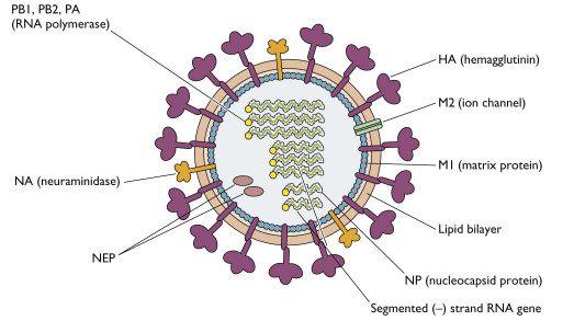
  • Size: It is a very small virus (approximately 40–50 nm in diameter).
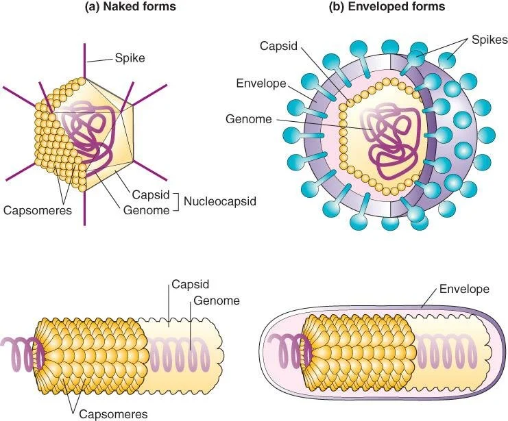
  • Structure: It is non-enveloped (meaning it lacks a delicate outer lipid bilayer). Because it lacks this lipid envelope, the virus is highly resistant to environmental degradation, heat, drying, and routine alcohol-based hand sanitizers. It consists purely of a tough, icosahedral protein capsid protecting its DNA.
  • Genome: It is a double-stranded DNA (dsDNA) virus.
  • Oncogenic Potential: Like other viruses in the polyomavirus family, it is naturally capable of inducing tumorigenesis (cancer formation) under specific host conditions.
  • Context: Polyomaviruses are known, well-documented pathogens of birds and mammals, including humans. The best-characterized and most heavily studied polyomavirus historically is SV40 (Simian Virus 40), which was discovered in monkeys and serves as the ultimate laboratory model for understanding how MCPyV behaves in humans.

II. Classification of the Virus & Clinical Anatomy

MCPyV belongs to a specific taxonomic lineage among the human polyomaviruses. Other members of this human-infecting family include the previously characterized BK virus (which causes nephropathy in transplant patients) and JC virus (which causes Progressive Multifocal Leukoencephalopathy [PML] in HIV/AIDS patients), as well as the novel WU and KI viruses.

  • Family: Polyomaviridae
  • Genus: Alphapolyomavirus
  • Species: Alphapolyomavirus quintihominis
  • Pathological Consequence: Causes Merkel cell carcinoma (MCC).
Clinical Anatomy

What is a Merkel Cell?

Merkel cells are specialized, oval-shaped receptor cells found deep in the stratum basale (the deepest layer of the epidermis) of the skin. They are densely innervated by somatosensory nerve endings and are uniquely responsible for high-resolution light touch sensation (e.g., feeling the fine texture of an object, reading Braille).

Because they share features with both epithelial cells (skin cells) and nerve cells (they contain neurosecretory granules filled with neurotransmitters), tumors arising from them exhibit highly specific neuroendocrine features. This makes MCC a cutaneous neuroendocrine carcinoma.


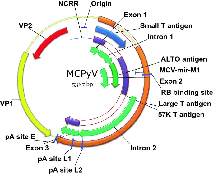
III. The Polyomavirus Genome & Life Cycle


Genomic Structure:

The viral genome is incredibly compact, highly organized, and circular. It contains approximately 5,000 base pairs. It is structurally divided into three distinct functional regions:

  1. The Regulatory Region: Contains the origin of replication (Ori) and bidirectional promoter/enhancer elements. This acts as the "control room" directing where and when transcription begins.
  2. The Early Unit: Transcribed immediately upon infection of the host cell. It encodes the non-structural, highly oncogenic proteins responsible for hijacking the cell: the Large T-antigen (LT) and the Small T-antigen (sT).
  3. The Late Unit: Transcribed later in the cycle, only after viral DNA replication has begun. It encodes the viral structural capsid proteins (VP1, VP2, and VP3) needed to physically build the new viruses.

The Viral Life Cycle (Lytic vs. Lysogenic):

The ultimate outcome of a polyomavirus infection depends entirely on whether the host cell is "permissive" (allows the virus to complete its lifecycle) or "nonpermissive."

In Permissive Cells

The Lytic Phase

The virus successfully infects the cell, commandeers the cellular machinery, and forces the cell to undergo massive, rapid viral replication. This intense manufacturing process culminates in complete cell lysis (death) and the bursting release of thousands of infectious progeny virions to infect neighboring cells. (Note: Dead cells cannot form tumors).

In Nonpermissive Cells

The Lysogenic / Transformation Phase

The virus infects the cell but cannot complete its full replication cycle. There is a total loss of lytic potential. In a rare, catastrophic biological event, the viral DNA accidentally requires integration into the host cell's chromosomal genome. Because the host cell does not die, the integrated viral DNA continually produces Early Unit proteins (T-antigens), constantly pushing the cell to divide. This leads directly to cellular transformation, immortality, and tumor formation.


IV. Virulence Factors: The Tumor Antigens (TAg)

The true oncogenic (cancer-causing) power of MCPyV lies hidden in its Early Unit proteins, specifically the Large and Small T-Antigens. These are functional polyomavirus proteins designed to bind to and degrade or sequester the host cell's natural tumor suppressors, thereby actively promoting S-phase entry (forcing the cell into the DNA synthesis phase of the cell cycle).

1. Large T Antigen (LT Antigen):

This is the major virulence protein and the primary driver of transformation.

  • Structure & Domains: The LT protein (approximately 817 amino acids long) contains several critical motifs (functional sections):
    • DnaJ (Hsc70-binding motif): Interacts with cellular chaperones to alter host protein folding and stability.
    • Rb-binding motif (LXCXE motif): The absolute critical domain for oncogenesis.
    • Ori binding domain: Binds the viral replication origin and regulatory elements to initiate viral replication.
    • Helicase domain: Unwinds DNA. Perpetuates the synthesis of a large number of progeny and induces cell lysis in permissive cells.
    • p53-binding sites: Interacts with the p53 tumor suppressor (though its exact role in MCPyV differs from SV40).
  • Mechanism of Action (The Rb Pathway): It heavily interferes with normal cell cycle regulation. It does this by aggressively binding and inactivating the ultimate tumor suppressor protein, Retinoblastoma (pRb).

Physiology Expansion: The pRb/E2F "Brakes"

Normally, pRb acts as the absolute "brakes" of the cell cycle. It holds tightly onto a transcription factor called E2F, preventing the cell from dividing. When the LT Antigen binds to pRb, it physically rips pRb away from E2F. The newly freed E2F travels straight to the nucleus and commands the cell to divide endlessly, pushing infected cells into uncontrolled, catastrophic divisions.

2. Small T Antigen (sT Antigen):

While originally thought to be secondary, sT is now considered a profoundly strong oncogenic virulence factor in its own right.

  • Mechanism of Action: Enhances viral replication and cell transformation by activating cellular pathways involved in rampant cell growth and survival. Specifically, it aggressively inhibits protein phosphatase 2A (PP2A).

Physiology Expansion: The PP2A "Off Switch"

PP2A is a vital cellular enzyme that removes phosphate groups from signaling proteins. Removing phosphates normally acts to turn "off" powerful growth signals (like the MAP kinase and mTOR pathways). By physically inhibiting PP2A, the sT Antigen ensures that cellular growth pathways stay permanently switched "on," leading to abnormal signaling and massively increased, unrestrained cellular proliferation.

🧠 Mnemonic: The Actions of the T-Antigens

To easily remember exactly how the virus causes cancer on an exam, think: "Large Rb, Small PP"

  • Large T-Antigen blocks pRb (Releasing the brakes on the cell cycle).
  • Small T-Antigen blocks PP2A (Keeping the growth signals permanently turned on).

V. Infection Dynamics: Immune Evasion, Persistence & Latency

How does a virus that infects almost the entire human population only cause cancer in a very select few? The answer lies in the dynamic, lifelong interplay between the virus's stealth mechanisms and the host's immune system.

1. Immune Evasion:

The virus usually causes a completely asymptomatic infection in healthy individuals. It evades immune detection through three primary mechanisms:

  • Reduced presentation of viral antigens to surveying T-cells.
  • Direct molecular interference with host immune signaling pathways.
  • Quiet persistence inside host skin cells as a circular episome without causing immediate damage (avoiding the triggering of inflammatory alarms).

2. Viral Integration into Host Genome:

In Merkel cell carcinoma, the viral DNA does not stay as a free-floating circle (episome). Instead, it undergoes clonal integration directly into the host cell's chromosomes. This accidental integration makes the infected cells continuously, permanently express oncogenic proteins (the T-antigens), which contributes directly to irreversible malignant transformation.

3. Persistence and Latency:

Most people in the general population are actually infected with MCPyV during early childhood (likely through skin-to-skin contact). The virus establishes long-term persistence (latency) in the skin and remains harmless (asymptomatic) for decades, as long as the host's robust immune system keeps it strictly in check.

The Trigger for Disease: MCC mainly develops when host cellular immunity is severely weakened, allowing the virus to reactivate and mutate. High-risk populations include:

  • Elderly individuals (due to natural immunosenescence—the aging of the immune system).
  • Immunocompromised patients (e.g., individuals living with uncontrolled HIV/AIDS).
  • Organ transplant recipients (who are actively and permanently taking immunosuppressive drugs, like Tacrolimus or Cyclosporine, to prevent organ rejection).

❓ Applied Clinical Question: Viral Integration

Question: A 65-year-old kidney transplant patient develops a rapidly growing, painless, purple nodule on his neck. A biopsy reveals Merkel Cell Carcinoma. If the virus has been living harmlessly in his skin since childhood, why did the tumor only form now, and what specific molecular event had to occur to the viral DNA for this to happen?

Answer: The tumor formed now because his immune surveillance was artificially suppressed by the lifelong anti-rejection drugs required for his kidney transplant. However, immunosuppression alone doesn't instantly cause the cancer. The absolutely required molecular event is Viral DNA Integration into the host genome. The virus transitioned from a quiet, latent, free-floating state to being permanently stitched into the host DNA. This resulting architectural change triggered the continuous, unstoppable production of Large and Small T-Antigens that forced the cell into malignant transformation.


VI. Merkel Cell Carcinoma (MCC): Clinical Features

Before the discovery of MCPyV, Merkel Cell Carcinoma was known only as the deadliest form of skin cancer with a totally unknown origin/etiology. It is characterized by a rapidly increasing number of cases globally, frequent early metastases to regional lymph nodes, and a historical lack of effective treatments.

Clinical Presentation & Risk Factors:

  • It presents as an aggressively fast-growing skin cancer.
  • Usually appears as a painless nodule that may be red, purple, pink, or skin-colored, frequently possessing a shiny surface.
  • Enlarged, firm lymph nodes are palpable if regional metastasis has already occurred (which is common at the time of initial diagnosis).
  • High-Risk Demographics: It is highly prevalent in immunosuppressed patients (HIV, organ transplants, chronic lymphocytic leukemia patients) and is vastly more common in fair-skinned individuals.
  • Location: Commonly occurs on severely, chronically sun-exposed areas such as the face, neck, and extensor surfaces of the arms. (UV radiation acts as a potent local immunosuppressant and a DNA-damaging mutagen).
🧠 Mnemonic

The Clinical Features of MCC: "AEIOU"

Dermatologists use the "AEIOU" mnemonic to rapidly identify suspected Merkel Cell Carcinoma in a clinical setting:

  • Asymptomatic (Painless nodule, lack of tenderness).
  • Expanding rapidly (Doubling in size in weeks, not months).
  • Immunosuppression (History of transplant, HIV, or chronic disease).
  • Older than 50 (Peak incidence in the 70s).
  • UV-exposed skin (Occurs predominantly on sun-damaged, fair skin).

VII. Diagnosis and Treatment of MCC

Diagnosis:

  • Clinical Examination: Suspicious, rapidly growing skin lesions matching the AEIOU criteria are thoroughly examined.
  • Skin Biopsy: Confirms the diagnosis histologically. Under the microscope, MCC presents classically as a "small round blue cell tumor" with high mitotic activity (many cells visibly dividing) and distinct neuroendocrine features.
  • Immunohistochemistry (IHC): Essential to differentiate MCC from other small round blue cell tumors (like metastatic small cell lung cancer, melanoma, or lymphoma). Typical markers include:
    • CK20 positive: Cytokeratin 20 shows a highly characteristic "dot-like" pattern (perinuclear accumulation of intermediate filaments). This is the hallmark diagnostic stain.
    • Chromogranin: Positive (confirms neuroendocrine origin by highlighting neurosecretory granules).
    • Synaptophysin: Positive (also confirms neuroendocrine origin).
    • Note: MCC is typically Thyroid Transcription Factor-1 (TTF-1) negative, which helps differentiate it from small cell lung cancer (which is TTF-1 positive).

Treatment Modalities:

  • Surgery: Wide local excision (WLE) with massive margins is the primary, main treatment for localized disease. Sentinel lymph node biopsy is universally performed to check for microscopic spread.
  • Radiotherapy: Often used after surgery (adjuvant) to kill any remaining microscopic cancer cells in the tumor bed or draining lymph nodes, as MCC is remarkably radiosensitive.
  • Immunotherapy: A massive, recent breakthrough for advanced, metastatic MCC. Important drugs include Pembrolizumab, Avelumab, and Nivolumab.
    Physiology Expansion: Because MCC is fundamentally driven by a foreign virus, the tumor cells are highly immunogenic (they look foreign to the body). To survive, tumors express PD-L1 to put the immune system to sleep. These immunotherapy drugs are PD-1/PD-L1 inhibitors that "take the brakes off" the patient's immune system, allowing circulating T-cells to recognize and destroy the virally infected cancer cells.
  • Chemotherapy: Used as a salvage therapy in advanced/metastatic disease, but clinical responses are notoriously short-lived, and the cancer almost always recurs.

VIII. The MCPyV Hypothesis & Genomic Evidence

Researchers investigating MCC tumors discovered low copy number polyomavirus-like transcripts in 80% to 90% of all MCC tumors globally. Furthermore, they definitively found that the viral DNA had undergone clonal DNA integration into the human genome, meaning the virus was present before the tumor expanded.

The T-Antigen Truncation Mutation (Extremely High-Yield Concept)

Sequencing of the integrated viral genome in MCC tumors revealed highly specific point mutations in the TAg-encoding gene. These specific mutations are predicted to result in the synthesis of a truncated (shortened) protein due to premature stop codons.

Two distinct truncating mutations were identified (labeled MCV350 and MCV339). Only two MCPyV genomes were initially sequenced to find this profound pattern.


💡 Why MUST the T-Antigen be truncated to cause cancer?

This is the core, beautiful paradox of MCPyV oncogenesis!

The wild-type (WT) virus wants to replicate. Its Large T-Antigen has two main functional ends: a front end that binds pRb (forcing the cell to replicate its DNA to provide building blocks for the virus) and a back end with a Helicase domain (which utilizes those building blocks to replicate the viral genome and ultimately kills/lyses the cell to escape).

If the virus replicates and kills the cell, cancer cannot form (dead cells don't form tumors). Therefore, for a tumor to develop, the viral DNA integrates into the host chromosome and suffers a highly specific truncating mutation that literally chops off the Helicase domain. The resulting truncated T-Antigen still possesses the front end to block pRb (forcing the cell to divide forever), but it can no longer replicate the virus or lyse the cell. The cell is permanently trapped in an infinite division cycle without dying, resulting in a tumor!

The Working Hypothesis:

  • Normal Pathway: Wild-type MCPyV infects permissive human cells, successfully replicates using intact helicase, and causes cellular lysis, which releases infectious progeny into the environment.
  • Oncogenic Pathway: Mutated MCPyV infects a cell and accidentally integrates into its genome. This results in tumor transformation because the truncated TAg forces division without lysis. The transformed cell initiates tumors, which rapidly expand and lead to further aggressive metastases.

IX. Research Aim 1: The Transforming and Oncogenic Potentials of MCPyV

Scientists utilized specific molecular assays to definitively prove that MCPyV could cause cancer.

Question: Does MCPyV T-antigen immortalize human cells in vitro?

  • Background: SV40 TAg immortalizes mammalian cells in vitro. Normally, primary laboratory cells divide a set number of times (the Hayflick limit) and senesce (die of old age). Transformed cells proliferate indefinitely. If MCPyV TAg causes cellular transformation, primary cells should become immortalized.
  • Experiment: MCPyV and SV40 TAgs were artificially delivered to cultured cells using modified retroviruses.
  • Result: MCPyV may cause cellular transformation, but NO, it does not immortalize cells exactly in the same robust manner as SV40. The p53-binding sites may be required (which MCPyV lacks or utilizes differently). Wild-Type (WT) TAg is continually used to further refine and answer this discrepancy.

Question: Can MCPyV cause tumors in laboratory animals?

  • Background: Murine (mouse) PyV causes tumors in mice; SV40, JC, and BK viruses rapidly induce brain tumors when injected into hamsters and rats. Is MCPyV also a definitive tumor-causing agent in animals?
  • Experiment: Inject newborn, immunologically naive hamsters with the virus and systematically examine their bodies for the development of tumors. Presence of a tumor strongly suggests in vivo oncogenic potential.
  • Result: NO direct tumors formed in standard animal models using simply the virus. Complex mutations in VP (viral protein) genes or artificial SV40/JC/BK/MC recombinants are strictly needed to answer this question definitively in animal models.

X. Research Aim 2: Identification of WT MCPyV & Infectivity

  • Question 1: Is Wild-Type (WT) MCPyV present in non-tumor tissues of MCC patients or the environment (air, dust, parasites)?
    • Action: Researchers utilized targeted PCR (Polymerase Chain Reaction) to amplify the TAg and VP-coding genes from various environmental and tissue swabs, and sequenced the resulting viral DNA to find out the virus's natural reservoir.
  • Question 2: Is WT MCPyV capable of lytic growth?
    • Background: Polyomaviruses natively cause lytic death of permissive cells. The WT (whether a clinical isolate or laboratory recombinant) MCPyV is theoretically expected to be capable of lytic growth. Lytic growth results in a massive abundance of free virions and allows measuring infectivity via a standard plaque assay.
    • Result: The truncated TAg found specifically in tumors is strictly and biologically incapable of driving the lytic phase. Recombinant WT viruses generated in the lab may contain additional, unknown mutations depriving such a virus from lytic potential in culture. Thus, while WT MCPyV easily infects humans globally, it ONLY causes tumors upon the catastrophic loss of lytic potential via mutation.

XI. Research Aim 3: Mechanism of MCPyV-Induced Transformation

This aim explores the exact, granular molecular biology of how the viral proteins hijack the human cell cycle.

  • Protein Interactions: Do MCPyV proteins interact with cellular partners?
    • Tested using: Co-immunoprecipitation (using antibodies against TAg, VP, and cellular proteins to pull them out of solution together), Yeast two-hybrid systems (to test for direct interactions between TAg and cellular tumor suppressors from a genetic expression library), and BiFC (Bimolecular Fluorescence Complementation).
    • Result: Yes, MCPyV proteins interact intimately with specific cellular partners, directly leading to transformation.
  • Stability & Expression: Is the stability of mRNA and expression of MCPyV proteins affected?
    • Tested using: Proteins were analyzed via Western hybridization (blot). mRNA stability was tested via quantitative rtPCR (DTS).
    • Result: MCV339 and MCV350 (the truncated, mutated tumor proteins) are highly stable and synthesized continuously without degradation.
High-Yield Molecular Biology

Epistasis Analysis of TAg

Goal: Identify the exact mechanism of MCPyV TAg-mediated transformation. SV40 T-antigen is historically known to interact with both pRb and p53 to deregulate the cell cycle and simultaneously prevent apoptosis.

Experiment: Introduce TAg to cells or animal models that have already been genetically depleted (knocked out) of pRb and p53. This is called an epistasis analysis.
Logic: If removing pRb/p53 causes transformation on its own, and adding TAg causes the exact same transformation without adding any extra effects, they are scientifically operating in the exact same genetic pathway (Epistatic).

Results: Transformation occurs strictly via the disruption/transactivation of these pathways. TAg-dependent degradation of pRb is mathematically and biologically sufficient for full tumor transformation. Unknown tumor suppressors/oncogenes may also be involved but pRb is the primary key.


The Role of p53: Are p53-binding sites necessary for transformation?

Conclusion: NO. TAg-dependent degradation of pRb is entirely sufficient for tumor transformation in MCPyV, setting it apart from SV40 which heavily relies on p53 disruption.

Interestingly, researchers found that truncating mutations likely occurred after initial transformation. Translation re-initiation allows some minute synthesis of the C-terminus of the protein, aiding the tumor's survival.


XII. Summary, Significance, and Future Directions


Summary:

Merkel Cell Carcinoma (MCC) is a deadly, aggressive skin cancer with a previously completely unknown etiology and a severe lack of effective historical treatments. The compelling, molecular evidence of a novel polyomavirus capable of directly causing cancer allows for the development of highly targeted MCC treatments. MCPyV provides a direct, undeniable association between a human cancer and infectious polyomaviruses, shifting the paradigm of dermatological oncology.

Future Directions of Research:

  • Test for the presence of MCPyV in other types of poorly understood human tumors.
  • Explore the exact mechanism by which MCPyV natively spreads among humans (e.g., respiratory, fecal-oral, or direct dermal contact).
  • Test whether TAg truncation is a universal, common feature of absolutely all MCPyV-induced tumors globally.
  • Estimate true seropositivity (how many people have circulating antibodies against it) among the general, healthy human population.
  • Develop effective, targeted antiviral drugs or preventative immunologic treatments (like vaccines similar to the HPV vaccine).

❓ Final Module Review Question

Question: Based on the epistasis analysis and molecular mechanics discussed, why is immunotherapy (specifically PD-1/PD-L1 inhibitors like Pembrolizumab) a logical and highly effective treatment for an advanced Merkel Cell Carcinoma, whereas it might completely fail in a standard, non-viral melanoma?

Answer: Because MCC is fundamentally an infection-driven tumor. The tumor cells are actively and continuously expressing viral antigens (the truncated Large and Small T-Antigens). Because these are inherently foreign viral proteins, the patient's immune system naturally recognizes them as non-self and desperately wants to attack them. However, the tumor survives by hiding, overexpressing PD-L1 checkpoint proteins to "turn off" the attacking T-cells. By administering Immunotherapy to chemically block those checkpoints, the immune system "wakes up," clearly recognizes the foreign viral proteins on the cancer cells, and aggressively destroys the tumor. Standard melanomas driven purely by UV damage may lack these highly visible foreign antigens, making them harder for the immune system to target.


XIII. References & Suggested Reading

  • Feng, H., Shuda, M., Chang, Y., & Moore, P. S. (2008). Clonal integration of a polyomavirus in human Merkel cell carcinoma. Science, 319(5866), 1096-1100. (The landmark paper discovering MCPyV).
  • Shuda, M., et al. (2008). T antigen mutations are a human tumor-specific signature for Merkel cell polyomavirus. Proceedings of the National Academy of Sciences, 105(42), 16272-16277.
  • Houben, R., et al. (2010). Merkel cell polyomavirus-infected Merkel cell carcinoma cells require expression of viral T antigens. Journal of Virology, 84(14), 7064-7072.
  • Schrama, D., et al. (2012). The role of Merkel cell polyomavirus in Merkel cell carcinoma. Current Opinion in Oncology, 24(2), 141-149.
  • Nghiem, P. T., et al. (2016). PD-1 Blockade with Pembrolizumab in Advanced Merkel-Cell Carcinoma. New England Journal of Medicine, 374(26), 2542-2552. (Landmark clinical trial for Immunotherapy in MCC).

Quick Quiz

Virology Intro Quiz

Microbiology - mobile-friendly and focused practice.

Privacy: Your details are used only for quiz tracking and certificates.

Merkel Cell Polyomavirus (MCPyV) Read More »

Rubella Virus (German Measles) (1)

Rubella Virus (German Measles)

Rubella Virus (German Measles)

Module Learning Objectives

By the conclusion of this exhaustive master guide, you will be deeply conversant with:

  • The complete Virology and Pathogenesis of the Rubella virus, including its recent taxonomic reclassification.
  • The critical epidemiological factors driving transmission and the rationale behind herd immunity.
  • The distinct clinical presentations differentiating Postnatal (Acquired) Rubella from the devastating Congenital Rubella Syndrome (CRS).
  • The definitive diagnostic protocols, including serology and molecular techniques.
  • Evidence-based Prevention Strategies, focusing on the MMR/MR vaccine administration schedules, contraindications, and debunking prevalent myths.

I. Introduction & Historical Context

Rubella, commonly known as German Measles or 3-Day Measles, is an acute, typically mild, highly contagious viral infection. While it rarely causes severe complications or prolonged illness in healthy postnatal populations (children and adults), it is clinically critical due to its devastating teratogenic (birth-defect causing) effects if contracted by a woman during early pregnancy.

Historical Milestones:

  • Origin of Name: The disease was first described as a distinct clinical entity by German physicians (including Friedrich Hoffmann and de Bergen) in the mid-18th century, which is why it was colloquially dubbed "German Measles." In 1814, George Maton officially suggested it be considered a distinct disease from standard measles and scarlet fever. The word "Rubella" was coined in 1866 by Henry Veale, derived from Latin, meaning "little red."
  • Teratogenic Discovery: The devastating fetal effects of Rubella were not realized until 1941. Australian ophthalmologist Dr. Norman McAlister Gregg meticulously documented a sudden, massive spike in congenital cataracts among infants whose mothers had contracted rubella during a massive 1940 outbreak. This was a watershed moment in medical history, proving that environmental/infectious agents could cause severe birth defects.

II. Virology & Classification

Historically classified under the Togaviridae family alongside mosquito-borne alphaviruses, modern genetic sequencing has recently forced virologists to reclassify the Rubella virus into its own completely distinct family.

Taxonomic Classification:

  • Domain / Kingdom / Phylum: Not applicable (viruses are acellular entities and do not fit into the standard cellular biological kingdoms).
  • Order: Hepelivirales
  • Family: Matonaviridae (Formerly Togaviridae). The family is named in honor of George Maton.
  • Genus: Rubivirus (For decades, Rubella was the sole member of this genus, though recently, rubella-like viruses such as Ruhugu virus and Rustrela virus have been discovered in animals).
  • Species: Rubella virus

Morphology & Genomic Structure:

  • Genome: It possesses a Single-stranded RNA (ssRNA) genome. It is Positive-sense (+), linear, and non-segmented, with a size of approximately 9.7 to 10 kilobases (kb).
  • Shape & Size: The virus is spherical and measures roughly 50 to 70 nanometers (nm) in diameter. It contains a dense, electron-rich inner core of 30-35 nm containing the RNA and capsid proteins.
  • Capsid: It exhibits strict icosahedral symmetry.
  • Envelope: It is a lipid-enveloped virus. The lipid bilayer is stolen (derived) directly from the host cell's intracellular membranes (like the Golgi apparatus) during the budding process. Because it relies on a delicate lipid envelope, the virus is highly labile , meaning it is fragile outside the body and easily destroyed by heat, UV light, lipid solvents, acidic pH (<6.8), and standard chemical detergents/soaps.
  • Surface Projections (Spikes): The envelope is studded with distinct spike-like heterodimeric glycoproteins, specifically E1 and E2.
    • E1 is the major structural protein acting as the primary immunogen (the target for neutralizing antibodies) and the hemagglutinin responsible for attaching to host cells and fusing membranes.
    • E2 assists in receptor binding and viral assembly.
Pathophysiology Expansion

The Mechanism of "Positive-Sense" (+) RNA

Why does it matter clinically and virologically that Rubella is a "positive-sense" (+) virus? Human cellular ribosomes can only read RNA that is oriented in the 5' to 3' (+) direction (which is what standard human mRNA looks like) to manufacture proteins.

Because Rubella's genome is already oriented in the (+) direction, it is "infectious" the moment it enters the host cell's cytoplasm. It does NOT need to enter the host's nucleus to replicate, nor does it need to carry its own polymerase upon entry. The host ribosomes are tricked; they immediately bind to the viral RNA and start reading it like regular mRNA, instantly manufacturing viral non-structural proteins (like RNA-dependent RNA polymerase) to kickstart massive viral replication.


III. Epidemiological Determinants

The transmission dynamics of the Rubella virus follow the classic epidemiological triad: Agent, Host, and Environment. While less contagious than Rubeola (standard measles), it remains highly infectious in non-immune populations.

1. Agent Factors

Asymptomatic Spread: A massive epidemiological challenge with Rubella is that a large proportion of infections (20% to 50%) are entirely sub-clinical (asymptomatic). People shed the virus and infect others without ever developing a rash or realizing they are sick. This makes contact tracing and isolating outbreaks extremely difficult.

2. Host Factors
  • Age: In unimmunized populations, it is primarily a mild disease of childhood (peaking between ages 3 and 10 years).
  • Immunity: Natural infection confers robust, lifelong immunity; second symptomatic attacks are incredibly rare.
  • Vulnerability: Males and females contract the virus equally. However, in regions lacking robust, sustained immunization programs (e.g., parts of Sub-Saharan Africa and Southeast Asia), up to 40% of women reach childbearing age remaining susceptible to the virus. This creates an unacceptably high risk for Congenital Rubella Syndrome.
3. Environmental Factors

Seasonality: In temperate zones, outbreaks follow a distinct seasonal pattern, peaking during the late winter and early spring. Historically, major epidemics occurred in cyclical waves every 4 to 9 years before vaccine introduction. In tropical climates (like Uganda), transmission can occur year-round but frequently peaks during dry seasons. Overcrowded environments (military barracks, schools, daycares) facilitate explosive localized outbreaks.

Transmission Dynamics:

  • Route of Transmission: Transmitted directly from person to person via inhalation of respiratory droplet nuclei (expelled from the nose and throat during coughing, sneezing, or talking). It can also cross the placental barrier (vertical transmission).
  • Incubation Period: Ranges from 14 to 21 days (with a reliable average of 18 days).
  • Period of Communicability: A patient is highly contagious from 1 week before the onset of any symptoms or rash, continuing until about 1 week after the rash fully appears. Furthermore, infants born with Congenital Rubella Syndrome act as massive reservoirs; they can shed live virus in their urine and pharyngeal secretions for up to 12 months after birth, posing a severe risk to non-immune healthcare workers and pregnant relatives.

IV. Pathogenesis: How the Virus Destroys Tissue

The journey of the Rubella virus from initial exposure to clinical manifestation involves several distinct phases of replication and systemic spread.

  1. Entry & Primary Replication: The virus is inhaled and implants in the respiratory epithelium. It replicates locally in the mucosa of the upper respiratory tract (nasopharynx, tonsils) and drains into the regional (cervical) lymph nodes, causing them to swell early in the disease course.
  2. Primary Viremia: Roughly 5 to 7 days post-exposure, the virus enters the bloodstream, disseminating to the reticuloendothelial system (spleen, liver) for further, massive replication.
  3. Secondary Viremia: A massive wave of virus re-enters the blood, seeding the skin, joints, kidneys, and, critically, the placenta.
  4. Rash Formation: Interestingly, the characteristic maculopapular rash is not primarily caused by direct viral destruction of skin cells. It is an immune-mediated reaction (Type III Hypersensitivity). As the body produces antibodies, they bind to viral antigens, forming antigen-antibody immune complexes that deposit in the skin's capillary beds, causing localized inflammation and the red rash.
  5. In Pregnancy (Teratogenesis): If maternal viremia occurs, the virus rapidly crosses the placenta, infecting the fetal chorion and establishing a persistent, chronic infection in fetal tissues. The virus does not simply kill cells outright; instead, it exhibits specific cytopathic effects:
    • Inhibition of Mitosis: It severely slows down cellular division.
    • Chromosomal Breakage: It causes severe DNA damage.
    • Apoptosis: It triggers programmed cell death in developing tissues.
    Because the first trimester (weeks 1–12) is the critical period of organogenesis (the initial formation of the heart, brain, and eyes), this cellular arrest leads to massive, irreversible organ malformations. The risk of congenital defects is >90% if contracted in the first 11 weeks of gestation, dropping to roughly 20% by week 16, and becomes negligible after week 20 as organ structures are already fully formed.
Mnemonic

The Rubella Lymph Nodes: "POP goes the Rubella"

Rubella is famous for distinct, tender swelling of specific lymph nodes that occurs before the rash even appears. To remember the locations, think POP:

  • Post-auricular (behind the ears)
  • Occipital (at the base of the skull)
  • Posterior cervical (down the back of the neck)

V. Clinical Presentation

The clinical manifestations of Rubella vary drastically depending on whether the infection is postnatal (acquired naturally after birth) or congenital (acquired in utero via maternal blood).

A. Postnatal (Acquired) Rubella

Often so mild that it goes unnoticed or is misdiagnosed as a common cold.

  • Prodromal Symptoms: Precede the rash by 1-5 days. Includes general malaise, low-grade fever (rarely exceeding 38.3°C / 101°F), headache, coryza (stuffy/runny nose), mild non-purulent conjunctivitis (red eyes), and the classic tender POP lymphadenopathy.
    Clinical Sign: Patients may exhibit Forchheimer Spots—small, pinpoint red/petechial macules located on the soft palate of the mouth, appearing just before the skin rash.
  • The Rash (3-Day Measles):
    • A pink-to-red maculopapular rash (consisting of flat and slightly raised spots). It is less aggressively red and less confluent than standard Measles.
    • Progression (Cephalocaudal spread): It begins on the face and hairline, then rapidly spreads downward to the neck, trunk, and extremities within 24 hours.
    • Resolution: It rarely lasts more than 5 days, reliably clearing up within 3 days. It fades and disappears in the exact same order it appeared (face first, then body). It is often accompanied by mild pruritus (itching) and occasionally fine desquamation (flaking, peeling skin) as it completely resolves. Adult Symptoms: While children usually brush off a postnatal rubella infection easily, adults (and especially adult females) experience a much more aggressive inflammatory response. They are highly prone to severe arthralgia (joint aching) and arthritis (active joint inflammation). This typically presents as a symmetrical polyarthritis affecting the fingers, wrists, and knees. It can persist for weeks or even months after the rash has disappeared, closely mimicking early Rheumatoid Arthritis.

  • B. Congenital Rubella Syndrome (CRS)

    This is the catastrophic core of why Rubella is so feared in medicine. If a pregnant woman contracts the virus (even if she is completely asymptomatic), the virus establishes a ferocious viremia and crosses the placental barrier to infect the developing fetus.

    Risk by Gestational Age: If the maternal infection occurs in the first trimester (the first 12 weeks), there is a >90% chance the baby will be born with severe CRS. The risk drops to ~20% by week 16, and fetal organ damage is exceedingly rare if the infection occurs after week 20 (because the organs have already finished primary organogenesis).

    The Classic CRS Triad

    Every medical and nursing board exam tests this foundational triad. If an infant has these three defects, suspect CRS:

    1. Sensorineural Deafness: The absolute most common major defect. It is often bilateral. If a mother is infected slightly later in pregnancy (weeks 13-16), deafness may be the only clinical manifestation the child is born with.
    2. Eye Defects: The hallmark is bilateral Nuclear Cataracts (the lenses are cloudy, white, and opaque at birth). Other ocular defects include "salt and pepper" retinopathy, congenital glaucoma, and microphthalmia (abnormally small eyes).
    3. Congenital Heart Disease: The virus profoundly disrupts the formation of fetal blood vessels. The most classic and heavily tested cardiac anomaly is a Patent Ductus Arteriosus (PDA) (a failure of the fetal vessel connecting the pulmonary artery to the aorta to close after birth, resulting in a continuous "machine-like" murmur). Pulmonary Artery Stenosis and Ventricular Septal Defects (VSD) are also common.
    Other Neonatal Features

    The virus causes widespread systemic damage beyond the triad:

    • Central Nervous System: Microcephaly (abnormally small head/brain size leading to severe intellectual disability) and meningoencephalitis.
    • Visceral Organs: Hepatosplenomegaly (massive enlargement of the liver and spleen) coupled with severe neonatal jaundice.
    • "Blueberry Muffin" Rash: A visually striking and pathognomonic sign. Because the virus suppresses the infant's bone marrow, the infant's skin attempts to manufacture blood cells itself (a process called extramedullary hematopoiesis). This creates dark blue/purple, raised purpuric lesions all over the infant's body, resembling a blueberry muffin.
    • Growth: Extreme Intrauterine Growth Restriction (IUGR), resulting in dangerously low birth weights.

    Late-Onset Complications of CRS:

    The tragedy of CRS is that the virus can linger in the child's tissues for years, triggering delayed autoimmune-like destruction. Conditions that may not appear until childhood or adolescence include delayed-onset Autism Spectrum Disorders, Schizophrenia, severe learning difficulties, autoimmune thyroiditis, and a significantly heightened risk of developing Type 1 Diabetes Mellitus.

    ❓ NCLEX-Style Question: Recognizing the Triad

    Case: A neonate is born to a mother who recently emigrated from a region with historically low immunization rates. The mother reports experiencing a mild, short-lived rash and severe wrist joint pain during her second month of pregnancy. During the newborn assessment, the infant is noted to have a continuous "machine-like" heart murmur, demonstrates no startle reflex to loud noises, and the nurse notes bilaterally absent red reflexes in the eyes. What syndrome does this describe?

    Answer & Rationale: Congenital Rubella Syndrome (CRS). The symptoms perfectly map to the classic triad:
    1. Congenital heart defect (machine-like murmur = PDA).
    2. Sensorineural deafness (no startle reflex to loud claps).
    3. Cataracts (white, cloudy lenses block light, causing an absent red reflex during an ophthalmoscope exam).
    Furthermore, the mother's history of a first-trimester rash coupled with joint pain (arthralgia) is the classic presentation of maternal Rubella.


    VI. Laboratory Diagnosis

    Because the classic Rubella rash looks practically identical to several other mild viral exanthems (like Parvovirus B19 / Fifth Disease, mild Rubeola, Adenovirus / Roseola, and even some drug allergies), a definitive diagnosis cannot be made on clinical features alone. Laboratory confirmation is absolutely legally and clinically required, especially in pregnant women.

    • 1. Serology (The Most Important Method):
      • Rubella-specific IgM: The body's "first responder" antibody. Its presence indicates an acute, recent, or currently active infection. It peaks at 7-10 days and fades within 4 weeks.
        Clinical Note for Neonates: If a newborn's blood tests positive for Rubella IgM, it definitively confirms CRS. Maternal IgM is physically too large to cross the placenta; therefore, if IgM is in the baby's blood, the baby's own immune system manufactured it in response to the virus inside them.
      • Rubella-specific IgG: The "memory" antibody. Indicates a past infection or established immunity from vaccination. (If a young infant has IgG that persists and actually rises beyond 6 months of age, it confirms infection, because passive maternally-derived IgG would have naturally degraded and faded by that time).
      • Rising IgG Titer: If you take a blood sample on day 1 (acute phase) and another sample 14-21 days later (convalescent phase), a 4-fold increase in the amount of IgG confirms a recent infection.
      • Common Assays: Enzyme-Linked Immunosorbent Assay (ELISA), Haemagglutination inhibition test (HAI), and specific radio-immune assays.
    • 2. Molecular Methods (RT-PCR):
      • Reverse Transcription Polymerase Chain Reaction (RT-PCR) detects the actual viral RNA. This is highly useful for very early infections (before antibodies have formed) and for diagnosing congenital cases. Viable specimens include blood serum, deep throat/nasopharyngeal swabs, or urine.
    • 3. Prenatal Diagnosis (For the Fetus):
      • Can be performed aggressively by drawing Amniotic Fluid via amniocentesis for RT-PCR analysis, or by conducting fetal blood sampling (Percutaneous Umbilical Blood Sampling - PUBS) to detect fetal IgM. (Note: The fetal immune system does not reliably produce IgM until roughly 22 weeks gestation, making early serology difficult).

    VII. Management & Complications

    General Treatment

    There is absolutely no specific antiviral therapy (no "Rubella-vir") capable of curing the Rubella virus. Management is strictly supportive. This involves Antipyretics (like Paracetamol/Acetaminophen) to control fever and relieve severe joint pain, maintaining hydration, and bed rest. Aspirin should be strictly avoided in children due to the risk of Reye's Syndrome.

    Management in Pregnancy

    This represents a profound medical and ethical challenge. If a pregnant woman is confirmed to have an acute Rubella infection, there is no medical treatment capable of saving the fetus or reversing the massive cellular damage already inflicted.
    Intervention: Intensive counseling is essential. Depending on the exact gestational age (especially if the infection occurs <12 weeks), pregnancy outcomes and potential medical termination (abortion) must be actively discussed with the parents due to the extreme, near-guaranteed risk of severe, life-altering congenital defects. In cases where termination is refused, Intravenous Immunoglobulin (IVIG) can theoretically be administered to the mother, but it does not guarantee prevention of viral transmission to the fetus.

    Complications of Rubella

    Adults / Children
    • Arthritis & Arthralgia: Very common in adult women.
    • Thrombocytopenic Purpura (ITP): The virus transiently attacks blood platelets (dropping counts to dangerous levels), causing spontaneous bleeding, petechiae, and bruising.
    • Post-infectious Encephalitis: A rare but lethal complication (occurring in roughly 1 in 6,000 cases) where the brain swells massively following the infection.
    • Guillain-Barré Syndrome: A rare, ascending autoimmune paralysis triggered by the viral infection.
    Pregnancy

    Aside from the teratogenic effects of CRS on a surviving infant, the virus poses an intense threat to the viability of the pregnancy itself.

    • Spontaneous Abortion: There is a staggering 20% risk of miscarriage.
    • Intrauterine Fetal Demise (Stillbirth).
    • Early Neonatal Death: Infants born with severe CRS often succumb to massive heart failure or overwhelming systemic infection within days of birth.

    VIII. Prevention and Control (The MMR Vaccine)

    Because there is no cure and CRS is so catastrophic, aggressive, universal, herd-immunity-driven vaccination is the only way to prevent Rubella and eradicate CRS from the human population.

    The Vaccine Formulation:

    The MMR vaccine (Measles, Mumps, Rubella) utilizes the highly effective RA27/3 strain of the rubella virus, which is grown in human diploid cell cultures.
    WARNING: Because it is a LIVE ATTENUATED (weakened but alive) virus, it is strictly CONTRAINDICATED in two major populations:

    1. Pregnant Women: Due to the theoretical risk that the live vaccine strain could cross the placenta and cause CRS. Women of childbearing age receiving the vaccine must be actively counseled to avoid becoming pregnant for at least 28 days post-vaccination.
    2. Severely Immunocompromised Patients: E.g., HIV patients with a CD4 count < 200, active leukemia patients, or those on high-dose immunosuppressive corticosteroid therapies. The weakened virus could run rampant in a host with no immune defenses.

    Administration & Schedule:

    • Route: Administered exclusively via Subcutaneous (SC) injection into the fatty tissue of the upper arm or thigh.
    • Standard International / CDC Schedule:
      • Dose 1: Administered at 12 to 15 months of age.
      • Dose 2: Administered before school entry, at 4 to 6 years of age.
    • Uganda (MOH / UNEPI) Schedule: The Ugandan Ministry of Health utilizes a bivalent MR (Measles-Rubella) formulation to combat high endemic rates.
      • MR 1: Administered at 9 months of age.
      • MR 2: Administered at 18 months of age (delivered in the left upper arm).
    Vaccine Physiology Note

    The "Second Dose" is NOT a Booster

    It is a common clinical misconception that the second MMR/MR dose is a "booster" meant to refresh waning immunity. This is false. A single dose of the Rubella vaccine yields robust immunity in about 95% to 98% of people. The second dose is given solely to provide a "second chance" to the 2% to 5% of individuals whose immune systems completely failed to seroconvert (failed to create antibodies) after the first dose—a phenomenon known as primary vaccine failure. By administering two doses, population immunity is pushed to ~99%.

    Side Effects vs. Myths:

    • Actual Side Effects: They are minimal and generally self-limiting. Approximately 15% of recipients may develop a mild, non-contagious fever 7-12 days after the injection. About 5% may develop a minor, transient rash. Teenage and adult women frequently experience temporary joint aches (arthralgia) reflecting the body's immune response to the rubella component. Severe, life-threatening reactions like anaphylaxis or severe thrombocytopenia are astronomically rare (< 1 in 1,000,000 doses).
    • The Autism Myth: It must be aggressively stated in all clinical counseling that there is absolutely no scientific, epidemiological, or biological link between the MMR vaccination and the development of autism. This myth was birthed from a fraudulent, retracted, and widely debunked 1998 paper by Andrew Wakefield. The dangers of remaining unvaccinated (permanent deafness, blindness, severe brain damage, and infant death) exponentially outweigh any theoretical or minor adverse effects of the vaccine.
    • The Logic of Herd Immunity: Why do we vaccinate young boys for a disease that primarily causes birth defects in pregnant women? Herd Immunity. By immunizing males and children, we eliminate the virus's ability to circulate in the community, creating a protective "shield" around vulnerable pregnant women and ensuring the virus never reaches the unborn fetus.

    References & Evidence-Based Guidelines

    • World Health Organization (WHO): Rubella vaccines: WHO position paper. Wkly Epidemiol Rec. (Provides the global rationale for MR vaccine integration into routine EPI schedules).
    • Centers for Disease Control and Prevention (CDC): The Pink Book: Epidemiology and Prevention of Vaccine-Preventable Diseases (Chapter on Rubella). (Excellent resource for detailed pathophysiology and MMR contraindications).
    • American Academy of Pediatrics (AAP) & Advisory Committee on Immunization Practices (ACIP): Recommended Child and Adolescent Immunization Schedule.
    • Uganda Ministry of Health (MOH) / UNEPI: National Routine Immunisation Schedule Guidelines. (Specific to the 9-month and 18-month MR administration protocols).
    • Mandell, Douglas, and Bennett's: Principles and Practice of Infectious Diseases. (The definitive textbook for deep-dive molecular virology, the Matonaviridae reclassification, and the pathogenesis of Congenital Rubella Syndrome).

Quick Quiz

Virology Intro Quiz

Microbiology - mobile-friendly and focused practice.

Privacy: Your details are used only for quiz tracking and certificates.

Rubella Virus (German Measles) Read More »

General Structure and Classification of Viruses

General Structure and Classification of Viruses

General Structure, Classification, and Life Cycle of Viruses

Module Learning Objectives

By the conclusion of this exhaustive master guide, you will be deeply conversant with:

  • The historical discovery and fundamental, defining characteristics of viruses as obligate intracellular parasites.
  • The detailed architectural components of a virion (capsids, envelopes, and internal enzymes) and their clinical implications.
  • The morphological symmetry of viruses (Icosahedral, Helical, and Complex).
  • The rigid frameworks of viral taxonomy, including the ICTV Hierarchical System and an expanded, comprehensive Baltimore Classification System.
  • The exact, step-by-step molecular pathophysiology of the Viral Life Cycle (from adsorption to release).
  • The advanced laboratory modalities utilized in the structural investigation of virology.

I. The Concept and History of the Virus

Viruses are incredibly unique, sub-microscopic infectious agents that bridge the gap between living biology and non-living chemistry. Unlike bacteria, fungi, or parasites, viruses are not strictly considered "living" organisms because they cannot reproduce independently, generate their own energy, or perform metabolic processes outside of a host cell.

Historical Milestones in Virology:

  • Edward Jenner (1798): Introduced the term "virus" (which literally translates to "poison" or "venom" in Latin/Greek) to the field of microbiology.
    • The First Vaccine: Jenner observed that milkmaids who contracted cowpox (a mild, localized disease) rarely contracted smallpox (a highly contagious, deadly systemic disease). He hypothesized that the vesicle fluid from the cowpox-infected maid contained a specific "poison" (virus) that stimulated biological resistance.
    • Jenner famously inoculated an eight-year-old boy, James Phipps, with this cowpox vesicle fluid. The boy developed a mild fever but sustained lifelong immunity against smallpox.
    • Clinical Note: This cross-immunity principle is the foundational basis for modern vaccinology and immunology. The word "vaccine" is derived directly from "vacca," the Latin word for cow, honoring Jenner's groundbreaking cowpox experiment.
  • Dmitri Ivanovsky & Martinus Beijerinck (1890s): Discovered that the agent causing Tobacco Mosaic Disease was much smaller than any known bacteria because it could easily pass through a Chamberland porcelain filter (which trapped all known bacteria). Beijerinck famously called it a "contagium vivum fluidum" (contagious living fluid), marking the true birth of virology as a discipline distinct from bacteriology.

II. General Characteristics of Viruses

To accurately understand viral pathology, modes of transmission, and pharmacological treatments, nurses, physicians, and microbiologists must thoroughly grasp the unique structural limitations of viruses.

1. Size and Visibility
  • Viruses are vastly smaller than bacteria. They typically range from 20 to 300 nanometers (nm) in diameter. (A nanometer is a milli-micron, or 10-9 meters).
  • To put this in perspective: A red blood cell is 7,000 nm, an E. coli bacterium is 1,000 nm, while the Poliovirus is merely 30 nm.
  • Due to this infinitesimal size, they cannot be seen with a standard light microscope and require electron microscopy for visualization.
2. Genetic Material (The Core Rule)
  • A virus contains only ONE type of nucleic acid—either DNA or RNA. A single viral particle will absolutely never contain both in its genome.
  • This genome can be single-stranded (ss) or double-stranded (ds), linear or circular, single-segment or multi-segmented.
3. Metabolic Incompetence
  • Viruses lack all cellular organelles, including mitochondria (cannot generate ATP/energy), ribosomes (cannot synthesize their own proteins), and a true cytoplasm.
  • Because of this profound incompetence, they do not multiply in chemically defined laboratory media (like the blood agar or MacConkey agar plates used for cultivating bacteria).
  • They do not undergo binary fission (cellular division). Instead, they replicate through the independent synthesis of their nucleic acid and viral proteins by hijacking the host cell's machinery, followed by complex self-assembly.
4. Obligate Intracellular Parasites
  • Because they entirely lack organelles, viruses are strictly obligate intracellular parasites. They can only replicate inside a living, susceptible, and permissive host cell (whether that is a human cell, an animal cell, a plant cell, or even a bacterium, known as a bacteriophage).

❓ Applied Clinical Question: Antimicrobial Stewardship

Case: A patient is diagnosed with an upper respiratory infection caused by the Rhinovirus. The patient forcefully demands an antibiotic prescription, stating, "I want medicine to kill the germs in my throat." What is the best physiological and pharmacological rationale the nurse should provide for refusing antibiotics?

Answer: Antibiotics work by targeting specific, living cellular structures that are unique to bacteria, such as peptidoglycan bacterial cell walls (targeted by Penicillin) or 70S bacterial ribosomes (targeted by Macrolides). Because viruses are not truly living cells, they completely lack cellular organelles (like ribosomes) and lack cell walls. Therefore, antibiotics have absolutely no anatomical or metabolic targets to attack on a virus. Giving an antibiotic for a viral infection will do nothing to the virus; it will only destroy the patient's normal, healthy bacterial flora, severely increasing the risk of gastrointestinal superinfections (like C. diff) and promoting global antimicrobial resistance.


III. Viral Terminology & Architectural Components

Understanding viral architecture is mandatory for grasping how viruses survive in the environment and how they infect humans. The following specific microbiological terminology is utilized worldwide:

  • Virion: The complete, fully assembled, physically intact, and infective virus particle that exists outside of a host cell. This is the vehicle that transports the viral genome from one host to another.
  • Capsid: The rigid, symmetrical protein shell/coat that entirely encloses and protects the fragile nucleic acid from physical, chemical, and enzymatic inactivation in the harsh extracellular environment.
  • Capsomeres & Structure Units: The capsid is not one solid piece of protein; it is meticulously constructed from smaller, repeating functional building blocks called structure units (or protomers). Clusters of these structure units group together to form visible, distinct morphological units on the particle's surface called Capsomeres. (e.g., Hexons and Pentons).
  • Nucleocapsid: The integrated, combined unit of the Capsid + the enclosed Nucleic Acid. In simple viruses, the nucleocapsid *is* the entire virion.
  • Tegument (Matrix Protein): An unstructured layer of proteins located between the nucleocapsid and the envelope in certain complex viruses (like Herpesviruses). These proteins are dumped into the host cell upon entry to rapidly shut down host defenses.
  • Envelope: An additional, highly sensitive outer lipoprotein layer that covers the nucleocapsid in *some* viruses. It is acquired when the virus "buds" out of the host cell, taking a piece of the host's plasma membrane, endoplasmic reticulum, or Golgi apparatus. The envelope is typically studded with virally encoded glycoprotein spikes (peplomers) that are strictly required for recognizing and attaching to new host cells.
  • Defective Virus: A mutated, damaged, or genetically incomplete virus that cannot replicate on its own. It requires a specific "helper virus" to coinfect the exact same cell simultaneously to supply the missing replication functions. (Classic Example: Hepatitis D virus is a defective virus that can only infect and replicate in a patient who is simultaneously infected with the Hepatitis B virus, which provides the necessary surface envelope proteins).
Clinical Rationale

Enveloped vs. Unenveloped (Naked) Viruses

Logically, one might assume that a virus with an extra outer "envelope" armor is much harder to kill in the hospital environment. In reality, the exact opposite is true. Enveloped viruses are much EASIER to destroy with standard hand sanitizers (alcohol), hand soap, and mild detergents.

Why? The envelope is made of a highly delicate lipid (fat) bilayer that must remain wet to survive. Soap and alcohol easily dissolve lipids, popping the envelope like a balloon and permanently destroying the glycoprotein spikes the virus relies on to infect you. (e.g., HIV, Influenza, Hepatitis B, and SARS-CoV-2 are all enveloped, making them fragile outside the body). If an enveloped virus dries out, it dies.

Unenveloped (Naked) viruses (e.g., Norovirus, Rotavirus, Adenovirus, HPV), on the other hand, do not have a fragile fat layer. They only have a tough, highly rigid, hydrophilic protein capsid. This makes them incredibly resistant to drying out, resistant to stomach acid, and highly resistant to standard alcohol hand sanitizers, requiring heavy friction, prolonged soap washing, or bleach for environmental decontamination. This is why naked viruses are frequently transmitted via the fecal-oral route (they easily survive stomach acid).


IV. Internal Viral Enzymes

While viruses lack organelles, some must pack their own specialized, pre-made enzymes inside the virion to successfully hijack the host cell. This is especially true if the human host cell doesn't naturally possess the specific biochemical machinery the virus requires.

  • Transcriptase (RNA-dependent RNA polymerase): Found in all single-stranded (ss) RNA viruses with negative polarity (e.g., Rabies, Ebola, Influenza). Humans only possess DNA-dependent RNA polymerases. We do not have enzymes that copy RNA directly into more RNA. Therefore, the virus must bring its own pre-packaged polymerase into the cell to immediately begin reading its genome.
  • Reverse Transcriptase (RNA-dependent DNA polymerase): A highly specialized enzyme that breaks the central dogma of biology by turning RNA backward into DNA. This is carried heavily by Retroviruses (like HIV) and Hepadnaviruses (Hepatitis B).
  • Integrase: Carried by retroviruses. Once the reverse transcriptase has created viral DNA, the Integrase enzyme slices open the human host's chromosomal DNA and seamlessly pastes the viral DNA permanently into the human genome (creating a "provirus").
  • Protease: An enzyme carried by many viruses (like HIV and Hepatitis C) that acts as a molecular pair of scissors. It cleaves massive, non-functional viral polyproteins into smaller, active, functional structural proteins during the final assembly stage of the viral life cycle.

Pharmacological Note: Because humans do not naturally possess Reverse Transcriptase, Integrase, or viral-specific Proteases, these enzymes act as perfect, highly selective targets for antiviral drugs (such as the HAART therapy used to treat HIV, which consists of Reverse Transcriptase Inhibitors, Integrase Inhibitors, and Protease Inhibitors).


V. Symmetry and Morphology of Viruses

Viruses are incredibly efficient. To save space in their tiny genomes, they use a single repeating protein to build their capsid. Based on how these capsomeres self-assemble, viruses are classified into three major structural groups.

1. Cubic (Icosahedral) Symmetry
  • The virus particle appears almost spherical under low magnification.
  • Structurally, it is an icosahedron—a highly efficient geometric shape composed of exactly 20 equilateral triangular faces and 12 vertices. This shape encloses the maximum volume of space with the absolute minimum amount of protein.
  • It features a mathematically perfect 2-3-5 rotational symmetry.
  • Examples: Adenoviruses, Papillomaviruses, Poliovirus (all Naked/Unenveloped); Herpesviruses (Enveloped).
2. Helical Symmetry
  • The virus particle is elongated, cylindrical, or pleomorphic (variable shape, not strictly spherical).
  • The nucleic acid is coiled in a continuous spiral, and the capsomeres are arranged tightly around the nucleic acid coil like steps on a spiral staircase, creating a long, tube-like structure. The length of the capsid is dictated precisely by the length of the nucleic acid.
  • All known human viruses with helical symmetry possess an outer lipid envelope.
  • Examples: Influenza virus, Rabies virus, Ebola virus, Measles virus (all Enveloped).
3. Complex Symmetry
  • The virus particle does not conform to either strict cubic or helical symmetry. It has a highly intricate, often multi-layered, brick-like, or ovoid structure.
  • Examples: Poxviruses (e.g., Smallpox, Cowpox) which are massive, brick-shaped viruses with a complex outer wall.
  • Another classic example is the Bacteriophage (a virus that infects bacteria), which looks like a lunar lander with an icosahedral head, a helical contractile tail sheath, and complex tail fibers used for drilling into bacterial walls.

VI. Classification Systems of Viruses

Because viruses are not cellular life forms, standard biological taxonomy (Kingdom, Phylum, Class) is incredibly difficult to apply. Instead, two primary, internationally recognized classification systems exist: The Hierarchical System and the Baltimore System.

A. The Hierarchical Virus Classification System

Advanced initially by Lwoff, R. W. Horne, and P. Tournier in 1962, this system is now strictly governed by the ICTV (International Committee on Taxonomy of Viruses). The ICTV dictates that viruses should be grouped based on their shared structural and genetic properties rather than the host cells they infect or the diseases they cause.

Four Main Characteristics Used for Grouping:

  1. Nature of the nucleic acid: (DNA or RNA, single or double-stranded, positive or negative sense).
  2. Symmetry of the capsid: (Cubic/Icosahedral, Helical, or Complex).
  3. Presence or absence of a lipid envelope.
  4. Dimensions/size: Of the overall virion and the internal capsid.

Nomenclature Rules:

  • Order: Suffix is -virales (e.g., Nidovirales).
  • Family: Always ends in the suffix -viridae (e.g., Picornaviridae, Reoviridae, Coronaviridae).
  • Subfamily: Ends in the suffix -virinae (e.g., Alphaherpesvirinae).
  • Genera: Always ends in the suffix -virus (e.g., within Picornaviridae, there are 5 genera: enterovirus, cardiovirus, rhinovirus, apthovirus, hepatovirus).

Defining a viral "species" involves significant subjectivity, but members within a family are now largely ordered by advanced Genomics (the deep evolutionary sequencing of nucleic acids and proteins). For example, SARS-CoV-2 falls under Order: Nidovirales, Family: Coronaviridae, Genus: Betacoronavirus, Species: Severe acute respiratory syndrome-related coronavirus.


VII. The Baltimore Classification System

Originated by Nobel laureate David Baltimore, this system is an incredibly practical, deeply molecular guide focused entirely on the mechanism of viral genome replication and mRNA synthesis.

💡 The Central Theme of Baltimore Classification

The Central Dogma of biology states that DNA makes RNA, and RNA makes proteins. No matter what bizarre genetic material a virus starts with, ALL viruses must eventually generate positive-strand (+) messenger RNA (mRNA).

Why? Because human host ribosomes only read (+) mRNA to produce proteins. If a virus brings DNA, double-stranded RNA, or negative-strand RNA, it cannot be read immediately. It must undergo distinct, extra enzymatic steps to convert its genome into (+) mRNA first. The Baltimore system breaks all viruses into 7 specific groups based precisely on the strategic pathway they use to reach this ultimate (+) mRNA goal.

Baltimore Group Genome Type Replication Strategy & Clinical Examples
Group I Double-Stranded DNA (dsDNA) The most straightforward group. They use standard transcription (DNA → RNA). Most replicate inside the host nucleus, hijacking the host's cellular DNA-dependent RNA polymerases.
Examples: Adenoviruses, Herpesviruses (HSV, VZV), HPV.
Exception: Poxviruses (Smallpox) are so massive they replicate entirely in the cytoplasm and must provide their own enzymes for DNA replication and transcription.
Group II Single-Stranded DNA (ssDNA) Replication occurs in the nucleus. Because ribosomes can't read ssDNA, the virus must first use host enzymes to synthesize a complementary (-) sense DNA strand, forming a dsDNA intermediate. This dsDNA then serves as the template for creating (+) mRNA.
Examples: Parvoviruses (e.g., Parvovirus B19 causing Fifth Disease).
Group III Double-Stranded RNA (dsRNA) These viruses feature highly segmented genomes. The human cell cannot transcribe dsRNA, so the virus brings an RNA-dependent RNA polymerase. Each segment is transcribed separately to produce monocistronic mRNAs (one mRNA codes for one specific protein).
Examples: Reoviruses (e.g., Rotavirus causing severe infant diarrhea), Birnaviruses.
Group IV Single-Stranded (+) RNA The viral genome is already structured exactly like human (+) mRNA! This means the naked viral RNA is immediately infectious the second it enters the cell, without needing to bring any pre-packaged virion polymerases.
Strategies:
a) Polycistronic: Creates one massive, continuous polyprotein that is subsequently cleaved by viral proteases into functional mature proteins (e.g., Picornaviruses like Polio, Flaviviruses like Hepatitis C).
b) Complex: Requires multiple rounds of translation and subgenomic RNA synthesis (e.g., Togaviruses, Coronaviruses like SARS-CoV-2).
Group V Single-Stranded (-) RNA Because the genome is (-) sense, it is "backward" and human ribosomes cannot read it. The naked RNA is non-infectious alone. The virus MUST carry its own pre-packaged RNA-dependent RNA polymerase to immediately transcribe the (-) genome into readable (+) mRNA.
Strategies:
a) Segmented: Orthomyxoviruses (Influenza virus, capable of antigenic shift).
b) Non-segmented: Rhabdoviruses (Rabies), Paramyxoviruses (Measles, Mumps), Filoviruses (Ebola).
Group VI Single-Stranded (+) RNA with Reverse Transcriptase Although they have a (+) RNA genome, they do not act like Group IV. Instead of being translated directly, their RNA is converted backward into dsDNA by a pre-packaged Reverse Transcriptase enzyme. This dsDNA is then integrated into the host's chromosome, where the host cell transcribes it into mRNA.
Examples: Retroviruses (HIV, HTLV).
Group VII Double-Stranded DNA with Reverse Transcriptase (Pararetroviruses) They have a partially double-stranded DNA genome. During replication inside the nucleus, they create a massive RNA intermediate (pregenomic RNA). This RNA is then reverse-transcribed back into DNA by a viral reverse transcriptase inside the newly forming viral capsid in the cytoplasm.
Examples: Hepadnaviruses (Hepatitis B).

VIII. The Viral Life Cycle (Replication Steps)

A virus replicating in a human cell undergoes 8 highly coordinated, distinct stages. Understanding these stages is paramount because all pharmacological antiviral therapies are engineered to chemically block one or more of these specific steps.

  1. Adsorption (Attachment):
    The virus must physically recognize and bind to highly specific cellular receptors on the host cell surface using its viral glycoproteins or capsid proteins. This interaction determines viral "tropism" (which explains why Hepatitis viruses only infect the liver, why the Rabies virus binds specifically to Acetylcholine receptors on neurons, and why HIV strictly targets CD4 receptors and CCR5 co-receptors on T-helper cells). If a human lacks the specific receptor, the virus cannot attach.
  2. Penetration (Entry):
    Once attached, the virus must breach the cell barrier.
    • A. Enveloped Viruses: Enter either through Receptor-Mediated Endocytosis (the cell is tricked into swallowing the virus in a vesicle) or Membrane Fusion. In fusion, the viral envelope lipid bilayer melts seamlessly into the host plasma membrane, dumping the nucleocapsid directly inside. Pathology note: Viruses that use fusion proteins can cause adjacent infected host cells to meld together into a massive, multi-nucleated giant cell called a Syncytia (e.g., Respiratory Syncytial Virus (RSV), Herpesviruses, HIV).
    • B. Unenveloped (Naked) Viruses: Enter primarily by Endocytosis. Once inside the endosomal vesicle, the virus must escape before the cell destroys it. It either lyses (bursts) the endosome entirely (e.g., Adenoviruses) or undergoes a conformational change to form a pore in the endosomal membrane to forcefully inject its RNA into the cytoplasm (e.g., Picornaviruses).
  3. Uncoating:
    The protective protein capsid is broken down by cellular or viral enzymes, completely releasing the naked viral genome into the cytoplasm or nucleus so it can be transcribed and replicated. (Pharmacological Note: The antiviral drug Amantadine works specifically by blocking the uncoating of the Influenza A virus inside the endosome).
  4. Transcription:
    The viral genome is transcribed to synthesize viral mRNA. Early transcription typically produces non-structural proteins (like enzymes and polymerases needed for replication), while late transcription produces structural proteins (capsomeres for the new viral shell).
  5. Translation:
    The viral mRNA officially hijacks the host cell's ribosomes, forcing them to translate the viral genetic code into massive amounts of viral structural and non-structural proteins, halting the host cell's normal protein synthesis.
  6. Replication of the Genome:
    The host cell machinery (or the newly synthesized viral polymerases) mass-produces thousands of identical copies of the viral nucleic acid. Rule of Thumb: Most DNA viruses replicate in the nucleus, while most RNA viruses replicate in the cytoplasm.
  7. Assembly (Maturation):
    The newly manufactured viral proteins and copied nucleic acids spontaneously self-assemble into complete, new nucleocapsids. This occurs either in the nucleus, cytoplasm, or at the plasma membrane depending on the virus.
  8. Release:
    The new virions must escape to infect new cells.
    • Enveloped Viruses: Are released primarily by Budding. They push outward through the host cell membrane, cloaking themselves in a piece of the host's lipid bilayer (which is now studded with viral glycoproteins) to form their envelope. Because the membrane seals behind them, the host cell may survive and continue shedding virus for a significant period.
    • Unenveloped Viruses: Are released primarily by Lysis. They build up inside the host cell until the structural integrity fails. The host cell ruptures and dies instantly, spilling thousands of mature virions into the surrounding tissue.

IX. Structural Investigations of Cells and Virions

How do microbiologists, pathologists, and laboratory technicians actually "see" viruses to study them or diagnose patients? Several highly specialized laboratory modalities are utilized worldwide.

1. Light Microscopy
  • The physical size of most viruses is far beyond the resolution limit of a standard clinical light microscope.
  • However, light microscopy is heavily and routinely used by pathologists to detect Cytopathic Effects (CPE)—the structural morphological changes and damage a virus causes to the infected host cells. Examples include observing syncytia formation, cell rounding, or the presence of viral Inclusion Bodies (e.g., Negri bodies in Rabies-infected neurons, or "Owl's eye" inclusions in Cytomegalovirus).
  • Confocal Microscopy: An advanced light technique that uses a spatial pinhole to exclude out-of-focus light and scans the specimen with a high-intensity laser. This produces exceptionally clear, high-resolution 3D images of thick, living, or fluorescently tagged specimens.
2. Electron Microscopy (EM)
  • Achieves the massive magnifications required to directly visualize virion structure, symmetry, and counting.
  • Negative Staining Technique: Because viruses are transparent to electrons, scientists use heavy-metal-containing compounds (such as potassium phosphotungstate, uranyl acetate, and ammonium molybdate). These stains do not penetrate the virus; instead, they pool around it and fill the crevices, creating a dark, electron-dense background. The virion appears light and translucent against the dark stain, beautifully revealing its overall shape, size, surface spikes, and structural hollows.
  • Cryo-Electron Microscopy (Cryo-EM): A modern breakthrough where samples are flash-frozen, preserving their native, hydrated state without the need for harsh chemical stains.
3. X-Ray Crystallography
  • Reveals highly detailed, atomic-level 3D structures of individual virions, DNA, proteins, and DNA-protein complexes.
  • A highly purified, solid crystal of the virion is placed in an intense beam of X-rays. The repeating arrangement of atoms within the crystal causes the X-rays to diffract (scatter) in highly specific mathematical patterns. Computational analysis of this diffraction pattern allows scientists to map the exact relative positions of every single atom.
  • Other advanced molecular techniques include Nuclear Magnetic Resonance (NMR) spectroscopy and Atomic Force Microscopy (AFM).
4. Electrophoretic Separation & Blotting
  • These vital diagnostic techniques separate nucleic acids and proteins by their size and charge using an electrical current through a gel matrix. The separated bands are then transferred (blotted) onto a nitrocellulose membrane for highly specific identification using targeted probes or antibodies.
Mnemonic

The Molecular Blotting Techniques: SNoW DRoP

A classic, indispensable medical mnemonic to effortlessly remember which specific blotting technique targets which macromolecule is SNoW DRoP:

  • Southern blot = DNA (Named directly after its inventor, molecular biologist Edwin Southern).
  • Northern blot = RNA (Named as a geographic play on "Southern").
  • o (ignore) = o (ignore)
  • Western blot = Protein. (Used heavily in clinical virology, for example, to definitively confirm the presence of specific HIV antibodies/proteins in a patient's serum after an initial positive ELISA screening test).

References

  • Jawetz, Melnick, & Adelberg's: Medical Microbiology (McGraw-Hill Education). An authoritative text on viral pathogenesis, Baltimore classification, and replication cycles.
  • Murray, P. R., Rosenthal, K. S., & Pfaller, M. A.: Medical Microbiology (Elsevier). Excellent resource for clinical correlations and viral life cycle diagrams.
  • Flint, S. J., et al.: Principles of Virology (ASM Press). The definitive, exhaustive guide to molecular virology, viral assembly, and X-ray crystallography structures.
  • Robbins & Cotran: Pathologic Basis of Disease (Elsevier). In-depth explanations of cellular cytopathic effects (CPE), syncytia formation, and inclusion bodies.
  • International Committee on Taxonomy of Viruses (ICTV): The official, updated global database for the hierarchical taxonomy of viruses (Order, Family, Genus).

Quick Quiz

Virology Intro Quiz

Microbiology - mobile-friendly and focused practice.

Privacy: Your details are used only for quiz tracking and certificates.

General Structure and Classification of Viruses Read More »

Infection Prevention and Control (IPC)

Infection Prevention and Control (IPC)

Infection Prevention and Control (IPC)

Module Learning Objectives

By the conclusion of this exhaustive master guide, you will be deeply conversant with:

  • The comprehensive integration of Standard Precautions in daily clinical practice.
  • The crucial, rigid hierarchy of Cleaning, Disinfection, and Sterilization.
  • The scientific rationale behind the Directional Workflow and the WHO 3-Bucket Technique.
  • The correct preparation, dilution, and clinical application of Chlorine-based disinfectants.
  • Mastery of the Spaulding Classification for medical instruments, supplemented with numerous clinical examples.
  • Exact, step-by-step protocols for managing biohazard spills and decontaminating specific hospital items.

I. Foundation of Infection Prevention: Standard Precautions

Before diving into the complex chemistry of decontamination, we must firmly establish where it fits into the broader picture. Standard Precautions are the absolute minimum infection prevention practices that apply to ALL patient care, everywhere, every time, regardless of whether the patient has a confirmed, suspected, or unknown infection status. The guiding principle is universal: Assume every patient, every bodily fluid, and every surface is potentially infectious.

The 8 Core Elements of Standard Precautions:

These elements work synergistically to break the chain of infection.

  1. Hand hygiene: The single most effective, fundamental action to stop Healthcare-Associated Infections (HAIs). (e.g., Washing with soap and water after removing gloves, or using alcohol-based rub before touching a patient).
  2. Respiratory hygiene & Cough Etiquette: Source control. This includes masking coughing patients, maintaining a spatial separation of at least 3 feet (1 meter), providing tissues, and offering no-touch receptacles for tissue disposal.
  3. Safe injection practices & Sharps management: The rule is "One needle, one syringe, one time." It also mandates the immediate disposal of used needles into puncture-proof, leak-proof sharps containers without ever manually recapping them.
  4. Personal Protective Equipment (PPE): Selected strictly based on a pre-task risk assessment. (e.g., If anticipating a splash of arterial blood, goggles and a face shield are mandatory; if just touching intact skin, gloves may not even be required).
  5. Environmental cleaning: Routine, scheduled wiping of floors, walls, overbed tables, and light switches to reduce the environmental bio-burden.
  6. Safe handling and cleaning of soiled linen: Preventing the aerosolization of pathogens. Dirty sheets must be rolled inward, never shaken, and held away from the uniform.
  7. Safe handling, cleaning, and disinfection of patient care equipment: The primary focus of this entire module. Ensuring stethoscopes, blood pressure cuffs, and surgical tools do not act as vectors for disease.
  8. Waste management: Proper segregation at the point of generation. (e.g., Blood-soaked gauze goes into the infectious/biohazard red bin, while the plastic wrapper from a syringe goes into the general/municipal black or green bin).

II. Core Definitions in Decontamination

In nursing, medicine, and microbiology, the terms "cleaning," "disinfecting," and "sterilizing" are absolutely NOT synonyms. They represent a strict, ascending hierarchy of microbial elimination. Decontamination is the umbrella term encompassing all of these processes; it simply means removing soil and pathogens from an object so that it is entirely safe to handle without protective equipment.

1. Cleaning (The Mandatory First Step)
  • Definition: The physical, mechanical removal of foreign material (dust, dirt) and organic matter (blood, vomit, feces, respiratory secretions, pus).
  • Mechanism: Uses water, detergent (soap), and mechanical friction (scrubbing).
  • Clinical Rationale: You absolutely cannot disinfect or sterilize a dirty surface. Organic matter like blood contains proteins that coagulate and act as an impenetrable physical shield for bacteria. Furthermore, organic matter chemically neutralizes active disinfectants (especially chlorine and bleach). Cleaning must ALWAYS happen first.
2. Disinfection (The Intermediate Step)
  • Definition: A chemical process that drastically reduces the number of viable (living) microorganisms on an inanimate object to a level previously deemed less harmful or safe.
  • Limitations: Disinfection kills most vegetative (actively growing) bacteria, fungi, and lipid-enveloped viruses. However, it does not reliably inactivate highly resistant bacterial spores (e.g., C. diff, Anthrax), prions, or some tough non-enveloped viruses (like Norovirus).
  • Common Agent: Chlorine is the gold standard for environmental hospital surfaces but is highly corrosive to metals, making it unsuitable for delicate surgical instruments.
3. Sterilization (The Ultimate Step)
  • Definition: A validated, extreme process used to render an object 100% free of ALL viable microorganisms, including the toughest, most resilient bacterial spores.
  • Methods: High-pressure steam (autoclaving), dry heat, Ethylene Oxide (EtO) gas, or prolonged immersion in heavy chemical sterilants (like severe Glutaraldehyde).
  • The Exception (Prions): Standard sterilization does not reliably destroy prions (infectious, misfolded proteins that cause neurodegenerative fatal diseases like Creutzfeldt-Jakob Disease/Mad Cow Disease). Prions are incredibly resilient and require highly specialized extreme incineration or extreme alkaline chemical processing (e.g., prolonged immersion in 1N Sodium Hydroxide).

❓ Applied Clinical Question: The Decontamination Hierarchy

Case: A patient is discharged after being treated for a severe Clostridioides difficile (C. diff) infection. The nurse is preparing the room for the next patient. The nurse immediately sprays a standard hospital-grade chemical disinfectant directly onto the bedside commode, which has visible feces on the seat, and wipes it off. Why is this a critical infection control failure?

Answer: The nurse failed on two massive fronts.
First, the nurse failed to CLEAN before disinfecting; the organic matter (feces) literally protects the pathogens and chemically neutralizes the disinfectant on contact.
Second, C. diff forms highly resistant spores. Standard surface disinfection does not kill spores; specialized sporicidal agents (like heavy bleach/chlorine solutions) and aggressive mechanical friction must be used, but ONLY after the visible feces is physically removed with soap and water.


III. Why is Decontamination Important?

Microorganisms do not just live inside patients; they thrive in the environment. They live on surfaces, on medical equipment, and suspended in microscopic droplets of body fluids. Over time, bacteria can even form Biofilms—a thick, slimy matrix of sugars and proteins that glues the bacteria to a surface and makes them nearly impervious to standard cleaning, requiring severe mechanical scrubbing to break.

High-Touch Zones (The "Red Dots" / Fomites):
Inanimate objects that carry disease are called fomites. Studies show that specific areas in a hospital room harbor massive, dangerous bacterial loads because they are touched hundreds of times a day. These include:

  • Wheelchair armrests and transport stretchers.
  • Commode armrests, toilet seats, and bathroom flush handles.
  • Bedrails, overbed tray tables, and call buttons.
  • IV pump keypads, ventilators, and cardiac monitor touchscreens.
  • Doorknobs and light switches.

Decontamination directly breaks the chain of transmission from these high-touch surfaces to the hands of Healthcare Professionals (HCP) and subsequently to the vulnerable next patient.


IV. Principles of Environmental Cleaning & Disinfection

Cleaning a hospital room is a highly scientific, protocol-driven process. If done in the wrong order or with the wrong tools, you will actively spread pathogens across the room instead of removing them.

1. Directional Workflow:

  • Highest to Lowest (Top to Bottom): Always start at the top of the room (e.g., IV poles, top of monitors, top of the bed frame) and work your way down to the floor.
    Rationale: Gravity pulls dust, droplets, and pathogens downward during the cleaning process. If you clean the floor first, the dust falling from the ceiling fixtures or monitors will immediately re-contaminate the freshly cleaned floor.
  • Cleanest to Dirtiest: Always start in the least contaminated areas (e.g., the visitor chair, the doorway, the clean supply cart) and move progressively toward the most contaminated areas (e.g., the patient bed, the bedside table, and finally the bathroom/commode).
    Rationale: This strict directional flow prevents your cleaning rag/mop from dragging high bacterial loads from the toilet or infected bed area out onto the clean visitor chair.

2. Equipment and Bucket Rules:

  • Equipment Decontamination: Clean and disinfect shared patient care equipment (like stethoscopes, pulse oximeters, and temporal thermometers) between every single patient. (e.g., wiping your stethoscope diaphragm with a 70% isopropyl alcohol prep pad).
  • One Bucket = One Task: Buckets must be strictly color-coded or explicitly labeled. A bucket specifically designated for floor mopping must NEVER be placed on a table or used to hold cloths for wiping bedside tables or food trays.
  • Isolation Rules: Cleaning products and tools (mops, rags) used in an isolation room must stay in that isolation room or be sent directly for sterilization. Furthermore, if you are cleaning a whole hospital ward, the isolation rooms (e.g., MRSA, TB, COVID-19) must always be cleaned LAST. This ensures you do not drag isolated, highly resistant pathogens out into the general, vulnerable ward.
Mnemonic

The Cleaning Flow: "Gravity and Grime"

To remember the strict directions of hospital cleaning, remember this simple rule:

  • Gravity: Clean Top to Bottom (Ceiling to Floor).
  • Grime: Clean Cleanest to Dirtiest (Table to Toilet).

V. The WHO 3-Bucket Technique

Developed to maintain the chemical integrity of disinfectants, this globally recognized technique prevents the rapid contamination of your chemical buckets. It ensures the chlorine actually works by keeping organic matter entirely out of it.

The Sequential Steps:

  1. Step 1: Soak your towel in Bucket #1 (Soapy Water) and clean the surface. This applies the necessary mechanical friction to remove the physical dirt and biofilms.
  2. Step 2: Rinse the dirty, soapy towel thoroughly in Bucket #2 (Clean Water). This strips the soap, fats, and dirt off the towel, capturing the grime in the rinse bucket.
  3. Step 3: Now that the towel is physically clean, soak it in Bucket #3 (Chlorine Water / Disinfectant). Because the towel is clean, it does not introduce organic matter into the chlorine, preserving the chlorine's chemical strength.
  4. Step 4: Disinfect the surface by wiping it down with the chlorinated towel. Leave it wet to air dry.
  5. Step 5: Rinse the towel in Bucket #2 (Clean Water) again before starting the process over at Step 1.

Critical Rule: You MUST change Bucket #2 (Clean Water) and the towel between every patient room, or the very moment the water begins to look cloudy or dirty. If Bucket 2 fails, Bucket 3 is destroyed.

💡 Critical "NEVER" Actions in Decontamination

  • NEVER spray disinfectant on PPE or in clinical areas: Spraying creates aerosolized, microscopic droplets of harsh chemicals and suspended pathogens that can easily be inhaled into the lungs or splashed into the eyes. Always apply liquid by pouring it or using a thoroughly soaked cloth.
  • NEVER soak a dirty towel in a bucket of chlorinated water: The organic soil (proteins, fats, blood) on the towel will instantly chemically react with and neutralize the hypochlorous acid in the chlorine, rendering the entire bucket biologically useless.
  • NEVER wipe surfaces with a dry towel: Dry wiping acts exactly like a broom; it agitates and aerosolizes settled dust and spreads pathogens into the air, allowing them to travel through the HVAC system rather than capturing them. Always use damp dusting.

VI. Chlorine Strengths and Preparation

Chlorine (often derived from HTH - High Test Hypochlorite powder, or Calcium Hypochlorite) is a powerful, cheap, and highly effective broad-spectrum disinfectant. However, the concentration must be perfectly matched to the clinical risk to prevent either therapeutic failure (too weak) or severe chemical burns/corrosion (too strong).

Chlorine Strength Clinical Indications (What is it used for?) Required Contact Time
0.05% (Mild) Routine Linen decontamination. Washing lightly soiled utensils. 30 minutes
0.1% (Standard) Routine environmental surfaces (floors, beds, tables) and routine reusable PPE. 10 minutes
0.5% (Strong) High-risk environmental surfaces, heavy reusable PPE, and general spills specifically in the Context of highly infectious outbreaks (e.g., Ebola, Marburg virus). Minimum 10 minutes
1.0% (Very Strong) Routine Blood and Body Fluid Spills (massive biohazard load). Severe outbreaks. 10 minutes

Preparation Protocols (Using standard 70% HTH Powder):

  • To make 0.5% (Strong) Solution: Mix 20 Litres of clean water + 10 tablespoons of HTH powder.
  • To make 0.05% (Mild) Solution: Mix 20 Litres of clean water + 1 soup spoon (tablespoon) of HTH powder.
  • To make Soapy Water: Mix 4 Litres of water + 1 Bar of soap (or 5 spoons of soap powder). You must stir aggressively until thick foam/suds are clearly visible to ensure the surfactants are active.
Chemistry Rationale

The "Wait 30 Minutes" Rule

After stirring the chlorine powder into the water, you must wait exactly 30 minutes before using it.

Why? The powder takes time to fully dissolve and undergo the chemical reaction that releases active free chlorine (hypochlorous acid) into the water. If you use it immediately, it is just gritty water; it will not kill the pathogens.
Storage Note: Chlorine solutions degrade rapidly when exposed to heat, sunlight, and organic matter. Solutions must be made fresh daily and kept in covered, opaque containers.


VII. Managing Blood and Body Fluid Spills

A blood or body fluid spill is a massive, concentrated biohazard risk capable of transmitting bloodborne pathogens like HIV, Hepatitis B, Hepatitis C, or Ebola. It requires a strict, sequential protocol to protect the nurse, the housekeeping staff, and the environment.

The Step-by-Step Spill Protocol:

  1. Hand Hygiene & PPE: Perform hand hygiene immediately. Put on heavy PPE (double gloves, a waterproof gown/apron, rubber rubber boots, a surgical mask, and eye protection/goggles).
  2. Absorb the Spill: Place an absorbent towel, spill-pad, or solidifying powder directly over the pool of blood/fluid to soak it up completely. Do not wipe yet, just absorb.
  3. Discard Safely: Carefully pick up the soaked towel and immediately discard it into a plastic bag designated for infectious waste (red bag).
    CRITICAL RULE: NEVER soak this dirty, blood-filled towel in your chlorine bucket or water bucket; it is considered highly infectious solid waste. Putting it in your bucket will instantly ruin your entire chemical supply!
  4. Clean: Use a dedicated mop or disposable cloth with detergent (soapy water) to clean the newly exposed floor area, then rinse with clean water to remove the soap residue.
  5. Disinfect: Liberally apply the facility's approved broad-spectrum disinfectant (or a 0.5% to 1.0% strong chlorine solution) to the entire spill area.
  6. Contact Time: You must leave the surface visibly wet for exactly 10 minutes. This is non-negotiable. If it dries before 10 minutes, the pathogens (especially Hepatitis B, which can live outside the body for 7 days) may survive. Reapply if it begins to dry.
  7. Doffing & Hygiene: Allow the floor to air dry. Remove disposable PPE directly into an infectious waste bin. Place reusable PPE (like heavy rubber aprons) into a designated decontamination bucket. Perform thorough hand hygiene with soap and water immediately.

VIII. Spaulding Classification of Instruments

Created by Dr. Earle Spaulding in 1968, the Spaulding Classification is a universal framework tested heavily on all nursing and medical boards. It categorizes medical instruments based entirely on the degree of infection risk they pose to the patient. This classification dictates exactly how the instrument must be decontaminated before it can be used on the next patient.

1. HIGH RISK

Critical Devices

  • Definition: Instruments that physically enter sterile human tissue, the vascular system, or sterile body cavities.
  • Examples: Surgical scalpels, bone saws, biopsy forceps, cardiac catheters, central venous catheters (CVCs), urinary catheters, orthopedic implants, laparoscopes, and hypodermic needles.
  • Required Decontamination: Sterilization. (e.g., Autoclave steam sterilization at 121°C or 134°C, or Ethylene Oxide gas). These items must be absolutely 100% free of all life, including the toughest bacterial spores, because they bypass all of the body's natural defenses.
2. INTERMEDIATE RISK

Semi-Critical Devices

  • Definition: Instruments that come into direct contact with mucous membranes (mouth, airway, vagina, rectum) or non-intact skin (burns, rashes), but do not penetrate sterile tissue.
  • Examples: Flexible endoscopes (gastroscopes, colonoscopes, bronchoscopes), endotracheal tubes, respiratory/anesthesia therapy equipment, laryngoscope blades, vaginal specula, and rectal thermometer probes.
  • Required Decontamination: High-Level Disinfection (HLD) at a minimum. HLD involves soaking the instrument in a potent chemical disinfectant (like 2% Glutaraldehyde or Ortho-phthalaldehyde) for a required amount of time to kill all vegetative bacteria, mycobacteria (TB), viruses, and fungi (though it may leave a very small number of resilient spores). Note: Sterilization is always preferred and acceptable if the item can tolerate the intense heat or pressure.
3. LOW RISK

Non-Critical Devices

  • Definition: Instruments that only come into contact with intact, healthy skin. (Intact skin acts as a natural, highly effective armor/barrier to most environmental pathogens).
  • Examples: Blood pressure cuffs, stethoscopes, pulse oximeter probes, axillary thermometers, crutches, bedpans, and electrocardiogram (ECG) leads.
  • Required Decontamination: Cleaning and Low-Level Disinfection. They should be wiped down with a towel soaked in 70% isopropyl alcohol or an approved EPA-registered hospital disinfectant between each use. Important Note: Non-critical instruments do NOT generally need to be washed with soap and water before alcohol disinfection unless they are visibly soiled with blood, sweat, or dirt.

❓ Applied Clinical Question: Spaulding Application

Case: A physician uses a reusable metal vaginal speculum during a routine pelvic exam, and then uses a standard stethoscope to listen to the patient's heart and lungs. According to the Spaulding Classification, how must the nurse process these two items after the patient leaves?

Answer:

  1. Vaginal Speculum: This contacts the vaginal mucous membranes, making it a Semi-Critical device. It must undergo, at an absolute minimum, High-Level Disinfection (HLD) using heavy chemical soaks, though autoclave sterilization is optimal if it is a heat-resistant metal speculum.
  2. Stethoscope: This only contacts the patient's intact chest skin, making it a Non-Critical device. It can be processed safely with Low-Level Disinfection (e.g., wiping the bell, diaphragm, and earpieces thoroughly with a 70% alcohol prep pad).

IX. Decontamination Protocols for Specific Items

Standardized, rote workflows exist for cleaning various hospital items to prevent cross-contamination between patients and wards.

Plates and Utensils (Dietary Items):

  1. Discard all leftover solid food directly into the appropriate waste bin. (Removes the bulk of organic matter).
  2. Wash thoroughly with warm soapy water and a sponge to remove grease, then rinse with clean water.
  3. Submerge and wash in 0.05% chlorinated water for 10 minutes to sanitize.
  4. Rinse deeply with clean water to remove the chlorine taste/smell, and let air dry on a clean rack. Pour used water carefully into patient latrines or designated sluice sinks.

Reusable PPE (Heavy Duty Boots, Rubber Aprons, Heavy Utility Gloves):

  1. Collect all used items in a designated dirty removal/doffing area.
  2. Remove visible body fluids (mud, blood, feces) by hosing or wiping down with clean water.
  3. Wash aggressively with soapy water and a brush, then rinse.
  4. Soak the PPE in a large bucket or tub of 0.5% chlorinated water for exactly 10 minutes.
  5. Rinse with clean water, hang on a line to air dry completely, and pour the infectious waste water into latrines.

Contaminated Linens (Bedsheets, Gowns, Blankets):

  1. If solid bodily fluids (feces, vomit, blood clots) are present, scrape it off gently with a solid, flat object (like a spatula or cardboard) directly into a patient latrine. Rule: Never wash solid feces down a standard handwashing sink.
  2. Place the linen in a designated leak-proof bag or bucket, disinfect the outside of the bucket with 0.1% chlorine, and transport it to the hospital laundry facility.
  3. Stir the cloth in hot, soapy water using a long stick (to avoid splashes and hand contact). Rinse heavily.
  4. Soak the linens in 0.05% chlorinated water for exactly 30 minutes to bleach and disinfect. Rinse again and spread to air dry in the sun (UV light provides additional disinfection).

Buckets of Excrement / Bedpans / Commodes:

Workflow: Wash with soap/water ➔ Rinse thoroughly ➔ Rinse/soak with 0.5% chlorinated water.

Crucial Environmental Rule: Always empty dirty patient water (with or without chlorine) directly into patient latrines or specialized deep sluice hoppers. NEVER pour this into general handwashing drains or kitchen sinks where splash-back could permanently contaminate clean areas.


X. Recommended Frequency of Cleaning

Routine scheduling prevents the invisible, dangerous buildup of bio-burden in healthcare settings. Cleaning is divided into Routine (Concurrent) Cleaning (done daily while the patient is admitted) and Terminal Cleaning (a massive, deep clean done when a patient is discharged, transferred, or dies).

Area / Item Required Minimum Frequency Notes & Rationale
General Surfaces (floors, bedside tables, visitor chairs) At least twice daily (and immediately when visibly soiled). Focus heavily on the high-touch "red dot" zones. Damp mop floors only.
Medical Equipment (stethoscopes, thermometers, BP cuffs) After every single patient encounter. Prevents vectoring pathogens from bed A to bed B.
Plates, Utensils, Linens, Mattresses After every single patient discharge/meal. Mattresses must have intact, waterproof covers wiped down with 0.1% chlorine or approved disinfectants during terminal cleaning.
Reusable PPE (heavy aprons, rubber boots) After high-risk procedures, after exiting an isolation area, or immediately when visibly soiled. Never walk between different wards wearing dirty PPE.
Screening Areas, Triage & Latrines/Toilets At least twice daily (plus immediately after any suspected highly infectious patient uses them). Toilets harbor immense loads of enteric pathogens (like Norovirus and C. diff).
Isolation Areas (e.g., MRSA, VRE, COVID, Ebola) At least once daily, and a massive Terminal Deep Clean after every patient discharge. Terminal cleaning here requires stripping the room bare, changing all curtains, and heavily disinfecting all walls and ceilings.
Non-Patient Care Areas (Offices, break rooms) Daily. Prevents staff-to-staff transmission during lunch/breaks.

XI. References & Evidence-Based Guidelines

  • World Health Organization (WHO): Guidelines on Core Components of Infection Prevention and Control Programmes at the National and Acute Health Care Facility Level.
  • Centers for Disease Control and Prevention (CDC): Guideline for Disinfection and Sterilization in Healthcare Facilities (Rutala, Weber, and the Healthcare Infection Control Practices Advisory Committee - HICPAC).
  • Association for Professionals in Infection Control and Epidemiology (APIC): Text of Infection Control and Epidemiology.
  • The Spaulding Classification Framework: Originally established by Dr. Earle H. Spaulding (1968), universally adopted by the FDA, CDC, and WHO for modern medical device reprocessing.

Quick Quiz

Infection Mitigation Quiz

Microbiology - mobile-friendly and focused practice.

Privacy: Your details are used only for quiz tracking and certificates.

Infection Prevention and Control (IPC) Read More »

Hand Hygiene

Hand Hygiene

Hand Hygiene


I. Introduction & Historical Context

Hand hygiene is universally recognized as the single most important, simplest, and least expensive means of reducing the prevalence of Healthcare-Associated Infections (HAIs) and combating the spread of antimicrobial resistance. It creates a mutually protective barrier, shielding both healthcare personnel (HCP) and patients from resistant pathogens.

Historical Milestone:
In 1822, a French pharmacist demonstrated that solutions containing chlorides of lime or soda could eradicate the foul odors of human corpses. This pivotal discovery proved that such solutions could be utilized effectively as disinfectants and antiseptics in medical practice.


II. Microbiology: Normal Bacterial Skin Flora

Normal human skin is naturally colonized with bacteria, but the concentration varies drastically depending on the anatomical region.

Bacterial Load by Body Area:

  • Scalp: 1 × 10⁶ Colony Forming Units (CFUs)/cm²
  • Axilla (Armpit): 5 × 10⁵ CFUs/cm²
  • Abdomen: 4 × 10⁴ CFUs/cm²
  • Forearm: 1 × 10⁴ CFUs/cm²
  • Hands of Medical Personnel: Range from 3.9 × 10⁴ to 4.6 × 10⁶ CFUs/cm²

Skin Squames & Environmental Contamination:

  • Humans shed approximately 1 million (106) skin squames (dead skin cells) containing viable microorganisms every single day.
  • Clinical Impact: These squames easily contaminate patient gowns, bed linens, bedside furniture, and other immediate environmental objects.

Transmission During "Clean" Activities:

Healthcare workers frequently contaminate their hands during seemingly "clean" tasks, such as lifting a patient, taking a pulse/blood pressure, measuring oral temperature, or simply touching a patient's hand, shoulder, or groin.
Note: Perineal or inguinal (groin) areas of normal, intact patient skin are usually the most heavily colonized.

Pathophysiology Expansion

Transient vs. Resident Flora

Bacteria recovered from hands fall into two distinct categories. Understanding the difference is critical for NCLEX and clinical practice:

  • Transient Flora: Colonize the superficial layers of the skin. They are easily picked up from patients and surfaces, but are highly amenable to removal by routine handwashing. Transient flora are the primary culprits behind HAIs.
  • Resident Flora: Attached to the deeper layers of the skin (and hair follicles). They are part of the body's normal defense but are much more resistant to removal.

III. Standard Definitions & Terminology

To standardize infection control protocols, precise terminology must be used.

  • Hand Hygiene (The Umbrella Term): A general term applying to handwashing, antiseptic handwash, antiseptic hand rub, or surgical hand antisepsis.
  • Handwashing vs. Antiseptic Handwash:
    • Handwashing: Washing hands with plain (non-antimicrobial) soap and water. Plain soap contains no antimicrobial agents (or only trace amounts as preservatives).
    • Antiseptic Handwash: Washing hands with water and soap/detergents that contain an active antiseptic agent.
  • Antiseptic Agents: Antimicrobial substances applied to the skin to reduce microbial flora (e.g., alcohols, chlorhexidine, chlorine, iodine, triclosan).
  • Alcohol-Based Hand Rub (ABHR): An alcohol-containing preparation (usually 60%–95% ethanol or isopropanol) applied to hands to reduce viable microorganisms.
  • Decontaminate Hands: The process of reducing bacterial counts by performing an antiseptic hand rub OR antiseptic handwash.
  • Surgical Hand Antisepsis: An intense antiseptic handwash or hand rub performed preoperatively to completely eliminate transient flora and temporarily reduce resident flora.

❓ Applied Clinical Question: Flora Management

Case: A nursing student finishes inserting an IV for a patient. The student washes their hands thoroughly with soap and water. Which type of skin flora is the student primarily removing, and why is this critical for patient safety?

Answer: The student is primarily removing Transient Flora. This is critical because transient flora sit on the superficial skin layers, are easily acquired from patient contact, and are the most frequent cause of Healthcare-Associated Infections (HAIs) transmitted via cross-contamination.


IV. Methods and Indications for Hand Hygiene

Hand hygiene primarily involves three types: Handwashing, Hand rubs, and Surgical scrubs. Each has specific indications based on clinical context.

Alcohol-Based Hand Sanitizers (ABHS) - The Preferred Method:

ABHS is the most effective product for reducing germs and is the preferred method in most clinical situations.

When to use ABHS:

  • Immediately before touching a patient.
  • Before performing an aseptic task or handling invasive medical devices.
  • Before moving from a soiled body site to a clean body site on the same patient.
  • After touching a patient or their immediate environment.
  • After contact with blood, body fluids, or contaminated surfaces (if hands are NOT visibly soiled).
  • Immediately after glove removal.

Handwashing (Soap and Water) - The Mandatory Alternative:

The fundamental practice of using soap and water to physically remove dirt, debris, and microorganisms.

When you MUST use Soap and Water:

  • Whenever hands are visibly dirty or soiled.
  • Before eating and after using the restroom.
  • After caring for a person with known/suspected infectious diarrhea.
  • After known/suspected exposure to spores (e.g., Bacillus anthracis, Clostridioides difficile).
Clinical Rationale

The Spore Exception

Why is ABHS ineffective against C. difficile? Alcohol denatures proteins, but spores have a tough, dormant outer coating that is highly resistant to alcohol. Washing with soap and water doesn't necessarily "kill" the spores, but the mechanical friction and rinsing action physically washes the spores off your hands and down the drain.


V. Proper Techniques for Hand Hygiene

Effective hand hygiene requires access to products, knowledge of when to perform it, and strict adherence to the correct technique.

Technique: Using ABHS (Hand Rub):

  1. Apply product to the palm and rub hands together.
  2. Cover all surfaces (palms, dorsum, between fingers, thumbs).
  3. Continue rubbing until hands feel completely dry (This should take around 20 seconds).

Technique: Washing with Soap and Water:

  1. Wet hands first with water, then apply the manufacturer-recommended amount of soap.
  2. Rub hands together vigorously for at least 15 seconds, covering all surfaces.
  3. Rinse with water and dry thoroughly using disposable towels.
  4. Crucial Step: Use the disposable towel to turn off the faucet (prevents re-contaminating clean hands).

Dermatology Note: Avoid using hot water. Hot water strips protective skin oils, leading to dryness, micro-tears, and dermatitis, which increases bacterial colonization.


VI. Hand Hygiene for Surgery (Surgical Scrubs)

Surgical scrubs are an intensive cleansing process designed to eradicate potential pathogens from the hands and forearms before invasive procedures.

  • Pre-Scrub Preparation: Remove all rings, watches, and bracelets. Remove debris from underneath fingernails using a nail cleaner under running water.
  • Using Antimicrobial Soap: Scrub hands and forearms for the length of time recommended by the manufacturer (usually 2–6 minutes).
  • Using Alcohol-Based Surgical Scrub (Persistent Activity): Prewash hands/forearms with non-antimicrobial soap, dry completely, then apply the alcohol solution as directed. Allow to dry thoroughly before donning sterile gloves.
  • Surgical Rationale: Scrubbing with an antiseptic slows bacterial growth. Reducing resident flora reduces the risk of bacteria releasing into the surgical field if gloves are punctured or torn. Double gloving is strongly advised during invasive surgeries due to increased blood exposure risk.

❓ NCLEX-Style Question: Surgical Prep

Question: A perioperative nurse is preparing to use an alcohol-based surgical hand-scrub product before a procedure. What must the nurse do before applying the alcohol scrub?

Answer: The nurse must prewash hands and forearms with a non-antimicrobial soap and dry them completely. Applying alcohol to wet hands dilutes the alcohol concentration, rendering it ineffective at achieving the required 60-95% germicidal threshold.


VII. Analysis of Preparations Used for Hand Hygiene

Different chemical agents have varied mechanisms of action, speeds of onset, and target pathogens. Healthcare facilities select agents based on these specific profiles.

Preparation Mechanism of Action & Characteristics Antimicrobial Efficacy
Plain Soap
(Non-Antimicrobial)
Contains esterified fatty acids and sodium/potassium hydroxide. Minimal intrinsic antimicrobial activity. Functions mechanically to remove loosely adherent transient flora.
Alcohols
(60%–95% ethanol or isopropanol)
Denatures proteins. Rapidly germicidal upon application, but has no persistent (residual) activity. Excellent against gram-positive & gram-negative vegetative bacteria, multidrug-resistant pathogens (MRSA, VRE), Mycobacterium tuberculosis, fungi, and viruses.
Chlorhexidine Attaches to and disrupts the cytoplasmic membrane, resulting in cellular content precipitation. Immediate activity is slower than alcohol, but it has excellent residual activity. Good against gram-positive bacteria. Less effective against gram-negative bacteria and fungi. Minimal activity against tubercle bacilli (TB).
Iodine and Iodophors
(5%–10%)
Rapidly penetrates cell walls; forms complexes with amino acids/fatty acids to impair protein synthesis and alter cell membranes. Bactericidal against gram-positive, gram-negative, and spore-forming bacteria.
Chloroxylenol (PCMX) Causes inactivation of bacterial enzymes and alteration of cell walls. Moderate general antimicrobial activity.
Hexachlorophene Inactivates essential enzyme systems. It is bacteriostatic (prevents growth rather than killing outright). Good activity against S. aureus. Weak against gram-negative bacteria, fungi, and mycobacteria.
Quaternary Ammonium Compounds Nitrogen atom linked to four alkyl groups (e.g., benzalkonium chloride, cetrimide). Widely used as general antiseptics; good surface-level decontamination.
Triclosan
(0.2%–2%)
Enters bacterial cells and affects the cytoplasmic membrane and synthesis of RNA, fatty acids, and proteins. Broad-spectrum antimicrobial activity.
Mnemonic

Alcohol vs. Chlorhexidine

To remember the clinical difference between the two most common agents:

  • Alcohol is the ASSASSIN: It strikes fast, kills everything instantly (MRSA, VRE, TB), but leaves the scene immediately (no residual activity).
  • Chlorhexidine is the GUARD: It takes a little longer to set up (slower immediate action), but stays on duty for hours (excellent persistent/residual activity).

VIII. Product Selection & Compliance

Choosing the right hand hygiene product is essential for institutional compliance. If products irritate the skin or are inconvenient, healthcare workers will not use them.

Factors Influencing Product Selection:

  • Relative efficacy, cost, availability, and dispenser convenience.
  • Dermal tolerance: Must minimize skin reactions.
  • Aesthetic preferences: Fragrance, color, texture, lack of "stickiness," and rapid drying time.
  • Freedom of choice by HCP at an institutional level (giving staff input increases compliance).

IX. Barriers to Hand Hygiene Adherence

Despite knowing the importance of hand hygiene, adherence is often suboptimal. Barriers are divided into observed risks, self-reported factors, and perceived institutional barriers.

1. Observed Risk Factors

Statistically linked to poor adherence:

  • Physician status or Nursing Assistant status (rather than Registered Nurse status).
  • Male sex.
  • Working in an Intensive Care Unit (ICU) or working during the week (vs. weekends).
  • Wearing gowns/gloves (creates a false sense of security).
  • Activities with high risks of cross-transmission.
2. Self-Reported Factors

Excuses/Complaints:

  • Irritation/dryness from agents.
  • Inconveniently located sinks or lack of soap/paper towels.
  • Understaffing, overcrowding, or insufficient time (patient needs taking priority).
  • Dangerous Beliefs: Skepticism regarding the value of hand hygiene, or the belief that wearing gloves obviates (removes) the need for hand hygiene.
3. Institutional Promotion Strategies

How to fix the problem:

  • Education, routine observation with feedback, and engineering controls (e.g., automated dispensers).
  • Administrative sanctions for non-compliers and rewards for compliers.
  • Avoiding overcrowding and understaffing.

X. The WHO "5 Moments of Hand Hygiene"

Developed by the World Health Organization, this is a key, evidence-based strategy to protect patients, HCP, and the environment against the spread of pathogens.

  1. Before touching a patient. (Protects patient against harmful germs carried on your hands).
  2. Before a clean/aseptic procedure. (Protects patient against harmful germs, including their own, from entering their body).
  3. After a procedure or body fluid exposure risk. (Protects you and the healthcare environment from harmful patient germs).
  4. After touching a patient. (Protects you and the healthcare environment).
  5. After touching a patient's surroundings. (Protects you and the healthcare environment, even if you didn't touch the patient directly).

XI. Hand Hygiene Assessment

Continuous assessment identifies areas for improvement and leads to successful interventions to boost compliance.

  • Assessments are based on the WHO Hand Hygiene Self-Assessment Framework.
  • Assessment Methods Include: Direct observation, self-reporting, and modern electronic monitoring systems.

❓ Applied Clinical Question: The Glove Myth

Case: A physician conducts a brief abdominal exam on a patient while wearing gloves. After the exam, the physician removes the gloves, throws them in the trash, and moves directly to type on the computer charting system. Based on hand hygiene adherence data, what error has occurred?

Answer: The physician has failed to perform hand hygiene after removing gloves. This is tied to a common self-reported barrier: the false belief that glove use negates the need for hand hygiene. Gloves often develop microscopic tears, and hands become moist incubators for bacteria while gloved. Hand hygiene is mandatory immediately after glove removal (WHO Moment 4).

Quick Quiz

Infection Mitigation Quiz

Microbiology - mobile-friendly and focused practice.

Privacy: Your details are used only for quiz tracking and certificates.

Hand Hygiene Read More »

infection mitigation measures

Infection Mitigation Measures

Introduction to Infection Mitigation Measures

Module Overview

Infection mitigation measures in healthcare refer to the specific, rigorously applied actions taken to prevent and control the spread of infections within healthcare settings. This comprehensive guide details the foundational protocols, hierarchies of safety, and transmission-based precautions required to keep both patients and providers safe.


I. Introduction & Definition of Terms

Primary Goals of Infection Control:

  1. To remove or mitigate infection risks: Proactively identifying hazards before they cause harm.
  2. To completely stop the "Chain of Transmission": Pathogens require a source, a mode of travel, and a susceptible host. Infection control aims to sever this chain at the most vulnerable link.

Clinical Importance: Hospital-Acquired Infections (HAIs)

Infection mitigation measures are of paramount importance in preventing and reducing the risk of Hospital-Acquired Infections (HAIs), historically known as nosocomial infections.

Clinical Expansion: The Burden of HAIs

HAIs are infections that a patient contracts while receiving care for another condition (strictly defined as an infection appearing 48 hours or more after hospital admission, or within 30 days of discharge). Hospitals are high-risk environments because they house highly vulnerable, immunocompromised patients and utilize invasive devices that bypass the body's natural anatomical barriers.

The "Big Four" Common HAIs:

  • MRSA (Methicillin-resistant Staphylococcus aureus): A highly antibiotic-resistant superbug often spread via contaminated hands of healthcare workers or shared equipment (like stethoscopes).
  • C. diff (Clostridioides difficile): A spore-forming bacterium causing severe, life-threatening diarrhea, usually triggered after normal gut flora is wiped out by broad-spectrum antibiotics.
  • CLABSI (Central Line-Associated Bloodstream Infections): Deadly infections occurring when bacteria travel down an intravenous central line directly into the heart/bloodstream.
  • CAUTI (Catheter-Associated Urinary Tract Infections): The most common HAI, occurring when urinary catheters act as a physical highway for bacteria to ascend into the bladder.

Dual Protection Principle

The implementation of infection control measures is never one-sided. It is a mutually protective framework crucial to ensure the safety of both the highly vulnerable patients and the frontline Healthcare Personnel (HCP) operating within the facility.


II. The Hierarchy of Controls

The Hierarchy of Controls is a step-by-step framework utilized by occupational health bodies (like NIOSH and the CDC) to eliminate or reduce hazards. It outlines a systematic approach to managing infectious agents.

Why a "Hierarchy"?

The hierarchy is strictly ordered from Most Effective to Least Effective. The premise is logical: controlling the hazard structurally at the source is vastly superior to relying on human memory, behavior, and compliance at the very end of the chain.

  • Benefits of the Hierarchy: Improved overall safety, increased productivity (fewer sick days for staff, shorter hospital stays for patients), strict regulatory compliance, and a proactive (rather than reactive) safety culture.

The Five Levels of Control

🧠 Mnemonic: To remember the order from Most Effective to Least Effective: "Every Smart Engineer Assesses PPE" (Elimination, Substitution, Engineering, Administrative, PPE).
  1. Elimination (Physically remove the hazard)

    This is the most effective measure because it completely removes the risk of exposure from the environment.

    • Laboratory Application: Discontinuing the use of a highly virulent pathogen strain in a teaching lab and replacing it with a completely harmless, non-infectious organism.
    • Clinical Application: If a patient has a highly contagious disease (e.g., active COVID-19) but needs a non-urgent elective surgery, you delay the surgery until they are no longer infectious.
    • Staff Protocol: Enforcing strict "stay home if sick" policies. An infected nurse cannot spread the flu to the ICU if they are physically eliminated from the ICU.
  2. Substitution (Replace the hazard)

    When a source of infection cannot be entirely eliminated, substitutions should be implemented to reduce the risk to a more manageable level.

    • Laboratory Application: Using alternative, less hazardous reagents, or using a less virulent, attenuated vaccine-strain of a virus for research instead of the wild-type virus.
    • Clinical Application: Utilizing Virtual Consultations (Telemedicine) via phone or video instead of in-person visits for routine checkups during a viral outbreak.
    • Requirement: Always compare the potential new risks of the substitute to the original risks to ensure a net safety gain.
  3. Engineering Controls (Isolate people from the hazard)

    These controls are physical, structural changes built into the facility. They reduce the risk of exposure directly at the source, without relying on human memory or behavior to act.

    • Airborne Infection Isolation Rooms (AIIRs): Specially designed hospital rooms with negative air pressure that physically trap airborne pathogens and vent them safely outside.
    • Biosafety Cabinets: Used in laboratories; they utilize laminar flow and HEPA filters to contain dangerous aerosols while a microbiologist works.
    • Automated Systems: Installing hands-free sinks, automated soap dispensers, and motion-sensor door openers so contaminated hands never touch physical surfaces.
  4. Administrative Controls (Change the way people work)

    These rely on facility policies, written procedures, training, and human adherence to the rules. Because humans make mistakes, this is less effective than engineering.

    • Policy Enforcement: Strict guidelines for hand hygiene, lab coat usage, and biohazard waste disposal.
    • Training & Education: Continuously training personnel on proper aseptic techniques and holding safety drills.
    • Workflow Alteration: Grouping (cohorting) patients with the same infection together, or assigning dedicated nurses to only care for infected patients to prevent cross-ward contamination.
  5. Personal Protective Equipment (Protect the worker)

    The least effective tier. PPE is the absolute final barrier between the pathogen and the healthcare worker. It relies 100% on perfect human compliance, perfect sizing/fit, and flawless technique (donning and doffing) to work.


III. Standard Precautions

Standard Precautions are the absolute minimum infection prevention practices that apply to ALL patient care, regardless of the suspected or confirmed infection status of the patient, in any setting where health care is delivered.

These practices are designed symmetrically: to protect the Healthcare Personnel (HCP) from the patient's flora, and to prevent the HCP from acting as a vector spreading infections among other patients.

The 5 Core Components
  1. Hand hygiene.
  2. Use of Personal Protective Equipment (PPE).
  3. Respiratory hygiene / cough etiquette.
  4. Sharps safety.
  5. Safe injection practices.

IV. Focus: Hand Hygiene

Hand hygiene is universally recognized by the WHO and CDC as the single most important measure to prevent the spread of infections among patients and healthcare providers.

Methods of Hand Hygiene

  • 1. Alcohol-Based Hand Rub (ABHR): The preferred, primary method for routine examinations and procedures when hands are NOT visibly soiled.
    • Mechanism: Alcohol rapidly denatures microbial proteins, killing pathogens instantly.
    • Advantages: It is much faster, more effective against most typical pathogens than soap, and contains emollients making it better tolerated by the skin during repetitive use.
  • 2. Soap and Water (Hand Washing): Must be used when hands are visibly soiled (e.g., stained with dirt, blood, feces, or body fluids).
    • Mechanism: Soap does not necessarily "kill" bacteria; instead, it is a surfactant. Combined with the mechanical friction of rubbing your hands, it physically lifts the microbes off the skin and washes them down the drain.
  • 3. Surgical Hand Scrub: A prolonged, highly specific, rigorous surgical scrub (often using Chlorhexidine) that must be performed to eliminate transient flora and reduce resident flora before donning sterile surgeon's gloves for the OR.

The WHO "5 Moments for Hand Hygiene"

When should you clean your hands? Memorize these 5 critical moments:

  1. Before touching a patient (Protects the patient from the nurse's germs).
  2. Before a clean/aseptic procedure (Protects the patient from harmful germs entering their body, like inserting an IV).
  3. After body fluid exposure risk (Protects the nurse and the healthcare environment).
  4. After touching a patient (Protects the nurse from the patient's flora).
  5. After touching patient surroundings (Even if you only touched the bedrail or monitor, you must clean your hands before leaving).
Applied Clinical Question

The Soap and Water Exception: C. diff

Case: You have just finished examining a patient who is suffering from severe, watery diarrhea caused by Clostridioides difficile (C. diff). You wore gloves, and upon removing them, your hands are NOT visibly soiled. Should you use the Alcohol-Based Hand Rub (ABHR) or wash with soap and water?

Answer: You MUST wash with Soap and Water.

Why? Because C. diff (as well as Norovirus and Bacillus anthracis) forms highly resilient, hard-shelled spores. These spores are practically armor-plated and are highly resistant to the alcohol in ABHR. The alcohol will simply wet the spores without killing them. The only way to remove them is via the mechanical friction and rinsing action of soap and water, which physically washes the spores down the drain. This is a massive, highly-tested exception to standard hand hygiene rules!


V. Standard Precautions: Personal Protective Equipment (PPE)

Personal Protective Equipment (PPE) refers to wearable equipment specifically designed to protect HCP from exposure to or contact with infectious agents, as well as biological, chemical, radiological, or physical hazards.

Appropriate Use of PPE

  • Gloves: Use in situations involving possible contact with blood, body fluids, mucous membranes, non-intact skin (e.g., rashes, open wounds), or heavily contaminated equipment.
  • Protective Clothing (Gowns, Lab coats, Aprons): Use to protect intact skin and personal clothing during procedures or activities where splashing or contact with blood/body fluids is anticipated (e.g., changing a heavily soiled wound dressing, assisting in trauma).
  • Mouth, Nose, and Eye Protection (Masks, Goggles, Face Shields): Use during procedures that are likely to generate splashes, sprays, or aerosols of blood or other body fluids (e.g., surgical procedures, dental work, suctioning an airway, or intubation).
Clinical Rationale

Why target Mucous Membranes & OPIM?

Why are we so incredibly protective of mucous membranes (the wet pink tissues of the eyes, inside the nose, and the mouth)?

Unlike intact skin on your arm—which is covered by a thick, dry layer of dead, keratinized cells acting as a literal brick wall against bacteria—mucous membranes are living, wet, highly vascular tissues. They readily absorb fluids. If a droplet of infected blood splashes into your eye, the pathogen can be absorbed directly into your bloodstream within seconds.

Furthermore, infection guidelines constantly mention OPIM (Other Potentially Infectious Materials). While everyone knows blood is dangerous, OPIM encompasses a wide range of dangerous fluids requiring full PPE, including: Semen, vaginal secretions, cerebrospinal fluid (CSF), synovial fluid (joints), pleural fluid (lungs), pericardial fluid (heart), peritoneal fluid (abdomen), and amniotic fluid.


VI. Key Recommendations for PPE Usage

Merely providing PPE is insufficient; HCP must be rigorously trained on how to select, put on (don), remove (doff), and dispose of PPE safely without self-contaminating.

Rules for Gloves & Preventing Cross-Contamination

  • Never reuse or wash gloves: Gloves are strictly single-use. Washing them with soap or alcohol degrades the latex/nitrile structural integrity instantly, creating microscopic holes you cannot see, rendering them useless.
  • One patient per pair: Never wear the same pair of gloves for the care of more than one patient.
  • Limit surface touching: Training must stress the dangers of cross-contamination. While wearing contaminated gloves, you must NOT touch clean environmental surfaces (e.g., do not type on the computer keyboard, adjust your glasses, answer your cell phone, or grab a door handle with bloody gloves!).
  • Hand hygiene is the final step: You must perform hand hygiene immediately after removing gloves. Why? Because gloves develop micro-tears during use, and the warm, moist environment inside the glove acts as a bacterial incubator for flora on your hands.
🧠 Mnemonic & Protocol

The Strict Order of PPE Donning & Doffing

Improper PPE removal is the #1 way healthcare workers accidentally infect themselves (e.g., pulling a contaminated gown over your head and rubbing the infectious material into your eyes). Memorize these standard sequences:

DONNING (Putting On): Work from the bottom up, hands are always last.

  1. Gown: Fully cover torso from neck to knees.
  2. Mask or Respirator: Secure ties or elastic bands at middle of head and neck.
  3. Goggles or Face Shield: Place over face and eyes.
  4. Gloves: Extend to cover the wrist of the isolation gown.

DOFFING (Taking Off): The dirtiest items are removed first.

  1. Gloves: The most contaminated. Peel off carefully without snapping.
  2. Goggles or Face Shield: Handle only by the clean headband or earpieces.
  3. Gown: Unfasten ties, pull away from neck and shoulders, rolling it inside-out into a bundle.
  4. Mask or Respirator: Grasp bottom ties/elastics, then top, and remove without touching the contaminated front.
  5. Hand Hygiene: Wash hands immediately!

VII. Standard Precautions: Respiratory Hygiene / Cough Etiquette

These infection prevention measures are designed to limit the transmission of respiratory pathogens spread by droplet or airborne routes. They apply to patients, visitors, and HCP alike.

  • Signage and Triage: Post visible signs at entrances instructing patients with respiratory symptoms (cough, runny nose) to alert staff immediately.
  • Source Control: This is highly critical. Offer a surgical mask to coughing patients as soon as they enter the facility. Putting the mask directly on the patient traps the pathogen at the source before it can aerosolize into the waiting room.
  • Spatial Separation: Provide space and encourage symptomatic persons to sit far away from others. The standard rule for basic droplet precautions is at least 3 to 6 feet (1 to 2 meters) of separation.
  • Resources: Supply tissues, no-touch foot-pedal trash cans, and highly visible hand sanitizer stations in all waiting areas.

VIII. Standard Precautions: Sharps Safety

Needlestick injuries are a massive occupational hazard. Engineering and work-practice controls are the primary methods used to reduce exposures to bloodborne pathogens.

  • Engineering Controls: Structurally isolating the hazard. Examples include heavy-duty, puncture-resistant red "Sharps Containers" mounted on walls, and syringes with built-in retractable safety shields.
  • Work-Practice Controls: Changing human behavior. The absolute golden rule is: NEVER recap used needles using both hands.
    • Physiology Expansion: Two-handed recapping is the leading cause of needle-stick injuries globally. If you hold the cap in one hand and the needle in the other, and you miss the tiny cap hole by a millimeter, you drive a hollow-bore needle filled with patient blood deeply into your own finger tissue.
    • The Solution: If recapping is absolutely necessary (e.g., drawing up medication away from the bedside), use the One-Handed Scoop Technique—leave the cap on the table, scoop it up with the needle, and press it against a hard surface to secure it.
Applied Clinical Question

The Needle-Stick Injury Probability

Case: While cleaning up a chaotic procedure tray, a nurse accidentally pricks their finger deeply with a needle that was just used to draw blood from a patient. The patient's chart reveals a history of active Hepatitis B (HBV), Hepatitis C (HCV), and HIV. Which of these three bloodborne viruses is the nurse statistically most likely to contract from this single needle-stick injury?

Answer: Hepatitis B Virus (HBV).

Rationale: The transmission risk is directly correlated to the viral load in the patient's blood and the hardiness of the virus. For a contaminated needle-stick, the risk of contracting HBV is terrifyingly high—up to 30% if the patient is highly infectious (HBeAg positive) and the HCP is unvaccinated. By contrast, the transmission risk for Hepatitis C is about 1.8%, and for HIV, the risk is surprisingly low at only about 0.3%. This high infectivity is exactly why the Hepatitis B vaccine series is strictly mandated for all clinical healthcare workers worldwide!


IX. Standard Precautions: Safe Injection Practices

Safe injection practices prevent the transmission of infectious diseases (specifically Hepatitis B, C, and HIV) between patients, or between a patient and HCP during parenteral (IV, IM, SubQ) medication administration.

Unsafe Practices Leading to Massive Patient Harm:

  1. Syringe Sharing: Administering medication to multiple patients using the same syringe. (Note: Even if you change the needle, the syringe is still contaminated! When you push fluid out, the release creates a microscopic vacuum effect that pulls invisible droplets of patient blood backward into the syringe barrel).
  2. Environmental Contamination: Preparing sterile IV medications in a "dirty" utility room or on a counter next to used blood tubes.
High-Yield Concept

The Multi-Dose Vial Trap

One of the most insidious ways infections spread in clinics involves multi-dose vials (e.g., a large vial of Insulin or Lidocaine used for multiple patients).

Historically, massive outbreaks of Hepatitis C have been traced back to nurses making a fatal error: They use a brand new, sterile needle to draw medication for Patient A, but they attach it to a used syringe. By plunging the needle into the vial, the used syringe permanently contaminates the entire multi-dose vial with Patient A's Hepatitis C virus.

Later that day, a second nurse uses a completely new, sterile needle AND a sterile syringe to draw medication from that same vial for Patient B. Because the vial fluid itself is now infected, Patient B contracts the virus.

The Golden Rule of Injections: One Needle, One Syringe, One Time. Every single time you puncture a medication vial, it must be with a brand-new needle attached to a brand-new syringe.


X. Transmission-Based Precautions

While Standard Precautions apply to everyone, Transmission-Based Precautions represent the rigorous second tier of infection control. They are NEVER used alone; they are always implemented in addition to Standard Precautions for patients known or suspected to be infected with highly transmissible or epidemiologically important pathogens.

There are three specific categories based on the route of transmission: Contact, Droplet, and Airborne.

1. Contact Precautions

Used for infections spread by direct skin-to-skin contact with the patient, or indirect contact with highly contaminated environmental surfaces (bedrails, call buttons, blood pressure cuffs).

  • Examples: MRSA, VRE, C. diff, Scabies, Shigella, Salmonella, RSV.
  • PPE Required: Gown and Gloves donned immediately upon room entry.
  • Equipment: Use dedicated, disposable patient-care equipment (e.g., leave a dedicated stethoscope and blood pressure cuff permanently inside the room).
  • Patient Transport: Limit transport. Ensure any infected areas (wounds) are heavily bandaged and contained before moving the patient.
2. Droplet Precautions

Designed to stop pathogens spread through close respiratory contact (sneezing, coughing, talking).

  • The Physics: Droplets are large, "heavy" respiratory particles (typically >5 microns). Because of their weight, gravity pulls them down quickly. They travel a maximum of 3 to 6 feet before falling to the ground or landing on surfaces.
  • PPE Required: Standard Surgical Mask (and eye protection if splashing is likely).
  • Room: Single room preferred, but patients can be cohorted with others having the same active infection. No special ventilation required.
  • Mnemonic: "PIMP my Ride"
    • Pertussis (Whooping Cough)
    • Influenza (Flu)
    • Meningitis (Neisseria meningitidis)
    • Pneumonia (Mycoplasma)
3. Airborne Precautions

The highest level of respiratory isolation, used for highly contagious pathogens that survive in the air over long distances and time.

  • The Physics: Airborne particles (droplet nuclei) are incredibly small, "light" particles (<5 microns). Evaporation turns them into microscopic dust. Like smoke, they defy gravity, remain suspended in the air for hours, and easily travel out into hallways and through HVAC ventilation systems.
  • PPE Required: Fit-tested N95 Respirator (or higher PAPR). A standard surgical mask is absolutely useless, as air flows around the loose edges and the paper cannot filter sub-micron particles.
  • Room: MUST be an AIIR (Airborne Infection Isolation Room). AIIRs feature Negative Air Pressure—air flows into the room from the hallway, never out. Air is exhausted directly outside or pushed through massive HEPA filters. The door must remain strictly closed.
  • Mnemonic: "MTV"
    • Measles (Rubeola)
    • Tuberculosis (Pulmonary TB)
    • Varicella (Chickenpox / Disseminated Shingles)
Applied Clinical Question

The Triage Decision

Case: A patient arrives at the clinic complaining of a chronic bloody cough, severe weight loss, and drenching night sweats. You suspect active Pulmonary Tuberculosis. You have two available rooms: a standard exam room and a room equipped with a negative-pressure ventilation system. Where do you place the patient, and what is your first immediate action?

Answer: Place the patient immediately in the Negative-Pressure Room (AIIR) and perform immediate Source Control by putting a standard surgical mask directly onto the patient's face.

Rationale: The mask traps the large infectious droplets as they exit the patient's mouth before they can evaporate into dangerous airborne droplet nuclei. The negative pressure room ensures that any "smoke-like" infectious particles they do exhale cannot draft out under the door and infect the immunocompromised patients sitting in the general waiting area.


XI. Care Bundles

Care bundles are a highly structured set of evidence-based guidelines, checklists, or interventions designed to dramatically improve the quality and safety of patient care.

  • Definition & Philosophy: They typically consist of a small, manageable group (3 to 5) of critical interventions.
  • The "Bundle" Principle (All-or-Nothing): Physiology dictates that these interventions work synergistically. When implemented together as a cohesive package, they have been shown to produce significantly better survival outcomes than when implemented individually or randomly. If a bundle has 5 required steps and the nurse only completes 4, the bundle is considered "failed," and the patient does not get the full protective benefit.

Examples of Critical Care Bundles

  • Sepsis Bundles (The Hour-1 Bundle): Sepsis is a deadly blood infection causing organ failure. The bundle mandates that within the first hour, the team must: 1. Measure lactate levels. 2. Obtain blood cultures (before antibiotics). 3. Administer broad-spectrum antibiotics. 4. Begin rapid IV fluid resuscitation. 5. Apply vasopressors if blood pressure drops.
  • Surgical Site Infection (SSI) Prevention Bundles: To prevent cutting open a patient and introducing bacteria, the bundle mandates: 1. Hair clipping instead of razor shaving (shaving creates microscopic skin cuts where bacteria breed). 2. Administering prophylactic IV antibiotics exactly 60 minutes prior to incision. 3. Maintaining strict blood glucose control during surgery.
  • Ventilator-Associated Pneumonia (VAP) Bundles: Patients on life-support breathing machines easily get pneumonia. The bundle mandates: 1. Elevating the head of the bed 30-45 degrees (prevents acid reflux/aspiration into the lungs). 2. Daily "sedation vacations" (waking the patient up to see if they can breathe on their own). 3. Strict oral care scrubbing with chlorhexidine to kill mouth bacteria.
Final Module Review Question

The Hierarchy of Controls & Checklists

Question: Why is a "checklist" considered an Administrative Control in the hierarchy of controls, and why is it absolutely essential for the success of Care Bundles?

Answer: A checklist is an Administrative Control because it fundamentally alters the "way people work" by enforcing a specific, written protocol and standardizing human behavior. It is essential for Care Bundles because bundles rely entirely on consistency and completeness. In a high-stress, fast-paced ICU environment, human memory is flawed. The checklist acts as an administrative safety net, ensuring that no single evidence-based step is forgotten, thereby maximizing patient survival.


List of References

  1. Centers for Disease Control and Prevention (CDC). (2020). Guidelines for Environmental Infection Control in Health-Care Facilities. Atlanta, GA: U.S. Department of Health and Human Services.
  2. World Health Organization (WHO). (2009). WHO Guidelines on Hand Hygiene in Health Care: First Global Patient Safety Challenge Clean Care is Safer Care. Geneva: World Health Organization.
  3. National Institute for Occupational Safety and Health (NIOSH). (2015). Hierarchy of Controls. U.S. Centers for Disease Control and Prevention.
  4. Siegel, J. D., Rhinehart, E., Jackson, M., & Chiarello, L. (2007). 2007 Guideline for Isolation Precautions: Preventing Transmission of Infectious Agents in Health Care Settings. American Journal of Infection Control.
  5. Institute for Healthcare Improvement (IHI). (2012). What is a Bundle? Cambridge, Massachusetts: IHI.
  6. Bennett, J. E., Dolin, R., & Blaser, M. J. (2019). Mandell, Douglas, and Bennett's Principles and Practice of Infectious Diseases (9th ed.). Elsevier.

Quick Quiz

Infection Mitigation Quiz

Microbiology - mobile-friendly and focused practice.

Privacy: Your details are used only for quiz tracking and certificates.

Infection Mitigation Measures Read More »

Influenza Virus

Influenza & Japanese Encephalitis Viruses

Module Learning Objectives

By the end of this highly detailed module, you will be deeply conversant with:

  • The taxonomic classification, genomic structure, and morphological features of both the Influenza Virus and the Japanese Encephalitis Virus (JEV).
  • The critical mechanisms of viral evasion, specifically Antigenic Drift vs. Antigenic Shift.
  • The step-by-step pathophysiology and lifecycle of these viruses within the human host.
  • How to identify, diagnose, and treat the resulting clinical syndromes, including managing severe complications like secondary bacterial pneumonia and viral encephalitis.

Part I: The Influenza Virus

The influenza virus is a highly infectious, microscopic agent that primarily targets and infects the upper and lower respiratory tracts. It causes an acute, highly contagious respiratory illness universally known as the "flu". Beyond humans, it acts as a widespread zoonotic agent, causing diseases in a wide variety of vertebrates (including aquatic birds, poultry, pigs, and horses).

1. Taxonomic Classification

Understanding the strict taxonomy helps us predict how the virus behaves and replicates.

  • Realm: Riboviria
  • Kingdom: Orthornavirae
  • Phylum: Negarnaviricota
  • Class: Insthoviricetes
  • Order: Articulavirales
  • Family: Orthomyxoviridae (The definitive family of all influenza viruses)
  • Genus: Influenzavirus
  • Group: Group V (Negative-sense ssRNA)

The 4 Main Species (Types) of Influenza:

Influenza Type Host Range Clinical Significance & Epidemiology
Influenza A Humans and multiple animals (birds, pigs, horses). The most virulent and dangerous type. It is the only type capable of causing massive global pandemics (due to its ability to mix genetics with animal strains).
Influenza B Almost exclusively infects humans. Causes significant seasonal epidemics (especially in children) but does not cause global pandemics because it lacks an animal reservoir to draw new genetics from.
Influenza C Humans, dogs, pigs. Causes very mild, often subclinical respiratory infections (similar to a common cold). It does not cause epidemics.
Influenza D Cattle and pigs. Primarily affects cattle. It is not currently known to infect or cause any illness in humans.

2. Morphology & Genomic Structure

The influenza virus is an enveloped, negative-sense, single-stranded RNA virus. Let us break down exactly what this means physically and genetically.

Physical Characteristics:

  • Shape: Roughly spherical (pleomorphic), but can frequently occur as elongated, filamentous forms (especially Type C) when observed under a dark-ground or electron microscope.
  • Size: Approximately 80 to 120 nm in diameter.
  • Outer Layer (The Envelope): It is an "enveloped" virus, meaning its outermost layer is composed of a lipid bilayer (a fat membrane). Deep Dive: The virus does not make this membrane itself! It physically steals it from the host cell's plasma membrane as the newly formed virus buds off and exits the cell.

The Genomic Structure:

  • Antisense (Negative-sense) ssRNA: The genome is written in "reverse." Because it is negative-sense, the viral RNA cannot be translated directly into proteins by human host ribosomes. Crucial Mechanism: The virus MUST carry its own special enzyme (RNA-dependent RNA polymerase) packed inside the virion. Once inside the host, this enzyme reads the negative strand and converts it into a positive-sense mRNA strand, which the host ribosomes can then read to build viral proteins.
  • Segmented Genome: Unlike human DNA which is continuous, the complete influenza genome is broken up into individual fragments.
    • Influenza A & B have exactly 8 segments.
    • Influenza C has exactly 7 segments.
    • The total genomic size is roughly 13.5 kilobase pairs (bp).

3. Antigenic Structure (Internal & Surface Proteins)

The virus is classified by its internal and surface antigens. Understanding these specific proteins is critical, as they dictate how the virus replicates, how it causes disease, and how we target it with pharmacological drugs.

A. Internal Antigens (Stable)

These proteins reside inside the viral envelope. They are highly stable and do not mutate rapidly.

  • Ribonucleic proteins (RNP): Formerly known as the soluble (S) antigen. It wraps tightly around the RNA segments to protect them. It does not exhibit antigenic variation, making it the perfect target for lab tests to determine if a patient has Type A, B, or C influenza.
  • Matrix (M) Proteins: These interact with the stolen host lipid envelope to give the virus its structure.
    • M1 Protein: Aligns the inside of the viral envelope, acting as a scaffold to promote the assembly of new viruses.
    • M2 Protein: Forms a critical protein ion channel straight through the viral membrane. Physiology Expansion: When the virus is swallowed by a host cell into a vacuole, the M2 channel pumps protons (H+) from the host into the viral core. This acidifies the virus, causing it to break apart (uncoat) and release its deadly RNA into the host cytoplasm.
B. Surface Antigens (The "Spikes")

These are the glycoprotein projections covering the outside of the envelope. They are highly mutagenic and are the primary targets for our immune system's antibodies.

  • Hemagglutinin (HA): The "Entry Key."
    • There are ~500 HA spikes per virus. Composed of HA1 and HA2 subunits.
    • Function: It perfectly fits into and binds with host mucoprotein receptors (specifically Sialic Acid) found on human respiratory epithelial cells. It allows the virus to attach and enter.
    • Lab relevance: It causes hemagglutination (clumping of red blood cells in laboratory diagnostic tests). There are 16 known subtypes.
  • Neuraminidase (NA): The "Exit Scissors."
    • There are ~100 NA spikes per virus.
    • Function: An enzyme that destroys host cell receptors via hydrolytic cleavage. When new viruses are built, they get stuck to the host cell's sialic acid. NA acts like "molecular scissors," snipping this connection so the new viruses can break free and spread to other cells. There are 9 known subtypes.

💡 High-Yield Virology: Naming the Virus

Viral strains are identified globally by the specific variation in their HA and NA surface antigens. The different forms are assigned numbers. For example: H1N1 (caused the 1918 Spanish Flu and 2009 Swine Flu), H2N2, H3N2, or H5N1 (highly lethal Avian Flu). Note: This strict numbering convention only applies to Influenza Type A!


4. Antigenic Variation: Drift vs. Shift

Influenza's defining and most dangerous characteristic is its unpredictable epidemiology. It constantly changes its viral surface proteins (HA and NA) to evade our immune system. This immune evasion occurs via two completely different genetic mechanisms.

Feature Antigenic Drift Antigenic Shift
Mechanism Minor changes. Gradual, steady point mutations on surface proteins caused by random copying errors during viral RNA replication. Major changes. Sudden, massive exchange of entire RNA segments between different Influenza A strains.
Rate of Change Slow, continuous, and progressive. Sudden, dramatic, and completely unpredictable.
Occurs In Influenza A and B. Influenza A only.
Results A slightly altered seasonal strain emerges. The population retains partial cross-immunity to it. A completely new subtype is generated (e.g., jumping from H1N1 to H3N2). There is absolutely zero existing immunity in the human population.
Consequences Causes routine seasonal epidemics. This is the exact reason we require annual vaccine reformulation. Causes massive global pandemics (e.g., Asian flu by H2N2 in 1958-1959). Occurs roughly every 30 to 40 years with devastatingly high mortality rates.

🧠 Memory Hack: Drift vs. Shift
Drift = Dinky (small) mutations. Happens all the time, drifts slowly, causes you to catch the normal flu every winter.
Shift = Shit just hit the fan! Huge genetic reassortment, causes terrifying global pandemics.

Deep Dive Example of Antigenic Shift: The Pig as a "Mixing Vessel". A pig is uniquely susceptible to being infected by both a Human Influenza virus and an Avian (Bird) Influenza virus at the exact same time. If a pig cell is co-infected with both viruses, the 8 RNA segments of the human virus and the 8 segments of the bird virus get shuffled together. When the viruses assemble to leave the cell, a brand new virus emerges with bird surface spikes (which humans have no immunity against) but human internal genes (allowing it to spread easily between people). This triggers a pandemic.


5. Transmission

Influenza is incredibly successful because it utilizes multiple avenues of transmission:

  1. Direct Transmission (Virus Aerosols): Sneezing, coughing, and speaking all produce aerosols carrying the virus directly from one respiratory tract to another. A powerful sneeze can generate up to 20,000 highly infectious aerosols!
  2. Respiratory Droplets: These are heavier droplets (larger than 5 microns) that fall quickly to the ground within a 6-foot radius but can infect someone nearby if they land in their mouth or nose.
  3. Indirect Transmission (Contact/Fomites): An infected person wipes their runny nose, then touches a physical object (a fomite) like a doorknob, toy, or light switch. The virus survives on hard surfaces for up to 24 hours. An uninfected person touches the object, then innocently rubs their own eyes, nose, or mouth, achieving self-inoculation.
  4. Airborne Transmission: Tiny, dried-out aerosolized particles (droplet nuclei) can remain suspended and floating in the air for hours in poorly ventilated rooms.

6. Pathophysiology & Lifecycle

The influenza virus infects both the upper respiratory tract (nose, throat) and lower respiratory tract (lungs). The typical incubation period is short and rapid: 24 hours to 4 days. Children, the elderly, and immunocompromised individuals are often more severely affected.

The 4 Stages of the Viral Lifecycle:

  1. Entry and Attachment: Influenza is inhaled. The virus uses its Neuraminidase (NA) to thin out and reduce the viscosity of the protective mucus lining the respiratory tract. Once through the mucus, the Hemagglutinin (HA) spike acts as a key, locking firmly onto the sialic acid receptors on the surface of the human respiratory epithelial cells.
  2. Penetration & Uncoating: The host cell is tricked into engulfing the virus via receptor-mediated endocytosis. Inside the cell's vacuole (endosome), the acidic environment triggers the HA to fuse the viral envelope with the endosome membrane. The viral M2 ion channel pumps acid inside the virus, melting the capsid away (uncoating) and releasing the naked viral RNA into the host cytoplasm.
  3. Biosynthesis & Replication Cycle: The viral RNA travels directly into the host cell's nucleus for replication. (Note: This is a highly unusual exam fact! Almost all other RNA viruses replicate exclusively in the cytoplasm, but Orthomyxoviruses replicate in the nucleus). The viral RNA-dependent RNA polymerase copies the negative RNA into positive Messenger RNA (mRNA).
  4. Assembly & Release: The host ribosomes read the mRNA and manufacture thousands of new viral proteins. New viral particles are assembled at the cell membrane. Finally, the virus buds off. To prevent the new viruses from getting stuck to the host cell, the viral Neuraminidase (NA) cleaves the sialic acid tether, releasing the swarm into the extracellular fluid to infect neighboring cells. The host cell continues pumping out viruses until it exhausts its resources and undergoes necrosis (cell death).

7. Immune Response & Tissue Damage

The symptoms of the flu are actually caused more by your own immune system's massive reaction than by the virus itself.

  • Systemic Symptoms (The Cytokine Storm): Once the virus is detected, immune cells release a massive wave of chemical messengers called cytokines (Interferons, Tumor Necrosis Factor). This systemic inflammation directly causes the classic, sudden-onset flu symptoms: spiking fever (100°F to 104°F), profound fatigue, severe myalgia (body/joint aches), malaise, headache, and a dry, hacking cough.
  • Mucosal Damage & The Loss of Defense: The replicating influenza virus physically shreds and destroys the ciliated epithelial lining of the respiratory tracts. It destroys the "mucociliary escalator"—the microscopic hairs that normally sweep dust and bacteria out of the lungs. This impairment leaves the lungs stripped bare, severely vulnerable, and exposed.
Applied Clinical Scenario

The "Double Sick" Phenomenon: Bacterial Superinfection

The Presentation: A 65-year-old patient has the flu for 4 days. Their fever breaks, and they start to feel slightly better. However, on day 6, they suddenly develop a massive secondary spike in fever, a deep productive cough with thick, rust-colored purulent sputum, and extreme shortness of breath. What happened?

The Answer: The patient developed a Secondary Bacterial Pneumonia. Because the flu virus annihilated the protective cilia in their lungs, highly opportunistic bacteria from the upper throat migrated down and invaded the naked lung tissue. Common culprits include Streptococcus pneumoniae, Staphylococcus aureus (notoriously lethal post-flu), and Haemophilus influenzae.

Clinical Note: This secondary bacterial superinfection is historically the most common fatal complication of influenza! Other severe complications include primary viral pneumonia, exacerbation of heart failure, and otitis media (ear infections) in children.


8. Diagnosis, Treatment & Prevention

Diagnosis:

  • Specimens: Nasopharyngeal (NP) swabs taken deeply from the back of the nose are the Gold Standard. Standard nasal or throat swabs can be used but are slightly less sensitive.
  • RT-PCR (Reverse Transcription Polymerase Chain Reaction): The most accurate test. It detects actual viral RNA. Because standard PCR requires DNA, the viral RNA is first converted into complementary DNA (cDNA) using the laboratory enzyme reverse transcriptase, and then amplified.
  • Other testing modalities: Rapid Influenza Diagnostic Tests (RIDTs/Antigen testing - fast but prone to false negatives), Immunofluorescence assays, and Viral culture (slow, used for epidemiological tracking).

Treatment (Antiviral Pharmacotherapy):

Antivirals are most effective if administered within the first 48 hours of symptom onset.

1. Neuraminidase Inhibitors

Examples: Oseltamivir (Tamiflu) - oral; Zanamivir (Relenza) - inhaled.

Mechanism: They bind directly to the active site of the neuraminidase proteins, acting as competitive inhibitors. This paralyzes the "molecular scissors." The newly formed influenza viruses remain physically glued to the dying host cell and cannot detach to spread the infection further. It curtails the duration and severity of the illness.

2. M2 Ion Channel Inhibitors

Examples: Amantadine and Rimantadine.

Mechanism: These antiviral drugs target Influenza A exclusively. They physically plug the viral M2 protein ion channel, preventing acid from entering the virion, thus stopping the virus from uncoating and releasing its RNA.

Note: Due to massive, widespread global viral resistance developed over the last few decades, these drugs are essentially obsolete and are rarely used today.

Prevention:

  • Vaccination: The cornerstone of prevention. The CDC heavily recommends annual flu vaccination for all individuals over 6 months of age. Effectiveness hovers around 40-60% depending on how well scientists predicted the circulating strains that year.
    • Vaccine Composition: Each seasonal vaccine is either trivalent or quadrivalent, containing antigens from three or four predicted viral strains (e.g., Type A H1N1, Type A H3N2, and one or two Type B lineages).
    • Side Effects: Generally extremely safe. Minor local reactions (sore arm) are common. Mild systemic symptoms (low-grade fever, runny nose, transient asthma exacerbation in children) may occur. Severe reactions like Guillain-Barré Syndrome are incredibly rare.
  • Hygiene & Public Health: Meticulous hand washing, covering the nose and mouth while coughing/sneezing (respiratory etiquette), avoiding touching the facial mucosa (eyes, nose, mouth), and isolation/limiting contact with sick individuals.


Part II: Japanese Encephalitis Virus (JEV)

Moving from the respiratory tract to the central nervous system, we examine the Japanese Encephalitis Virus (JEV). This is a highly dangerous, neurotropic virus that causes profound, life-threatening inflammation of the brain parenchyma (encephalitis). Unlike Influenza, which spreads directly from human to human, JEV relies entirely on an insect vector—it is an arbovirus (arthropod-borne virus).

1. Introduction & Taxonomic Classification

  • Realm: Riboviria
  • Kingdom: Orthornavirae
  • Phylum: Kitrinoviricota
  • Class: Flasuviricetes
  • Order: Amarillovirales
  • Family: Flaviviridae (Bachelor's Expansion: This is the exact same terrifying family of viruses that includes Dengue, Zika, West Nile, and Yellow Fever!)
  • Genus: Flavivirus
  • Species: Japanese encephalitis virus

2. Viral Structure & Morphology

The JEV Virion has a highly efficient structure perfectly adapted for its lifecycle between mosquitoes and mammals.

  • Envelope: It is an Enveloped virus. It acquires its lipid bilayer membrane internally from the host cell's endoplasmic reticulum (ER) and Golgi apparatus before exocytosis.
  • Genome: It contains Positive-sense single-stranded RNA (+ssRNA) and it is non-segmented.
    • Physiology Expansion: Because it is positive-sense, the viral RNA acts exactly like natural human messenger RNA (mRNA). The very second it enters the host cell cytoplasm, it is immediately translated into functional proteins by the human host ribosomes. It does not need to carry a polymerase enzyme with it, making infection incredibly rapid and efficient.
  • Capsid Symmetry: It utilizes Icosahedral symmetry.
    • An icosahedron is a geometric shape possessing 20 flat triangular faces, appearing roughly spherical under a microscope.
    • Function: This highly complex geometric shape provides extreme structural stability, protects the fragile viral RNA core from environmental degradation, and efficiently packages the genetic material using the absolute minimum amount of building-block protein.

3. Epidemiology & The Transmission Cycle

JEV is a disease of geography, environment, and agriculture.

  • Epidemiology: JEV is strictly endemic to rural regions of Asia and the Western Pacific. It thrives particularly in agricultural areas utilizing flooded rice paddies (which serve as massive mosquito breeding grounds). It is the leading cause of viral encephalitis in Asia, causing an estimated 68,000 devastating clinical cases annually.

The Transmission Cycle (Mosquito – Animal – Human):

  • The Vector: Transmission occurs exclusively via the bite of infected mosquitoes. The primary culprit is the Culex mosquito species (specifically Culex tritaeniorhynchus), which typically feed at dusk and during the night.
  • Natural Reservoirs (The Sylvatic Cycle): Wild wading water birds (like water fowl, egrets, and herons) act as the natural maintenance reservoirs. The virus circulates silently and continuously in nature back and forth between these water birds and Culex mosquitoes without causing disease.
  • Amplifying Hosts: Domestic Pigs are the primary amplifying hosts in agricultural communities. When an infected mosquito bites a pig, the virus replicates to massively high titers (concentrations) in the pig's blood. The pig does not usually die from the virus, acting as a massive biological factory. Dozens of uninfected mosquitoes then bite the pig, become highly infectious, and swarm the nearby human communities.
High-Yield Epidemiological Concept

The "Dead-End Host"

Epidemiological diagrams explicitly label Humans and Horses as "Dead-end hosts." What does this mean clinically?

When an infectious Culex mosquito bites a human, the human can get severely ill and suffer massive brain damage from encephalitis. However, the viral load (viremia) in the human's bloodstream never gets high enough to pass the virus on to a new, uninfected mosquito that might bite them. Therefore, the virus cannot spread from human-to-human, nor can it spread backward from human-to-mosquito. Once the virus enters a human, its lifecycle ends there. Humans are a strict biological dead end!


4. Pathogenesis

How does a simple mosquito bite on the arm lead to severe, life-altering brain damage? The pathogenesis of JEV follows a strict, step-by-step systemic invasion pathway:

  1. Entry: The virus enters the human body through the bite of a female Culex mosquito, injected directly into the dermis along with mosquito saliva.
  2. Local Replication: The virus begins its assault by replicating locally in specialized immune cells of the skin (dendritic cells / Langerhans cells) and eventually migrates to the regional draining lymph nodes.
  3. Viremia: Having multiplied, the virus aggressively spills out of the lymph nodes and into the systemic bloodstream, causing transient viremia (virus in the blood).
  4. CNS Invasion: The virus successfully crosses the highly restrictive Blood-Brain Barrier (BBB), likely by infecting endothelial cells of the brain capillaries or "Trojan-horsing" its way in inside infected immune cells.
  5. Infection & Damage: Once securely inside the brain parenchyma, the virus displays extreme neurotropism—it specifically targets and infects neurons, replicating wildly. This dual-action assault results in:
    • Direct Neuronal damage: The physical destruction and lysis of brain cells by viral replication.
    • Cytokine release & Inflammation: The brain's resident immune cells (microglia) detect the virus and overreact violently, causing a localized "cytokine storm." This triggers massive cerebral inflammation (Encephalitis), leading to cerebral edema (brain swelling), increased intracranial pressure, and crushing of vital brain structures.

5. Clinical Features

While the vast majority of JEV infections (nearly 99%) are completely asymptomatic or present merely as a mild, non-specific flu-like illness, approximately 1 in 250 infections progresses to severe, life-threatening clinical illness. The mortality rate in symptomatic encephalitis patients is up to 30%, and many survivors suffer permanent neurological deficits.

When the virus attacks the brain, the clinical features are rapid and devastating:

  • Fever: Sudden, acute onset of extremely high temperatures.
  • Seizures: Very common, particularly in young children, due to severe irritation of the cerebral cortex.
  • Confusion & Altered Sensorium: Ranging from mild disorientation and lethargy to stupor and profound coma.
  • Paralysis & Motor Deficits: The virus notoriously targets the brain's basal ganglia and thalamus. This leads to distinct motor abnormalities including flaccid paralysis, or Parkinsonian-like features such as severe muscular rigidity, uncoordinated tremors, and a blank, mask-like facial expression.
Applied Clinical Scenario

Differentiating CNS Infections

The Presentation: A 7-year-old boy living in a rural agricultural village in Southeast Asia presents with a high fever, a stiff neck, and sudden onset of generalized tonic-clonic seizures. He rapidly slips into a coma. A lumbar puncture is performed to evaluate the cerebrospinal fluid (CSF).

The Differential: If this child had acute Bacterial Meningitis (e.g., Neisseria meningitidis), his CSF would show drastically elevated neutrophils, very high protein, and critically low glucose (because the bacteria are eating the sugar).

The Viral Answer: Since this is Viral Encephalitis caused by JEV, his CSF will present a classic viral profile: Lymphocytic pleocytosis (an elevation in white blood cells, predominantly lymphocytes/mononuclear cells), a normal or only slightly elevated protein level, and critically, a completely normal glucose level.


6. Diagnosis & Treatment

Diagnosis:

  • Samples: Common clinical samples collected for laboratory evaluation include Serum (blood) and CSF (Cerebrospinal fluid). CSF analysis is of paramount importance in patients presenting with overt neurological symptoms to confirm Central Nervous System involvement.
  • The Gold Standard Test: IgM-ELISA (Enzyme-Linked Immunosorbent Assay). This highly sensitive test detects JEV-specific IgM antibodies floating in the serum or CSF. Because IgM is the "first responder" antibody, its presence definitively indicates a recent, acute, active infection.

Treatment:

  • No Specific Antivirals: Unlike Influenza (which has Tamiflu) or Herpes (which has Acyclovir), there is absolutely no specific, targeted antiviral medication capable of curing or treating Japanese Encephalitis once it begins.
  • Supportive Care: Medical management relies entirely on aggressive, high-level supportive care in an Intensive Care Unit (ICU). This includes airway management/mechanical ventilation, aggressively controlling seizures with intravenous anticonvulsants, pharmacologically reducing intracranial pressure (e.g., using Mannitol), and maintaining strict IV fluid and electrolyte balance.

7. Prevention & Nursing Role

Because there is no cure, prevention is the absolute primary strategy against JEV.

  • Vaccination: This is by far the most effective preventative measure. Safe and highly efficacious vaccines exist. They are universally recommended as part of the routine childhood immunization schedule for residents of endemic Asian nations, and highly advised for travelers, military personnel, or expatriates spending significant time (especially a month or more) in rural Asia.
  • Mosquito Control (Vector Management):
    • Environmental modification to eliminate stagnant water breeding sites around homes.
    • Utilizing insecticide-treated bed nets while sleeping.
    • Applying DEET-containing insect repellents to the skin and wearing long-sleeved clothing, particularly from dusk to dawn when Culex mosquitoes actively feed.
  • The Nursing Role: In a clinical setting managing an infected patient, the nursing role is intense and critical. It involves:
    • Continuous, rigorous monitoring of the patient's neurological status (utilizing the Glasgow Coma Scale).
    • Implementing strict seizure precautions (padding bed rails, having suction ready).
    • Maintaining airway patency and preventing aspiration pneumonia in lethargic or deeply comatose patients.
    • Providing physical therapy and rehabilitation support for survivors suffering from severe, lasting motor deficits.

8. Summary Comparison: Influenza Virus vs. JEV

A high-yield breakdown comparing the defining characteristics of the two entirely distinct viruses covered in this master guide:

Feature Influenza Virus Japanese Encephalitis Virus (JEV)
Viral Family Orthomyxoviridae Flaviviridae
Genomic Structure Negative-sense RNA (ssRNA), Segmented (7-8 pieces) Positive-sense RNA (+ssRNA), Non-segmented (1 continuous strand)
Mode of Transmission Respiratory droplets / Aerosols / Fomites (Direct Human-to-Human spread) Culex Mosquito bite (Arbovirus / Zoonotic spread). Humans are dead-end hosts.
Primary Target Organ Respiratory tract mucosa (Upper and lower) Brain parenchyma / Central Nervous System (Neurons)
Vaccine Strategy Annual reformulation required (Due to constant Antigenic drift/shift) Standard 2-dose series (Provides highly stable, long-lasting immunity)
Medical Treatment Targeted Antivirals available (e.g., Neuraminidase inhibitors like Oseltamivir/Tamiflu) Strictly Supportive care only (No specific antivirals exist)


List of References

  1. Carroll, K. C., Hobden, J. A., Miller, S., Morse, S. A., Mietzner, T. A., & Detrick, B. (2019). Jawetz, Melnick, & Adelberg's Medical Microbiology (28th ed.). McGraw-Hill Education.
  2. Murray, P. R., Rosenthal, K. S., & Pfaller, M. A. (2020). Medical Microbiology (9th ed.). Elsevier.
  3. Centers for Disease Control and Prevention (CDC). (2023). Influenza (Flu) Information for Health Professionals. Retrieved from official CDC guidelines.
  4. Centers for Disease Control and Prevention (CDC). (2024). CDC Yellow Book 2024: Health Information for International Travel. Chapter 4: Travel-Related Infectious Diseases (Japanese Encephalitis). Oxford University Press.
  5. World Health Organization (WHO). (2019). Japanese Encephalitis Fact Sheet. Geneva: WHO.
  6. Treanor, J. J. (2015). Influenza Viruses, Including Avian Influenza and Swine Influenza. In Mandell, Douglas, and Bennett's Principles and Practice of Infectious Diseases (8th ed., pp. 2000-2024). Elsevier.

Quick Quiz

Influenza Virus Quiz

Microbiology - mobile-friendly and focused practice.

Privacy: Your details are used only for quiz tracking and certificates.

Influenza Virus Read More »

infective

Infective Endocarditis

Infective Endocarditis (IE)

Infective endocarditis is a severe, life-threatening infection of the inner lining of the heart and its associated structures. Before the advent of the antibiotic era, this condition was universally fatal. Even today, despite advanced antimicrobial therapy and surgical interventions, it carries a very high morbidity and mortality rate, requiring meticulous clinical, microbiological, and nursing management.

I. Definition & Scope

  • Definition: IE is defined strictly as an infection of the endocardial surface of the heart.
  • Pathologic Hallmark: It implies the physical presence of microorganisms in a specific, destructive lesion known as a vegetation—a chaotic mass of platelets, fibrin, microcolonies of microorganisms, and scant inflammatory cells.
  • Anatomical Location: Although the heart valves are affected most commonly (especially the mitral and aortic valves due to extreme high pressure and blood flow turbulence), the disease also may occur within septal defects (e.g., Ventricular Septal Defects - VSDs, Patent Ductus Arteriosus - PDA), on the mural endocardium (the flat wall of the heart chambers), or on intravascular devices.
Anatomy & Physiology Expansion

Valves of the Heart & The Hemodynamics of Infection

To truly understand Infective Endocarditis, you must remember blood flow, valve pressures, and the Venturi Effect.

  • Left-Sided Valves (Mitral and Aortic): These valves are under extreme systemic pressure. This high pressure causes intense turbulence, making the left-sided valves highly susceptible to continuous, microscopic endothelial injury. This makes them the absolute most common sites for IE in the general population. Due to the Venturi effect, bacteria are typically deposited on the low-pressure side of a high-pressure jet (e.g., on the atrial surface of an incompetent mitral valve, or the ventricular surface of an incompetent aortic valve).
  • Right-Sided Valves (Tricuspid and Pulmonary): These are under significantly lower pressure. However, Tricuspid Valve IE is the classic, hallmark presentation in Intravenous Drug Users (IVDU). Why? Because non-sterile, contaminated drugs (and particulate matter like talc) injected into the veins hit the right side of the heart first, directly battering the tricuspid valve!
  • Congenital Lesions: Patients with a bicuspid aortic valve, coarctation of the aorta, or unrepaired VSDs have chronically turbulent blood flow, drastically increasing their lifelong risk of IE.

II. Clinical Classification of IE

Historically, IE was strictly classified as acute or subacute. This distinction was based on the usual progression of the untreated disease and the inherent virulence of the infecting organism. While modern classification also heavily factors in whether the valve is native or prosthetic, the acute/subacute clinical paradigms remain essential for diagnosis.

1. Acute Infective Endocarditis
  • Clinical Course: Follows a rapid, highly destructive, fulminant course.
  • Presentation: Usually presents with high, spiking fevers, severe systemic toxicity, rigors, and marked leukocytosis.
  • Prognosis: If untreated, valve destruction is rapid, and death occurs rapidly (in several days to less than 6 weeks) from cardiogenic shock or massive embolic strokes.
  • Pathophysiology: The causative organisms are highly virulent bacteria that have the power to attack and destroy completely healthy, previously undamaged valves.
  • Typical Causative Organisms:
    • Staphylococcus aureus: The absolute most common cause of acute IE. Possesses potent enzymes and toxins.
    • Streptococcus pyogenes
    • Streptococcus pneumoniae
    • Neisseria gonorrhoeae (rare today, but historically notable).
2. Subacute & Chronic Infective Endocarditis
  • Clinical Course: Slow, indolent (lazy/sluggish) course. Subacute is defined as death occurring in 6 weeks to 3 months without treatment. Chronic is defined as death occurring later than 3 months.
  • Predisposition: They rarely attack normal valves. They commonly occur in the setting of prior valvular disease (e.g., rheumatic heart disease causing mitral stenosis, congenital defects, mitral valve prolapse with regurgitation).
  • Presentation: Low-grade fever, extreme night sweats, progressive weight loss, anorexia, and vague systemic complaints (fatigue, severe malaise).
  • Typical Causative Organisms: Low-virulence organisms that require a pre-existing "foothold." Classically Viridans group streptococci (normal, harmless oral flora that become opportunistic).

III. Pathogenesis of Native Valve Endocarditis

Infective endocarditis does not happen randomly; it requires a specific "perfect storm" of structural damage and bacterial invasion. It follows a highly sequential, step-by-step pathophysiological pathway:

Step 1: Endothelial Alteration & NBTE Formation

  • The highly smooth, non-stick valve surface first must be altered. This occurs via direct trauma, chronic turbulent blood flow, or metabolic changes (like hypoxia). This alteration destroys the protective endothelial layer.
  • The destruction exposes highly reactive underlying collagen, tissue factor, and von Willebrand factor.
  • This rapidly results in the immediate deposition of circulating platelets and fibrin.
  • This forms a sterile, sticky, microscopic vegetation—known as the lesions of Nonbacterial Thrombotic Endocarditis (NBTE). (Note: NBTE can also happen in hypercoagulable states, such as advanced malignancies, known as marantic endocarditis).

Step 2: Bacteremia & Adherence

  • Bacteria then must reach this sterile site. This occurs via transient bacteremia—a temporary showering of bacteria into the blood. This can happen from major trauma (surgery) or micro-trauma to mucous membranes/colonized tissues (e.g., vigorous tooth brushing, chewing hard candy, colonoscopy, or urinary catheterization).
  • The circulating bacteria adhere to the sticky NBTE tissue to produce colonization. This is mediated by specific bacterial adherence factors (e.g., dextran production by Streptococcus, or Fibronectin-binding proteins in S. aureus) and local ecological factors (bacteriocins, IgA proteases).

Step 3: Colonization & Mature Vegetation

  • After successful colonization, the bacteria stimulate the surrounding tissue to deposit even more fibrin and platelets over them. The surface is covered rapidly with a protective sheath.
  • This sheath produces a shielded, avascular environment highly conducive to massive bacterial division and vegetative growth. Crucially, because valves lack their own direct blood supply, neutrophils, macrophages, and host complement antibodies cannot penetrate the vegetation effectively.
  • The bacteria release extracellular proteases and toxins that further digest and damage the valve tissue, leading to a mature, highly destructive vegetation that can cause chordae tendineae rupture or valve perforation.

IV. Etiologic Agents of IE

The microbial landscape of IE is diverse, heavily dependent on the patient's specific risk factors, geographic location, and valve status (native vs. prosthetic).

Common Bacterial Pathogens

  • Viridans group streptococci (e.g., S. mutans, S. sanguinis, S. mitis): These are part of the normal oral flora. They are the classic cause of subacute IE after dental procedures. They produce complex extracellular dextrans that bind tightly to fibrin.
  • Staphylococcus aureus: Highly virulent, possessing coagulase, hemolysins, and superantigens. It is the classic cause of acute IE, Intravenous Drug Use (IVDU) right-sided endocarditis, and early prosthetic valve infections.
  • Coagulase-negative staphylococci (CoNS) - e.g., S. epidermidis: These are ubiquitous skin flora. They are the absolute most common cause of infections on prosthetic valves, pacemakers, and implanted cardiovascular lines!
  • Enterococci spp (e.g., E. faecalis, E. faecium): Found heavily in the GI and GU (genitourinary) tracts. Often seen in older men who have recently undergone urinary tract procedures (like cystoscopy or transurethral resection of the prostate - TURP), or women after obstetric procedures.
Extremely High-Yield Clinical Pearl

Streptococcus gallolyticus (formerly Streptococcus bovis)

If a patient develops Infective Endocarditis and the blood culture reveals Streptococcus bovis / gallolyticus, you MUST immediately suspect an underlying Colorectal Carcinoma (Colon Cancer) or advanced gastrointestinal polyps. This specific bacterium thrives in the environment of colon tumors and leaks into the bloodstream from the bleeding gut. A colonoscopy is mandatory in these patients!

Gram-Negative Aerobic Bacilli & The HACEK Group

  • Non-HACEK Gram-Negatives: Pseudomonas aeruginosa, Serratia marcescens, Acinetobacter, and Stenotrophomonas spp. (Often seen in severely immunocompromised patients, nosocomial settings, or IVDU using contaminated tap water).
  • HACEK Group: These are fastidious, incredibly slow-growing Gram-negative bacteria that are part of the normal oropharyngeal flora. They are a classic cause of culture-negative endocarditis because they can take up to 21 days to grow in standard lab broths.
    • Haemophilus parainfluenzae / aphrophilus
    • Actinobacillus actinomycetemcomitans
    • Cardiobacterium hominis
    • Eikenella corrodens (often associated with human bite wounds)
    • Kingella kingae

Fungi

Fungal endocarditis is exceedingly rare but extremely lethal. It often forms massive, bulky vegetations that embolize frequently and block major arteries. Because antifungal drugs cannot penetrate the massive fungal balls, urgent surgical valve replacement is almost always required.

  • Causes include: Candida spp (especially in patients on prolonged IV antibiotics, total parenteral nutrition (TPN), or immunosuppression), Aspergillus spp, Cryptococcus neoformans, and Histoplasma capsulatum.

Other Rare Organisms

  • Corynebacterium spp. (Skin flora)
  • Coxiella burnetii: The agent of Q-Fever, acquired from infected sheep/cattle products. It is a major cause of culture-negative IE and requires diagnosis via serologic antibody titers.
  • Bartonella spp: Acquired via cat scratches or body lice. Also a classic cause of culture-negative IE.

V. Infections of Prosthetic Valves & Devices

Technologic advances in medicine since the 1950s have included the development of numerous implantable devices (mechanical valves, bioprosthetic pig/cow valves, pacemakers, ICDs, VADs) that improve or sustain life. However, these foreign bodies act as perfect, non-living scaffolds for profound infection.

Routes of Device Infection

Infections of cardiovascular devices can occur by one of three primary ways:

  1. Direct Operative Contamination: Microbial contamination of the device occurs at the time of surgical placement (nosocomial setting) via the surgeon's hands or operating room air.
  2. Hematogenous Dissemination: Devices can become infected as a result of transient bacteremia from a distant source (like a dental infection or UTI) hitting the device months or years later.
  3. Contiguous Spread: An adjacent infection in the mediastinum or sternum (e.g., post-operative sternal osteomyelitis) can spread and infect an indwelling cardiovascular device secondarily.

Pathogenesis on Devices (The Role of Staphylococci)

  • Microbial Adherence: Staphylococci attach to host extracellular matrix proteins (like fibrinogen and fibronectin) that quickly coat the synthetic foreign body almost immediately after it is implanted in the blood.
  • MSCRAMMs: S. aureus uses highly specific adhesions called MSCRAMMs (Microbial Surface Components Recognizing Adhesive Matrix Molecules). These are genetically engineered grappling hooks responsible for absolute attachment to molecular molecules like fibronectin and collagen.
  • Gene Regulators: This entire attachment process is heavily controlled by complex global bacterial gene regulators, including the accessory gene regulator (agr), and staphylococcal accessory regulator (sar).
Extreme High-Yield Mechanism

Biofilm Formation

Once attached to the cardiovascular device, staphylococci do not just float around; they construct a Biofilm.

  • A biofilm is an incredibly dense assemblage of surface-associated microbial cells that is permanently enclosed in a self-produced Extracellular Polymeric Substance (EPS) matrix (a slimy, impenetrable sugar-protein shield).
  • Once established, they are attached irreversibly to the surface of the foreign body.
  • Clinical Danger: Intravenous antibiotics physically cannot penetrate biofilms effectively. Furthermore, the bacteria deep inside the biofilm enter a dormant, slow-metabolizing state, rendering antibiotics that target cell wall synthesis (like Penicillins/Cephalosporins) useless. Therefore, device removal (re-do open heart surgery) frequently is strictly required for the cure of the infection, alongside massive doses of antimicrobial therapy.

Causative Factors in Prosthetic IE

Staphylococci: S. aureus and Coagulase-negative staphylococci (CoNS - notably S. epidermidis) are the primary culprits.

Because device contamination often occurs in the nosocomial (hospital) setting at the time of placement, these bacteria frequently exhibit profound multidrug resistance (e.g., MRSA - Methicillin-Resistant S. aureus, MRSE - Methicillin-Resistant S. epidermidis), necessitating the use of heavy-duty antibiotics like Vancomycin.

Early vs. Late Prosthetic Valve Endocarditis (PVE)

  • Early PVE (< 1 year post-op): Almost exclusively caused by hospital-acquired skin flora introduced during the surgery itself. The valve sewing ring is not yet fully healed. The dominant bugs are S. epidermidis (CoNS) and S. aureus. Gram-negatives and fungi can also occur.
  • Late PVE (> 1 year post-op): The valve has had over a year to completely heal and be covered in native host endothelial tissue (endothelialization). Therefore, the vulnerability profile changes. The bacterial profile shifts to match community-acquired Native Valve Endocarditis—mostly Viridans group streptococci from the mouth entering via transient bacteremia!

VI. Clinical Manifestations & The Duke Criteria

The clinical presentation of IE is highly variable and depends intrinsically on the causative organism. Low-grade fever, weight loss, progressive malaise, and myalgias are seen with CoNS, Corynebacterium spp., viridans group streptococci, and HACEK (Subacute). Acute presentation associated with rapid cardiovascular collapse and findings of severe sepsis is seen with S. aureus, β-hemolytic streptococci, Pseudomonas aeruginosa, and Candida spp (Acute).

The Four Pillars of Clinical Manifestation

The signs and symptoms of IE are produced by four distinct pathophysiologic processes:

  1. The Local Infectious Process on the Valve: This causes progressive valve destruction leading to severe valvular regurgitation (presenting as a new or changing heart murmur), and local intracardiac complications like paravalvular abscesses (which can eat into the conduction system, causing sudden AV heart blocks on an ECG), and congestive heart failure.
  2. Bland or Septic Embolization: Fragments of the brittle vegetation constantly break off and travel through the blood to virtually any organ. Left-sided vegetations shoot to the brain (causing catastrophic strokes), spleen (splenic infarcts), kidneys, and limbs. Right-sided vegetations shoot straight to the lungs (causing septic pulmonary emboli).
  3. Constant Bacteremia: The vegetation pumps bacteria into the blood 24/7. This constant shedding often results in metastatic foci of infection (seeding the infection in distant sites like the spine causing osteomyelitis, joints causing septic arthritis, or kidneys).
  4. Immunopathologic Factors: The body generates a massive, sustained antibody response. These antibodies bind to the circulating bacterial antigens, forming Immune Complexes (Type III Hypersensitivity Reaction). These massive complexes get trapped in small capillaries worldwide, causing glomerulonephritis (kidney damage), Rheumatoid factor positivity, and classic peripheral skin lesions.

Diagnosis & Treatment Overview

  • Diagnosis: Heavily reliant on two primary modalities: Blood cultures (to definitively identify the organism and determine its antibiotic susceptibilities) and Echocardiography (to visualize the physical vegetation, assess valve destruction, and guide surgical planning). Transesophageal Echocardiography (TEE) is far superior to Transthoracic (TTE) for visualizing tiny vegetations and abscesses.
  • Treatment: Requires prolonged, pathogen-specific, bactericidal intravenous antimicrobial therapy. Complete eradication takes weeks to achieve (typically 4-6 weeks of continuous IV antibiotics), and relapse is not unusual.

Why is it so hard to treat? The infection exists in an area of impaired host defense (valves have no blood supply of their own, so WBCs cannot march in). It is encased tightly in a fibrin meshwork in which bacterial colonies divide relatively free from interference from phagocytic cells. Bacteria in these vegetations reach tremendous, staggering population densities (often 109 to 1010 CFU/g), creating a massive bioburden. Device removal/Valve Replacement is often strictly indicated for fungal IE, resistant staph, massive >10mm vegetations, or intractable heart failure due to valve destruction.

The Modified Duke Criteria for Clinical Diagnosis

Because IE can mimic hundreds of other diseases, we use the Duke Criteria to objectively secure the diagnosis. A definitive diagnosis requires 2 Major criteria, OR 1 Major + 3 Minor, OR 5 Minor criteria.

MAJOR CRITERIA (The "BE" - Blood & Echo)

  1. Positive Blood Culture
    • Typical microorganism consistent with IE from 2 separate blood cultures (Viridans, S. bovis, HACEK, S. aureus, or community-acquired enterococci).
    • Persistently positive cultures drawn >12 hours apart, or all of 3 (or a majority of 4+) separate cultures drawn over at least an hour.
    • Single positive blood culture for Coxiella burnetii or anti-phase I IgG antibody titer > 1:800.
  2. Evidence of Endocardial Involvement
    • Positive echocardiogram showing: an oscillating intracardiac mass on a valve or supporting structure, in the path of regurgitant jets, or on implanted material in the absence of an alternative anatomic explanation.
    • Myocardial Abscess.
    • New partial dehiscence (tearing away/detachment) of a prosthetic valve.
    • New valvular regurgitation (a worsening or change in preexisting murmur is NOT sufficient; it must be a completely new regurgitant jet).

MINOR CRITERIA

  1. Predisposition: Predisposing heart condition (e.g., prosthetic valve, VSD, rheumatic disease) or Injection Drug Use (IVDU).
  2. Fever: Temperature ≥ 38.0°C (≥ 100.4°F).
  3. Vascular Phenomena: Major arterial emboli, septic pulmonary infarcts, mycotic (infected) aneurysms, intracranial hemorrhage, conjunctival hemorrhages, and Janeway lesions (painless, erythematous hemorrhagic lesions on the palms/soles caused by septic micro-emboli).
  4. Immunologic Phenomena: Glomerulonephritis, Osler's nodes (painful, raised nodules on the finger/toe pads caused by immune complex deposition), Roth's spots (retinal hemorrhages with pale, immune-complex centers), and Rheumatoid factor positivity.
  5. Microbiologic Evidence: Positive blood culture but not meeting major criteria, or serologic evidence of active infection with an organism consistent with IE.
🧠 Mnemonic: Signs of Endocarditis -> "FROM JANE"
Fever
Roth's spots (Immunologic - seen in the eyes)
Osler's nodes (Immunologic - "Ouch!" because they are painful)
Murmur (New regurgitation)
Janeway lesions (Vascular - painless on palms/soles)
Anemia (Anemia of chronic disease)
Nail-bed (splinter) hemorrhages (Dark red streaks under the nails from microemboli)
Emboli (Leading to strokes or ischemia)

VII. Comprehensive Clinical Application: Case Scenario

📋 The Clinical Case

A 45-year-old male with a history of prosthetic valve replacement presents with persistent fever (3 weeks), drenching night sweats, and a 10 lb weight loss. He reports having a recent, complicated dental extraction one month ago. On physical examination: temperature 38.5°C, a loud new diastolic murmur, petechiae scattered across his chest, and mild splenomegaly palpated on abdominal exam. Blood cultures are drawn and rapidly yield Streptococcus viridans. Transesophageal Echocardiography (TEE) shows large, mobile vegetations attached to the sewing ring of the prosthetic valve.

1. Most Likely Diagnosis & Justification

Diagnosis: Infective Endocarditis (IE), specifically Prosthetic Valve Endocarditis (PVE).

Clinical Justification:

  • Persistent fever for 3 weeks with night sweats and weight loss: This classic triad indicates a deep, chronic, smoldering infectious/inflammatory process demanding intense metabolic energy.
  • New cardiac murmur: Suggests severe, acute valvular damage or mechanical dysfunction of the prosthesis—highly characteristic of a destructive IE vegetation altering blood hemodynamics.
  • Petechiae: These are micro-embolic or immunologic vascular phenomena caused by tiny septic emboli showering the capillaries or widespread immune complex deposition vasculitis.
  • Splenomegaly: Results from massive, chronic antigenic stimulation. The spleen enlarges as it attempts to filter out the relentless immune complexes and bacteria.
  • Recent dental extraction: A well-documented, high-risk factor for inducing transient bacteremia, especially showering the blood with oral flora.

Microbiological & Imaging Justification:

  • Blood cultures yielding Streptococcus viridans: This is a classic, textbook causative organism for subacute bacterial endocarditis (SBE), especially after aggressive dental procedures. It is a dominant part of the normal oral microbiota.
  • Echocardiographic vegetations: This is absolute, direct visual evidence of infected thrombotic masses on the valve, definitively confirming endocardial infection.

2. Classification of IE in this Patient

This patient has Subacute Prosthetic Valve Endocarditis (Late PVE).

  • Onset (Subacute): Symptoms developed gradually over 3 weeks (not violently over hours/days). The patient is not in acute septic shock. S. viridans is a low-virulence organism that gradually, stealthily damages the valve.
  • Valve Type (Prosthetic): The patient has an artificial valve. IE on non-living prosthetic material behaves differently from native valve IE, heavily involving biofilms, EPS matrices, and attacking the suture ring.
  • Timing (Late PVE >1 year post-surgery): Early PVE (<1 year) is usually caused by Staphylococcus epidermidis or S. aureus (skin flora introduced during the open-heart surgery). Late PVE (>1 year) has epidemiology exactly similar to native valve IE, with oral streptococci being highly common—suggesting community acquisition via transient bacteremia from his recent dental work.

3. Pathogenesis of Infective Endocarditis (Step-by-Step for this Case)

  1. Step 1: Endothelial Injury/Scaffold: Pre-existing valve damage, extremely turbulent blood flow (especially across the rigid struts of prosthetic valves), or the synthetic sewing ring itself provides a non-smooth surface.
  2. Step 2: Non-Bacterial Thrombotic Endocarditis (NBTE): Circulating host platelets and fibrin constantly deposit on the damaged endothelium or prosthetic ring, forming sterile, microscopic vegetations. This creates a highly "sticky" landing zone.
  3. Step 3: Bacteremia (The "Seed"): Microorganisms enter the bloodstream (in this case, massive amounts of bacteria pushed into the blood from the bleeding gums during the dental extraction). Even transient bacteremia is incredibly dangerous if the heart has a predisposing sticky lesion.
  4. Step 4: Adhesion and Colonization: The bacteria physically crash into and adhere to the NBTE. S. viridans utilizes specific surface adhesins and produces heavy extracellular dextrans (glucans) that act like biological superglue, helping it stick relentlessly to the fibrin-platelet aggregates.
  5. Step 5: Vegetation Maturation: The bacteria multiply exponentially within the vegetation, protected by an outer, constantly thickening fibrin layer. This creates an impenetrable biofilm-like environment that absolutely shields them from passing host immune cells (neutrophils) and makes antibiotic penetration difficult.
  6. Step 6: Local and Systemic Damage: Locally, the growing mass causes valve dysfunction, tears the sutures (dehiscence), or forms deep paravalvular abscesses. Systemically, it sheds septic emboli (to the brain, spleen, kidneys, skin) and triggers circulating immune complex vasculitis.

4. Why Streptococcus viridans? (The Dental Connection)

  • Normal Oral Flora: These bacteria live harmlessly in the mouth, upon the teeth, and deep in the gingival crevices. A violent dental extraction completely disrupts the protective mucosal barrier, releasing millions of bacteria directly into the oral capillaries and bloodstream (transient bacteremia).
  • Adhesion Molecules: S. viridans evolved to stick to teeth (causing dental plaque). They express surface proteins (adhesins) and synthesize tough extracellular polysaccharides (dextrans) from dietary sucrose. These molecules act like "biological glue," allowing the bacteria to stick just as strongly to fibrin-platelet thrombi in the heart as they do to tooth enamel.
  • Low Virulence = Subacute Course: Unlike aggressive S. aureus (which destroys tissue in days), S. viridans lacks potent flesh-eating toxins. It grows slowly, causing a smoldering, weeks-long illness. This prolonged timeline gives the patient's immune system extensive time to form massive amounts of antibodies and immune complexes, producing the classic immunologic signs (splenomegaly, petechiae, Osler's nodes).
  • Prosthetic Valve Susceptibility: Artificial valves completely lack living, defensive endothelial cells. The Dacron or Teflon sewing ring provides an ideal, defenseless scaffold for bacterial attachment once bacteremia occurs.

5. Duke Criteria Application for this Patient

Major Criteria Present:

  • Blood cultures positive for IE: S. viridans isolated from the blood cultures (a typical, classic organism).
  • Evidence of endocardial involvement: The Transesophageal Echocardiogram shows definitive vegetations on the prosthetic valve, and the clinical exam reveals a new, loud diastolic murmur indicating massive new valvular regurgitation.

Minor Criteria Present:

  • Predisposition: Presence of a prosthetic valve replacement.
  • Fever: Temperature 38.5°C (meets the ≥ 38.0°C requirement).
  • Vascular phenomena: Cutaneous petechiae on the chest.
  • Immunologic phenomena: Splenomegaly (evidence of chronic, massive immune system stimulation).
Conclusion: This patient meets 2 Major + 4 Minor criteria = Definite Infective Endocarditis.

6. Comprehensive Nursing Management Plan

  • A. Antimicrobial Therapy Management: Administer strict, prolonged IV antibiotics (typically 4–6 weeks via a PICC line). Crucially, obtain all baseline blood cultures BEFORE starting any empiric antibiotics to avoid sterilizing the blood and missing the organism. Monitor renal/hepatic function diligently (synergistic drugs like Gentamicin are highly nephrotoxic and ototoxic, while Vancomycin requires strict trough level monitoring).
  • B. Hemodynamic Monitoring: Continuously monitor vital signs (temp, HR, BP). Assess obsessively for insidious signs of worsening heart failure (dyspnea, orthopnea, JVD, peripheral edema, new lung crackles). Monitor the cardiac rhythm continuously on telemetry for new conduction blocks (e.g., a sudden first-degree AV block strongly indicates the infection has eaten into the septum, creating a myocardial abscess).
  • C. Fever and Comfort Management: Administer antipyretics for comfort. Provide cooling measures and ensure strict bed rest to decrease cardiac workload. Monitor for sudden "fever spikes" paired with a change in condition (possible fresh septic emboli shower).
  • D. Nutrition and Hydration: Provide a high-calorie, high-protein diet to combat the severe, chronic catabolism and weight loss associated with smoldering infection. Monitor strict Intake/Output (I/O).
  • E. Embolic Precautions: Maintain bed rest initially to reduce the hemodynamic risk of large emboli dislodgement. Avoid any unnecessary invasive procedures (e.g., IM injections, Foley catheters) to prevent introducing secondary infections or bleeding risks.
  • F. Patient Education: Educate the patient forcefully that lifelong antibiotic prophylaxis is strictly required before any future dental, respiratory, or invasive mucosal procedures! Teach them to recognize the signs of embolic complications (sudden severe headache, unilateral limb pain, chest pain) to report immediately.

7. Potential Complications & Early Nursing Detection

Complication Category Specific Complication Early Detection by Nurse (Red Flags)
Cardiac Complications Heart Failure (Most common cause of death) Monitor for sudden dyspnea, orthopnea, tachycardia, new/worsening murmurs, basilar lung crackles, JVD, pitting edema, and decreased SpO₂.
Valvular Destruction / Dehiscence Development of a radically new or changing murmur, sudden profound hemodynamic instability, or cardiogenic shock.
Myocardial Abscess / Conduction System Invasion Persistent, unyielding fever despite optimal antibiotics; new conduction blocks on the ECG (e.g., prolonged PR interval or complete heart block).
Systemic Embolic/Immunologic Complications Septic Emboli to the Brain (Stroke) Perform strict neurological checks q4h: Watch for sudden severe headache, confusion, focal weakness, facial droop, aphasia, visual changes, or seizures.
Splenic Infarction / Abscess Sudden, severe Left Upper Quadrant (LUQ) abdominal pain, referred left shoulder pain (Kehr's sign), and persistent spiking fever.
Renal Emboli / Immune Glomerulonephritis Sudden decreased urine output (oliguria), gross hematuria (cola-colored or tea-colored urine), rapidly rising serum creatinine, systemic edema, and new-onset hypertension.
Peripheral Arterial Septic Emboli Assess limbs frequently for sudden pain, pallor, pulselessness, paresthesia, paralysis, and a cold extremity (the classic "6 Ps" of acute arterial occlusion).

Key Nursing Action: The nurse must maintain an exceptionally high index of suspicion. Any sudden, acute change in the patient's condition—whether it is neurologic (stroke), respiratory (heart failure), abdominal (splenic infarct), or limb-related (peripheral embolus)—must be considered a catastrophic embolic or hemodynamic event and reported to the physician immediately for life-saving intervention.


List of References

  • Loscalzo, J., Fauci, A. S., Kasper, D. L., Hauser, S. L., Longo, D. L., & Jameson, J. L. (2022). Harrison's Principles of Internal Medicine (21st ed.). McGraw Hill.
  • Kumar, V., Abbas, A. K., & Aster, J. C. (2020). Robbins & Cotran Pathologic Basis of Disease (10th ed.). Elsevier.
  • Hinkle, J. L., & Cheever, K. H. (2018). Brunner & Suddarth's Textbook of Medical-Surgical Nursing (14th ed.). Wolters Kluwer.
  • Baddour, L. M., Wilson, W. R., Bayer, A. S., et al. (2015). Infective Endocarditis in Adults: Diagnosis, Antimicrobial Therapy, and Management of Complications: A Scientific Statement for Healthcare Professionals From the American Heart Association. Circulation, 132(15), 1435-1486.

Quick Quiz

Infective Endocarditis Quiz

Microbiology - mobile-friendly and focused practice.

Privacy: Your details are used only for quiz tracking and certificates.

Infective Endocarditis Read More »

Poliovirus

PolioVirus

Poliovirus

Module Learning Objectives

By the end of this exhaustive module, you will possess a comprehensive mastery of:

  • The historical milestones, taxonomic classification, and robust morphological characteristics of the Poliovirus.
  • The intricate pathogenesis, from the initial fecal-oral transmission to the devastating neurological destruction of the Anterior Horn cells.
  • The full clinical spectrum of Poliomyelitis, ranging from asymptomatic shedding to bulbospinal flaccid paralysis and Post-Polio Syndrome.
  • Diagnostic gold standards, including viral cultivation and RT-PCR methodologies.
  • The critical pharmacological and immunological differences between Salk (IPV) and Sabin (OPV) vaccines, alongside their respective immunization schedules.

I. Introduction & History of Polio

Poliovirus is a highly infectious, exceptionally hardy viral agent and the sole causative pathogen of the disease poliomyelitis. The term itself is derived from Greek: "Polio" meaning grey matter, "Myelon" meaning spinal cord, and "-itis" meaning inflammation. It primarily affects humans by systematically invading the central nervous system (CNS), leading to the targeted destruction of lower motor neurons, which results in acute flaccid paralysis.

Serotypes & Global Epidemiology

There are exactly three serotypes of wild poliovirus (Type 1, Type 2, and Type 3). Understanding their differences is crucial for global health and epidemiology:

  • Type 1 (Mahoney strain): This is the most common and by far the most neurovirulent (most likely to cause paralysis). It is the only wild serotype still circulating in the world today.
  • Type 2 (Lansing strain): Officially declared globally eradicated by the WHO in 2015.
  • Type 3 (Leon strain): Officially declared globally eradicated by the WHO in 2019.

Crucial Immunological Note: Immunity to one serotype does NOT provide significant cross-immunity to the others! You must be vaccinated against all three distinct serotypes (trivalent immunity) to be fully protected from poliomyelitis. Currently, wild Type 1 remains actively endemic in only two countries globally: Afghanistan and Pakistan, largely due to geopolitical instability hindering vaccination efforts.

Historical Context & Milestones

The journey to understanding and defeating polio is one of the greatest triumphs in medical history:

  • 1894: The first major documented and medically investigated polio outbreak in the United States occurred in Rutland County, Vermont, during June. It caused 18 deaths and left 132 individuals with permanent, life-altering paralysis.
  • 1905: Swedish physician and scientist Ivar Wickman made a groundbreaking epidemiological discovery. He proved that the disease was infectious and could be spread directly from person to person. More notably, he realized the concept of "subclinical spread"—that the vast majority of people infected with polio do not develop severe paralysis but act as silent carriers, unknowingly spreading the pathogen through communities.
  • 1908: Karl Landsteiner (who also discovered human blood groups) and Erwin Popper conducted experiments using porcelain filters designed to trap bacteria. They discovered that the infectious agent causing polio passed right through these filters, proving that an infectious particle infinitely smaller than a bacterium caused the disease. They rightfully concluded it was a virus.
  • 1950s: The invention and refinement of the electron microscope finally enabled scientists to visually observe the physical structure of the poliovirus directly, paving the way for targeted vaccine development by Jonas Salk and Albert Sabin.

II. Classification & Morphology

Taxonomic Classification

The classification of the poliovirus highlights its genetic lineage as a tiny, RNA-based pathogen:

  • Realm: Riboviria (RNA viruses utilizing an RNA-dependent RNA polymerase)
  • Kingdom: Orthornavirae
  • Phylum: Pisuviricota
  • Class: Pisoniviricetes
  • Order: Picornavirales
  • Family: Picornaviridae (From "Pico" = small, "RNA" = ribonucleic acid)
  • Genus: Enterovirus (Denoting its transmission and replication in the enteric/intestinal tract)
  • Species: Enterovirus C (Virus type: Poliovirus)

Viral Morphology & Structural Integrity

  • Size & Shape: The virion is an incredibly small, spherical particle, measuring only about 27-30 nm in diameter, exhibiting perfect icosahedral symmetry (a 20-sided geometric shape).
  • Genome: It contains a single strand of positive-sense RNA (+ssRNA), roughly 7478 base pairs in length.
Bachelor's Expansion: Positive-Sense RNA

Why does "positive-sense" matter clinically? Because the viral genome is positive-sense (+ssRNA), it acts exactly like native human messenger RNA (mRNA). The moment the virus breaches the host cell, it does not need to bring its own polymerase into the cell; the host's own ribosomes immediately bind to the viral RNA and begin translating viral proteins. It hijacks the factory instantly!

  • Capsid Shell: The protective protein shell (capsid) consists of 60 repeating subunits (protomers). Each individual subunit is meticulously folded and contains four distinct viral proteins:
    • VP1, VP2, VP3: These form the exterior surface of the capsid. They contain the specific binding sites (canyons) that attach to human cells and are the primary targets for our neutralizing antibodies.
    • VP4: Located completely internally, serving to anchor and stabilize the core RNA structure.
  • Envelope Status: Poliovirus is a Naked Virus (it lacks a lipid envelope). While this might sound like a vulnerability, it is actually its greatest strength. Enveloped viruses (like HIV or COVID-19) are easily destroyed by soap, alcohol, and stomach acid. Because poliovirus is naked, it possesses massive resistance to heat, chemical detergents, and harsh environments.
  • Crystallization: The virus structure is so uniform and simple that it can be crystallized in a laboratory, and magnificent arrays of virus crystals can be seen in the cytoplasm of heavily infected host cells under microscopy.

Resistance & Viability (High-Yield Data)

The poliovirus is an environmental survivor. Understanding its viability explains why it spreads so efficiently through populations lacking modern sanitation.

Chemical & Acid Resistance
  • Resistant to ether, chloroform, harsh lipid-solvents, and all the proteolytic enzymes of the human intestinal tract.
  • Acid Stability: It remains completely stable and infectious at a pH as low as 3. (Physiological significance: This is precisely why it survives passage through the highly acidic human stomach to successfully reach and infect the intestines!).
Environmental Survival Times
  • Can survive in human feces for months at 4°C.
  • Can survive for years in deep freeze at -20°C.
  • At standard room temperature, it remains fully infectious for several weeks on surfaces or in water.
Inactivation (How to destroy it)
  • Destroyed when heated to 55°C for 30 minutes.
  • Inactivated by ultraviolet (UV) light and severe drying/desiccation.
  • Formaldehyde and strong oxidizing disinfectants destroy the virus.
  • Chlorination: Destroys it in municipal water supplies, BUT the presence of organic matter (like heavy raw sewage) shields the virus and severely delays inactivation.
  • Does not survive lyophilization (freeze-drying) well.

III. Transmission & Pathogenesis

Transmission Routes

  • Fecal-Oral Route (Main Mode): The primary driver of global epidemics. It occurs via the ingestion of contaminated food and water, exacerbated by poor hand hygiene and inadequate sewage/sanitation systems. An infected person sheds millions of viral particles heavily in their feces, which then leach into the water table or contaminate food handled by unwashed hands, which healthy individuals subsequently ingest.
  • Person-to-Person Contact: Oral-to-oral transmission can occur through saliva, shared utensils, or respiratory droplets, especially during the very early stages of the infection when the virus is replicating in the throat.

Pathogenesis: The Pathway to Paralysis

The timeline of infection follows a very specific, aggressive anatomical pathway inside the human body:

  1. Alimentary Phase: The virus enters via the mouth and begins its initial replication primarily in the oropharynx (tonsils) and the mucosal lining of the intestines.
  2. Lymphatic Phase: The virus actively seeks out and attaches to specific host cell receptors called CD155 (also known as the Poliovirus Receptor or PVR). These receptors are found abundantly in lymphoid tissue, especially within the Peyer's patches of the ileum (intestine). The virus replicates massively here. From the Peyer's patches, it drains directly into the deep cervical and mesenteric lymph nodes.
  3. Viremic Phase: Overwhelming the lymph nodes, the virus spills into the bloodstream, creating a state of viremia. It spreads systemically throughout the body, seeding extraneural tissues (like brown fat and muscle).
  4. Neurologic Phase: In a small percentage of unfortunate patients, the virus manages to breach the Central Nervous System (CNS). It does this either by crossing the Blood-Brain Barrier (BBB) directly during high viremia, or by traveling via retrograde axonal transport (climbing backward up peripheral nerves from infected muscles directly into the spinal cord).
  5. Neuronal Destruction: Once inside the CNS, the virus exhibits absolute, devastating tropism (preference) for the Anterior Horn cells of the spinal cord (which house the vital lower motor neurons) and the motor nuclei located in the bulbar region of the brainstem.
  6. Result: The replication of the virus inside the nerve cell causes the cell to swell, lyse, and die. The death of the nerve cell breaks the connection to the muscle it controls, resulting in a permanent failure of muscle contraction. This manifests as acute flaccid paralysis of the limbs and potential respiratory failure. Meanwhile, the virus continues to be massively excreted in the patient's feces, contributing to further community spread.
Physiology Expansion

Anterior Horns & Flaccid Paralysis

Why does Polio cause flaccid paralysis and not spastic paralysis? The spinal cord grey matter is divided into two main sections: the Dorsal Horns (which receive Sensory information from the body) and the Anterior/Ventral Horns (which send Motor commands to the muscles).

Poliovirus specifically and exclusively attacks the Anterior Horns. Because these cell bodies are Lower Motor Neurons (LMNs), their destruction leads to classic, textbook LMN signs:

  • Flaccid (limp/floppy) paralysis
  • Profound muscle atrophy (wasting away of the limb)
  • Absent deep tendon reflexes (areflexia)
  • Fasciculations (muscle twitching as nerves die)

Crucially, because the virus spares the dorsal horns entirely, the patient's sensation remains perfectly intact! They can feel a pinprick on a paralyzed leg perfectly well, but they cannot move the leg.


IV. Clinical Features & Syndromes

The incubation period for poliovirus is relatively brief, ranging from 2 to 4 days for minor symptoms, though it can take up to 35 days for paralytic forms to fully manifest. The disease presents in a wide, highly variable spectrum of severity:

Syndrome Incidence Clinical Presentation & Pathology
1. Asymptomatic Illness ~95% of cases The virus stays strictly in the gastrointestinal tract and does not attack the nerves or cause any symptoms. The patient feels completely normal but aggressively sheds the virus in their stool, serving as a silent vector for further community infection.
2. Abortive Poliomyelitis
(The Minor Illness)
1-2% of cases The virus causes a mild, non-specific viral syndrome but is defeated by the immune system before it can spread to the CNS. Symptoms in adults include sneezing, nasal obstruction/discharge, sore throat, headache, mild cough, malaise, a chilly sensation, low-grade fever, nausea, vomiting, and abdominal discomfort. It does not lead to paralysis; recovery is rapid and complete.
3. Nonparalytic Poliomyelitis
(Aseptic Meningitis)
~1-2% of cases The virus manages to breach the CNS, causing inflammation of the meninges (the protective layers of the brain/spinal cord). It presents with intense back pain and muscle spasms, plus classic meningitis signs (like nuchal rigidity/neck stiffness and photophobia), but stops short of actual neuronal destruction. No paralysis occurs.
4. Paralytic Poliomyelitis
(The Major Illness)
0.5-1% of cases Usually begins with the manifestation of the minor illness, followed by a brief recovery of a few days, and then a sudden, severe relapse characterized by high fever (a "biphasic" or dromedary illness curve). After 3-8 days, paralytic manifestations occur due to the invasion and explosive destruction of motor nerves.

Sub-types of Paralytic Poliomyelitis (The Major Illness)

  • Spinal Polio: Causes acute flaccid paralysis in the limbs. It is characteristically asymmetrical (e.g., one leg is completely paralyzed and wasting away, while the other leg remains perfectly fine and strong).
  • Bulbar Polio: The virus attacks the cranial nerve motor nuclei in the brainstem (the bulb). This is incredibly dangerous. It causes dysphagia (inability to swallow, leading to choking on own saliva), dysphonia (nasal speech), a weak cough, and severe respiratory distress as the diaphragm loses enervation.
  • Bulbospinal Polio: A devastating combined form affecting both the spinal limbs (arms/legs) and the brainstem respiratory/swallowing muscles simultaneously.
Late Complication

5. Progressive Postpoliomyelitis Muscle Atrophy (Post-Polio Syndrome)

This is a cruel condition that occurs decades (typically 15 to 40 years) after the patient has survived the initial paralytic infection. When the original motor neurons died during childhood polio, the surviving, healthy motor neurons sprouted extra axonal branches (collaterals) to re-innervate the orphaned muscle fibers, compensating for the loss.

However, decades later, these massively overworked "giant motor units" eventually succumb to metabolic exhaustion and burn out. As these compensating neurons die late in life, the patient experiences new, progressive muscle weakness, extreme fatigue, joint pain, and renewed muscle atrophy. It is a strictly mechanical exhaustion, not a reactivation of the virus.


V. Laboratory Diagnosis & Cultivation


Specimen Collection & Handling

Rapid and precise sample collection is critical for diagnosing polio, especially to differentiate it from other causes of acute flaccid paralysis (like Guillain-Barré syndrome).

  • Samples Required: Blood, Cerebrospinal Fluid (CSF), throat swabs, and feces.
  • Timing: Polioviruses may be readily isolated from the pharynx (throat swabs) during the first 3 to 5 days of illness. However, because shedding is heaviest in the gut, the virus can be isolated from feces for up to 30 days post-infection. They are rarely found in the CSF itself. Post-mortem, high concentrations can be obtained directly from the spinal cord and brain tissue.
  • Cold Chain: Because RNA viruses degrade, feces must be transported strictly frozen or on ice to the reference laboratory.

Viral Cultivation (The Historical Gold Standard)

  • Cell Lines: Primary monkey kidney cells are traditionally employed as they express the necessary CD155 receptors in abundance.
  • Cytopathic Effects (CPE): Virus growth is rapid, indicated by typical CPE within 2 to 3 days. Under a microscope, the normally flat, healthy cells violently round up, become highly refractile (shiny), shrink, and become pyknotic (dense dying nuclei).
  • Inclusions: Eosinophilic intranuclear inclusion bodies may be clearly demonstrated in stained slide preparations. Well-formed viral plaques develop in infected tissue monolayers that have an agar overlay.

Modern Antigen & Molecular Detection

  • RT-PCR (Reverse Transcriptase Polymerase Chain Reaction): This is the modern diagnostic gold standard. Multiplex RT-PCR is incredibly rapid and highly sensitive; it directly detects the viral RNA in the stool and can even sequence it to differentiate between wild-type poliovirus and a mutated vaccine-derived strain.
  • Antigen detection: Utilizes specific anti-serum to identify the viral proteins.
  • Serological Tests: Less often employed today due to the speed of PCR. However, an antibody rise can be demonstrated in paired sera (blood drawn weeks apart) by neutralization tests or complement fixation tests. If specific antibody titers show a 4-fold rise in two sera collected at an interval of one week, acute poliomyelitis is serologically confirmed.

VI. Treatment and Management

Despite decades of research, no antiviral cure exists for poliomyelitis once the nerve destruction begins. Treatment is purely supportive, palliative, and rehabilitative.

  • Acute Phase Respiratory Care: In severe bulbar or bulbospinal polio, the diaphragm becomes paralyzed. Historically, patients were placed inside massive negative-pressure ventilators known as "Iron Lungs" to physically force them to breathe. Modern medicine utilizes positive pressure mechanical ventilators via endotracheal intubation or tracheostomy.
  • Pain Control & Muscle Spasms: Hot packs, analgesics, and muscle relaxants are used to alleviate the severe back pain and agonizing muscle spasms that occur during the active meningeal phase.
  • Prevention of Deformities (Contractures): Because the paralysis is usually asymmetrical, healthy muscles will pull against paralyzed, limp muscles. If left unchecked, this unopposed pulling causes permanent, severe joint deformities known as contractures. This requires immediate splinting and orthotic bracing.
  • Physiotherapy: Extensive, long-term physical therapy is mandated to retrain surviving muscles, maintain joint mobility, and assist the patient in regaining as much functional independence as possible.

Because there is absolutely no cure for the paralysis, Vaccination is the absolute, undisputed key preventive strategy.


VII. Polio Vaccines (Prophylaxis)

Two distinct types of vaccines exist. Understanding their pharmacological mechanisms, advantages, and limitations is a massive priority for all medical and nursing board exams.

Feature 1. Oral Polio Vaccine (OPV) - "Sabin" 2. Inactivated Polio Vaccine (IPV) - "Salk"
Type of Virus Live-attenuated (weakened but alive) virus. Killed (inactivated by formaldehyde) virus.
Route of Administration Given orally (drops in the mouth). Given by injection (intramuscular or subcutaneous).
Mechanism of Action The live virus actually replicates in the patient's intestine without invading the nervous system. This stimulates robust mucosal (gut) immunity (Secretory IgA) as well as systemic blood immunity (IgG). Because it is dead, it does NOT replicate in the body. It stimulates strong blood/humoral immunity (IgG) but produces very poor gut immunity.
Major Advantages
  • Easy to administer (no needles/syringes required, great for mass campaigns).
  • Extremely low cost.
  • Excellent at stopping community transmission. Because the gut is coated in IgA, if the wild virus enters the mouth, it is destroyed in the intestines before it can replicate and be shed in the stool.
  • Completely safe for all patients. There is Zero risk of vaccine-derived infection because the virus is literally dead.
  • Excellent protection against paralytic disease (the IgG in the blood neutralizes the virus before it reaches the brain).
Major Limitations
  • Risk of Vaccine-Derived Poliovirus (VDPV): As the live virus replicates in the gut, it can undergo genetic mutation, revert back to a virulent, paralyzing form, and be shed in the stool, paralyzing unvaccinated contacts.
  • Strictly contraindicated in immunocompromised individuals.
  • Weaker intestinal mucosal immunity.
  • Does not stop transmission well. If exposed to wild polio, the IPV patient will not get paralyzed (blood IgG protects them), but the wild virus can still successfully replicate in their unprotected gut and be shed in their stool, infecting others.
  • Requires trained staff and sterile needles.
🧠 Mnemonic: The Polio Vaccines
"Sabin is Alive and in the Saliva (Oral)."
"Salk is dead and needs a Stalk (Needle/Injection)."

VIII. Immunization Schedule

Because each vaccine has unique strengths (OPV stops community spread via gut immunity; IPV guarantees safety from paralysis via blood immunity), modern global health initiatives utilize a synergistic schedule combining both.

  • At Birth: OPV (0) - Given as drops by mouth before the infant leaves the hospital.
  • 6 Weeks: OPV (1) (Drops by mouth) + IPV (1) (Intramuscular injection, usually in the left thigh).
  • 10 Weeks: OPV (2) (Intramuscular/left thigh). (Note: While standard global schedules usually administer OPV strictly orally at this stage, this specific prompt guideline dictates acknowledging intramuscular application here per institutional charts).
  • 14 Weeks: OPV (3) + IPV (2).
  • 18 Months: IPV Booster dose to ensure lifelong systemic immunity.

General Rule Noted in Lectures: The primary protective course is universally structured as 3 distinct doses of OPV administered at precise one-month intervals commencing at 6 weeks of age, with one essential booster dose of OPV recommended between 12 to 18 months of age to solidify the immunological memory.


List of References

  • Essentials of Microbiology (Betterversion edition), Chapter on Picornaviruses, pages 493–499.
  • Textbook of Microbiology, 7th Edition, authored by R. Ananthanarayan & C.K.J. Paniker, pages 491–496.

Quick Quiz

Polio Virus

Microbiology - mobile-friendly and focused practice.

Privacy: Your details are used only for quiz tracking and certificates.

PolioVirus Read More »

Genital Tract Infections

Genital Tract Infections

Genital Tract Infections


I. Introduction & Outline

Genital tract infections represent a massive burden of disease globally, contributing significantly to morbidity, infertility, and even mortality. Our comprehensive study of these infections will systematically cover their Aetiology (exact microbial causes), Pathogenesis (the precise mechanisms of how they cause disease), Clinical Presentation (signs and symptoms), and the crucial laboratory steps of Specimen Collection, Transportation, and Processing.

Understanding these aspects is vital for clinicians to prevent long-term sequelae such as pelvic inflammatory disease (PID), ectopic pregnancies, chronic pelvic pain, and the facilitation of HIV transmission.


II. Classification of Genital Tract Infections

Infections of the genital tract are broadly classified into three major categories based on their origin (where they come from) and their mode of transmission (how they spread):

Category Origin (Where they come from) Transmission (How they spread) Common Examples & Details
Endogenous Infections Organisms that are normally found living harmoniously in the vagina (Normal Flora / Commensals). Usually not spread from person to person. Disease occurs when environmental changes (like pH shifts, uncontrolled diabetes, pregnancy, or broad-spectrum antibiotic use) allow an aggressive overgrowth of these organisms, leading to symptoms. Yeast infections (Candida albicans overgrowth), Bacterial Vaginosis (overgrowth of Gardnerella vaginalis replacing protective Lactobacilli).
Sexually Transmitted Infections (STIs) Acquired from external sexual partners carrying the infectious agent. Spread via direct sexual contact (vaginal, anal, or oral) with an infected partner. The mucosal barriers are breached or directly colonized. Gonorrhea, Chlamydia, Syphilis, Chancroid, Trichomoniasis, Genital Herpes (HSV), Genital Warts (HPV), Human Immunodeficiency Virus (HIV).
Iatrogenic Infections Can originate from inside (endogenous vaginal flora) or outside the body (contaminated instruments/hands). Spread by medical procedures or following examinations/interventions. Infection is accidentally pushed physically through the protective cervical barrier into the sterile upper genital tract. Occurs during pregnancy, childbirth, postpartum, or family planning if infection control is poor or contaminated needles/uterine sounds are used. Pelvic Inflammatory Disease (PID) following an induced abortion, dilation and curettage (D&C), or transcervical procedures like Intrauterine Device (IUD) insertion. Puerperal sepsis (postpartum infections).

III. Pathogenesis of Genital Infections

Pathogens do not all behave the same way once they reach the genital tract. They possess specific virulence factors and utilize four main mechanisms to establish infection and cause disease:

1. Local Invasion

The pathogen remains strictly at the site of entry. It utilizes cytotoxins and destructive enzymes to destroy local epithelial tissue, often forming distinct ulcers, chancres, or fluid-filled vesicles on the skin and mucous membranes.

  • Examples: Treponema pallidum (Primary Syphilis chancre), Haemophilus ducreyi (Chancroid ulcer), Klebsiella granulomatis (Donovanosis), and Herpes Simplex Virus (vesicles).
2. Dissemination (Systemic Spread)

Some locally invading pathogens don't stay local; they are highly invasive. They penetrate deeper through the mucosal layers to access the lymphatic system and bloodstream, disseminating (spreading systemically) to distant sites and organs.

  • Examples: Treponema pallidum (causing secondary and tertiary systemic syphilis affecting the brain and heart), Human Immunodeficiency Virus (HIV), and disseminated gonococcal infection (DGI) causing systemic arthritis.
3. Ascending Infection

The pathogen enters the lower tract (vagina/cervix) and physically climbs upward along the mucosal surfaces. It moves through the urethra and cervix to aggressively infect the normally sterile environment of the uterus, fallopian tubes, and sometimes the peritoneal cavity.

  • Examples: Neisseria gonorrhoeae and Chlamydia trachomatis. They "hitchhike" on sperm or utilize their own motility/pili to climb, leading to Pelvic Inflammatory Disease (PID).
4. Vertical Transmission

This refers to transmission from mother to child. Infants born through a genital tract infected with these pathogens may become infected during vaginal delivery (intrapartum) or across the placenta (transplacental) during pregnancy.

  • Examples: Neonatal conjunctivitis (Ophthalmia neonatorum) acquired from Gonorrhea or Chlamydia in the birth canal. Transplacental transmission of Syphilis (Congenital Syphilis), HIV, or Cytomegalovirus (CMV).

IV. Lower Reproductive Tract Infections


1. Vaginitis (Vulvovaginitis / Bacterial Vaginitis)

Vaginitis is the inflammation of the vaginal and vulvar mucosa. It is frequently characterized by abnormal discharge, and can be extremely painful or pruritic (itchy).

  • Causes: Usually caused by endogenous infections (e.g., Candida fungal overgrowth) due to a drop in protective Lactobacilli, or sometimes by STIs (e.g., Trichomonas vaginalis).
  • Complications: If left untreated, the severe local inflammation alters the cervical mucous barrier, allowing organisms to bypass the cervix and resulting in an upward tract infection (ascending infection).
  • Facilitating Factors: This upward spread is heavily facilitated by medical interventions such as abortion, IUD insertions, and menstrual regulation procedures, which mechanically break the cervical seal.
High-Yield Diagnostics: Infectious Causes of Vaginitis

You must absolutely memorize the clinical presentation and laboratory signs to differentiate the three main causes of Vaginitis. Correct diagnosis dictates entirely different treatments (Antifungal vs. Antibacterial vs. Antiprotozoal).

Condition Standard Clinical & Laboratory Signs
Candida albicans (Yeast Infection / Candidiasis)
  • Discharge: Abnormal thick, white, curd-like (cottage cheese) discharge. Extreme vulvar itching (pruritus).
  • Microscopy: Fungi (pseudohyphae and budding yeast cells) are clearly visible on a wet preparation slide treated with 10% Potassium Hydroxide (KOH). The KOH dissolves the human skin cells but leaves the fungal chitin walls intact.
  • Vaginal pH: Usually < 4.5 (Remains normal/acidic).
Bacterial Vaginosis (BV)
(Gardnerella vaginalis overgrowth)
Diagnosed strictly by The Amsel Criteria (Requires at least 3 of the following 4 to be positive):
  1. Speculum exam shows a homogeneous, thin, grey-white vaginal discharge smoothly coating the vaginal walls.
  2. "Clue cells" are found on microscopy (>20% of epithelial cells). Clue cells are vaginal squamous epithelial cells completely studded and obscured by adhering tiny bacteria (Gardnerella).
  3. Vaginal pH is > 4.5 (Alkaline shift due to loss of lactic-acid producing Lactobacilli).
  4. A positive Whiff Test: A pungent "fishy" amine odor is immediately produced when 10% KOH is added to the vaginal secretions on a slide.
Trichomonas Infection (Trichomoniasis)
  • Discharge: Presents with a copious, frothy, malodorous, green-yellow discharge.
  • Clinical Sign: Can present with a "Strawberry Cervix" (Colpitis macularis) due to punctate mucosal hemorrhages.
  • Microscopy: Motile, bi-flagellated, pear-shaped trichomonads seen actively darting and jerking around on a wet mount microscopy.
  • Vaginal pH: Usually > 4.5 (Alkaline).

2. Cervical Infections (Cervicitis)

These are primary infections heavily localized to the columnar epithelium of the cervix. They present with mucopurulent (mucus and pus) discharge emerging directly from the cervical os (opening). They are most frequently caused by the intracellular obligate Chlamydia trachomatis and the diplococcus Neisseria gonorrhoeae.


V. Upper Reproductive Tract Infections

Involves highly dangerous infections ascending from the lower tract up into the Uterus (Endometritis), Fallopian tubes (Salpingitis), and Ovaries (Oophoritis). They are usually caused as a direct complication of lower tract infections, mainly untreated STIs.

  • Pelvic Inflammatory Disease (PID): A severe, broad-spectrum syndrome resulting from complicated Gonorrhea or Chlamydiasis ascending into the normally completely sterile upper tract. It presents with severe lower abdominal pain, cervical motion tenderness (Chandelier sign), and high fever.
  • Severe Sequelae (Long-term consequences): Because the infection causes intense inflammation, it physically destroys the delicate ciliated cells inside the fallopian tubes, leading to massive scarring and strictures. This scarring is highly associated with:
    • Ectopic pregnancy: A fertilized egg gets physically stuck in the scarred, narrowed tube and implants there instead of the uterus, leading to a life-threatening rupture.
    • Permanent Infertility: Complete blockage of the fallopian tubes prevents sperm from ever reaching the egg.
    • Tubo-ovarian Abscesses: Dangerous pockets of pus forming between the tube and ovary.

VI. Jock Itch / Tinea Cruris


Pathogenesis and Clinical Presentation

Tinea cruris is an opportunistic fungal infection of the groin region, commonly known as "Jock Itch".

  • Aetiology: Usually caused by the anthropophilic dermatophyte Trichophyton rubrum. Other common dermatophyte pathogens include: Candida albicans (which causes a similar intertrigo but features distinct "satellite lesions"), Trichophyton mentagrophytes, and Epidermophyton floccosum.
  • Pathogenic Mechanism: Dermatophytes possess special enzymes called keratinases, which allow them to actively digest and live off the keratin protein found in human skin, hair, and nails.
  • Risk Groups: While it can affect anyone, invasive or highly persistent infections mainly occur in immunocompromised individuals. It is significantly more frequent in men than in women due to anatomical friction and moisture retention.
  • Predisposing Factors: Sweaty athletic sports, improper drying of the skin around the groin region after bathing, sharing of sanitary towels or unwashed clothing, and having an active athlete's foot infection (Tinea pedis - which spreads to the groin when pulling underwear over infected feet).
  • Lesion Morphology: The infection involves the groin folds, inner thigh skin, or perianal area. It begins with an area in the groin presenting with sharply defined, raised, red-patched advancing borders that may blister. The center of the lesion often clears up, leaving a ring-like appearance.

Laboratory Diagnosis & Treatment

1. Sample Collection:

  • Cleanse the infected area with 70% ethanol (v/v) to remove surface bacterial contaminants.
  • Collect the sample by actively scraping the surface of the margin of the lesion using a blunt sterile scalpel. (Crucial detail: The active, multiplying fungi live at the red advancing borders, not in the cleared, healed center).
  • Collect the skin scrapings on a clean dark piece of paper. Fold the paper enclosing the sample, label it clearly, and send it to the laboratory.

2. Microscopic Examination:

  • Place the skin scrapings on a microscope slide in a drop of KOH 20% (V/W). The Potassium Hydroxide dissolves the human keratin and skin cells but leaves the tough fungal chitin walls completely intact for easy viewing. Cover with a cover slip.
  • Under the microscope, look for distinct fungal elements: Septate hyphae (branching tubes with cross-walls), conidiophores, and microconidia.

3. Macroscopic Culture Examination:

  • When cultured on specialized fungal media (e.g., Sabouraud Dextrose Agar - SDA), the texture grows waxy or cottony/fluffy.
  • Front view of plate: White to brightish yellow.
  • Reverse view (underside of plate): Pale, yellowish, or brown depending on the specific dermatophyte species.

4. Treatment: Managed with topical or systemic antifungals such as Ketoconazole, Clotrimazole, or oral Terbinafine for stubborn cases.

Applied Clinical Scenario: Vaginitis vs. Tinea

Case: A 24-year-old female presents with severe vaginal itching and a thick, white discharge. Her vaginal pH is 4.0. The physician performs a wet mount using 10% KOH. What is the specific purpose of the KOH in this wet mount, and what organism is she looking for?

Answer: The 10% KOH dissolves all the human epithelial cells, WBCs, and vaginal debris, making it much easier to clearly visualize the branching pseudohyphae and budding yeast cells. Based on the thick white "curd-like" discharge and a normal acidic pH (< 4.5), the organism is undoubtedly Candida albicans.


VII. Trichomoniasis


Aetiology & Pathogenesis

It is a highly prevalent, curable Sexually Transmitted Infection (STI).

  • Causative Agent: Caused by the highly motile, flagellated protozoan parasite, Trichomonas vaginalis.
  • Microbiology Expansion: Unlike many other protozoa (like Giardia or Entamoeba), T. vaginalis exists only as a trophozoite; it does not possess a hardy, protective cyst stage. Therefore, it is extremely fragile, cannot survive well in the external environment, and strictly requires direct, wet sexual contact for transmission. ("Ping-pong" transmission between partners is common, so both partners must be treated simultaneously to cure it).

Pathogenic Mechanisms

  • Adherence: It binds tightly onto host squamous epithelial cells, a process strongly facilitated by parasitic cysteine proteases.
  • Contact-independent factors: It releases cytotoxic factors to induce local tissue damage.
  • Beta-hemolysis: It actively lyses and breaks down host erythrocytes (red blood cells). It does this to steal and acquire lipids and iron, which are absolutely necessary for its own survival and replication.
  • Host Macromolecule Acquisition: It actively strips and acquires host macromolecules (e.g., glycoproteins) from the tissues, which is directly associated with massive epithelial cell detachment and mucosal ulceration.

Clinical Presentation

  • In Females: Presents as acute Vaginitis. Hallmark symptoms include a copious, green or yellow, foamy (purulent) discharge with a distinct, highly offensive fishy smell. The vaginal pH is distinctly elevated to > 4.5 (often around pH 5, whereas normal is acidic 3.5-4.5). The cervix may show punctate hemorrhages ("Strawberry Cervix").
  • In Males: Males are usually entirely asymptomatic, though it can sometimes cause mild discomfort during urination (mild non-gonococcal urethritis) or prostatitis. Crucially, asymptomatic males act as silent reservoirs, continuously re-infecting their partners.

Complications if Untreated

  • Neonatal complications: In expectant mothers, it can lead to low birth weight and premature infants.
  • Postpartum complications: Postpartum endometritis and premature rupture of membranes (PROM).
  • HIV Transmission Catalyst: The severe local inflammation, accumulation of target immune cells, and physical micro-ulcerations severely compromise the mucosal barrier, heavily facilitating both HIV transmission and acquisition.

Laboratory Diagnosis, Processing, and Transport

  • Collection & Transport: Swabs from the posterior vaginal fornix must be collected by a medical officer or an experienced nurse. Because the trophozoite is fragile and lacks a cyst stage, the specimen must be transported in Amies transport medium and carried in a cool box to preserve organism viability. Must be transported to the laboratory immediately.
  • Direct Wet Mount Preparation (Immediate): A swab is smeared on a microscope slide, a drop of physiological saline is added. Examined immediately for the classic motile trophozoites. You will see them actively darting, spinning, or jerking around the slide. This rapid method is standard but has a lower sensitivity of 38-82%.
  • Broth Culture (Gold Standard): Uses specialized media (like Diamond's medium). It is easy to interpret and highly sensitive, though it is slower and requires 48 to 72 hours of incubation.
  • Immune-based Techniques: ELISA, Agglutination tests, Complement fixation, and Fluorescent antibody (Fl.ab) stains.

Morphology Note: The trophozoite of Trichomonas vaginalis shows four anterior flagella used for motility and a single nucleus. A dark median rod running through the center is the axostyle, which acts like a structural spine and is characteristic of trichomonads. Approximate size = 26 µm.


VIII. Gonorrhea


Aetiology & Pathogenesis

A globally major STI caused by the bacterium Neisseria gonorrhoeae (often referred to as the Gonococcus).

  • Microbiology: It is an intracellular Gram-negative diplococcus (appears as red/pink pairs of kidney-bean or coffee-bean shaped spheres under a Gram stain, frequently found hiding entirely inside the cytoplasm of human neutrophils).

Pathogenesis & Immune Evasion Mechanisms

  • Colonizes the columnar epithelium of the male urethra and the female cervix.
  • Pili (Fimbriae): Upon entry, the pathogen uses hair-like pili for rigid, strong attachment onto the mucosal epithelial cells, preventing them from being washed away by the strong mechanical flow of urine.
  • Antigenic Variation: The bacteria constantly mutate and alter the proteins on their pili and outer membrane in different populations. (Biological Expansion: This is why you never develop long-term immunity to Gonorrhea and can be infected repeatedly over a lifetime—the host immune system makes antibodies, but the bacteria changes its "face," becoming a constantly moving target!)
  • IgA Protease: It secretes an enzyme that actively chops up and destroys human IgA antibodies, which are the primary mucosal defense weapon of the genital tract.
  • Surface Protein (PorB): An important virulence factor involved in adhesion, resisting serum killing, and inducing apoptosis (cell death) of the epithelial barrier during systemic infection.
  • Systemic Spread: While it usually causes massive local suppurative (pus-forming) inflammations, it can go systemic (Disseminated Gonococcal Infection - DGI). DGI leads to severe septic Arthritis, endocarditis, meningitis, and skin lesions.

Clinical Presentation

  • General Signs: Presents with a copious greenish-yellow discharge with an unpleasant odor.
  • In Males: Primarily causes acute urethritis. Hallmarked by extreme burning/pain during urination (dysuria) and a thick, purulent (pus-like) discharge dripping from the penis. "The drip."
  • In Females: Primarily causes cervical infection. Often milder symptoms initially, but can present with dysuria, increased vaginal discharge, and intermenstrual bleeding.

Complications if Untreated

  • Pelvic Inflammatory Disease (PID) in women, leading to chronic pelvic pain.
  • Permanent Infertility in both men and women (due to severe scarring of the epididymis in men, or fallopian tubes in women).
  • Materno-neonatal complications: Membrane rupture, premature delivery, and most notably, blinding neonatal conjunctivitis (Ophthalmia neonatorum) acquired as the baby's eyes are bathed in bacteria while passing through the infected birth canal. (Prophylactic erythromycin eye drops are given to newborns to prevent this).

Laboratory Diagnosis & Processing

Specimen collection requires specific swabs (Dacron or Rayon, not cotton which can be toxic to the bug) and rapid transport as Neisseria is extremely fragile and susceptible to drying and cold temperatures.

  1. Gram-Stained Smear: Look for inflammatory exudate containing polymorphonuclear leukocytes (PMNs / pus cells) heavily packed with Gram-negative diplococci located inside the white blood cells. This is highly diagnostic in symptomatic males.
  2. Culture (Crucial Step):
    • Cultured on enriched Chocolate agar or the highly selective Thayer-Martin medium (Chocolate agar containing Vancomycin, Colistin, Nystatin, and Trimethoprim to kill all competing normal flora and fungi).
    • Plates MUST be incubated in a 5% Carbon dioxide (CO2) enriched atmosphere at 35-37°C for 48 hours.
    • Colonies appear translucent and tiny. The bacteria are biochemically Oxidase positive.
  3. Confirmation by Carbohydrate Utilization:
    • N. gonorrhoeae ONLY ferments Glucose.
    • Result: Glucose (Positive); Lactose, Sucrose, and Maltose (Negative).
  4. Rapid Methods: Antigen detection or Nucleic Acid Amplification Tests (NAATs), which are now the gold standard for screening.
Mnemonic: Neisseria Sugar Fermentation
To successfully distinguish the two major pathogenic Neisseria species in the lab:
-> Neisseria Gonorrhoeae ferments Glucose only.
-> Neisseria Meningitidis ferments Maltose AND Glucose.

IX. Chlamydia


Aetiology & Pathogenesis

A major, ubiquitous STI caused by the bacterium Chlamydia trachomatis. It is the most frequently reported bacterial infectious disease in many countries.

High-Yield Biology: The Obligate Intracellular Pathogen

Your slides note that Chlamydia is grown on "cell culture lines." Why not normal agar plates like Gonorrhea? Because Chlamydia is an Obligate Intracellular pathogen—it lacks the machinery to make its own ATP (an "energy parasite") and must live completely inside living host cells to survive! It has a unique, highly specialized biphasic life cycle:

  • The Elementary Body (EB): The tough, metabolically inactive, extracellular form. It is the infectious form that Enters the host cell.
  • The Reticulate Body (RB): Once inside the cell, it transforms into the fragile, metabolically active form. It is the form that Replicates inside the cell, before converting back to EBs, rupturing the cell, and spreading.
  • Cellular Tropism & Receptors: Infection begins by colonization using sialic acid receptors, which serve as the primary binding sites on the host columnar epithelial cells.
  • Immune Evasion Mechanisms:
    • There is a distinct absence of phagocytes, T cells, or B cells initially in the local genitalia, allowing prolonged silent colonization.
    • Crucial Factor: Once the Elementary Body enters the host cell and sits in an endosome, the bacterial cell wall strongly inhibits phagolysosome fusion. This means the host cell is physically blocked from dumping its destructive acidic lysosomes onto the bacteria, allowing the Reticulate Body to safely survive and multiply inside the cell!
  • Serotypes & Disease Presentation: There are 15 distinct serotypes (serovars) of C. trachomatis, which completely dictate the specific disease presentation:
    • Serovars D-K: Cause classic sexually transmitted urogenital infections (urethritis, cervicitis, PID).
    • Serovars L1, L2, L3: Cause Lymphogranuloma venereum (LGV). This is a severe, invasive systemic infection that specifically targets the lymphatics. It causes massive, highly painful, suppurative swelling of the lymph nodes in the groin (buboes), often divided by the inguinal ligament creating a classic "Groove sign."
    • Serovars A, B, Ba, C: Cause Trachoma (the leading infectious cause of blindness globally, transmitted by flies/hands, not sexually).

Clinical Presentation

  • Often completely silent (asymptomatic), especially in women (up to 80% of cases), earning it the dark title of the "silent epidemic."
  • When symptomatic, it causes Cervical infection and Urethral infection, presenting with painful urination (dysuria) in both men and women.
  • In men, it can cause a discharge, though it is typically much more watery, clear, or mucoid than the thick, purulent, dripping discharge of Gonorrhea.

Complications if Untreated

  • Pelvic Inflammatory Disease (PID).
  • Perihepatitis: Also known as Fitz-Hugh-Curtis syndrome. The infection tracks all the way up the abdomen, causing inflammation of the liver capsule, resulting in classic "violin string" adhesions between the liver and the abdominal wall.
  • Reactive Arthritis (Reiter's Syndrome): An autoimmune cross-reaction triggered by Chlamydia, causing the classic triad of Urethritis, Conjunctivitis, and Arthritis ("Can't see, can't pee, can't climb a tree").
  • Neonatal Complications: If transmitted during birth, it causes severe Neonatal Conjunctivitis (inclusion conjunctivitis) and atypical Neonatal Pneumonia (often presenting weeks later with a classic staccato cough).

Laboratory Diagnosis

  • Nucleic Acid Amplification Tests (NAATs): The absolute gold standard due to its incredibly high sensitivity and specificity. Uses vaginal swabs or first-catch urine.
  • Enzyme-linked immunosorbent assay (ELISA) & Direct fluorescent antibody (DFA) test.
  • Chlamydia culture: Seldom done routinely now, but if required, must be performed using living cell culture lines (e.g., McCoy cells or HeLa cells) because it absolutely cannot grow on artificial agar media. Look for intracytoplasmic inclusion bodies stained with iodine or Giemsa.
Applied Clinical Scenario: Co-infection

Case: A 22-year-old male is diagnosed with Gonorrhea via a Gram stain showing intracellular Gram-negative diplococci. The physician prescribes a Ceftriaxone injection to treat the Gonorrhea, but also routinely prescribes a 7-day course of Doxycycline as well. Why?

Answer: Because Gonorrhea and Chlamydia frequently co-infect patients (up to 40% of the time). Since Chlamydia is an intracellular pathogen that does not show up on a standard Gram stain, physicians must empirically treat for both pathogens whenever a patient tests positive for Gonorrhea to prevent the silent progression of Chlamydia into PID, epididymitis, or infertility.


X. Syphilis


Aetiology & Pathogenesis

It is a highly dangerous, systemic Sexually Transmitted Infection (STI) caused by the spirochete bacterium Treponema pallidum.

  • Microbiology Expansion: It is a highly motile, tightly coiled, corkscrew-shaped bacterium. It relies on endoflagella for a distinct spinning motility. It is so extraordinarily thin that it cannot be seen on standard light microscopy Gram stains and strictly requires dark-field microscopy or fluorescent antibody staining.

Clinical Manifestations (The Stages of Syphilis)

Syphilis is notoriously known as "The Great Imitator" because its symptoms mimic many other diseases. It progresses through distinct, predictable stages if left untreated.

  1. Primary Syphilis:
    • Characterized by the appearance of a single ulcer (called a chancre) at the exact site of inoculation (genitals, rectum, or mouth), appearing 10-90 days after exposure.
    • The classic chancre has raised, firm, indurated (hard) borders and a clean base. Because it is completely painless, it often goes entirely unnoticed, especially if it occurs deep inside the vagina or rectum. It heals spontaneously after a few weeks, leading the patient to falsely believe they are cured.
  2. Secondary Syphilis:
    • Occurs 4 to 8 weeks after the appearance of the primary ulcer. The spirochetes have now widely disseminated via the bloodstream.
    • Presents with highly contagious, generalized lesions on the skin and mucous membranes. The hallmark is a diffuse maculopapular rash that specifically includes the palms of the hands and soles of the feet.
    • Other signs include Condylomata lata (smooth, painless, wart-like white lesions on the genitals), generalized swollen lymph nodes (lymphadenopathy), fever, patchy hair loss (alopecia), and malaise.
  3. Latent Syphilis:
    • An asymptomatic period lasting years to decades where the bacteria remain dormant in the body. Blood tests remain positive, but the patient feels fine.
  4. Tertiary (Late) Syphilis Complications:
    • Can develop in about 1/3 of untreated patients years later and manifests systemically with devastating consequences.
    • Neurologic conditions (Neurosyphilis): General paresis (insanity), paralysis, severe dementia, and tabes dorsalis (demyelination of the posterior columns of the spinal cord causing loss of proprioception/balance). Features the classic Argyll Robertson pupil (pupil accommodates to near vision, but does not react to light).
    • Cardiovascular disease: Syphilitic aortitis (inflammation of the aorta causing a "tree-bark" appearance and leading to a massive, deadly aneurysm of the ascending aorta).
    • Severe lesions (Gummas): Destructive, rubbery, soft granulomatous lesions on the skin, mucous membranes, liver, and bones. Can lead to severe disfigurement and death.
  5. Materno-Neonatal (Congenital Syphilis):
    • T. pallidum easily crosses the placenta. This can result in late abortion, stillbirth, or severe congenital syphilis.
    • Early signs in the infant include "Snuffles" (highly infectious blood-tinged nasal discharge) and a desquamating rash.
    • Late developmental signs include Hutchinson teeth (notched, peg-shaped incisors), Mulberry molars, Saber shins (anterior bowing of the tibia), and deafness.

Specimen Collection, Transport, and Processing

Collection Steps (From a Primary Chancre):

  1. Wearing strict protective rubber gloves (as the fluid is incredibly contagious), cleanse around the ulcer using a swab moistened with physiological saline.
  2. Carefully remove any crust or scab present over the lesion.
  3. Gently squeeze the base of the lesion to express the clear serous fluid (avoid getting too much blood, which obscures the view).
  4. Collect a drop of the expressed fluid on a cover slip and invert it directly onto a microscope slide.

Processing & Diagnosis:

  • Dark Field Microscopy: Immediately deliver the wet preparation to the laboratory. It MUST be viewed quickly (within 20 minutes) to successfully see the actively motile, spinning, corkscrew-shaped spirochetes against a dark background before they die!
  • UV Microscopy: Done after fixing and staining the sample with fluorescein-labeled anti-treponemal antibodies (Direct Fluorescent Antibody - DFA testing).
  • Serologic Detection (The most common diagnostic route):
    • Non-Treponemal Tests (Screening): VDRL (Venereal Disease Research Laboratory) and RPR (Rapid Plasma Reagin). These test for antibodies against cardiolipin (released by damaged cells). They are cheap and good for tracking treatment success (titers go down when cured), but prone to false positives (e.g., from pregnancy, lupus, or viral infections).
    • Treponemal Tests (Confirmatory): FTA-ABS (Fluorescent Treponemal Antibody Absorption) or TP-PA. These test directly for antibodies against the actual bacteria. Once positive, these remain positive for the patient's entire life, even after successful treatment.

XI. Chancroid

  • Aetiology: Caused by the bacterium Haemophilus ducreyi (a fastidious Gram-negative coccobacillus). On a Gram stain, it classically appears as parallel chains, described as a "School of Fish" or "railroad tracks" arrangement.
  • Pathogenesis: The bacteria gain access through minute (microscopic) breaks in the mucosal epithelium during sexual intercourse. The organism is drained into the regional inguinal lymph nodes. This causes massive, highly painful, pus-filled nodes that can rupture through the skin (called suppurative buboes). Unlike Syphilis, H. ducreyi does not disseminate further into the body (it remains a strictly localized infection).
  • Clinical Presentation: Presents as a "Soft Sore." It is a genital ulcer which tends to be highly painful, non-indurated (soft), and shallow with ragged, undermined edges and a yellow-grey exudate base. (This is in stark contrast to the painless, hard-edged, clean chancre of syphilis).
  • Diagnosis: Difficult to view microscopically and notoriously not easily cultured in the lab (strictly requires special chocolate agar enriched with specific factors like Factor X (hemin) and Factor V (NAD)). Diagnosis is often primarily clinical based on the presentation of painful ulcers and painful buboes.

XII. Donovanosis (Granuloma Inguinale)

  • Aetiology: Caused by the intracellular, encapsulated Gram-negative bacterium Klebsiella granulomatis (formerly known as Calymmatobacterium granulomatis).
  • Clinical Presentation: Causes ulcerative granuloma inguinale. This presents as slowly progressive, extensive ulceration of the genitalia. The classic description is a "beefy-red," highly vascular, painless, easily bleeding ulcer.
  • Crucial distinction: Unlike Chancroid or LGV, the lymph nodes are LESS involved. True suppurative buboes do not form, although massive subcutaneous swelling (pseudobuboes) can mimic them.
  • Diagnosis: The disease is definitively diagnosed by taking deep tissue smears/crush preparations from the infected tissue edges and utilizing Giemsa or Wright stains to find Donovan bodies. These are bacteria safely clustered inside vacuoles within large mononuclear macrophages, classically resembling closed safety pins.
High-Yield Pathology: The Genital Ulcer Differential

You MUST be able to rapidly differentiate the three main bacterial genital ulcers for your exams based on Pain and Lymph Node (LN) involvement:

  • Syphilis (Chancre): PAINLESS ulcer, hard edges, clean base, accompanied by painless, rubbery LNs.
  • Donovanosis (Granuloma Inguinale): PAINLESS ulcer, classic "beefy-red" appearance, NO massive true LN involvement.
  • Chancroid: PAINFUL ulcer, ragged edges, accompanied by very painful, rupturing suppurative LNs (buboes). (Mnemonic: "Haemophilus ducreyi makes you do cry" because it hurts!)

XIII. Herpes Simplex (Genital Herpes)

  • Aetiology: Caused by the Herpes Simplex Virus (HSV). Historically, HSV-2 was strictly responsible for genital infections, while HSV-1 caused oral cold sores. However, due to changing sexual practices (oral sex), HSV-1 is now a frequent cause of primary genital herpes as well.
  • Pathogenesis & Latency: Upon infecting the genital mucosa, the virus travels up the local sensory nerves and establishes permanent, lifelong latency in the Sacral dorsal root ganglia. During times of stress, immune suppression, or menstruation, the virus travels back down the nerve to cause recurrent outbreaks.
  • Clinical Manifestation:
    • The lesions initially begin as small red bumps (papules).
    • These rapidly develop into extremely painful vesicles (fluid-filled blisters) grouped tightly on an erythematous (red) base.
    • The fragile vesicles rupture within days, leaving behind severely painful, shallow, wet ulcers that crust over and heal.
    • Systemic symptoms (Primary outbreak): The first ever outbreak is usually the worst, characterized by swollen, extremely tender regional lymph nodes, fever, headache, myalgia, and severe malaise. Recurrent outbreaks are usually milder.
  • Diagnosis:
    • Viral Culture/PCR: The virus can be isolated directly from fresh vesicle fluids and ulcer swabs. PCR is the test of choice for high sensitivity.
    • Immunofluorescence: The isolate is typed definitively by using type-specific monoclonal antibodies (to determine if it is HSV-1 or HSV-2).
    • Tzanck Smear: An older, rapid bedside test where the base of a freshly unroofed vesicle is scraped and stained. A positive result shows giant, multi-nucleated epithelial cells with intranuclear inclusions, confirming a Herpesvirus infection (though it cannot differentiate between HSV-1, HSV-2, or Varicella/Shingles).

XIV. Iatrogenic Infections

These are serious infections introduced artificially to the normally sterile upper genital tract (uterus, fallopian tubes, peritoneal cavity) through medical interventions.

  • Causes & Mechanisms: The cervix normally contains a thick mucus plug that acts as a physical and chemical barrier against ascending vaginal bacteria. Usually secondary to medical procedures, e.g., insertion of Intrauterine Devices (IUDs), induced abortions, D&C, or during vaginal delivery/cesarean section, this barrier is mechanically breached. If sterile technique is broken, contaminated instruments or normal vaginal flora are pushed directly into the uterus.
  • Clinical Symptoms:
    • Severe, deep pain in the pelvic/lower abdominal region.
    • Sudden high spiking fevers accompanied by chills.
    • Menstrual disturbances.
    • Pain during sexual intercourse (dyspareunia).
    • Unusual, abundant, and often foul-smelling (putrid) vaginal/cervical discharge (especially if caused by anaerobes like Bacteroides or Clostridium).

XV. Pathogen and Commensal Swab Profiles

Different anatomical areas of the genital tract harbor completely different normal flora (commensals) and are susceptible to specific pathogens. This information is vital for the correct laboratory interpretation of clinical swabs.

1. Possible Pathogens by Swab Site

Source of Swab Expected Pathogens to Look For
Urethral Swabs Neisseria gonorrhoeae, Chlamydia trachomatis (serovars D-K), and occasionally Ureaplasma urealyticum, Mycoplasma genitalium, and Trichomonas vaginalis.
Cervical Swabs (Non-puerperal women) Neisseria gonorrhoeae, Chlamydia trachomatis (D-K), Streptococcus pyogenes (Group A Strep), Herpes Simplex Virus.
Cervical Swabs (Puerperal Sepsis / Septic Abortion) A highly invasive, mixed flora environment causing massive tissue necrosis: Streptococcus pyogenes, other beta-hemolytic streptococci (like Group B Strep), Staphylococcus aureus, Enterococcus species, anaerobic cocci, Clostridium perfringens (gas gangrene), Bacteroides fragilis, E. coli, Proteus, Listeria monocytogenes.
Genital Ulcer Specimens Treponema pallidum, Haemophilus ducreyi, Klebsiella (Calymmatobacterium) granulomatis, Chlamydia trachomatis (serovars L1, L2, L3 - causing LGV), Herpes Simplex Virus.

2. Normal Commensal Flora by Swab Site

  • Urethral Swabs: Diphtheroids, Acinetobacter species, non-pathogenic enterobacteria, and general skin commensals (like coagulase-negative staphylococci).
  • Cervical Swabs: The normal, healthy upper endocervix is largely sterile, acting as a barrier to the uterus!
  • Vaginal Swabs - The Role of Estrogen & pH:
    • From Puberty to Menopause (Acidic pH): High systemic Estrogen levels strongly drive the massive production of glycogen in the vaginal mucosal cells. Highly beneficial Lactobacilli (specifically Döderlein's bacilli) ferment this plentiful glycogen into lactic acid, dropping and keeping the vaginal pH very acidic (3.5 - 4.5). This acid suppresses the growth of other bacteria. Other minor flora kept in check include: anaerobic/microaerophilic streptococci, Clostridium species, Bacteroides, Acinetobacter, fusobacteria, Gardnerella vaginalis, Mycoplasma, and small numbers of diphtheroids and yeasts like Candida.
    • After Menopause or Before Puberty (Alkaline pH): Estrogen levels plummet or are absent, meaning glycogen production drops, and the protective Lactobacilli starve and disappear. Without lactic acid, the vagina becomes alkaline (pH > 6.0), fundamentally altering the flora. Commensals aggressively shift to a mixed skin/gut type profile: Diphtheroids, micrococci, Staphylococcus epidermidis, viridans streptococci, massive enterobacteria, C. albicans and other yeasts.
Applied Clinical Scenario: Commensal vs. Pathogen

Case: A 28-year-old woman is evaluated for a severe pelvic infection (PID) following a poorly managed, non-sterile IUD insertion at an unlicensed clinic. A deep cervical/endometrial swab is taken. The laboratory isolates Bacteroides fragilis and Escherichia coli.

Question: Based on your knowledge of normal flora, are these organisms normally found deep in the cervix, and what type of infection is this?

Answer: No! The healthy cervix and uterus are normally completely sterile. Bacteroides and E. coli are normal gut and lower vaginal flora. The presence of these organisms high up in the genital tract following a medical procedure represents a classic Iatrogenic Infection. The practitioner physically pushed the patient's own lower vaginal/perineal bacteria into the sterile uterine cavity during the IUD insertion, causing a massive internal infection.


List of References

  1. Murray, P. R., Rosenthal, K. S., & Pfaller, M. A. (2020). Medical Microbiology (9th ed.). Elsevier. (Excellent resource for pathogenesis and specific microbial virulence factors).
  2. Kumar, V., Abbas, A. K., & Aster, J. C. (2020). Robbins & Cotran Pathologic Basis of Disease (10th ed.). Elsevier. (Definitive guide on the pathology of STIs, PID, and cellular morphological changes).
  3. Levinson, W., Chin-Hong, P., Joyce, E. A., Nussbaum, J., & Schwartz, B. (2020). Review of Medical Microbiology and Immunology (16th ed.). McGraw-Hill Education. (Highly recommended for high-yield laboratory diagnosis differentials).
  4. Centers for Disease Control and Prevention (CDC). (2021). Sexually Transmitted Infections Treatment Guidelines. MMWR Recomm Rep 2021;70(No. RR-4). (The global gold standard for diagnostic criteria, such as Amsel criteria, and current STI management).

Quick Quiz

Genital Tract Infections Quiz

Microbiology - mobile-friendly and focused practice.

Privacy: Your details are used only for quiz tracking and certificates.

Genital Tract Infections Read More »

Want notes in PDF? Join our classes!!

Send us a message on WhatsApp
0726113908

Scroll to Top
Enable Notifications OK No thanks