Nurses Revision

Microbiology

rota virus

Rota Virus

Rotavirus

Module Overview

Welcome to the exhaustive master guide on Rotavirus. This comprehensive material covers everything from the foundational virology and unique triple-layered structure to its devastating clinical presentation in pediatric populations. We will extensively explore the exact mechanisms of its dual-action diarrhea (osmotic and secretory), its intriguing links to oncogenesis, and the life-saving impact of Oral Rehydration Salts (ORS) and modern vaccines.


I. Introduction & Epidemiology

First identified as a cause of diarrhea in 1973 by Dr. Ruth Bishop in Melbourne, Australia, Rotavirus holds the grim title of being the most common cause of severe, fatal diarrheal disease in infants and young children globally. Before the introduction of vaccines, it was an unavoidable rite of passage for almost every child on earth.

Global Burden of Disease:

  • Universal Infection: It is nearly universal in causing infection by 5 years of age. Regardless of whether a child lives in a clean, high-income country or a low-income setting, they will almost certainly encounter Rotavirus.
  • Massive Scale: It is estimated to cause 3 to 5 billion infections globally.
  • Mortality: It is responsible for approximately 200,000 child deaths annually. However, it is crucial to note that historical datasets and specific peak years noted >1.0 million diarrheal deaths each year worldwide before widespread vaccine rollouts.
  • Geographic Disparity: While infection rates are similar globally, the mortality burden is overwhelmingly highest in developing countries (Sub-Saharan Africa, South Asia, Southeast Asia) due to lack of immediate access to intravenous hydration and advanced medical care.

Epidemiological Patterns:

  • Seasonality: The winter season (cooler and drier months) is highly predisposing in temperate climates. In tropical climates (like Uganda), the disease occurs year-round but often peaks during drier months.
  • Settings: Spreads incredibly rapidly in settings where many children are congregated together. Day-care centers, pediatric hospital wards (nosocomial infections), and orphanages are notorious hotspots.
  • Adults: Adults too can get infected, though it is usually milder (often asymptomatic or presenting as mild "traveler's diarrhea" or stomach flu) because of pre-existing immunity. However, if the adult is immunocompromised (e.g., HIV/AIDS, transplant recipients), it can lead to severe, life-threatening diarrhea, chronicity, and even viremia (virus entering the bloodstream).

II. Virology & Structural Features

Rotavirus gets its name from the Latin word 'rota' meaning 'wheel'. When viewed under an Electron Microscope, it has a characteristic sharp-edged, wheel-like appearance with short spokes grouped around a central hub.

Viral Classification & Genome
  • Family: Reoviridae.
  • Genus: Rotavirus.
  • Genome: Double-stranded RNA (dsRNA). It contains exactly 11 distinct genome segments. Because it is segmented, Rotavirus can undergo genetic reassortment (just like the Influenza virus), leading to the sudden emergence of new, highly virulent strains. These 11 segments encode 6 structural proteins (VP) and 6 non-structural proteins (NSP).
  • Size & Envelope: It is 60–80 nm in size and is a Non-enveloped virus (meaning it has no fragile lipid outer coat, making it incredibly tough).

The Triple-Layered Protein Capsid (TLP)

Because it lacks a lipid envelope, Rotavirus evolved a unique, armor-like triple-protein shell for extreme environmental protection, specifically designed to survive the harsh, highly acidic environment of the human stomach:

  • Outer Layer: Contains VP4 (the spike protein that acts like a key to enter cells; it determines the 'P' genotype, standing for Protease-sensitive) and VP7 (the major outer glycoprotein that determines the 'G' serotype).
  • Middle Layer: Composed of VP6. This is the most abundant structural protein and determines the Group classification (Groups A through H). It is highly antigenic.
  • Inner Layer: Composed of VP2. This tightly surrounds and protects the fragile dsRNA genome and the viral RNA-dependent RNA polymerase enzymes (VP1 and VP3).

Strains and Groupings:

  • Group A is the most important human pathogen (Groups A-G/H exist, but B and C cause only rare, sporadic outbreaks).
  • G and P typing is perfectly analogous to influenza strain naming (like H1N1). The 5 predominant strains (G1-G4, G9) account for 90% of all isolates globally.
  • Strain G1 accounts for 73% of infections. Specifically, the combination G1P[8] is the most common global strain causing severe disease.

Environmental Characteristics:

  • The virus is incredibly stable in the environment and may remain viable on hard surfaces (toys, doorknobs, changing tables) for hours, days, weeks, or even months if not actively disinfected!
  • It is relatively resistant to standard hand-washing agents and mild soaps. (Because it lacks a lipid envelope, alcohol-based hand sanitizers are often less effective against it than they are against enveloped viruses like COVID-19).
  • It is susceptible to proper, strong disinfection protocols (95% ethanol, 'Lysol', formalin, and bleach solutions).

🧠 Exam Mnemonic: Rotavirus Features

Remember the acronym ROTA:

  • Right round (Wheel-like on Electron Microscopy) / RNA (dsRNA, 11 segments)
  • Oral-fecal transmission
  • Triple-layered capsid (No envelope!)
  • Atrophy of villi (The core of its Pathogenesis)

III. Transmission & Pathogenesis


Transmission Dynamics:

  • Reservoir: Strictly the Human GI tract.
  • Route: Mainly person-to-person via the fecal-oral route and fomites (inanimate objects like toys, due to poor hygiene). Food and water-borne spread is possible but less common. Spread via the respiratory route (aerosolized vomit or feces) is heavily speculated due to how rapidly it spreads in enclosed wards.
  • Infectivity: Exceedingly highly contagious! The infectious dose is as low as 10 to 100 viral particles. (To put this in perspective, an infected child sheds up to 100 billion viral particles per gram of stool).
  • Communicability window: From 2 days before the onset of diarrhea up to 10 days after the onset of symptoms.

Cellular Pathogenesis & The Enterotoxin:

Once ingested, the virus must survive the acidic stomach. The stomach acid actually activates the virus by cleaving the VP4 spike protein (using proteases like trypsin in the gut), priming it for cellular invasion.

  • Entry & Target: The virus enters through the mouth and exclusively infects and replicates within the mature villous enterocytes of the small intestine. (Note: The gastric and colonic mucosa are NOT infected. Viremia is uncommon in healthy hosts.)
  • Attachment & Multiplication: VP4 spikes attach to the intestinal lining. The outer shell is shed, and the subparticle enters the cytoplasm where the virus multiplies and produces toxic proteins. Damaged cells eventually lyse (burst), releasing millions of new viruses into the lumen, appearing in the stool.

Mechanism of Diarrhea (High-Yield Core Concept)

Rotavirus is uniquely destructive because it employs a multi-pronged attack to cause diarrhea. It does not just rely on one mechanism; it uses three:

  1. Villous Blunting/Atrophy: The virus strips and kills the mature, highly functional enterocytes at the very tips of the villi. This heavily decreases the surface area of the gut (reducing specific absorptive capacities by >50%). The body responds by rushing immature, non-absorbing cells from the crypts to replace them (Crypt hyperplasia). Because these immature cells cannot absorb nutrients or digest complex sugars (like lactose), food rots in the gut, pulling water with it. This leads to profound malabsorption and Osmotic Diarrhea.
  2. The Viral Enterotoxin (NSP4): This is a landmark discovery in virology. Rotavirus produces a protein called NSP4 that acts as a direct viral enterotoxin (the first viral enterotoxin ever discovered). It triggers massive chloride secretion and calcium dysregulation, leading to massive Secretory Diarrhea independent of cellular destruction!
  3. Enteric Nervous System: NSP4 also physically activates the enteric nervous system (the "brain of the gut"), heavily increasing intestinal motility (peristalsis), rushing fluids out of the body before they can be absorbed.

Result: Profuse, watery, isotonic diarrhea leading to severe, rapid, life-threatening dehydration. Viral excretion lasts for 2–12 days.

Physiology Expansion

Diarrhea Classification & Secondary Lactose Intolerance

Your physiological pathology classifies diarrhea into two main types. Rotavirus uniquely causes BOTH simultaneously!

  • Osmotic Diarrhea: Increased amounts of poorly absorbed, osmotically active solutes in the gut lumen interfere with water reabsorption. Clinical Scenario: A baby with Rotavirus is given breast milk. Because the mature villi are dead, the baby has no Lactase enzyme to break down the milk sugar (lactose). The lactose remains in the gut, ferments, pulls massive amounts of water from the blood into the intestines, and causes explosive, frothy diarrhea. This is called transient secondary lactose intolerance.
  • Secretory Diarrhea: Excess secretion of electrolytes/fluid across the mucosa, usually driven by toxins. In Rotavirus, the NSP4 protein acts exactly like the infamous Cholera toxin, forcing the intestinal cells to dump chloride and water into the lumen!

IV. Rotavirus & Oncogenesis

While Rotavirus is not a classical oncogenic (cancer-causing) virus like HPV (which directly mutates p53 and Rb tumor suppressor genes) or HBV, growing modern evidence links it to cancer through indirect, inflammatory mechanisms.

Indirect Mechanisms:

  • Chronic Inflammation: Chronic gut inflammation from repeated, unresolved infections causes sustained mucosal damage and a high turnover of cells, increasing the chance of random genetic mutations.
  • Oxidative Stress: Persistent infection creates massive amounts of Reactive Oxygen Species (ROS) that physically damage intestinal epithelial DNA.
  • NSP4 Disruption: The NSP4 enterotoxin continuously disrupts cell signaling pathways relevant to cancer biology (specifically pathways involved in calcium homeostasis, apoptosis, and cell death).

Hepatic Links & Inflammatory Carcinogenesis:

Studies have suggested a mysterious but persistent association with biliary atresia (a severe pediatric condition where bile ducts are destroyed) and hepatocellular changes. In chronically immunosuppressed patients who cannot clear rotavirus, there is a distinct risk of dysplastic (pre-cancerous) cellular changes occurring in the gut and biliary tree.

The Model: This follows the inflammatory carcinogenesis model (very similar to how Helicobacter pylori causes chronic inflammation that eventually leads to gastric cancer). Research is ongoing; the link is less rigidly established than HCV/HBV, but it is a major area of pediatric research.


V. Clinical Presentation & Immunity

Clinical Syndromes & Features:

  • Incubation period: Very rapid, usually 1–3 days.
  • Acute Phase Symptoms:
    • Vomiting: This is a hallmark and often precedes the onset of diarrhea by 12 to 24 hours. This makes oral rehydration extremely difficult early on.
    • Profuse watery diarrhea: Non-bloody, no leukocytes (no pus, unlike Shigella). A child can have up to 20 massive explosive episodes per day!
    • Low to moderate-grade fever.
    • Severe abdominal cramping and irritability.
  • Duration: Typically 3–8 days. GI symptoms generally resolve spontaneously in 3 to 7 days if the child survives the dehydration.
  • Severity: The very first infection after age 3 months (when maternal antibodies wane) is generally the most severe, often leading to severe dehydrating diarrhea with fever and vomiting.
Clinical Scenario

Recognizing Severe Dehydration

The leading cause of morbidity and mortality in Rotavirus is not the virus itself, but hypovolemic shock. A baby presenting to the clinic will show classic signs of severe fluid loss:

  • Sunken fontanelle: The soft spot on the baby's skull dips inward.
  • Sunken eyes and cheeks.
  • Decreased skin turgor: If you pinch the skin on the baby's abdomen, it stays tented up ("skin tenting") instead of snapping back immediately.
  • No tears when crying.
  • Dry, parched mouth and tongue.
  • Lethargy and unconsciousness.

Complications:

  • Severe Dehydration: As described above.
  • Metabolic Acidosis & Shock: From the massive loss of bicarbonate in the stool (diarrhea causes you to lose base, leading to acid buildup in the blood).
  • Electrolyte imbalances: Severe Hyponatremia (low sodium) and Hypokalemia (low potassium, which can trigger cardiac arrhythmias).
  • Rare: Encephalopathy, viral seizures, and transient hepatitis.

Immunity Profile:

  • Antibodies specifically directed against VP7 and VP4, as well as mucosal Secretory IgA in the gut, are absolutely the most important factors for mucosal protection.
  • The first infection usually does not lead to permanent, lifelong immunity. Re-infection can occur at any age. Young children in endemic areas may suffer up to five distinct re-infections by 2 years of age!
  • However, subsequent infections act as "boosters" and are generally much less severe. By age 3, 90% of children globally have serum antibodies to one or more rotavirus types, rendering future infections relatively mild.

VI. Laboratory Investigations & Diagnosis

Because the clinical presentation of Rotavirus is visually indistinguishable from other viral diarrheas (like Norovirus or Adenovirus), definitive diagnosis relies on laboratory confirmation.

  • Direct Antigen Detection: Feces collected early in the illness (when viral shedding is at its absolute peak) is the ideal specimen. Detecting viral antigen in the stool by ELISA (Enzyme-Linked Immunosorbent Assay) and Latex Agglutination is the best, most cost-effective, and most common clinical method globally. ICT (Immunochromatographic test, similar to a rapid pregnancy test) is also widely used at the bedside.
  • Molecular Methods (RT-PCR): Reverse Transcription Polymerase Chain Reaction is the absolute most sensitive and specific detection method for Rotavirus Nucleic Acid (NA) from stool specimens. It is highly precise but more expensive.
  • Genotyping: Advanced typing methods. G serotypes and P genotypes can be detected by RNA sequence typing and neutralization types, respectively. Crucial for tracking outbreaks and vaccine efficacy.
  • Microscopy (EM): Immunoelectron microscopy (IEM) helps in early disease. It visually shows the diagnostic sharp-edged, triple-shelled capsid, but is largely reserved for research due to the need for expensive equipment.
  • Culture & Histology: Group A Rotaviruses can be cultured in monkey kidney cells (though they are notoriously difficult to grow). Histopathology of the gut shows mature enterocytes lining the tips of villi heavily affected, massive villous atrophy/blunting, mononuclear inflammatory cell infiltration, and reactive crypt hyperplasia.
  • Serology: ELISA can detect antibodies (IgG and IgA) in the blood to establish a rising titer, but this is retrospective and mostly used for broad epidemiologic studies, not acute patient diagnosis.

VII. Treatment, Prevention & Vaccines

Treatment & Fluid Replacement:

There is absolutely no specific antiviral treatment for Rotavirus. Furthermore, because it is a virus, Antibiotics are NOT indicated and may actually worsen the diarrhea by destroying healthy gut flora.

Management is entirely supportive. The absolute goal is the rapid, aggressive correction of the loss of water and electrolytes! Failure leads to acidosis, hypovolemic shock, and death.

  • Oral Rehydration Salts (ORS): The cornerstone, gold-standard, and most important medical intervention of the 20th century. Highly effective in reducing morbidity and mortality. It is cheap, safe, and can be administered by mothers at home.
  • IV Fluids: Required immediately for severe dehydration, for children who are unconscious, or for those whose vomiting is so severe they cannot keep ORS down. Used alongside strict electrolyte correction.
  • Adjunct Therapies:
    • Zinc supplementation: Highly recommended by the WHO. Zinc profoundly reduces the severity and duration of the diarrheal illness and promotes rapid healing of the damaged intestinal epithelium.
    • Probiotics (e.g., Lactobacillus): Have shown some proven benefit in repopulating the gut and slightly reducing the duration of diarrhea.
  • Nutrition: Continued breastfeeding (which contains protective maternal IgA) and regular feeding should be aggressively maintained during illness to prevent malnutrition. Do not starve a child with diarrhea!

💡 High-Yield Physiology: WHO / UNICEF O.R.S. Formula

For your exams, you must know exactly what goes into standard Oral Rehydration Salts. This is not just random salty water; it is a meticulously calculated pharmacological fluid. To be dissolved in exactly one liter of clean drinking water:

  • Sodium Chloride (NaCl): 3.5 grams (To replace massive sodium losses).
  • Sodium Bicarbonate: 2.5 grams (To fight the deadly metabolic acidosis caused by bicarbonate loss in the stool).
  • Potassium Chloride (KCl): 1.5 grams (To replace K+ lost in diarrhea and prevent cardiac arrhythmias).
  • Glucose: 20 grams (Crucial! Remember the SGLT1 transporter from gut physiology? The gut cannot absorb sodium without glucose present. They must be co-transported together across the gut wall. Giving a baby plain water or plain salt water will fail; giving water + sugar + salt forces the SGLT1 pump to pull water back into the blood, saving the child's life!).

Prevention and Control:

  • Basic Measures: Waste water management, safe drinking water supplies, and rigorous sanitation. Keep hands clean (wash often with soap and warm water, especially after toilet use, diapering, and before food preparation).
The Vaccines

Modern Miracles in Pediatrics

Unlike Hepatitis C or HIV, a highly effective vaccine exists and is now a mandatory part of the routine immunization schedule (EPI), including in Uganda (via GAVI support). They are live, oral vaccines (given as sweet drops in the mouth, not as painful injections) that directly stimulate local mucosal IgA immunity in the gut where it is needed most.

  • Rotarix™ (RV1): Live attenuated, monovalent vaccine containing the G1P[8] human strain. Administered as 2 oral doses.
  • RotaTeq™ (RV5): Live attenuated, pentavalent vaccine covering strains G1-G4 & P[8]. Administered as 3 oral doses. (Fascinatingly, this was created by the genetic re-assortment of human and bovine (cow) antigens in tissue culture to create a safe hybrid).
  • Schedule: Administered early at 2, 4, and (if RV5) 6 months of age. In many African schedules, this aligns with 6, 10, and 14 weeks alongside other EPI vaccines like DTaP and Oral Polio.
  • Efficacy & Impact: They are 85–98% effective against severe disease in high-income settings, and roughly 50–64% effective in low-income/high-burden settings (due to concurrent gut infections and malnutrition). Despite the lower percentage in developing nations, because the burden is so high, they have caused a dramatic, historic 30–50% reduction in childhood diarrheal deaths globally!
  • Adverse Effect Note (Intussusception): Older, discontinued vaccines (Rotashield) caused an increased risk of intussusception (a severe medical emergency where the bowel telescopes into itself). The modern vaccines (Rotarix/RotaTeq) are incredibly safe, with an exceptionally rare risk (roughly 1 to 5 in 100,000) that is vastly outweighed by the millions of lives saved.

List of References

  • Bishop, R. F. (1996). Natural history of human rotavirus infection. Archives of Virology, 12, 119-128.
  • Crawford, S. E., Ramig, R. F., Broughman, J. R., et al. (2001). Rotavirus protein structure and pathogenesis. Microbiology and Molecular Biology Reviews.
  • World Health Organization (WHO). (2021). Rotavirus vaccines WHO position paper. Weekly Epidemiological Record.
  • Parashar, U. D., Gibson, C. J., Bresse, J. S., & Glass, R. I. (2006). Rotavirus and severe childhood diarrhea. Emerging Infectious Diseases.
  • Estes, M. K., & Greenberg, H. B. (2013). Rotaviruses. In Knipe D.M., Howley P.M. (Eds.), Fields Virology (6th ed.). Lippincott Williams & Wilkins.
  • Uganda Ministry of Health (MoH) & UNEPI. Routine Immunization Schedules and Guidelines for Rotarix Administration.

Quick Quiz

Measles Virus Quiz

Microbiology - mobile-friendly and focused practice.

Privacy: Your details are used only for quiz tracking and certificates.

Rota Virus Read More »

SEVERE ACUTE RESPIRATORY SYNDROME (SARs)

SEVERE ACUTE RESPIRATORY SYNDROME (SARs)

Severe Acute Respiratory Syndrome (SARS)

Module Overview

This master guide provides an exhaustive, deeply detailed exploration of Severe Acute Respiratory Syndrome (SARS). Moving beyond basic summaries, this material covers the etiology, exact pathophysiological mechanisms, definitive diagnostic criteria, and the precise intensive care management protocols required for this highly lethal viral pneumonia. This guide is tailored for advanced clinical understanding and rigorous exam preparation.


I. Introduction & Problem Statement

Severe Acute Respiratory Syndrome (SARS) is a highly contagious, notifiable viral respiratory illness. It is characterized by an acute, overwhelming inflammatory syndrome that leads to massive pulmonary capillary leakage. This systemic inflammatory storm inevitably results in severe interstitial and alveolar pulmonary edema, rapidly suffocating the patient.

The 2002-2004 Epidemic: A Global Crisis

The sudden emergence of SARS represented the first major global pandemic of the 21st century, fundamentally altering modern infectious disease protocols and international travel regulations.

  • Origin and Initial Spread: The earliest recognized case was traced retrospectively to a healthcare worker in Foshan, Guangdong Province, China, in November 2002. The disease was officially recognized globally in early 2003 after a physician unknowingly carried the virus to the Metropole Hotel in Hong Kong, sparking a massive super-spreader event.
  • Rapid Global Dissemination: Fueled by modern globalization and international flights, the virus spread from the Hong Kong hotel corridor to Singapore, Vietnam, Taiwan, and as far as Toronto, Canada, within a matter of days.
  • Epidemiological Statistics: By August 2003, when the primary outbreak was contained, exactly 8,422 cases were reported to the World Health Organization (WHO) across 30 different countries. This resulted in approximately 744 to 916 confirmed fatalities.
  • Case Fatality Rate (CFR): The global CFR hovered alarmingly between 10% to 14%. However, this mortality was sharply age-dependent, rising past 50% in the elderly.
  • Current Status: Rigorous global public health interventions successfully eradicated the virus from the human population. Since 2004, NO natural SARS outbreaks have been reported anywhere in the world. (A few subsequent cases were strictly related to laboratory laboratory accidents).

II. Etiology & Zoonotic Origins

The Pathogen

SARS is caused by the SARS-associated coronavirus (SARS-CoV-1). This is an enveloped, positive-sense, single-stranded RNA virus belonging to the Coronaviridae family. Prior to 2002, coronaviruses were only known to cause mild, self-limiting upper respiratory tract infections (the common cold) in humans.

Zoonotic Transmission Cycle

SARS-CoV-1 is a classic zoonosis (a disease transmitted from animals to humans). The emergence of the virus is a textbook example of viral recombination and interspecies jumping.

  • Natural Reservoir: Extensive virological tracing led researchers to cave-dwelling horseshoe bats (genus Rhinolophus) in Yunnan, China. Multiple SARS-related coronaviruses continuously recombine and evolve within this specific bat population without making the bats sick.
  • Intermediary Amplifying Hosts: Direct bat-to-human transmission is rare. In 2003, researchers tested animals in Guangdong's crowded live animal "wet markets." They successfully isolated an almost genetically identical virus from masked palm civets and raccoon dogs sold for human consumption.
Pathophysiology Expansion

The ACE2 Receptor Mutation

Why did a bat virus suddenly become a human killer? The virus naturally mutated while replicating inside the intermediary civet cats. Specifically, the mutation occurred in the viral Spike (S) glycoprotein. This structural shift gave the Spike protein a massive, highly specific affinity for the human Angiotensin-Converting Enzyme 2 (ACE2) receptor. Because ACE2 receptors are highly expressed on the surface of human ciliated respiratory epithelium and alveolar type II pneumocytes, the virus was suddenly able to effortlessly dock, fuse, and invade human lungs.

Environmental Survival

The SARS virus is exceptionally resilient, which heavily contributed to its ability to cause massive hospital outbreaks and environmental spread.

  • It can survive for hours on common dry surfaces outside the human body.
  • It can survive at least 24 hours on plastic surfaces at room temperature.
  • It can survive for up to four days in human waste (feces and urine), which led to massive plumbing-related aerosolized outbreaks (such as the infamous Amoy Gardens apartment complex outbreak in Hong Kong).
  • It can live for highly extended periods in cold environments, preserving its viability in refrigerated conditions.

III. Transmission & Infection Control

SARS is classified as a strict notifiable infectious disease under the International Health Regulations (IHR 2005). This is due to its terrifying potential for super-spreading events, where a single highly infectious individual transmits the virus to dozens of others in a single setting.

Modes of Transmission (MOT)

  • Primary Mode: The virus is primarily spread via direct or indirect contact with large respiratory droplets (generated by coughing or sneezing) or via fomites (contaminated environmental surfaces).
  • Aerosol-Generating Procedures (AGPs): This is a critical clinical concept. Medical procedures such as endotracheal intubation, bronchoscopy, CPR, and nebulizer treatments artificially aerosolize the virus, turning heavy droplets into microscopic, floating droplet nuclei that bypass standard surgical masks. This high-risk aerosolization heavily amplified transmission in hospitals. Consequently, Healthcare Workers (HCWs) accounted for a staggering 21% of all global SARS cases!

Epidemiological Timing of Transmission

Understanding when a patient is most infectious is crucial for quarantine protocols.

  • Unlike typical viruses that peak in shedding on day 1 or 2, maximum virus excretion from the respiratory tract in SARS-CoV-1 occurs on Day 10 of the illness, and then steadily declines.
  • Transmission efficiency is greatest following exposure to severely ill patients experiencing rapid clinical deterioration during the second week of illness.
  • Crucially, there is no evidence that a patient can transmit the infection 10 days after their fever has completely resolved.
Unique Epidemiological Demographics

SARS exhibited highly unusual demographic behaviors compared to standard respiratory viruses (like RSV or Influenza):

  • Children were rarely affected by SARS, and when they were, the clinical course was exceptionally mild.
  • There were NO reports of child-to-child transmission globally.
  • Astonishingly, there was NO evidence of vertical (maternal-fetal) transmission in infants born to mothers who were infected during pregnancy!

Infection Control & Public Measures

Without a vaccine or cure, the 2003 outbreak was halted entirely through medieval-style public health measures enhanced by modern epidemiology:

  • Prompt identification, triaging, and strict isolation of suspected cases.
  • Strict respiratory isolation utilizing airborne precautions (mandatory use of N95 particulate respirators, eye protection/face shields, gowns, gloves, and placing patients in Negative Pressure isolation rooms).
  • Simple but aggressive hygienic measures: Frequent, fastidious hand washing (especially after touching patients or removing PPE), and avoiding touching the facial mucosa with unwashed hands.
  • Implementation of mandatory exit screening (temperature checks) of international travelers at airports to sever the chains of global spread.

IV. Clinical Manifestations & Disease Phases

The Incubation Period (IP) ranges broadly from 1 to 16 days, but the vast majority of patients fall into a tighter window of 2 to 7 days (averaging 3-5 days). Following the incubation period, the disease reliably follows a distinct triphasic clinical course.

Classification by Clinical Phase

Phase Timing Key Clinical Features & Pathophysiology
1. Early Phase (Prodrome) Days 1 - 7 Fever (> 38°C) is the predominant, cardinal sign and is almost universal (90-100% of patients). It is accompanied by profound systemic flu-like symptoms: severe headache, myalgia (muscle aches), chills, rigors, malaise, dizziness, and intense fatigue. Crucial Note: Respiratory symptoms are notably ABSENT initially. A dry, non-productive cough typically only appears later, around Day 3-7. Watery diarrhea may also occur a few days after the onset of the fever.
2. Progressive Phase Days 10 - 14 This phase marks rapid and terrifying clinical deterioration. The viral load peaks, and the immune system launches a cytokine storm. Patients experience worsening dyspnea (shortness of breath), profound hypoxemia (low blood O2 saturation), chest tightness, and the potential development of Acute Respiratory Distress Syndrome (ARDS).
3. Recovery Phase Week 2 - 3 In patients who survive without requiring prolonged mechanical ventilation (mild-to-moderate cases), there is a gradual resolution of the fever and a slow, steady improvement in respiratory symptoms.

Classification by Disease Severity

  • Mild Cases (Majority): Present with the aforementioned mild clinical symptoms, a relatively short disease duration, and no significant long-term respiratory complications.
  • Severe Cases (10-20%): Characterized by rapid, aggressive disease progression. These patients suffer massive alveolar damage leading directly to Acute Respiratory Distress Syndrome (ARDS), inevitably requiring invasive mechanical ventilatory support in an ICU setting.

Physical Examination Findings

  • Auscultation: The chest may initially sound deceivingly clear, but as the disease progresses, auscultation reveals fine, moist rales (crackles) predominantly in the lower lung fields.
  • Percussion/Palpation: Dullness to percussion and bronchial breath sounds indicating frank lung consolidation may be observed in severe cases with heavy exudate.
  • Inspection: Visible tachypnea (rapid, shallow breathing), nasal flaring, intercostal retractions, and the use of accessory respiratory muscles indicating impending respiratory failure.

V. Laboratory & Radiographic Diagnosis

Laboratory Abnormalities

Routine blood work reveals a specific pattern indicative of severe viral stress and widespread tissue destruction.

Hematology
  • WBCs: A mild to moderate decrease in the total white blood cell count (Leukopenia).
  • Lymphopenia: A highly specific and severe decrease in absolute lymphocytes. SARS-CoV-1 actively induces apoptosis (cell death) in T-lymphocytes, crippling the immune response.
Hepatic & Cellular Markers
  • Liver Enzymes: Elevated AST and ALT (Normal range is typically 10-40 U/L). This transaminitis indicates viral-induced hepatic stress and bystander damage.
  • Tissue Damage Markers: Massively increased Lactate Dehydrogenase (LDH) and elevated Creatine Phosphokinase (CPK). (Deep Clinical Expansion: High LDH and CPK are classic, ominous markers. They indicate widespread, severe cellular necrosis and muscle destruction caused by the systemic viral inflammatory storm and profound hypoxemia).

Radiographic Findings (Chest X-Ray / CT Scan)

Radiographic progression mirrors the clinical deterioration of the patient.

  • Early Findings: Typically begin as small, unilateral, focal patchy shadowing or ground-glass opacities, often located in the peripheral or lower lung zones.
  • Progression: Over the course of just 1-2 days, these opacities rapidly progress to become bilateral, generalized, and confluent, featuring severe interstitial and alveolar infiltration.
  • Conclusion: The overall radiographic features are perfectly consistent with severe atypical viral pneumonia progressing to ARDS.
Crucial Care Criteria

ARDS Classification (The Berlin Criteria)

The severity of Acute Respiratory Distress Syndrome (ARDS) is strictly classified based on the PaO2/FiO2 ratio (the ratio of the partial pressure of arterial oxygen obtained from an ABG, divided by the fractional percentage of inspired oxygen the patient is breathing). Memorize these cutoffs for clinical examinations:

  • Normal Healthy Lungs: > 400 (e.g., PaO2 of 90 on 21% room air = 90 / 0.21 = 428)
  • Mild ARDS: ≤ 300 (and > 200)
  • Moderate ARDS: ≤ 200 (and > 100)
  • Severe ARDS: ≤ 100 (e.g., The patient requires 100% pure oxygen just to maintain a poor arterial O2 of 60. Ratio = 60 / 1.0 = 60. This patient is dying).

VI. Case Definition & Diagnostic Confirmations

Clinical Case Definition (Suspect Case)

A patient meets the clinical criteria for SARS if they present with the following combination:

  1. A reliable history of fever, or a currently documented high fever (> 38°C) AND
  2. One or more symptoms of lower respiratory tract illness (such as cough, difficulty breathing, shortness of breath) AND
  3. Radiographic evidence (CXR/CT) of lung infiltrates consistent with pneumonia or ARDS (or corresponding autopsy findings) AND
  4. No alternative diagnosis that fully explains the illness (e.g., ruling out bacterial pneumonia or influenza).

Laboratory Confirmation

Because the positive predictive value of a SARS-CoV test is extremely low in the absence of an active global outbreak (leading to false positives), diagnosis must be independently verified by maximum-security WHO reference laboratories. Official confirmation requires ONE of the following strict criteria:

A. Molecular (RT-PCR)

Conventional or real-time Reverse Transcription Polymerase Chain Reaction detecting viral RNA present in:

  • At least 2 different clinical specimens (e.g., a nasopharyngeal swab AND a stool sample).
  • OR The exact same clinical specimen type collected on 2 or more separate occasions during the course of the illness.
B. Serology (ELISA / IFA)

Testing for the immune system's antibody response (IgM/IgG) to the virus:

  • A negative antibody test on acute-phase serum (early in disease), followed by a definitive positive test on convalescent-phase serum (weeks later).
  • OR A 4-fold or greater rise in quantitative antibody titer between acute and convalescent sera.
C. Viral Culture

The definitive, physical isolation of the live SARS-CoV-1 virus from any clinical specimen, grown in a BSL-3 laboratory tissue culture.


VII. Management & Reparatory Support

Despite massive global research efforts, there is NO specific antiviral therapy that has demonstrated clear, reproducible efficacy against SARS-CoV-1, and there is NO available vaccine. Furthermore, because SARS is a viral pathogen, broad-spectrum antibiotics are completely ineffective (though they may be given prophylactically to prevent secondary bacterial superinfections).

Therefore, the absolute mainstay of treatment is purely high-level Supportive Care designed to keep the patient alive while the viral illness naturally runs its course.

General Supportive Measures

  • Intravenous Fluid Administration: Meticulously calculated to maintain systemic hydration and blood pressure without overloading the already leaking, edematous pulmonary capillaries.
  • Electrolyte Monitoring: Frequent laboratory checks to correct life-threatening imbalances (e.g., hypokalemia or hyponatremia caused by severe diarrhea).
  • Nutritional Support: Enteral or parenteral feeding for sedated ICU patients.
  • Comorbidity Management: Aggressively controlling underlying contributing factors such as diabetes, hypertension, or heart disease.

Reparatory (Respiratory) Support Protocols

This is the most critical intervention for severe cases facing respiratory collapse.

  • Supplemental Oxygen: Ranging from nasal cannula to high-flow oxygen masks to correct acute hypoxemia.
  • Mechanical Ventilation: Required in 20% to 30% of hospitalized patients who progress to severe respiratory failure. The patient is sedated, paralyzed, and intubated.
  • Protective Ventilation Strategies: Crucially, ventilators must be set to deliver low tidal volumes (e.g., 6 mL/kg of ideal body weight). Because the ARDS lung is stiff, inflamed, and non-compliant, forcing large volumes of air into it will cause devastating barotrauma (blowing out the alveoli).
  • Prone Positioning: Patients with moderate to severe ARDS must be physically flipped over into the prone position (face down) for 12-16 hours a day to improve oxygenation. (Deep Physiology: Proning relieves the heavy physical compression exerted on the posterior, dependent lung zones by the heart and abdominal organs. This pops open collapsed posterior alveoli, massively improving V/Q (Ventilation/Perfusion) matching and saving the patient's life!)
  • ECMO (Extracorporeal Membrane Oxygenation): Venovenous (VV-ECMO) acts as an artificial, external lung. It pumps the patient's blood out, oxygenates it through a membrane, and pumps it back in. This may improve survival in the most extreme, refractory cases, though strict adherence to indication/contraindication criteria is absolutely required.
Pharmacology Review

Historical Empirical Treatments (The 2003 Experience)

During the height of the 2003 epidemic, physicians were desperate and utilized several experimental agents off-label:

  • Ribavirin (Administered at massive doses of 400-600mg/d up to 4g/d).
  • Lopinavir/Ritonavir (HIV protease inhibitors).
  • Interferon Type 1 and Intravenous Immunoglobulin (IVIg).
  • Systemic Corticosteroids (Used aggressively in an attempt to blunt the deadly cytokine storm).

The Conclusion: Post-epidemic analysis revealed that the treatment efficacy of these agents remains largely inconclusive. Specifically, Ribavirin demonstrated no proven clinical benefit and was associated with significant toxicities (like hemolytic anemia). Massive steroid use led to long-term severe sequelae (such as avascular necrosis of the hip joints in survivors).


VIII. Complications & Prognosis

Severe Clinical Complications

Surviving the initial viral assault does not guarantee safety. The cascade of complications is vast:

  • Pulmonary Decompensation: This is the most feared and common problem, as the lungs fill with protein-rich fluid.
  • ARDS: Occurs in approximately 16% of all patients, marking the transition to critical illness.
  • Intensive Care Sequelae: Prolonged intubation and ICU stays invite secondary disasters:
    • Nosocomial Infections: Hospital-acquired pathogens causing Ventilator-Associated Pneumonia (VAP) or deadly bloodstream sepsis.
    • Tension Pneumothorax: A catastrophic complication resulting from ventilation at high peak inspiratory pressures, causing the stiff, brittle lung tissue to rupture, trapping air in the chest cavity and crushing the heart.
    • Non-cardiogenic Pulmonary Edema: Widespread fluid leaking into the lungs purely from capillary inflammation, independently of heart failure.

Prognosis & Independent Risk Factors

The virus does not kill equally. Mortality and clinical outcomes are heavily stratified based on specific demographic and biochemical markers:

  • Age-Dependent Mortality:
    • Less than 1% mortality in healthy persons under 24 years of age.
    • Greater than 50% mortality in persons over 65 years of age.
  • Established Poor Prognostic Factors Include:
    • Advanced age (The single greatest risk factor).
    • Pregnancy (Especially devastating during the third trimester, leading to high maternal mortality and spontaneous abortion).
    • Chronic Hepatitis B infection treated with Lamivudine: This indicates an already stressed hepatic system and an immunocompromised state.
    • High initial or peak Lactate Dehydrogenase (LDH) concentrations (correlating with the sheer volume of destroyed tissue).
    • High absolute neutrophil count on initial presentation.
    • The presence of underlying comorbidities: specifically Diabetes mellitus, cardiovascular disease, and acute/chronic kidney disease.
    • Abnormally low counts of CD4 and CD8 T-cells on presentation, indicating a severely crippled adaptive immune system unable to mount a defense.

IX. References

  • World Health Organization (WHO). (2003). Consensus document on the epidemiology of severe acute respiratory syndrome (SARS). Department of Communicable Disease Surveillance and Response.
  • Peiris, J. S., Yuen, K. Y., Osterhaus, A. D., & Stöhr, K. (2003). The severe acute respiratory syndrome. New England Journal of Medicine, 349(25), 2431-2441.
  • Centers for Disease Control and Prevention (CDC). (2004). Public Health Guidance for Community-Level Preparedness and Response to Severe Acute Respiratory Syndrome (SARS).
  • Guan, Y., Zheng, B. J., He, Y. Q., et al. (2003). Isolation and characterization of viruses related to the SARS coronavirus from animals in southern China. Science, 302(5643), 276-278.
  • The ARDS Definition Task Force. (2012). Acute Respiratory Distress Syndrome: The Berlin Definition. JAMA, 307(23), 2526-2533.

Quick Quiz

Measles Virus Quiz

Microbiology - mobile-friendly and focused practice.

Privacy: Your details are used only for quiz tracking and certificates.

SEVERE ACUTE RESPIRATORY SYNDROME (SARs) Read More »

mumps virus

Mumps Virus

The Mumps Virus

Module Overview

Mumps is an acute, highly contagious systemic viral infection of childhood and young adulthood. While it is most famously recognized by the painful, massive swelling of one or both parotid salivary glands, it is a highly invasive pathogen that can infect multiple glandular and neural organs across the body.

In this exhaustive module, we will explore the virology, pathogenesis, clinical manifestations, complications, diagnostic modalities, and vaccine strategies associated with the Mumps virus. Mastering this requires understanding how the virus enters the body and why it chooses specific organs to attack.


I. Introduction & Virological Structure

To understand how mumps causes disease, we must first look at its microscopic architecture. The structure of the virus dictates how it attaches to our cells, how it replicates, and why our vaccines are so effective.

Virological Classification

  • Viral Family: Paramyxoviridae. This is a notorious family of respiratory viruses. It notably includes the parainfluenza viruses, the measles virus, and Respiratory Syncytial Virus (RSV).
  • Genus: Rubulavirus (formally updated in recent taxonomy to Orthorubulavirus).

Viral Architecture & Proteins

The mumps virus (MuV) is an enveloped RNA virus. Its structural components are highly specialized for respiratory invasion.

  • Genome (The Blueprint): It contains a non-segmented, negative-sense, single-stranded RNA genome.
    • Deep Dive / Examiner's Note: What does "negative-sense" mean? Human ribosomes can only read "positive-sense" mRNA to make proteins. Because the mumps genome is backwards (negative), it is entirely useless on its own. Therefore, the virus must carry its own pre-made enzyme called RNA-dependent RNA polymerase (RdRp) into the host cell. This enzyme immediately transcribes the negative RNA into readable positive mRNA before viral replication can even begin!
  • Helical Nucleocapsid: This is the protective protein shell that tightly wraps and coils the fragile RNA genome, shielding it from destruction by host cell nucleases.
  • Viral Envelope: A lipid bilayer. The virus does not make this lipid itself; rather, it steals it directly from the host cell's plasma membrane as the newly formed virus "buds" and exits the infected cell.

Key Surface Proteins (Crucial for Infection)

Protruding from the viral envelope are glycoprotein spikes. These are the "keys" the virus uses to break into human cells:

1. Hemagglutinin-Neuraminidase (HN)

The Attachment Protein.

This protein acts as the grappling hook. It is responsible for docking and chemically attaching the virus to specific sialic acid receptors located on the surface of human respiratory and glandular cells. Without HN, the virus simply bounces off the cell.

2. Fusion (F) Protein

The Entry Protein.

Once HN attaches to the cell, the F protein activates. It undergoes a massive physical shape change (conformational change) that violently pulls the viral lipid envelope and the human cell membrane together, forcing them to fuse. This blows a hole in the cell, allowing the deadly viral core to drop into the host cytoplasm.

Serotypes and Immunity

There is exactly one known serotype of the mumps virus. This is incredibly important for medicine.

  • Clinical Significance: Viruses like Influenza or HIV have multiple serotypes and mutate their surface proteins constantly, rendering vaccines obsolete every year. Because the mumps virus has only one stable serotype, your immune system only has to learn to fight it once. An infection confers permanent, lifelong immunity, and a single vaccine formulation provides broad, lifelong protection!

II. Epidemiology and Transmission

Mumps is an endemic disease in most unvaccinated populations worldwide. It affects both biological sexes equally. While epidemics can occur in all seasons, they are historically slightly more frequent in the late winter and early spring.

Transmission Routes

The mumps virus is strictly a human pathogen; there are no animal reservoirs, insects, or vectors involved. If we vaccinate all humans, the virus goes extinct.

  • Respiratory Droplets: The primary mode of spread. When an infected person coughs, sneezes, or talks, they expel thousands of aerosolized droplets loaded with the virus, which are then inhaled by a nearby susceptible host.
  • Direct Contact: Sharing saliva-contaminated items. This includes sharing water bottles, kissing, sharing lip balm, or using the same eating utensils.
  • Fomites: Touching inanimate surfaces (doorknobs, desks, toys) contaminated with infected salivary secretions, and then touching one's own mouth or nose.
  • Urine: The virus is remarkably systemic. It has been successfully isolated from the urine from the 1st day to the 14th day after the onset of salivary gland swelling. This means urine is a viable, though less common, transmission vector (e.g., in daycare centers or shared restrooms).

The Infectivity Window (When are they contagious?)

The virus is incredibly stealthy. It has been isolated from human saliva as long as 6 days before and up to 9 days after the appearance of the physical salivary gland swelling.

  • However, peak, rapid transmission usually occurs no more than 24 hours before the swelling appears, and ceases about 3 days after the swelling has subsided.
  • The Danger of Asymptomatic Spread: Up to 30-40% of mumps infections are completely subclinical (asymptomatic). These patients feel entirely fine but are walking viral factories, shedding the virus and highly contagious to others. This makes quarantine efforts exceptionally difficult.

Epidemiological Shifts (The Vaccine Effect)

The introduction of the vaccine drastically altered the demographics of who gets the mumps.

  • Pre-1967 (Before Vaccine): Mumps was universally a childhood disease. Peak incidence occurred in children 5-9 years of age, and 85% of all infections occurred in children younger than 15.
  • Post-1967 (Vaccine Era): The USA reported massive, population-wide declines after the live vaccine was introduced in 1967, its routine use mandated in 1977, and a reinforced 2-dose MMR schedule adopted in 1989.
  • Current Outbreaks: Ironically, when outbreaks happen today, they primarily occur in young adults, sweeping through colleges, university dormitories, and tight-knit workplaces. These outbreaks are primarily related to a lack of complete immunization (especially in the under-immunized cohort of children born during the transition period from 1967-1977) and the intense, close-contact environment of college dorms, rather than a total waning of immunity.

III. Pathogenesis and Viral Spread

How does the virus travel from a sneeze into the testicles or the brain? It follows a highly predictable, multi-step systemic pathway.

  1. Primary Site of Infection: After inhalation or oral entry, initial viral multiplication occurs locally in the epithelial cells of the upper respiratory tract (nasopharyngeal epithelium) and the nearby regional lymph nodes in the neck.
  2. Primary Viremia: After multiplying locally, the virus dumps into the bloodstream. This makes it bloodborne (viremia).
  3. Tissue Tropism & Secondary Spread: The mumps virus has a severe tropism (biological affinity or preference) for glandular and nervous tissue. The blood carries it directly to these preferred sites: the salivary glands, testes, ovaries, pancreas, and the meninges (brain lining). Once there, it multiplies heavily.
  4. Incubation Period: Because it takes time for the virus to multiply in the throat, enter the blood, find a gland, and multiply again enough to cause swelling, the incubation period is long. It typically ranges from 12 to 25 days (with a specific peak commonly seen at 17-18 days).

IV. Clinical Manifestations

While mumps is generally considered mild and self-limiting in young children, it can be exceedingly severe, painful, and complicated in post-pubertal adolescents and adults.

1. The Prodrome (The Warning Signs)

In symptomatic patients, a prodromal phase typically precedes the actual parotid gland swelling by 12 to 24 hours. (Note: In young children, this phase is often skipped or so mild it is missed).

  • Manifests as a low-grade fever, headache, severe malaise (exhaustion), loss of appetite (anorexia), and muscular pain (myalgia)—especially in the neck muscles.

2. Parotitis (Salivary Gland Swelling)

This is the classic hallmark of the disease, occurring in 70% of all symptomatic cases. The massive swelling of one or both parotid glands causes the distinctive "chipmunk cheek" facial deformity.

  • Physical Signs: The parotid gland is located situated in front of and below the ear. When it aggressively swells, it physically lifts the earlobe upward and outward. Furthermore, the sharp angle of the jawbone (mandible) is completely obscured by the puffy tissue.
  • Symptoms: Patients complain of a deep earache on the side of involvement, and pronounced, throbbing parotid pain during the first few days as the gland capsule stretches.
  • Oral Exam: If a doctor looks inside the patient's mouth, the opening of the Stensen duct (the tube that drains saliva from the parotid into the mouth, located on the buccal mucosa near the upper molars) will appear edematous (puffy) and erythematous (red/inflamed).
  • Trismus: Severe swelling can cause painful spasms of the masticatory (chewing) muscles, making it difficult or impossible for the patient to open their mouth fully.
Diagnostic Pearl: Discomfort with Acidic Food

Why does eating sour or acidic food (like pickles, lemon juice, or salt and vinegar chips) cause intense, shooting pain in Mumps patients?

The Pathophysiology: Acidic food is a potent secretagogue—it commands the brain to instantly flood the mouth with saliva to dilute the acid. The parotid gland immediately ramps up saliva production. However, the infected gland is heavily inflamed and trapped inside a tight, unyielding connective tissue capsule (the parotid fascia). The sudden surge of fluid trapped inside a swollen, blocked gland severely spikes the internal tissue pressure. This stretching of the fascia causes sharp, agonizing pain. This is a classic, textbook bedside diagnostic clue!

3. Other Clinical Features

  • Other Glands: The submandibular and sublingual salivary glands may also be involved. In 10-15% of patients, ONLY the submandibular gland is swollen, mimicking swollen lymph nodes.
  • Presternal Edema: Swelling extending down the neck to the front of the chest can occasionally be notable, caused by lymphatic blockage.
  • Rash: A faint, morbilliform (measles-like, macular) rash has been reported in rare association with the infection.
  • Timeline: Systemic symptoms (fever, malaise) usually resolve within 3 to 5 days. The massive parotid swelling generally subsides and flattens out within 7 to 10 days.

V. Complications of Mumps

Because the mumps virus has a strong tropism for glandular and nervous tissues beyond just the salivary glands, systemic complications are frequent, especially in adults. Mumps does not just stay in the face.

Mnemonic: Complications of Mumps
Remember that Mumps makes your organs "POMP" up!
P - Pancreatitis
O - Orchitis / Oophoritis
M - Meningoencephalitis / Myocarditis
P - Parotitis (The primary feature)

1. Meningoencephalomyelitis (Nervous System)

This is the most frequent severe complication in childhood, occurring in more than 10% of patients. While mortality is relatively low (about 2%), the incidence is alarming at 250 per 100,000 cases.

  • Clinical Presentation: Clinically indistinguishable from viral meningoencephalitis of other origins. Moderate neck stiffness (nuchal rigidity), headache, and drowsiness are seen. Other severe neurological findings are usually normal.
  • Pathogenesis (Two Distinct Forms):
    1. Primary viral infection of neurons: The virus directly attacks the brain cells. This appears at the exact same time as, or shortly following, the onset of parotitis.
    2. Post-infectious encephalitis with demyelination: This is an autoimmune reaction. The immune system, revved up to fight the mumps, accidentally attacks the myelin sheath of the patient's own nerves (molecular mimicry). This typically follows parotitis by an average delay of 10 days.
  • CSF Findings: If a lumbar puncture (spinal tap) is performed, the cerebrospinal fluid reveals lymphocytic pleocytosis (elevated white blood cells, specifically lymphocytes) of less than 500 cells/mm³, though occasionally it heavily exceeds 2,000 cells/mm³.

2. Orchitis and Epididymitis (Testicular Inflammation)

This is exceptionally rare in prepubescent boys, but it is a notorious and very common complication (14-35%) in post-pubertal adolescents and adult men.

  • Timeline: Usually follows the parotitis within 8 days. Warning: It may occur entirely without any evidence of salivary gland infection!
  • Symptoms: The onset is abrupt. The patient experiences a sudden secondary temperature rise, violent chills, headache, nausea, and lower abdominal pain. The affected testis becomes exquisitely tender, massively swollen (up to 3-4 times normal size), and the adjacent scrotal skin becomes red, warm, and edematous. Bilateral orchitis (both testicles) occurs in roughly 30% of these patients. Hydrocele (fluid around the testicle) is rare but possible.
  • Outcomes & Pathophysiology: The average duration of the acute illness is 4 days. Because the testicle is encased in a rigid fibrous shell (the tunica albuginea), the massive inflammatory swelling cuts off its own blood supply (ischemia). As a result, approximately 30-40% of affected testes undergo permanent atrophy (shrinkage, leaving a cosmetic imbalance and softer texture).
    Good News: Despite the terrifying presentation, complete infertility or absolute sterility is remarkably rare, even with bilateral orchitis, because the destruction is patchy rather than total.
Clinical Scenario: The College Outbreak

A 20-year-old unimmunized male college student presents to the ER with a sudden high fever, vomiting, and agonizing, swollen pain in his right testicle. He reports that a week ago, his jaw was swollen, which he thought was just a toothache. He is terrified he has testicular cancer or torsion.

Diagnosis: The physician recognizes the timeline: Jaw swelling 8 days prior, followed by acute testicular pain in a young adult living in a dorm. This is a classic presentation of Mumps Orchitis. The treatment is supportive (scrotal elevation, ice, NSAIDs).

3. Oophoritis (Ovarian Inflammation)

The female equivalent of orchitis. Pelvic pain and lower quadrant tenderness are noted in about 7% of post-pubertal female patients. Unlike orchitis, there is absolutely NO evidence of impairment to future female fertility.

4. Pancreatitis (Pancreas)

Mild or subclinical pancreatic involvement is common, though severe hemorrhagic pancreatitis is rare. It occurs because the virus directly invades and destroys the acinar cells of the pancreas.

  • Diagnostic Trap: It may be entirely misdiagnosed as standard viral gastroenteritis or food poisoning if it is unassociated with facial salivary gland symptoms.
  • Symptoms: Severe epigastric pain (often radiating to the back), exquisite upper abdominal tenderness, fever, chills, vomiting, and severe prostration (exhaustion).

5. Rare but Severe Complications

  • Myocarditis: Serious clinical cardiac manifestations are extremely rare, but mild, subclinical myocardial infection is likely very common. ECGs reveal ST-segment depression in 13% of adults. It causes precordial (chest) pain, bradycardia (slow heart rate), and profound fatigue.
  • Arthritis: Migratory polyarthralgia or frank arthritis is rare in children but seen in adults. It affects large joints like knees, ankles, shoulders, and wrists. It lasts anywhere from a few days to 3 months before resolving without permanent joint destruction.
  • Thyroiditis: Uncommon in children. Manifests as diffuse, exquisitely tender swelling of the thyroid gland ~1 week after parotitis. The immune system may subsequently develop antithyroid antibodies, leading to transient dysfunction.
  • Deafness: Unilateral (and rarely bilateral) sensorineural nerve deafness. The pathogenesis involves endolymphatic labyrinthitis (inflammation of the inner ear fluid pathways). While the incidence is low (1 in 15,000 cases), before the vaccine, Mumps was historically one of the leading causes of acquired unilateral nerve deafness in children. It can be transient, but is often permanent.
  • Ocular Complications: Dacryoadenitis (painful, usually bilateral swelling of the lacrimal/tear glands above the eyes) and Optic Neuritis (papillitis causing symptoms ranging from mild visual blurring to severe vision loss, generally recovering in 10-20 days).

VI. Diagnosis & Differential Diagnosis

Diagnostic Modalities

While often diagnosed simply by looking at the patient, laboratory confirmation is vital during outbreaks or atypical presentations.

  • Clinical Diagnosis: Classic Mumps parotitis is usually readily apparent from clinical symptoms (chipmunk cheeks, lifted earlobes) and a history of exposure.
  • Routine Labs: Highly nonspecific. Complete Blood Count (CBC) usually shows leukopenia (low overall white blood cells) with a relative lymphocytosis (a higher percentage of lymphocytes, typical of viral infections).
  • Serum Amylase: An elevation in serum amylase is extremely common. The rise tends to perfectly parallel the parotid swelling and returns to normal within 2 weeks.
    Deep Dive: Amylase is produced by both the salivary glands (S-type isoenzyme) and the pancreas (P-type isoenzyme). Because mumps attacks both organs, amylase is a brilliant dual-marker for the disease!
  • RT-PCR (Reverse Transcription Polymerase Chain Reaction): The preferred, rapid, and definitive microbiological method. Done via a buccal swab (swabbing the inner cheek directly over the Stensen duct to catch shedding virus) or a deep throat swab.
  • Serology: Enzyme immunoassay (EIA) for Mumps Immunoglobulins.
    • IgM Antibodies: Detectable in the first few days of illness. Presence of IgM is considered definitive diagnostic evidence of an acute, current infection.
    • IgG Antibodies: A significant rise in IgG titers (a 4-fold increase between acute and convalescent phase) indicates infection, or lifelong immunity from a past infection/vaccine.
  • Viral Culture: Historically used (growing the virus on primary cultures of human or monkey kidney cells) from saliva, CSF, blood, urine, or brain tissue. This is highly accurate but much slower than PCR, rendering it less useful for immediate outbreak management.
  • Note: The historical "mumps skin test" (injecting antigen under the skin) is highly unreliable and no longer used in modern medicine.

Differential Diagnosis (What else could it be?)

Not all swollen cheeks are the mumps. The physician must rule out:

  • Bacterial (suppurative) Parotitis: Usually caused by Staphylococcus aureus in dehydrated elderly patients. How to tell the difference: Bacterial parotitis is usually intensely red, hot to the touch, and if you press on the gland, thick yellow pus will squirt out of the Stensen duct into the mouth. Mumps does NOT produce pus.
  • Parotid Duct Stone (Sialolithiasis): A calcium stone blocking the saliva tube. Causes swelling purely when eating, usually unilateral, and no fever is present.
  • Drug Reactions: Iodine, phenylbutazone, or heavy metal poisoning can cause salivary swelling.
  • Sjogren Syndrome: An autoimmune disease causing chronic, painless, bilateral parotid swelling accompanied by severe dry mouth and dry eyes.
  • Other Viruses: Influenza A, coxsackievirus A, echovirus, Epstein-Barr Virus (EBV), and parainfluenza viruses 1 and 3 can all cause mild parotitis. If a fully vaccinated child gets "recurrent mumps," it is almost always one of these other viruses.

VII. Treatment & Prognosis

Because Mumps is a viral infection, antibiotics are completely useless. Furthermore, there is no specific antiviral therapy (drugs like acyclovir or oseltamivir do not work on Paramyxoviridae). Treatment is entirely supportive and palliative.

  • General Care: Strict hydration to prevent dehydration from fever and painful swallowing. Antipyretics and analgesics (acetaminophen or ibuprofen) for fever and pain relief.
    Warning: Salicylates (Aspirin) should NEVER be used in children with viral illnesses due to the severe risk of Reye's Syndrome (a fatal liver and brain disease).
  • Dietary Modifications: Diet should be adjusted to the patient's ability to chew. Provide soft foods, and strictly avoid acidic, sour, or tart foods (no citrus, no tomatoes) to prevent agonizing salivary spasms.
  • Local Relief: Application of warm or cold compresses over the swollen parotid glands provides physical comfort.
  • Complication Management: Bed rest does not magically prevent complications from arising. If Orchitis develops, it is treated conservatively with physical local support (a jockstrap or scrotal hammock), ice packs, and strict bed rest to prevent agonizing movement. Mumps arthritis is usually self-limiting but may respond well to a 2-week course of NSAIDs or oral corticosteroids to suppress joint inflammation.

Prognosis: The prognosis is exceptionally excellent in childhood. Fatalities are exceedingly rare. The infection usually confers permanent, lifelong immunity (though incredibly rare reinfections have been documented in severely immunocompromised individuals).


VIII. Prevention: The MMR Vaccine

The eradication of Mumps relies entirely on the global administration of the vaccine. Mumps cannot hide in animals, so human vaccination is our ultimate weapon.

Vaccine Composition

  • Derived primarily from the Jeryl Lynn strain. (Named after the scientist's daughter from whose throat the virus was originally isolated in 1963).
  • It is a Live-Attenuated Virus Vaccine.
    Deep Dive: "Live-attenuated" means the virus is still "alive" and capable of reproducing, but it has been heavily weakened in a lab (by passing it through chick embryo cells repeatedly) so it has lost its human virulence. It produces a massive, natural-feeling immune response without causing the actual disease. It is almost always administered as the trivalent MMR vaccine (Measles, Mumps, Rubella) or quadrivalent MMRV (adding Varicella/Chickenpox).

Efficacy and Schedule

  • Efficacy: It induces protective antibodies in 96% of seronegative recipients. It provides 88% to 97% broad protective efficacy following the standard two-dose regimen. Because the virus is so contagious, exceptionally high vaccination rates (~90%) are mathematically necessary to maintain Herd Immunity (protecting the few who cannot be vaccinated).
  • Schedule:
    • Dose 1: Recommended routinely at 12-15 months of age. Why wait a year? Maternal IgG antibodies passed through the placenta protect the infant for the first 6-9 months of life. If we give the live vaccine too early, the mother's antibodies will hunt down and instantly destroy the weak vaccine virus before the baby's own immune system can learn from it!
    • Dose 2: Recommended routinely at 4-6 years of age (just before entering elementary school), or at 11-12 years if previously missed.

Contraindications (Who CANNOT get the vaccine?)

Because it is a "live" virus, if you give it to someone with no immune system, the weakened virus can wake up, multiply unchecked, and kill the patient. Absolute contraindications include:

  • Pregnancy: Live viruses can cross the placenta and infect the fetus. Women should avoid becoming pregnant for 30 days after receiving a monovalent mumps vaccine, or 3 months if receiving the MMR combination.
  • Severe Allergies: A history of anaphylaxis to any vaccine component, most notably the antibiotic neomycin or gelatin (used as preservatives in the vial).
  • Moderate/Severe Acute Illness: Wait until the patient has recovered from their current severe illness so their immune system is free to process the vaccine.
  • Immunodeficiency: Patients undergoing active cancer chemotherapy, patients on systemic high-dose corticosteroids, or severely immunocompromised HIV patients (e.g., CD4 count below 200).
  • Recent Immune Globulin: Patients who recently received blood transfusions or IVIG therapy (the donor antibodies will neutralize the live vaccine).

ACIP 2006 Recommendations & Adult Guidelines

The Advisory Committee on Immunization Practices (ACIP) updated guidelines to address modern outbreaks:

  • Documentation of actual immunity now requires proof of 2 doses (not just 1) for all school-aged children and high-risk adults (including healthcare workers, international travelers, and university students).
  • Outbreak Settings: In the event of a local outbreak, health officials should consider administering a rapid 2nd dose for children 1-4 years of age and low-risk adults who previously only had one shot.
  • Healthcare Workers Specifics:
    • Born before 1957 without proof of immunity: Assume they were exposed naturally as kids, but hospitals should consider giving 1 dose routinely, and strictly require 2 doses during an active outbreak.
    • Born after 1957 without proof of immunity: Mandatory 2 doses required for employment.

IX. References and Further Reading

  • Levinson, W. (2018). Review of Medical Microbiology and Immunology (15th ed.). McGraw-Hill Education.
  • Murray, P. R., Rosenthal, K. S., & Pfaller, M. A. (2020). Medical Microbiology (9th ed.). Elsevier.
  • Centers for Disease Control and Prevention (CDC). (2021). Epidemiology and Prevention of Vaccine-Preventable Diseases (The Pink Book, 14th ed.). Public Health Foundation.
  • Jameson, J. L., Fauci, A. S., Kasper, D. L., Hauser, S. L., Longo, D. L., & Loscalzo, J. (2018). Harrison's Principles of Internal Medicine (20th ed.). McGraw-Hill Education.
  • Kumar, V., Abbas, A. K., & Aster, J. C. (2020). Robbins & Cotran Pathologic Basis of Disease (10th ed.). Elsevier.

Quick Quiz

Measles Virus Quiz

Microbiology - mobile-friendly and focused practice.

Privacy: Your details are used only for quiz tracking and certificates.

Mumps Virus Read More »

Measles virus (1)

Measles Virus

The Measles Virus (Rubeola)

Tutor's Note & Terminology Trap

Always remember that Rubeola is Measles. Do not confuse it with Rubella (German Measles) or Roseola (caused by Human Herpesvirus 6 / HHV-6). Measles is an acute, highly contagious, generalized viral infection primarily affecting children, characterized by a distinct prodrome (runny nose, cough, red eyes) and a classic maculopapular rash.


I. Classification and Etiology

Understanding the exact taxonomy of the measles virus helps predict its behavior, structure, and vulnerability.

  • Family: Paramyxoviridae. (This family also includes Mumps, Respiratory Syncytial Virus (RSV), and Parainfluenza viruses).
  • Genus: Morbillivirus.
  • Species/Cause: Measles virus.

Host Range & Serotypes

  • Host: Humans are the only natural host and reservoir. There is absolutely no animal or environmental reservoir.
  • Serotype: Only one single serotype is known to exist globally.
  • Public Health Significance: Because there is only one serotype (it does not mutate its surface proteins rapidly like Influenza) and no animal reservoir, Measles is an excellent theoretical candidate for global eradication (just like Smallpox and Rinderpest)! Once a person is immunized, they are immune for life.

II. Morphology and Viral Structure

Measles is an enveloped, pleomorphic (often spherical) viral particle. Its structural components are highly testable on microbiology and pathology exams.

1. The Genome
  • It is a Single-stranded, linear, non-segmented, negative-sense RNA virus.
  • Molecular Expansion: Because it is "negative-sense," the raw viral RNA cannot be directly translated into proteins by the host cell's ribosomes. The virus is forced to carry its own specialized enzyme—RNA-dependent RNA polymerase (RdRp)—inside its virion to transcribe a positive-sense mRNA strand first!
2. The Capsid
  • Possesses Helical symmetry, enclosing the RNA genome securely to protect it from environmental nucleases.
  • Surrounded by a lipid envelope derived from the host cell membrane during viral budding.

Surface Spikes (Peplomers)

The viral envelope contains two highly critical glycoprotein spikes that act as keys to enter human cells:

  • Hemagglutinin (H protein): Mediates the initial attachment of the virus to the host cell. It specifically binds to the CD46 cellular receptor (found on all nucleated human cells), SLAM (CD150) (found on immune cells), and Nectin-4 (found on epithelial cells).
    Crucial Note: Unlike many other paramyxoviruses (like Mumps or Parainfluenza), the measles H protein strictly lacks neuraminidase activity.
  • Fusion (F protein): Responsible for fusing the viral lipid envelope with the host cell membrane to allow the viral genome to slip inside.
    Pathological Consequence: It is also responsible for cell-to-cell fusion, creating giant multinucleated cells (syncytia), and causes viral hemolysis.

🧠 Mnemonic: Paramyxovirus Features

Think "Paramyxo is PaRa-M-X-O"

  • Pleomorphic/Spherical
  • RNA (Single-stranded, Negative sense)
  • Morbillivirus (Measles)
  • X (No segmented genome - it is non-segmented)
  • One serotype

III. Epidemiology

Historical & Global Context

Measles is highly endemic throughout the world. Despite the incredible effectiveness of the live-attenuated vaccine, it still causes approximately 2 million deaths annually worldwide, largely in developing nations lacking robust vaccination infrastructure.

  • Pre-Vaccine Era: Epidemics tended to occur irregularly, appearing in the spring in large cities at 2-to-4-year intervals as new cohorts of susceptible children were born, lost maternal antibodies, and were exposed. The peak incidence was among children 5-10 years of age.
  • Natural Immunity: Individuals born before 1957 are generally considered to have survived natural infection and are presumed to possess lifelong natural immunity. Most women of childbearing age in the US and developed nations now rely on immunity via vaccination rather than natural disease.

Maternal & Infant Immunity

  • Transplacental Protection: Infants acquire immunity transplacentally because maternal IgG antibodies readily cross the placenta. This applies to mothers who have had natural measles or the measles immunization.
  • Waning Immunity: This maternal immunity is usually robust for the first 4-6 months of life but wanes at a variable rate. However, trace amounts of protection can persist up to 11 months. Why does this matter? Because these lingering maternal antibodies will immediately attack and destroy a live measles vaccine, rendering it useless! This interference is exactly why routine live MMR immunizations are delayed until 12-15 months of age in developed countries.
  • Clinical Note: Infants of mothers with vaccine-induced immunity actually lose passive antibody protection at a younger age than infants of mothers who survived the natural, wild-type measles infection.
  • Zero Protection: Infants born to completely susceptible mothers (no vaccine, no prior disease) have zero protection and can contract the disease simultaneously with the mother before or even after delivery.

Clinical Presentation Rate: Measles is almost never subclinical. If a non-immune individual is infected by the virus, they will almost certainly manifest symptomatic disease.


IV. Transmission Dynamics

Measles is widely considered one of the most highly contagious infectious diseases known to humanity. It has an estimated R0 (Basic Reproduction Number) of 12 to 18, meaning one infected person will, on average, infect 12 to 18 completely susceptible people.

  • Method of Spread: Spread predominantly via airborne respiratory droplets (droplet spray) produced by coughing, breathing, or sneezing. Fomite transmission (touching a contaminated surface and then touching mucous membranes) also occurs.
  • Airborne Survival: The virus stays active and highly contagious suspended in the air and on surfaces for up to 2 hours after the infected person has physically left the room!
  • Attack Rate: Approximately 90% of susceptible household contacts will acquire the disease.
  • Contagious Window: An infected person can actively spread the virus from 4 days BEFORE the rash appears to 4 days AFTER the rash appears.
  • Maximal Dissemination: The absolute highest viral shedding occurs by droplet spray during the prodromal period (the catarrhal stage, when the patient is coughing and sneezing profusely, well before the classic rash is even visible!). Transmission to contacts almost always occurs prior to the diagnosis of the index case.
Applied Clinical Scenario: Transmission Window

Scenario: A mother brings her 4-year-old unvaccinated child to the clinic on Tuesday with a distinct maculopapular rash that began that morning. She asks when her child was likely spreading the virus at his crowded daycare.

Answer & Rationale: Based on the strict contagious period of measles, the child was highly contagious starting 4 days before the rash appeared (meaning the previous Friday). The child will remain contagious until 4 days after the rash appeared (the upcoming Saturday). This extended window is exactly why measles outbreaks spread like wildfire—children are highly infectious during the prodrome when teachers and parents simply assume they have a regular, harmless cold!


V. Pathogenesis & Cellular Effects

Entry and Viral Spread

  1. Step 1: Initial infection and viral replication occur deep in the respiratory tract epithelium and conjunctiva.
  2. Step 2: The virus drains into regional lymph nodes, replicates further, and spills into the blood, causing a primary viremia (virus in the bloodstream).
  3. Step 3: Massive dissemination to many organ systems occurs, including massive infection of the reticuloendothelial system (RES - spleen, liver, lymph nodes), followed by a massive secondary viremia that drives the virus into the skin, respiratory mucosa, and conjunctivae.

The Essential Lesions & Cellular Effects

The essential pathologic lesions of measles are found in the skin, conjunctivae, mucous membranes of the nasopharynx, bronchi, and the intestinal tract. A serous exudate and proliferation of mononuclear cells (macrophages/lymphocytes) occur around the local capillaries in these regions.

Warthin-Finkeldey Giant Cells (Pathological Hallmark)

Hyperplasia of lymphoid tissue occurs extensively (particularly notable in the appendix and lymph nodes). The measles virus—specifically via the action of its F-protein (Fusion protein)—causes infected cells to literally melt and fuse together with neighboring healthy cells.

This induces the formation of massive, multinucleated giant cells (syncytia) known pathologically as Warthin-Finkeldey reticuloendothelial giant cells. These massive cells can contain up to 100 nuclei, measure up to 100 µm in diameter, feature eosinophilic intranuclear and intracytoplasmic inclusions, and are an absolute hallmark of measles infection in lymphoid tissues.

Why does the virus do this? By fusing cells together, the virus can spread its genome from cell to cell without ever entering the extracellular space, effectively hiding from circulating neutralizing antibodies!

Pathogenesis of the Rash (The Exanthem)

Surprisingly, the characteristic red rash of measles is not directly caused by the virus killing skin cells. The rash is an immune-mediated response!

  • Mechanism: The rash is primarily caused by host cytotoxic CD8+ T cells actively hunting down and attacking the measles virus-infected vascular endothelial cells located in the skin's capillary beds.
  • Clinical Proof: In severely immunocompromised HIV patients who lack functional T-cells, they may contract a fatal case of measles pneumonia and encephalitis, yet never develop the classic rash because they lack the T-cells required to create the inflammatory skin response!

Other Mucosal Lesions

  • Koplik Spots: These are the classic oral lesions of measles. Pathologically, they consist of serous exudate, proliferation of endothelial cells, and focal necrosis, similar to the skin lesions but occurring in the mouth.
  • A general inflammatory reaction of the buccal and pharyngeal mucosa extends deep into the lymphoid tissue and the tracheobronchial mucous membrane, causing severe cough.

VI. Clinical Manifestations Overview

Measles follows a highly predictable, textbook clinical course. It is classically divided into three distinct clinical stages:

Stage 1

The Incubation Stage: The silent period of initial viral replication, lymph node spread, and primary viremia.

Stage 2

The Prodromal Stage: Characterized by an enanthem (mucous membrane rash, specifically Koplik's spots) and mild-to-severe systemic respiratory symptoms.

Stage 3

The Exanthem Stage: Characterized by a spreading maculopapular skin rash accompanied by a remarkably high fever peak.


VII. Stage 1: The Incubation Period

  • Duration: Lasts approximately 10 to 12 days from the moment of respiratory exposure to the onset of the first prodromal symptoms. It takes another 2-4 days for the rash to appear (making the rash predictably appear around Day 14 post-exposure). Rarely, the incubation may be as short as 6-10 days.
  • Physiological Temperature Shifts: Body temperature may increase slightly 9-10 days from the date of infection (as the massive secondary viremia begins), and then magically subside for 24 hours or so, lulling parents into a false sense of security before the severe prodrome strikes.
  • Transmission Window: The patient is shedding virus from the respiratory tract and may transmit it by the 9th-10th day after exposure (and occasionally as early as the 7th day). They are highly contagious before any doctor can clinically diagnose them.

VIII. Stage 2: The Prodromal Phase

The prodromal phase usually lasts 3 to 5 days and represents the peak of viral dissemination and respiratory mucosal inflammation.

🧠 Mnemonic: The Prodrome of Measles

Always remember the "3 C's and a K" for Measles diagnosis:

  • Cough (Usually a severe, dry, hacking cough due to tracheobronchial inflammation).
  • Coryza (Severe rhinitis / profoundly runny nose).
  • Conjunctivitis (Red, inflamed eyes accompanied by intense photophobia/light sensitivity).
  • Koplik's Spots.

Diagnostic Ocular Signs

The conjunctival inflammation and photophobia may strongly suggest measles even before Koplik's spots appear. A pathognomonic eye sign—a transverse line of conjunctival inflammation, sharply demarcated along the eyelid margin (often called the Stimson line)—may be of immense diagnostic assistance in the early prodromal stage. As the entire conjunctiva becomes completely red and involved later, this specific line disappears.

Progression of Severity

  • Occasionally, the prodromal phase may be exceedingly severe, ushered in by a sudden high fever, sometimes accompanied by febrile convulsions and even early viral pneumonia.
  • Usually, the coryza, fever, and cough become increasingly severe right up to the exact moment the rash covers the body.
  • The Fever Peak: The temperature rises abruptly as the rash begins to appear, often reaching a dangerously high 40°C (104°F) or higher!

IX. Koplik's Spots (The Enanthem)

Koplik's spots are the absolute pathognomonic sign of measles. If a physician identifies them in a febrile child, the diagnosis of measles is essentially 100% confirmed; no other known disease causes them.

  • Appearance: They are an enanthem (mucosal rash). Classically, they appear as tiny grayish-white or bluish-white dots, often vividly described as resembling "grains of salt on a wet red background". They are extremely small (1-3mm), surrounded by a slight, reddish inflammatory areola. Occasionally, they can be hemorrhagic.
  • Location: Tend to occur on the buccal mucosa (inside of the cheeks) directly opposite the lower 1st and 2nd molars. They may spread irregularly over the rest of the buccal mucosa. Rarely, they are found within the midportion of the lower lip, on the hard/soft palate, and even on the lacrimal caruncle (inner corner of the eye).
  • Timing: They nearly always precede the appearance of the typical skin rash by 2 to 3 days (specifically peaking 12-24 hours before the rash). They appear and disappear very rapidly, usually resolving within 12-18 hours. As they fade, a red, spotty discoloration of the mucosa may temporarily remain.

X. Stage 3: The Exanthem (The Rash)

Pattern of Spread (Cephalocaudal Progression)

The measles rash moves in a highly specific, downward directional flow (Head to Toe).

  • Day 1 (Onset): Usually starts as faint macules on the upper lateral parts of the neck, directly behind the ears, along the hairline, and on the posterior parts of the cheek.
  • First 24 Hours: The individual lesions become increasingly maculopapular (raised, bumpy, and red) as the rash spreads rapidly over the entire face, neck, upper arms, and upper part of the chest.
  • Succeeding 24 Hours (Day 2): The rash continues to spread strictly downward over the back, abdomen, entire arms, and thighs.
  • Days 2-3 of Rash: It finally reaches the feet. As it newly hits the feet, it simultaneously begins to fade on the face!

Characteristics & Severity

  • Confluence: The severity of the clinical disease is directly proportional to the extent and confluence (merging together) of the rash.
    • Mild Measles: Rash tends to remain discrete (not confluent). Very few, if any, lesions on the lower legs.
    • Severe Cases: Rash is highly confluent, merging into solid red sheets. The skin is completely covered, including the palms and soles. The face becomes distinctly swollen and disfigured.
  • Hemorrhagic Variations: The rash is often slightly hemorrhagic. In severe cases with a confluent rash, petechiae and extensive ecchymoses (bruising) may be present. "Black Measles" is a dreaded, severe hemorrhagic type of measles where frank bleeding occurs from the mouth, nose, or bowel, carrying a very high mortality rate.
  • Atypical Appearances: In mild cases, the rash may be less macular and more pinpoint, somewhat resembling scarlet fever or rubella. Infrequently, a faint macular or scarlatiniform rash may appear during the early prodromal stage, disappearing entirely before the typical measles rash arrives.

Resolution of the Illness

  • Absence of Rash: Complete absence of the rash is extremely rare. It primarily happens in patients who received immunoglobulin (Ig) during the incubation period, in some severe HIV-infected patients, or occasionally in infants <9 months old who still have partial maternal antibodies blunting the immune response.
  • Fading: The rash fades strictly downward in the exact same sequence in which it appeared (Face → Chest → Abdomen → Legs).
  • Desquamation (Peeling): As the rash fades, branny desquamation (fine, bran-like skin peeling) and a brownish discoloration occur, completely disappearing within 7-10 days. (Physiology note: The brownish color is due to hemosiderin deposition from the slight capillary hemorrhaging during the cytotoxic T-cell attack on the skin endothelial cells!).
  • Clinical Recovery: In uncomplicated cases, systemic symptoms rapidly subside within about 2 days as the rash hits the legs/feet, marked by an abrupt, dramatic drop in temperature to normal. Patients may appear desperately ill up to this point, but within 24 hours after the temperature drops, they miraculously appear well. Itching is generally very slight.

XI. Laboratory Diagnosis

Because the clinical picture is so classic (The 3 C's + Koplik spots + Cephalocaudal rash), the diagnosis is usually apparent clinically; laboratory confirmation is rarely needed in standard practice. However, it is rigorously utilized for epidemiological tracking, outbreak control, and atypical difficult cases.

  1. Serology (IgM & IgG):
    • Antibodies become detectable at the exact moment the rash appears.
    • IgM Testing: Highly recommended. Measles-specific IgM indicates a current/recent acute infection. It is detectable for 1 month after illness.
      Warning: Sensitivity of IgM assays may be limited (producing false negatives) during the first 72 hours of the rash!
    • IgG Titers: Testing of acute and convalescent sera (taken 2-4 weeks apart) that demonstrates a fourfold increase in IgG titer definitively confirms the diagnosis. Significant titers of measles antibodies in blood and CSF are crucially used to diagnose rare late complications like SSPE.
  2. Molecular Detection (RT-PCR): Reverse transcriptase PCR is highly sensitive and specific. It is used to detect viral RNA in respiratory secretions, throat swabs, or blood. It is excellent for early detection before IgM rises.
  3. Virus Isolation & Culturing: Isolation from clinical samples is useful to identify the specific genotype of the strain to track epidemiological transmission patterns globally. Grown in tissue culture using human embryonic cells, rhesus monkey kidney cells, or Vero/hSLAM cell lines.
  4. Cytopathology & Secretion Smears: During the prodromal stage, smears of the nasal mucosa can be directly examined to demonstrate the presence of multinucleated Warthin-Finkeldey giant cells. Cytopathic Effect (CPE) in culture is visible in 5-10 days, characterized by the formation of multinucleated giant cells containing both intranuclear and intracytoplasmic inclusions. (Measles is unique in producing inclusions in both the nucleus AND the cytoplasm!).
  5. General Laboratory Findings:
    • White Blood Cell Count: Tends to be surprisingly low (Leukopenia) with a relative lymphocytosis.
    • CSF findings (in Measles Encephalitis): Usually shows an increase in protein and a small increase in lymphocytes (pleocytosis). The glucose level remains strictly normal.

Public Health Rule: ALL suspected measles cases MUST be reported immediately to local or state health departments to mobilize outbreak response teams and initiate contact tracing!

Applied Clinical Question: Serology Interpretation

Scenario: A 6-year-old child presents with a fever, cough, and a confluent maculopapular rash that began on his face yesterday. The physician orders a Measles IgM test, which comes back negative. Should the physician rule out Measles based on this lab result?

Answer: No! As stated in the notes, the sensitivity of IgM assays is highly limited during the first 72 hours of the rash illness. Because the rash only began yesterday, this could easily be a false negative. The child must remain isolated, and the test should be repeated in a few days, or an RT-PCR should be utilized immediately.


XII. Differential Diagnosis

The maculopapular rash of rubeola (measles) must be carefully differentiated from a host of other viral, bacterial, and immune-mediated rashes:

  • Rubella (German Measles): Milder systemic symptoms, much shorter duration (3-day measles), prominent postauricular/suboccipital lymphadenopathy, and lacks Koplik spots.
  • Roseola Infantum (Exanthem Subitum): Caused by Human Herpesvirus 6 (HHV-6). Classic presentation is a high fever for 3-4 days that breaks abruptly, followed by the sudden appearance of a rash (whereas measles rash appears during the peak of the fever).
  • Infectious Mononucleosis (EBV): Can cause a maculopapular rash, especially if the patient is erroneously given ampicillin/amoxicillin. Features severe sore throat and massive splenomegaly.
  • Enteroviral Infections (Echovirus, Coxsackievirus) & Adenovirus: Can cause non-specific viral exanthems.
  • Bacterial Infections:
    • Scarlet Fever (Group A Strep): Rash feels like rough sandpaper, spares the area around the mouth (circumoral pallor), and features a strawberry tongue.
    • Meningococcemia: Petechial/purpuric rash that does not blanch on pressure. This is a rapidly fatal medical emergency!
    • Rickettsial diseases (e.g., Rocky Mountain Spotted Fever): Rash classically starts on the wrists/ankles and moves inward.
  • Other: Toxoplasmosis, Kawasaki disease (features strawberry tongue, red cracked lips, and desquamation of hands/feet), Serum sickness, and allergic Drug rashes.

XIII. Treatment and Management

There is currently no specific antiviral therapy (like acyclovir for herpes) available for measles. Treatment is entirely supportive and aimed at preventing severe complications.

General Supportive Care

  • Antipyretics: Acetaminophen or Ibuprofen for fever control. (Aspirin is avoided in children due to the risk of Reye syndrome).
  • Hydration & Rest: Bed rest and maintenance of an adequate fluid intake (IV fluids if oral intake is poor) are strictly indicated.
  • Symptom Relief: Humidification may alleviate symptoms of laryngitis, croup, or an excessively irritating cough. It is best to keep the room comfortably warm rather than cool.
  • Photophobia Management: Patients with severe photophobia should be protected from exposure to strong light (keep the hospital room dim/drawn curtains).

Medical Interventions

  • Antibiotics: Strictly used ONLY for proven secondary bacterial complications like otitis media and bronchopneumonia. They require appropriate targeted antimicrobial therapy; they do not treat the viral measles infection itself and should not be used purely prophylactically.
  • Severe Complications: Encephalitis, Subacute Sclerosing Panencephalitis (SSPE), giant cell pneumonia, and Disseminated Intravascular Coagulation (DIC) must be assessed and managed individually. Good supportive care in a Pediatric Intensive Care Unit (PICU) is essential.
  • Ineffective Therapies: Immunoglobulin and corticosteroids are of highly limited value once the disease has fully manifested. Currently available antiviral compounds (like Ribavirin) are not definitively effective in standard practice.

Vitamin A Supplementation (Critical Intervention!)

The American Academy of Pediatrics and the WHO strongly recommend urgent Vitamin A supplementation for all children 6 months to 2 years of age hospitalized for measles/complications, and children >6 months with any immunodeficiency.

Physiological Expansion: Measles infection rapidly depletes the body's Vitamin A stores. Vitamin A is crucial for the rapid turnover, repair, and maintenance of epithelial cells (the lining of the gut and lungs) and retina function. Supplementation drastically reduces overall morbidity, prevents blindness (corneal ulceration/xerophthalmia), and significantly reduces mortality, especially in developing countries where baseline nutrition is poor.

Dosage Regimen (Single Oral Dose):

  • 100,000 IU orally for children 6 months to 1 year of age.
  • 200,000 IU for children 1 year of age or older.
  • Ophthalmologic involvement: If there is clinical evidence of severe Vitamin A deficiency (e.g., Bitot's spots, corneal clouding), additional identical doses should be given the next day and exactly 4 weeks later.

XIV. Potential Complications

Measles itself is brutal, but the chief complications of measles are what actually dictate the severe morbidity and mortality of the disease.

A. Respiratory Tract Complications

  • Commonest Cause of Death: Secondary bacterial pneumonia. The virus destroys the respiratory ciliated epithelium, creating a perfect breeding ground for bacteria.
  • Viral Pneumonia: Interstitial pneumonitis directly caused by the measles virus takes the form of Hecht giant cell pneumonia.
  • Bacterial Superinfection: Bronchopneumonia and Otitis Media (the most common overall complication) are frequent. Usually involves Pneumococcus, Group A Streptococcus, Staphylococcus aureus, and Haemophilus influenzae type b.
  • Airway Inflammation: Laryngitis, tracheitis, and bronchitis are common and may be due to the virus alone, causing a severe croup-like stridor.
High-Yield Immunology

T-Cell Anergy & Tuberculosis Reactivation

Measles exerts a profound, temporary immunosuppressive effect on the body, specifically blinding and destroying memory T-cells for weeks to months after infection. This leads to two massive clinical consequences:

  1. Measles may radically exacerbate or reactivate a latent underlying Mycobacterium tuberculosis infection, leading to miliary TB.
  2. There is a temporary loss of hypersensitivity reaction to tuberculin skin testing (PPD/Mantoux test). If you test a child for TB while they have measles (or shortly after), you will get a False Negative because their cellular immunity is temporarily paralyzed (a state called Anergy)!

B. Neurologic Complications

Neurologic issues are significantly more common in measles than in any of the other exanthematous (rash-causing) childhood diseases.

  • Encephalomyelitis: Incidence is 1-2 per 1,000 cases of measles (carrying a 10% mortality rate and high rates of permanent brain damage/deafness). There is absolutely no correlation between the severity of the rash and the likelihood of neurologic involvement, nor the initial encephalitic process and the ultimate prognosis.
Type of Encephalitis Timing Pathophysiology
Early Onset Pre-eruptive or 2-5 days after rash appearance. Caused by direct viral invasion, replication, and destruction of the brain tissue.
Late Onset Occurs 1-2 weeks later. Predominantly a post-infectious demyelinating disease. It reflects an immunologic cross-reaction (autoimmune molecular mimicry where the host's activated immune system attacks its own myelin).
Subacute Sclerosing Panencephalitis (SSPE) Occurs years later (typically 1-15 years post-infection). A rare, fatal, slow brain infection (Dawson's encephalitis). Occurs due to a defective, mutated measles virus that lacks the M-matrix protein required to bud out of the cell. Trapped, it slowly jumps from neuron to neuron via fusion. Marked by degeneration of cortex/white matter and profound inclusion bodies. Causes personality changes, myoclonic jerks, coma, and inevitable death.
  • Fatal Encephalitis: Has occurred aggressively in children receiving immunosuppressive treatment for leukemia or organ transplants.
  • Rare CNS Complications: Guillain-Barré syndrome, Hemiplegia, Cerebral thrombophlebitis, and Retrobulbar neuritis.

C. Cardiovascular & Other Complications

  • Myocarditis: Infrequent but serious. Transient electrocardiographic (ECG) changes may be relatively common during the febrile peak.
  • Noma (Cancrum Oris): A devastating, rapidly progressive polymicrobial gangrenous infection of the cheeks and mouth tissues. It is rare, seen mostly in severely malnourished children following measles immunosuppression.
  • Gangrene: Can appear elsewhere on extremities secondary to purpura fulminans or Disseminated Intravascular Coagulation (DIC) triggered by the severe inflammatory state.
  • Blindness: Due to severe corneal ulceration, aggressively exacerbated by concurrent Vitamin A deficiency.
  • Pregnancy: Measles infection in a pregnant woman results in fetal death or premature labor in up to 20% of cases. Importantly, unlike Rubella (which causes Congenital Rubella Syndrome), Measles does not typically cause specific birth defects (teratogenesis).

XV. Prognosis

  • United States / Developed Nations: Case fatality rates have decreased to exceptionally low levels for all age groups, largely due to improved socioeconomic conditions, baseline nutrition, and highly effective antibacterial therapy for secondary infections. However, despite the decline, the baseline fatality rate is still 1-3 deaths per 1,000 reported cases. Deaths are primarily due to respiratory pneumonia or bacterial superinfections.
  • Developing Countries: Frequently occurs in very young infants. Because of concomitant severe malnutrition, crowding, and prevalent Vitamin A deficiency, the disease is far more severe and carries a terrifyingly high mortality rate (sometimes exceeding 10% in refugee camp settings).
  • Susceptible Populations: Historically, when measles is introduced into a highly susceptible, previously unexposed isolated population (e.g., indigenous island populations), the results are disastrous, wiping out significant percentages of the population.

XVI. Prevention and Vaccination

Isolation Precautions

Especially in hospitals, clinics, or institutional settings, strict airborne isolation (negative pressure rooms, N95 masks for staff) must be maintained from the 7th day after exposure until 5 days after the rash has appeared.

Active Immunization (The Vaccine)

The primary and most powerful tool for control. It is a Live Attenuated Vaccine, almost universally delivered as the MMR (Measles, Mumps, Rubella) or MMRV (plus Varicella) combined vaccine.

  • Schedule (US/Developed Nations): Initial immunization is recommended at 12-15 months of age (delayed to prevent interference from maternal IgG antibodies). A second dose is recommended routinely at 4-6 years of age (prior to school entry) to catch the 5% of children who fail to develop immunity from the first dose. Adolescents entering college or the workforce who have not received the second dose should be immediately immunized.
  • Schedule (Global/Developing Nations - WHO guidelines): First dose (MR1) is given earlier at 9 months old due to the extremely high risk of infant mortality in these regions overriding the risk of maternal antibody interference. Second dose (MR2) is given at 18 months old.

Vaccine Contraindications (DO NOT GIVE TO)

Because the MMR is a Live vaccine (the virus can still replicate, albeit weakly), it is absolutely contraindicated in:

  1. Pregnant women (theoretical risk of infecting the fetus).
  2. Children with primary immunodeficiency (e.g., severe combined immunodeficiency).
  3. Patients with active, untreated tuberculosis, active cancer, or recent organ transplantation.
  4. Those receiving long-term heavy immunosuppressive therapy (e.g., high-dose steroids, chemotherapy).
  5. Severely immunocompromised HIV-infected children.

Exception for HIV: HIV-infected children who do NOT have severe immunosuppression (e.g., CD4 counts >15%) and lack evidence of measles immunity may and should receive the measles vaccine, as wild-type measles would be far more deadly to them!


XVII. Post-Exposure Prophylaxis (PEP)

If a highly susceptible person is inadvertently exposed to measles, we can prevent or attenuate the disease using two specific methods, strictly depending on elapsed time and patient health status.

1. Passive Immunization

Immune Globulin (Ig)

Must be given within 6 days (144 hours) of exposure.

  • Indications & Dosing: Susceptible household/hospital contacts who are younger than 12 months or pregnant should receive Ig at a dose of 0.25 mL/kg (maximum 15 mL) intramuscularly.
  • Immunocompromised: Immunocompromised persons should receive a much higher dose of 0.5 mL/kg (maximum 15 mL) IM, absolutely regardless of their prior immunization status.
  • Infants under 6 months: If born to a completely nonimmune mother, they must receive Ig. If born to a verified immune mother, they are considered naturally protected by lingering maternal antibodies and do not need it.
2. Active Immunization

Vaccine as PEP

Must be given within 72 hours (3 days) of exposure to outpace the wild-type virus.

  • Susceptible children 6-12 months of age should be actively vaccinated as an emergency measure. However, this early emergency dose does not count towards their required routine childhood series; they must be revaccinated at 12-15 months.
  • Susceptible children 12 months or older should receive the vaccine alone if within the 72 hr window.
  • Golden Rule: Pregnant women and immunocompromised persons must receive Immune Globulin, NEVER the live vaccine.
Applied Clinical Question: Post-Exposure Prophylaxis

Scenario: A 7-month-old infant is exposed to a confirmed case of measles at a daycare center. The mother brings the infant to your clinic 4 days (96 hours) after the exposure occurred. What is the appropriate Post-Exposure Prophylaxis for this infant?

Answer: The infant must receive Immune Globulin (Ig). A live vaccine must be given within 72 hours (3 days) to work effectively as PEP, and we are at 96 hours. Furthermore, this infant is too young (under 12 months) for standard routine vaccination anyway. Therefore, they must receive passive immunization with Immune Globulin, which remains highly effective if given within 6 days of exposure!


XVIII. References & Source Literature

  • Centers for Disease Control and Prevention (CDC): Epidemiology of measles – United States, 2001–2003. Morbidity and Mortality Weekly Report (MMWR), 2004.
  • Centers for Disease Control and Prevention (CDC): Progress towards measles elimination, western hemisphere, 2002–2003.
  • Yeung LF, et al.: A limited measles outbreak in a highly vaccinated US boarding school. Pediatrics, 2005.
  • Bellini WJ, et al.: Subacute sclerosing panencephalitis: more cases of this fatal disease are prevented by measles immunization than was previously recognized. Journal of Infectious Diseases, 2005.

Quick Quiz

Measles Virus Quiz

Microbiology - mobile-friendly and focused practice.

Privacy: Your details are used only for quiz tracking and certificates.

Measles Virus Read More »

hepatitis b

Hepatitis b Virus (HbV)

Hepatitis B Virus (HBV)

Module Learning Objectives

By the end of this exhaustive, step-by-step master guide, you will be deeply conversant with:

  • The historical timeline and virological classification of all hepatotropic viruses.
  • The unique structure and replication cycle of the Hepatitis B Virus (including cccDNA and reverse transcription).
  • The precise mechanisms of pathogenesis, including immune-mediated damage and the development of Hepatocellular Carcinoma (HCC).
  • The detailed interpretation of HBV Serological Markers (the key to clinical diagnosis).
  • The exact phases of Chronic Hepatitis B and how they dictate treatment protocols.
  • The WHO guidelines for antiviral pharmacology, special populations, and post-exposure prophylaxis.

I. History and Introduction to Hepatitis

Hepatitis fundamentally describes the inflammation of the liver. While clinical medicine focuses heavily on infectious viral causes, it is highly vital to remember that hepatitis is a multi-etiological condition (it has many different causes).

Etiologies of Hepatitis:

  • Chemicals and Toxins: Chronic alcohol abuse, hepatotoxic drugs (e.g., a massive overdose of Paracetamol/Acetaminophen, or prolonged use of Statins), and industrial poisons.
  • Autoimmune Diseases: Autoimmune Hepatitis (Type 1 and Type 2), where the body generates anti-smooth muscle antibodies or anti-liver-kidney microsomal antibodies that attack the liver.
  • Metabolic Diseases: Wilson's Disease (toxic copper accumulation) or Hemochromatosis (toxic iron accumulation).
  • Infectious Viruses: Viral infections account for more than half the cases of acute hepatitis globally. Viral hepatitis is a systemic infection affecting the liver predominately, caused by any one of a heterogeneous group of hepatotropic (liver-seeking) viruses.

Historical Timeline:

  • It is an ancient disease first described in the 5th century B.C. by early physicians.
  • The earliest recognized blood-borne outbreak of hepatitis occurred in Germany in 1883. It happened after hundreds of people received a smallpox vaccine that had been accidentally contaminated by infectious human lymph fluid.
  • In 1947, medical researchers MacCalum and Bauer officially introduced the specific terms Hepatitis A (originally termed "infectious hepatitis" because it spread through food/water) and Hepatitis B (originally termed "serum hepatitis" because it spread via blood transfusions).
  • This clinical terminology was officially adopted by the World Health Organization (WHO) in 1973.

The Hepatotropic Viruses (Discovery Dates):

The alphabet of hepatitis viruses represents completely different viral families that simply share the liver as their primary target.

  • Hepatitis A (HAV): Discovered in 1973.
  • Hepatitis B (HBV): Discovered in 1970.
  • Hepatitis C (HCV): Discovered in 1988 (previously known merely as "Non-A, Non-B Hepatitis").
  • Hepatitis D (HDV): Discovered in 1977.
  • Hepatitis E (HEV): Discovered in 1983.
  • Hepatitis F: Not a separate entity! It was discovered to simply be a mutant strain of the B Virus.
  • Hepatitis G (HGV): Discovered in 1995.
Virology Mnemonic

RNA vs. DNA Viruses

"All Hepatitis viruses are RNA, except for B!"

Hepatitis A, C, D, E, and G are all RNA viruses (relying on RNA polymerases). Hepatitis B is the ONLY DNA virus in this group, belonging exclusively to the Hepadnaviridae family.

Transmission Rule of Thumb: Vowels for the Bowels! Hepatitis A and E are transmitted via the fecal-oral route (contaminated food and water passing through the bowels). Hepatitis B, C, and D are blood-borne pathogens (transmitted via Blood, Birth, and sexual Copulation).


II. Clinical Classification and Global Burden


Clinical Classification:

  • Acute Hepatitis: A sudden, highly symptomatic, but self-limited liver injury lasting less than 6 months. The immune system typically mounts a massive response and clears the virus.
  • Chronic Hepatitis: Hepatic inflammation persisting for more than 6 months without viral clearance. The virus has successfully evaded the immune system and settled into the liver permanently.

Epidemiology & Prevalence:

Hepatitis B is a serious, highly infectious disease affecting millions worldwide, posing a massive public health crisis.

  • Global Stats: More than 2,000 million (2 billion) people alive today have serological evidence of past or present infection with HBV.
  • Carriers: Of these, about 240 to 350 million individuals fail to clear the virus, remaining infected chronically. They become permanent "carriers" of the virus, capable of spreading it to others.
  • Mortality: Every year, there are over 4 million acute clinical cases of HBV. Roughly 25% of all chronic carriers (which translates to over 1 million people a year) will eventually die from the severe pathological consequences of chronic active hepatitis, cirrhosis (scarring), or primary liver cancer (Hepatocellular Carcinoma - HCC).

Geographic Distribution of Chronic HBV:

The world is divided into three distinct epidemiological areas based on the prevalence of chronic hepatitis infection:

Endemicity Level Prevalence Regions & Characteristics
High >8% South-East Asia, Pacific Basin, sub-Saharan Africa, parts of the Middle East, some Eastern European countries. In these areas, 70-90% of the population becomes infected before age 40, usually due to high rates of mother-to-child transmission at birth. 8-20% become lifelong chronic carriers.
Intermediate 2-8% The rest of the world falls into this range. Transmission is a mix of vertical (birth) and horizontal (childhood play, unsterile medical practices).
Low <2% North America, Western/Northern Europe, Australia. Less than 20% of the population is ever infected. The carrier rate is very low (about 2%). Infections here usually occur in high-risk adult populations (IV drug users, unprotected sex).

III. Structure of the Hepatitis B Virus (HBV)

HBV is a remarkably compact, highly complex virus classified within the family Hepadnaviridae (Hepa = liver, dna = DNA virus).

The Genome:

  • It possesses a small, circular, partially double-stranded DNA genome.
  • It is incredibly efficient, measuring only 3,200 base pairs in size (one of the smallest known viral genomes).
  • It utilizes overlapping reading frames to code for exactly four sets of viral products: Surface proteins (S), Core proteins (C), Polymerase (P), and the X protein (X).

The Virion (The Dane Particle):

The complete, fully mature, infectious HBV virion is 42 nm in diameter. Under an electron microscope, it looks like a sphere. It is officially referred to as the Dane particle (named after the scientist who first identified it).

  • Nucleocapsid Core (27 nm): The inner protective shell of the virus. It securely contains the viral DNA, the highly vital viral DNA polymerase (reverse transcriptase), protein kinase, and the Core antigen (HBcAg).
  • Envelope (Outer Coat): An outer lipo-protein coat that surrounds the core, derived from the host cell's endoplasmic reticulum. It contains the highly immunogenic Surface antigen (HBsAg), which the virus uses to dock onto healthy liver cells.

The Three Key Antigens (Crucial for Diagnosis):

1. HBsAg (Surface Antigen): The surface (coat) protein. This is the primary serological marker of infection. If it is in the blood, the virus is currently in the body (whether acute or chronic).

2. HBcAg (Core Antigen): The inner core protein. It remains trapped inside the hepatocyte (liver cell) and the intact virion. It is not typically found free-floating in the blood serum.

3. HBeAg (e-Antigen): A secreted protein. Virology Expansion: This is a truncated (cut-short) version of the core protein that is actively pushed out of the cell. Its presence in the blood indicates incredibly high active viral replication and extreme infectivity.

High-Yield Concept

Antigenic Decoy Particles

During the intense phase of HBV replication, the virus produces massive, overwhelming excess amounts of the surface protein (HBsAg). These excess proteins clump together to form spherical and elongated (tubular) particles measuring about 22nm in the blood.

The Catch: These particles are completely empty! They contain no viral core and absolutely no DNA. Therefore, they cannot cause infection.

Why does the virus do this? They act as immunological Decoys! By flooding the bloodstream with billions of empty ghost shells, the virus effectively soaks up and distracts all of the host body's neutralizing antibodies. The immune system attacks the empty shells, allowing the real, fully infectious Dane particles to sneak by unnoticed and infect other healthy liver cells.


IV. Replication Cycle of Hepatitis B Virus

Although HBV is classified as a DNA virus, it behaves completely uniquely because its replication strictly occurs via an RNA intermediate, utilizing the enzyme Reverse Transcriptase (very similar to the replication cycle of HIV).

  1. Attachment & Entry: The complete virion (Dane particle) utilizes its HBsAg envelope to attach to a highly specific receptor (NTCP) on the surface of the human hepatocyte (liver cell) membrane. The virus is then pulled inside via endocytosis.
  2. Nuclear Transport & Repair (cccDNA formation):
    • The viral envelope is stripped away, and the bare nucleocapsid delivers its partially double-stranded DNA deep into the host cell's nucleus.
    • Inside the nucleus, the host cell's own repair enzymes mistakenly "fix" the partial viral DNA strand, converting it into a perfectly seamless, covalently closed circular DNA known as cccDNA.
    • Deep Physiology Expansion: This cccDNA acts as a highly stable "mini-chromosome." It physically anchors itself inside the nucleus and hides there permanently. This is exactly why chronic Hepatitis B is so incredibly difficult to completely "cure" with current antiviral drugs—drugs can stop the virus from multiplying, but they cannot reach inside the nucleus to destroy the hidden cccDNA templates!
  3. Transcription: The host cell's RNA polymerase binds to the cccDNA and reads it, producing several viral messenger RNAs (mRNAs). The most important of these is a massive RNA strand called the pre-genomic RNA (pgRNA).
  4. Translation: The mRNAs exit the nucleus and enter the cytoplasm, where the host's ribosomes are hijacked to translate them into viral proteins: Surface proteins (HBsAg), Core proteins (HBcAg), e-antigen (HBeAg), and the viral DNA Polymerase enzyme.
  5. Reverse Transcription (The Unique Step!):
    • The massive pre-genomic RNA (pgRNA) is packaged inside a newly forming shell made of core proteins.
    • Inside this secure core, the newly manufactured viral Reverse Transcriptase reads the RNA strand and synthesizes a brand new, partially double-stranded viral DNA genome! (Translating RNA backwards into DNA).
  6. Assembly & Release:
    • The newly minted DNA core travels to the endoplasmic reticulum, where it steals a piece of membrane packed with HBsAg to serve as its new envelope.
    • The cell then assembles these "live" copies of the virus and releases them via exocytosis into the bloodstream to infect other liver cells.
    • Simultaneously, the massive excess of empty surface proteins (HBsAg) is released as the spherical and elongated antigenic decoy particles.
  7. Viral Integration (Carrier State & Cancer Risk): During this chaotic replication, some of the HBV DNA accidentally integrates directly into the host hepatocyte's native human genome. While not necessary for viral survival, this accidental integration causes the chronic carrier state and is the primary molecular trigger that causes cellular mutations, eventually leading to Hepatocellular Carcinoma (HCC).

V. Pathogenesis & "Ground Glass" Hepatocytes

1. Immune-Mediated Damage:

A crucial fact in hepatology is that HBV is generally not directly cytopathic. This means the virus does not kill the liver cell simply by replicating inside it. It merely turns the cell into a quiet virus factory.

However, as the cell produces viral proteins, it displays fragments of these proteins on its surface (via MHC Class I molecules). The host's own immune system (specifically CD8+ Cytotoxic T-cells) recognizes these foreign proteins and mounts a violent attack. The severe pathological damage, the inflammation (hepatitis), the cell necrosis, and the resulting clinical pain are actually caused entirely by your own immune system indiscriminately destroying your liver to eradicate the virus hiding inside!

2. Ground Glass Appearance:

Because of the massive, unbridled production of excess surface proteins (HBsAg), these lipo-proteins accumulate and stick together inside the cytoplasm (specifically swelling the Endoplasmic Reticulum). If a pathologist looks at an infected liver biopsy under a microscope using an H&E stain, this protein accumulation gives the cytoplasm of the infected liver cell a characteristic, finely granular, hazy "ground glass" appearance.

3. Cancer Risk (Oncogenesis):

Because the liver is subjected to constant chronic inflammation, continuous cell death, and rapid, desperate cellular regeneration, mistakes happen in the DNA. Combine this rapid cell turnover with the fact that viral DNA integrates directly into the host genome (often disrupting tumor suppressor genes like p53), HBV becomes a highly oncogenic virus. It is the direct cause of up to 80% of all cases of Hepatocellular Carcinoma (HCC) worldwide.

Applied Clinical Question

Antiviral Targets

Case: A patient with highly active chronic Hepatitis B is started on a daily oral medication called Tenofovir. Given the unique replication cycle of the Hepatitis B virus, what specific viral enzyme is this drug most likely targeting to stop the virus from multiplying?

Answer: Reverse Transcriptase (Viral DNA Polymerase). Since HBV must convert its pre-genomic RNA intermediate back into DNA to create new infectious virions, drugs like Tenofovir (which are Nucleotide Reverse Transcriptase Inhibitors) perfectly block this exact step. By causing chain termination, they halt viral replication instantly, protecting the remaining healthy liver cells.


VI. Modes of Transmission

HBV is notoriously highly infectious—it is estimated to be 100 times more infectious than the HIV virus. It can survive outside the body for at least 7 days and remain fully capable of causing infection. It is transmitted strictly via exposure to blood or mucous membranes containing infectious bodily fluids.

Concentration of Virus in Body Fluids:

  • High Concentration: Blood, blood serum, and weeping wound exudates.
  • Moderate Concentration: Semen, vaginal fluid, and saliva.
  • Low / Not Detectable: Urine, feces, sweat, tears, and breast milk. (HBV cannot be spread by hugging, sneezing, coughing, or sharing food).

Transmission Routes & Risk Groups:

  1. Perinatal (Vertical) Transmission:
    • Transmission directly from an infected mother to her infant during the trauma of childbirth (exposure to maternal blood in the birth canal).
    • Mothers who are highly infectious (HBeAg positive) are up to 90% more likely to transmit the virus to their offspring compared to those who are HBeAg negative.
    • This route represents the primary means of transmission in high-prevalence populations (e.g., Sub-Saharan Africa and rural Asia), heavily contributing to the massive global burden of chronic carriers.
  2. Parenteral / Percutaneous Transmission:
    • Direct exposure of the bloodstream to infectious blood via contaminated sharps.
    • Extremely high risk for Intravenous Drug Abusers (IVDA) sharing needles, healthcare workers (accidental needle-stick injuries in the ward), patients undergoing prolonged hemodialysis, individuals receiving unsanitary tattoos or piercings, and recipients of unscreened blood transfusions or organ transplants.
  3. Sexual Transmission:
    • Exposure to semen or vaginal fluids during unprotected intercourse.
    • High risk for commercial sex workers, men who have sex with men (MSM), and individuals with multiple, concurrent sexual partners.
  4. Horizontal Transmission (Person-to-Person):
    • Can occur in settings involving close, continuous interpersonal contact over an extended period.
    • High risk for household contacts of chronically infected individuals, and developmentally disabled persons living in crowded long-term care facilities.
    • In children, this can easily occur through minor scratches during play, iatrogenic events, folk remedies (e.g., unsterile tribal scarification or acupuncture), or simply sharing blood-contaminated items like toothbrushes or razors.

VII. Clinical Presentation of Acute Hepatitis B

The incubation period for HBV is exceptionally long, ranging widely from 45 to 180 days (Average: 60-90 days before any symptoms appear). Once symptomatic, the clinical illness progresses sequentially through three distinct phases:

1. Preicteric Phase (Before Jaundice):

  • Begins an average of 3 months after the initial infectious exposure.
  • Characterized by generalized, non-specific, flu-like viral symptoms: overwhelming tiredness, severe fatigue, anorexia (total loss of appetite), nausea, vomiting, vague right upper quadrant abdominal pain (due to liver swelling stretching the liver capsule), and low-grade fever.
  • Serum sickness-like illness: In 10-20% of patients, the massive amounts of HBsAg bind to the body's antibodies, forming circulating "immune complexes." These heavy complexes crash out of the bloodstream and deposit in joints and skin, causing severe arthralgias (joint pain) and hives/rash weeks before liver symptoms even appear.

2. Icteric Phase (Jaundice Phase):

  • Typically begins within 10 days of the initial vague symptoms.
  • Characterized by a striking yellowish discoloration of the mucous membranes, the sclera (white of the eyes), and eventually the skin, known clinically as Jaundice (or Icterus).
  • Dark urine and Pale/Clay-colored stool: As the liver fails, conjugated bilirubin is blocked from entering the intestines (hence the stool loses its brown pigment and turns clay-colored). The excess, water-soluble conjugated bilirubin spills back into the blood and is filtered by the kidneys, turning the urine as dark as Coca-Cola.
  • Laboratory tests show total serum bilirubin massively exceeding 20 to 40 mg/L.
  • Physical examination by a physician frequently reveals Hepatosplenomegaly (a physically enlarged, highly tender liver and a swollen spleen).

3. Convalescent Phase (Recovery):

  • Begins slowly after the disappearance of jaundice and the return of normal stool color.
  • Symptoms like fatigue typically last for several weeks but can persist stubbornly for up to 6 months.
  • Biochemically, it is characterized by the transient presence of HBsAg, HBeAg, and viral DNA, followed by the highly desired, successful seroconversion (the production of protective anti-HBs and anti-HBe antibodies).
High-Yield Outcome Predictor

The Age Factor

The clinical outcome of an HBV infection (whether you successfully clear it or become a chronically infected lifelong carrier) depends almost entirely on the age at which you are infected!

  • Adults: Have a fully mature, aggressive immune system. Upon sensing the virus, the adult immune system attacks the liver violently. This causes severe, highly symptomatic acute hepatitis (severe jaundice, intense pain), but because the attack is so thorough, <5% of adults become chronic. Over 95% of adults clear the virus completely and gain lifelong immunity!
  • Infants (Perinatal Infection): Have a highly immature immune system. The infant immune system completely fails to recognize the HBV virus as foreign (a phenomenon known as Immune Tolerance). Therefore, infants show absolutely zero acute symptoms when infected, but tragically, >90% of infected infants become chronically infected carriers for life, leading to cirrhosis and cancer decades later.
  • Children (1-5 years): Fall in the middle; 25% to 50% will fail to clear the virus and develop chronic infection.

VIII. Markers of Infection & Diagnosis

Because HBV can hide silently in the body for decades without causing pain, accurate diagnosis relies on a comprehensive panel of four specific investigative domains:

1. Virological Markers (Measuring The Virus Itself)

  • Viral Load (HBV DNA): Directly correlates with active viral replication. Quantification of serum HBV DNA (measured in IU/mL via PCR) is the absolute pivotal tool used by hepatologists to select candidates for antiviral therapy and to monitor whether the drug treatment is successfully working.
  • HBV Genotypes: The virus is not entirely uniform. There are at least nine different genotypes globally (A through I), classified based on >8% genomic differences. Genotypes C and F are known to have a significantly higher rate of progression to Hepatocellular Carcinoma (HCC). Note: Antiviral therapy and standard vaccines remain equally effective against all genotypes.
  • Mutant Viruses (High-Yield Clinical Challenge):
    • Precore Mutants: A terrifying genetic mutation that completely abolishes the virus's ability to produce HBeAg. The virus still replicates wildly and destroys the liver, but standard blood tests for HBeAg come back negative. This is extremely dangerous because it fools inexperienced doctors into thinking the virus is inactive. Clinically known as "HBeAg-negative Chronic Hepatitis B."
    • Core Mutants: Down-regulates, but does not abolish, HBeAg production.
    • YMDD Mutant: A specific, highly targeted mutation in the active site of the viral DNA polymerase. This mutation develops specifically during long-term treatment, causing absolute drug resistance to the older antiviral drug Lamivudine.

2. Biochemical Markers (Liver Function Tests - LFTs)

  • Liver Enzymes (ALT - Alanine Aminotransferase): The most critical cellular enzyme to look for. When liver cells die, they burst and spill ALT into the blood. Normal physiological limits are <30 U/L (men) and <19 U/L (women).
    • Acute HBV: ALT is temporarily, but massively, elevated (often in the thousands).
    • Chronic HBV: ALT fluctuates wildly. Consistently increased ALT indicates a severe, ongoing immune war, carrying a much higher risk of long-term permanent liver damage. Requires strict longitudinal monitoring.
  • Other secondary markers include Serum Albumin (drops if the liver stops producing proteins), Prothrombin Time (PT - prolongs if the liver stops making clotting factors), and Serum Bilirubin.

3. Histological & Non-Invasive Markers (Assessing Liver Damage)

  • Liver Biopsy: Invasive and painful. Involves driving a needle into the liver to extract tissue. Shows chronic hepatitis with necroinflammation (graded clinically by a Knodell score ≥ 4). Indicated today only when non-invasive tests are confusing or inconclusive.
  • Transient Elastography (FibroScan®): A rapid, painless, non-invasive specialized ultrasound technique that sends a mechanical wave into the liver to evaluate "liver stiffness." A stiff liver heavily predicts advanced fibrosis or cirrhosis.
  • Serum Marker Panels: Advanced algorithms like FibroTest® or the APRI (AST-to-Platelet Ratio Index) can accurately predict the exact stage of liver fibrosis based on simple blood draws, avoiding needles entirely.

4. Serological Markers (Antigens & Antibodies)

  • HBsAg (Surface Antigen): The general marker of active infection. If it remains present in the blood for >6 months, the patient officially has Chronic Hepatitis B.
  • Anti-HBs (Surface Antibody): Indicates clinical recovery and protective immunity. Crucial Note: It is the ONLY marker that will be positive in a person who is immune because of a VACCINE!
  • HBcAg (Core Antigen): Found strictly locked inside the nuclei of infected liver cells; no free HBcAg is ever found floating in blood serum.
  • Anti-HBc (Core Antibody):
    • IgM type: The massive, fast-acting antibody. It is the definitive marker of a recent, Acute infection.
    • IgG type: The long-term memory antibody. It is the marker of past recovery OR ongoing chronic infection.
  • HBeAg (e-Antigen): Soluble antigen indicating highly active viral replication and extreme infectivity (the patient is highly contagious).
  • Anti-HBe (e-Antibody): Indicates the host immune system has forced the virus to stop heavily replicating (except in precore mutants). It is a highly favorable marker of reduced infectivity.

IX. Interpretation of Serologic Test Results

This is arguably the most highly tested concept in all of hepatology. You must be able to read an HBV serology panel and diagnose the patient's exact clinical status flawlessly.

HBsAg (Surface Ag) Total Anti-HBc (Core Ab) IgM Anti-HBc (Acute Core) Anti-HBs (Surface Ab) Clinical Interpretation / Diagnosis
- (Negative) - (Negative) - (Negative) - (Negative) Never Infected / Susceptible. This patient has zero markers and needs the vaccine immediately.
+ (Positive) - (Negative) - (Negative) - (Negative) Early Acute Infection. Transient, occurs up to 18 days after initial exposure before the immune system has even reacted.
+ (Positive) + (Positive) + (Positive) - (Negative) Acute Infection. The HBsAg proves the virus is there. The presence of the fast-acting IgM Core Ab absolutely proves the infection is recent and acute!
- (Negative) + (Positive) + (Positive) + or - Acute Resolving Infection (The Window Period). See detailed explanation below.
- (Negative) + (Positive) - (Negative) + (Positive) Recovered from Past Infection (Natural Immunity). The Surface Ab provides immunity. The presence of the Core Ab proves they actually fought off the real, wild virus in the past.
+ (Positive) + (Positive) - (Negative) - (Negative) Chronic Infection. HBsAg is heavily positive, but the acute IgM is totally gone (replaced by IgG), meaning >6 months have passed and the patient failed to clear it.
- (Negative) - (Negative) - (Negative) + (Positive) Immune due to VACCINATION. The vaccine only contains isolated surface proteins. Therefore, only Surface Ab is produced. No Core Ab is present because the core of the virus was never injected! (Requires a titer of > 10 mIU/mL for protection).
Applied Physiology

The "Window Period"

Look carefully at the "Acute Resolving Infection" row in the table above. Notice that HBsAg is officially NEGATIVE, and Anti-HBs is also NEGATIVE. How is this physiologically possible if the patient is currently in the middle of fighting off the virus?

The Answer: During the crucial acute recovery phase, the body begins massively producing protective Anti-HBs antibodies. However, as soon as they are produced, they immediately collide and bind tightly to the massive amounts of HBsAg antigens still floating in the blood, physically pulling them out of circulation. Because they are locked together in giant immune complexes, standard laboratory machines cannot detect either the free antigen OR the free antibody! During this confusing "Window Period," the ONLY positive marker in the blood that can prove the patient currently has Hepatitis B is the IgM Anti-HBc (Core Antibody)!


X. Natural History & Phases of Chronic Hepatitis B (CHB)

Chronic Hepatitis B is a highly dynamic and extraordinarily complex disease. It progresses non-linearly through several recognizable clinical phases. These phases are of highly variable duration, do not always happen in a neat sequence, and strictly dictate whether aggressive antiviral treatment is necessary.

Phase 1: "Immune Tolerant"
  • Serology: HBeAg Positive.
  • Pattern: Insanely high levels of HBV DNA replication (often >200,000 IU/mL or even in the billions). However, ALT is persistently NORMAL, and biopsies show minimal histological liver disease.
  • Mechanism: Seen heavily in children infected at birth. The immature immune system "tolerates" the virus, ignoring it completely. Because the T-cells do not attack the infected liver cells, there is no inflammation or cell death (hence the perfectly normal ALT).
  • Treatment: Not generally indicated (but extremely close monitoring is required). Giving drugs here is useless because the immune system won't assist the drugs.
Phase 2: "Immune Active"

(HBeAg-Positive CHB)

  • Serology: HBeAg Positive (but the body may slowly start developing anti-HBe).
  • Pattern: High or wildly fluctuating HBV DNA (>2000 IU/mL). ALT is severely abnormal or intermittently elevated. Liver biopsy shows highly active necroinflammation.
  • Mechanism: As the patient ages, the immune system finally "wakes up" and recognizes the massive viral threat. It starts actively slaughtering infected liver cells to kill the virus, causing intense inflammation.
  • Treatment: May be heavily indicated here to assist the immune system and prevent permanent cirrhosis from the collateral damage.
Phase 3: Inactive Chronic Hepatitis

("Immune Control")

  • Serology: HBeAg turns Negative, Anti-HBe turns Positive.
  • Pattern: Low or totally undetectable HBV DNA (<2000 IU/mL). Persistently normal ALT.
  • Mechanism: The immune system definitively won the active battle! It forced the virus into deep hiding and stopped replication. The ongoing risk of developing cirrhosis and HCC is greatly reduced.
  • Treatment: Not generally indicated (but continuous monitoring for sudden reactivation is absolutely required).
Phase 4: "Immune Escape"

(HBeAg-Negative CHB)

  • Serology: HBeAg Negative, Anti-HBe Positive.
  • Pattern: Moderate to high HBV DNA (>20,000 IU/mL) returning, accompanied by heavily Abnormal ALT.
  • Mechanism: The virus mutated under pressure! (This is the terrifying Precore Mutant). The mutated virus successfully evades immune control and begins aggressively destroying the liver again, but successfully hides its HBeAg marker from doctors. Older persons are especially at risk for rapid, progressive fibrosis and cirrhosis.
  • Treatment: Highly indicated to prevent end-stage liver disease.
Phase 5: "Reactivation"

(Acute-on-Chronic)

  • Serology: HBeAg Positive OR Negative.
  • Pattern: Skyrocketing HBV DNA, massively Abnormal ALT. Seroreversion (HBeAg turns positive again) can unexpectedly occur.
  • Mechanism: The sleeping virus violently wakes up. This occurs spontaneously or is precipitated directly by profound immunosuppression (e.g., undergoing Chemotherapy, acquiring HIV, receiving an organ transplant), severe antiviral drug resistance, or suddenly stopping antiviral therapy without a doctor's order. It carries an insanely high risk of deadly liver decompensation and outright failure.
  • Treatment: Indicated immediately as a life-saving measure.

XI. Disease Progression & Complications

  • Acute Outcomes: Approximately 90% of adults resolve completely, roughly 9% become chronic (HBsAg+ > 6 months), and roughly 1% develop Fulminant Hepatitis (a devastating, massive, lightning-fast necrosis of the entire liver substance, leading to hepatic encephalopathy, coma, and usually death within days without an emergency liver transplant).
  • Chronic Outcomes: 15-40% of all chronic patients will eventually progress relentlessly to End-Stage Liver Failure, Cirrhosis, or Liver Cancer over several decades.

Hepatocellular Carcinoma (HCC):

  • HBV is a proven, highly dangerous oncogenic (cancer-causing) virus! It integrates its DNA directly into the host genome via insertional mutagenesis, often breaking the cell's natural tumor suppressors.
  • While only 5% of patients with generalized alcoholic cirrhosis develop HCC, HBV is directly responsible for a staggering 90% of all primary malignant tumors of the liver globally.
  • It represents the 7th most common cancer in males globally and 9th in females, causing >500,000 deaths annually. It typically appears after a mean duration of about 35 years of chronic, silent infection.

Extra-Hepatic Manifestations (Immune-Complex Mediated):

Because of the massive amounts of virus and antibodies floating in the blood, they form clumps that get stuck in tiny blood vessels, causing damage far outside the liver:

  • Polyarteritis Nodosum: Severe, necrotizing inflammation of medium-sized blood vessels leading to ischemia.
  • Glomerulonephritis: The immune complexes get stuck in the kidney filters, causing severe kidney damage and protein in the urine.
  • Papular acrodermatitis: Also known as Gianotti-Crosti syndrome, a distinct, blistering rash frequently seen on the limbs and face of infected children.

Management of Hepatitis B: Diagnosis, Pharmacology, Guidelines & Prevention

XII. Diagnostic Criteria & Initial Evaluation

Before initiating any potentially lifelong treatment, a patient with Chronic Hepatitis B (CHB) must be meticulously and thoroughly evaluated to determine their exact disease phase and the physical extent of their liver damage.

Diagnostic Criteria for CHB:

  1. HBsAg Positive consistently for > 6 months.
  2. Elevated Serum HBV DNA (quantifiable via PCR).
  3. Liver Enzymes: Persistent or intermittent elevation of ALT > 2x the Upper Limit of Normal (ULN) for 3 to 6 consecutive months.
  4. Liver Biopsy: Showing definitive chronic hepatitis with moderate or severe necroinflammation (Knodell score ≥ 4).

Initial Evaluation of Patients:

  • Comprehensive History and Physical Examination (looking for signs of advanced disease like spider angiomas or ascites).
  • Detailed Family History of liver disease or Hepatocellular Carcinoma (HCC).
  • Comprehensive laboratory tests to assess baseline liver function (Albumin, Bilirubin, Prothrombin time).
  • Specific tests for HBV replication status: HBeAg, anti-HBe, and heavily quantifying the exact HBV DNA load.
  • Co-infection Screening (Mandatory): Must definitively rule out other viral co-infections that complicate treatment—anti-HCV, anti-HDV, and anti-HIV in those at risk.
  • HCC Screening: Baseline Abdominal Ultrasound (USG) and Alpha-Fetoprotein (AFP) tumor marker levels, especially in older, high-risk patients.
  • Consider a liver biopsy to precisely grade and stage liver disease for patients who meet the criteria for chronic hepatitis but have confusing blood panels.

XIII. Antiviral Pharmacology: Treatment Options

The primary clinical goals of CHB treatment are to aggressively suppress HBV replication to totally undetectable levels, decrease hepatic necroinflammation and fibrosis, and definitively prevent progression to cirrhosis, liver failure, and HCC. Currently, there are seven major antiviral agents approved globally.

Class of Drug Specific Drugs & Dosing Mechanism of Action Key Advantages & Disadvantages
1. Immunomodulators (Injectables) Interferon alpha-2b (IFN) and Pegylated-interferon alpha-2a/2b (PEG-IFN).
Dose: PEG-IFN is 180 µg once per week (subcutaneous injection).
They do NOT kill the virus directly. They act as massive immune-system boosters. They enhance the phagocytic activity of macrophages, inhibit viral replication inside already virus-infected cells, and dramatically increase the aggressive cytotoxicity of T-lymphocytes against the infected liver cells. Advantages: Finite treatment duration (usually 48 weeks), zero risk of viral resistance, and higher rates of achieving actual HBeAg and HBsAg loss (a true "cure").
Disadvantages: Highly toxic. Less than 50% actually respond, very high cost, requires painful injections, and causes severe, debilitating side effects (flu-like symptoms, bone marrow suppression, suicidal depression, alopecia/hair loss).
2. Nucleoside/Nucleotide Analogues (Oral NAs) Tenofovir (TDF), Entecavir (ETV), Lamivudine (3TC), Adefovir (ADV), Telbivudine (LdT).
Doses: Tenofovir (300 mg once daily), Entecavir (0.5 mg daily; 1.0 mg if decompensated).
These are highly advanced "fake DNA building blocks." They act by specifically inhibiting the HBV DNA polymerase (Reverse Transcriptase) enzyme. When the virus tries to build new DNA, it accidentally inserts the drug instead of a real nucleotide, causing instant chain termination and a massive, rapid decrease in viral replication. Advantages: Extremely simple one-pill-a-day oral administration with very few side effects. Highly effective at dropping viral load fast.
Disadvantages: The major limitation is the development of viral resistance (the virus mutates to avoid the drug), requiring strict lifelong, uninterrupted therapy in most cases to keep the virus suppressed.

Absolute Contraindications for Interferon (IFN) Therapy

Because IFN causes massive immune system stimulation, it is absolutely contraindicated and extremely dangerous in patients with: Decompensated cirrhosis (it will push the failing liver over the edge), hypersplenism, uncontrolled thyroid disease, active autoimmune diseases (it will worsen the autoimmune attack), severe coronary artery disease, organ transplants (it will cause the body to reject the transplant!), pregnancy, severe psychiatric illness (due to profound depression side effects), and infants < 1 year of age.


XIV. WHO Treatment Guidelines (Updated Benchmarks)

A. First-Line Therapies & Managing Resistance

  • The WHO strongly, universally recommends Tenofovir (TDF) or Entecavir (ETV) as absolute first-line therapy because both possess an exceptionally high genetic barrier to drug resistance (the virus rarely mutates to beat them).
  • Entecavir is specifically highlighted and recommended for use in children aged 2–11 years.
  • Older NAs with a notoriously low barrier to resistance (Lamivudine, Adefovir, Telbivudine) predictably lead to rapid drug resistance and are no longer recommended as first-line options.
  • Rescue Therapy: If a patient previously on old drugs develops profound resistance to Lamivudine (developing the notorious YMDD mutation), the required treatment adaptation is to immediately switch the patient to Tenofovir (TDF), which remains effective against the mutant.

B. Who TO Treat (Treatment Priority)

  • Priority 1 (Mandatory): All adults, adolescents, and children with CHB who show clinical evidence of cirrhosis (or an APRI score > 2 in adults) MUST be treated immediately, completely regardless of what their ALT levels, HBeAg status, or HBV DNA levels are! The liver is scarred and needs immediate protection.
  • Priority 2: Adults with CHB who do NOT yet have cirrhosis, but are > 30 years old, and demonstrate persistently abnormal ALT levels AND highly active viral replication (HBV DNA > 20,000 IU/mL).

C. Who NOT To Treat (But continue to aggressively monitor)

  • Antiviral therapy is strictly deferred in persons without cirrhosis (APRI ≤ 2) who have persistently normal ALT and low/undetectable HBV DNA (< 2000 IU/mL). (This is the safe Immune Control phase).
  • Also strongly defer treatment in young patients (≤ 30 years old) without cirrhosis who possess insanely high DNA (> 20,000) but maintain persistently normal ALT. (This is the classic Immune Tolerant phase! Giving drugs here is futile and wastes resources).

D. When to STOP Treatment

  • Lifelong Therapy: Unconditionally required for all persons with established cirrhosis. They should never discontinue therapy because the inevitable viral reactivation rebound can cause severe acute-on-chronic liver injury, massive decompensation, and rapid death.
  • Discontinuation: May be considered highly exceptionally in non-cirrhotics ONLY if there is strict serological evidence of HBeAg loss AND clear seroconversion to protective anti-HBe, followed by at least one full additional year of "consolidation" treatment.
  • If stopped, relapse may fiercely occur. Retreatment is immediately recommended if HBsAg/HBeAg becomes positive again, ALT begins to rise, or DNA becomes heavily detectable again.

E. Strict Monitoring Guidelines

  • Disease Progression: Monitor ALT, HBsAg, HBeAg, HBV DNA, and non-invasive fibrosis tests (APRI/FibroScan) at least annually. If the patient is untreated and blood values are fluctuating wildly, monitor tightly every 3 months.
  • Toxicity Monitoring: The premier drugs TDF and ETV are cleared exclusively by the kidneys. Therefore, baseline renal function (creatinine) must be assessed before starting. Renal function must be monitored annually (due to the risk of insidious nephrotoxicity), and bone growth must be monitored carefully in children. Mandatory dose reductions and adjustments are required for patients with a creatinine clearance < 50 mL/min.
  • HCC Surveillance (Cancer Checks): Routine, lifelong surveillance utilizing high-resolution Abdominal Ultrasound and Alpha-Fetoprotein (AFP) blood testing every 6 months is heavily recommended for:
    • All persons with cirrhosis.
    • Persons with a known family history of HCC.
    • Persons > 40 years old without cirrhosis but with high, smoldering HBV DNA (> 2000 IU/mL).

XV. Special Populations & Clinical Coinfections

Treating HBV becomes exponentially more complicated when the patient has overlapping diseases or unique physiological conditions.

1. HBV / HIV Coinfection

Because they share the exact same transmission routes (blood/sex), coinfection is extremely common. In these individuals, Antiretroviral Therapy (ART) should be initiated in ALL those with severe liver disease (completely regardless of how high their CD4 count is), and in all those with a CD4 count ≤ 500 cells/mm³.

Recommended Regimen: Tenofovir + Lamivudine (or Emtricitabine) + Efavirenz as a combined fixed-dose. (Note: Tenofovir and Lamivudine are highly effective at treating BOTH HIV and HBV simultaneously!).

2. HBV / HDV (Delta Virus) Coinfection

Persistent HDV replication is the absolute most important predictor of severe mortality. Bachelor's Physiology: HDV is a "defective" RNA virus. It cannot survive on its own; it literally steals the HBsAg envelope generated by HBV to coat itself and infect cells!

Treatment: PEG-IFN (Interferon) is the ONLY drug currently effective against HDV. Highly advanced oral NAs (like Tenofovir) have virtually zero effect on HDV replication because HDV is an RNA virus that doesn't use reverse transcriptase!

3. HBV / HCV Coinfection

In patients with both viruses, HBV DNA levels are usually surprisingly low or totally undetectable. This is because the Hepatitis C Virus (HCV) is highly dominant and suppresses HBV, making HCV responsible for the active hepatitis.

Treatment: Patients should generally receive targeted treatment for HCV first. However, HBV DNA must be strictly and aggressively monitored during HCV therapy, because curing the HCV instantly removes the suppression, causing the silent HBV to terrifyingly reactivate!

4. HBV / Tuberculosis Coinfection

Treating TB requires heavily toxic drugs (isoniazid, rifampicin, pyrazinamide). Drug-induced liver injury (hepatotoxicity) from these anti-TB drugs is three- to six-fold higher and far more dangerous in persons concurrently coinfected with HBV, HCV, or HIV. Extreme caution and LFT monitoring is required.

5. Pregnant Women & Neonates

Treatment: Tenofovir (TDF) is the absolute preferred antiviral because it boasts a better resistance profile and extensive, proven safety data in pregnancy. Entecavir (ETV) safety is unknown, and IFN-based therapy is absolutely contraindicated.

Prevention of MTCT (Mother-to-Child): To prevent the 90% chronic infection rate in babies, deliver the first dose of the Hepatitis B vaccine as soon as physically possible after birth (strictly within 24 hours). Add HBIG (Hepatitis B Immune Globulin) if available to provide instant, passive circulating immunity (achieves >90% total efficacy).

6. Dialysis & Renal Transplant Patients

Because oral NAs are cleared by the kidneys, all NAs require massive dose adjustments in dialysis patients to prevent toxic overdose.

HBsAg-positive persons successfully undergoing renal transplantation must receive prophylactic, preventative NA therapy. Why? Because the heavy immunosuppressants required to prevent kidney rejection will cause life-threatening HBV reactivation post-transplant! IFN is strictly prohibited here because it aggressively stimulates the immune system, virtually guaranteeing rejection of the new kidney!


XVI. Levels of Prevention & Vaccination


Levels of Prevention:

  • Primary Prevention: Government advocacy, strict blood safety strategies (relying on voluntary non-remunerated donations to avoid infected paid donors), rigorous hospital infection control precautions, promoting safe injection/safe sex practices, and vital harm reduction programs (needle exchanges) for IV drug users.
  • Secondary Prevention: Aggressive early diagnosis to provide medical support, prevent spread to partners, and counsel the patient to protect the already compromised liver from additional compounding harm (strictly abstaining from all alcohol, tobacco, and hepatotoxic drugs like excessive Tylenol).
  • Tertiary Prevention: There is absolutely no surgical treatment for the virus itself. However, for fulminant hepatitis or end-stage advanced cirrhosis/liver failure, the only definitive treatment choice remaining to prevent death is a highly complex full Liver Transplant.

Hepatitis B Vaccination (The Ultimate Shield):

The HBV vaccine is the most overwhelmingly effective tool in preventing transmission. By successfully preventing Hepatitis B, the vaccine also completely eliminates the risk of acquiring Hepatitis D! It was originally introduced in the early 1980s.

  • Types of Vaccines:
    • Plasma Derived (Historical): Originally derived directly from the plasma of heavily infected HBsAg-positive donors. It utilized highly purified, formalin/heat-inactivated, alum-absorbed sub-virion surface particles (22nm). Because it was meticulously stripped of all detectable nucleic acid, it was completely non-infectious, though public fear of blood products remained high.
    • Recombinant DNA (Modern): Developed to replace plasma vaccines. It is genetically engineered (the yeast Saccharomyces cerevisiae is reprogrammed to mass-produce pure HBsAg). This is the safest, most common type used today (e.g., Recombivax HB, Engerix-B).
    • Combination Vaccines: To reduce needle sticks in children, HBsAg vaccines can be highly combined with BCG, measles, mumps, rubella, Hib, diphtheria, tetanus, and polio.
  • Schedule & Dosage:
    • The WHO heavily recommends universal infant vaccination with a 3-dose schedule: Month 0 (Birth), Month 1, and Month 6.
    • Administered via deep intramuscular (IM) injection in the anterolateral thigh for newborns/infants, or the deltoid muscle in the shoulder for older children/adults. Crucial Rule: Never inject subcutaneously or in the glutes (buttocks), as injecting into deep fat drastically reduces the absorption and destroys the protective immune response!
  • Sero-protection & Non-Responders:
    • Complete, lifelong protection is guaranteed if a blood test shows an anti-HBs titer ≥ 10 mIU/mL.
    • Some individuals fail to respond to the vaccine. Known factors for a decreased vaccine immune response include: Smoking, morbid obesity, HIV/immunocompromised state, hemodialysis, severe prematurity, genetic unresponsiveness, chronic disease, improper subcutaneous injection, or accidentally freezing the vaccine vial before use.

XVII. Dental Considerations for Hepatitis B

For practicing dentists and oral surgeons, HBV and HCV are critically important public health threats, as they represent the most common blood-borne infections transmitted via contaminated sharps, drills, and surgical instruments.

  • Associated Clinical Features in the Mouth: Severe HBV infection and subsequent liver failure manifest highly specific signs in the oral cavity:
    • Sjögren’s syndrome (severe dry mouth).
    • Lichen planus (white, lacy patches on the mucosal lining).
    • Glossitis (swollen, beefy red tongue) and/or angular cheilosis (cracking at the corners of the mouth) due to poor liver nutrient processing.
    • Mucosal Ecchymosis (Spontaneous Bleeding): The liver is the factory responsible for producing critical blood clotting factors (Factors II, VII, IX, and X). As the liver fails, these factors disappear, leading to uncontrolled bleeding and massive bruising inside the mouth after minor trauma or brushing.
  • Management for Dentists:
    • Pre-exposure vaccination is absolutely, legally mandatory for all dental practitioners and students (e.g., Engerix-B 3-dose series).
    • Strict, uncompromising adherence to sterilization and universal infection control (using disposable latex gloves, protective eyewear, heavy mouth masks, and rigid, puncture-proof sharps/needle disposal bins).
  • Role of the Public Health Dentist: Actively educating people about hidden transmission routes, dispelling cultural myths, heavily encouraging vaccination drives, and promoting healthy lifestyles to prevent compounding liver diseases.
Emergency Clinical Scenario

Occupational Needlestick Injury

Case: An unvaccinated, first-year dental student is rushing and accidentally violently sticks themselves deep in the finger with a sharp explorer tool that was just used on a patient. A rapid chart review reveals the patient is known to be highly infectious with chronic Hepatitis B (HBeAg heavily positive). What is the immediate, life-saving post-exposure prophylaxis (PEP) protocol?

Answer: Because the student has zero immunity and the viral load exposure is massive, the student must be rushed to occupational health and given a combination of Passive AND Active immunization.

  • Passive: They must receive a large intramuscular injection of HBIG (Hepatitis B Immunoglobulin) strictly within 48 hours (and no later than 7 days). These are pre-made, donor-derived antibodies that will circulate immediately, acting as a temporary shield to hunt down the virus currently swimming in the student's blood.
  • Active: Because HBIG only lasts a few weeks, the student MUST simultaneously receive their first dose of the Recombinant Hepatitis B Vaccine at a completely different anatomical site (e.g., opposite arm) to force the student's own immune system to begin building long-term, permanent immunity to finish the fight.

XVIII. Comprehensive References & Recommended Reading

  • Kumar, V., Abbas, A. K., & Aster, J. C. (2020). Robbins & Cotran Pathologic Basis of Disease (10th ed.). Elsevier. (Definitive reference for HCC, pathology, and cellular ground-glass morphologies).
  • World Health Organization (WHO). (2015). Guidelines for the Prevention, Care and Treatment of Persons with Chronic Hepatitis B Infection. Geneva: WHO Press. (Core reference for Priority 1 & 2 treatment algorithms and TDF/ETV recommendations).
  • Jameson, J. L., Fauci, A. S., Kasper, D. L., Hauser, S. L., Longo, D. L., & Loscalzo, J. (2018). Harrison's Principles of Internal Medicine (20th ed.). McGraw-Hill Education. (Core reference for clinical manifestations, virology, and extra-hepatic complications).
  • Katzung, B. G., & Trevor, A. J. (2021). Basic & Clinical Pharmacology (15th ed.). McGraw-Hill Education. (Pharmacodynamics and detailed mechanism of action for Nucleoside Analogues and Interferons).
  • Lok, A. S., & McMahon, B. J. (2015). Chronic hepatitis B: update 2009. Hepatology, 50(3), 661-662. (AASLD practice guidelines supplementing serological interpretation).

Quick Quiz

Hepatitis B Virus Quiz

Microbiology - mobile-friendly and focused practice.

Privacy: Your details are used only for quiz tracking and certificates.

Hepatitis b Virus (HbV) Read More »

Hepatitis C Virus (HCV)

Hepatitis C Virus (HCV)

Microbiology: Oncogenic Viruses & Hepatitis C (HCV)

Module Overview

This master guide provides an exhaustive, deeply detailed exploration of oncogenic viruses, with a primary focus on the Hepatitis C Virus (HCV). We will explore the precise molecular mechanisms of viral replication, the complex immunological warfare between the virus and the human host, the pathophysiology of viral-induced cancer, and the advancements in diagnostic and antiviral therapies.


Principles of Tumor Viruses and Oncogenesis

Before diving into the specifics of Hepatitis C, it is absolutely essential to establish a foundational understanding of how microscopic viruses can hijack cellular machinery to cause macroscopic tumors (cancer).

What is Oncogenesis?

Oncogenesis (also referred to interchangeably as carcinogenesis or tumourigenesis) is the complex, multi-step biological process by which normal, healthy, carefully regulated cells are fundamentally transformed into chaotic, rapidly dividing cancer cells.

Pathophysiology: This malignant transformation does not happen overnight. It results from a combination of severe genetic mutations, a state of chronic, unresolved inflammation, or the catastrophic disruption of normal cell cycle regulation and apoptosis (programmed cell suicide). When the brakes (tumor suppressor genes) are removed, and the accelerator (oncogenes) is jammed down, cancer develops.

Key Triggers of Oncogenesis:

  • Chemical carcinogens: e.g., Tobacco smoke, asbestos, aflatoxin.
  • Radiation: e.g., Ultraviolet (UV) light, X-rays, gamma radiation.
  • Inherited Genetic mutations: e.g., BRCA1/BRCA2 mutations in breast cancer.
  • Oncogenic Viruses: The focus of this module.

What are Oncogenic Viruses (Tumour Viruses)?

An oncogenic virus is an infectious viral agent that is inherently capable of causing or significantly contributing to the development of cancer within a host organism. These viruses orchestrate cancer through three primary, distinct mechanisms:

  1. Direct Oncogenesis: The viral DNA physically inserts (integrates) itself directly into the host cell's genome. In doing so, it can inadvertently place highly active viral promoters next to human proto-oncogenes, mutating them into hyperactive oncogenes (genes that drive uncontrolled cell division).
  2. Indirect Oncogenesis: The virus does not alter the host DNA directly. Instead, it causes decades of chronic inflammation. The constant immune attack destroys tissue, forcing the remaining cells to rapidly regenerate. This endless cycle of tissue destruction and rapid, panicked regeneration dramatically increases the statistical likelihood of spontaneous DNA replication errors.
  3. Tumor Suppression Sabotage: The virus produces specific malignant proteins that physically seek out, bind to, and deactivate the host's critical tumour suppressor genes (such as the p53 guardian protein or the Retinoblastoma (Rb) protein). Without these cellular "police officers," mutated cells are allowed to divide freely.

Recognized Oncogenic Viruses in Human Medicine:

Virus Associated Malignancy Mechanism Type
HPV (Human Papillomavirus) Cervical, Anal, and Oropharyngeal cancers. Direct (E6 and E7 proteins degrade p53 and Rb).
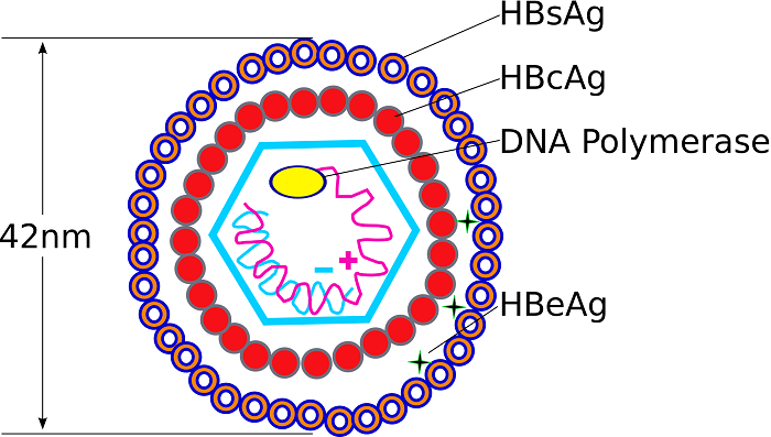
HBV (Hepatitis B Virus) Liver cancer (Hepatocellular Carcinoma). Direct Integration + Indirect Inflammation.
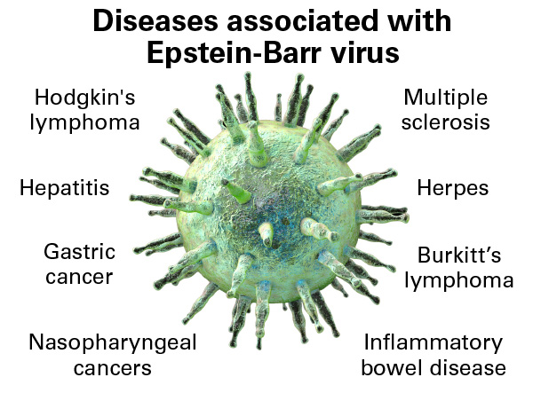
EBV (Epstein-Barr Virus) Burkitt's Lymphoma, Nasopharyngeal carcinoma, Hodgkin's Lymphoma. Direct (Immortalizes B-lymphocytes).
HTLV-1 (Human T-lymphotropic virus) Adult T-cell leukemia/lymphoma. Direct (Tax protein drives T-cell proliferation).
HCV (Hepatitis C Virus) Liver cancer (Hepatocellular Carcinoma). Strictly Indirect (Chronic inflammation/ROS).

Note: The World Health Organization (WHO) explicitly classifies HCV as a Group 1 Carcinogen (known to cause cancer in humans).

Virology Expansion: Direct vs. Indirect

A crucial distinction for your microbiology exams: Hepatitis B (HBV) is a DNA virus that integrates directly into the host genome, making it a Direct Oncogen. Hepatitis C (HCV) is an RNA virus. Its replication happens entirely in the cytoplasm, and it never integrates into the host DNA. Therefore, HCV causes cancer strictly through Indirect Oncogenesis (driven by decades of chronic inflammation, cirrhosis, oxidative stress, and viral proteins disrupting normal cell cycle checkpoints).


Introduction to Hepatitis C & Epidemiology


Basic Viral Classification

  • Full Name: Hepatitis C Virus (HCV).
  • Family: Flaviviridae. (To put this into context, this family also includes highly dangerous classical flaviviruses like Yellow Fever, Dengue Virus, Zika Virus, West Nile Virus, and various animal pestiviruses).
  • Genus: Hepacivirus.
  • Viral Architecture: Enveloped, positive-sense single-stranded RNA (+ssRNA) virus.
  • History: Discovered relatively recently in 1989. Before its official identification, it was mysteriously referred to in medical literature as "Non-A, Non-B Hepatitis."

Epidemiological Statistics and Global Burden

HCV is an insidious, silent pandemic. It represents a massive global health concern and remains a leading indication for liver transplantation worldwide.

  • Historical Exposure: Approximately 170 million people worldwide have been exposed to or infected by the virus at some point in their lives.
  • Active Chronic Burden: Currently, an estimated 50 million people are living with chronic, active HCV infections globally.
  • High-Burden Geographic Regions: Central and East Asia, North Africa & the Middle East, Sub-Saharan Africa, and Eastern Europe. (Specific Example: Egypt has historically had one of the highest HCV prevalence rates in the world—sometimes exceeding 10-15% of the population—due to historical mass-treatment campaigns for schistosomiasis using unsterilized, shared glass syringes in the mid-20th century).

Historical WHO Prevalence Data (1999 estimates):

  • Africa: 5.3%
  • Eastern Mediterranean: 4.6%
  • Western Pacific: 3.9%
  • South-East Asia: 2.15%
  • Americas: 1.7%
  • Europe: 1.03%
  • Total Global Average: 3.1%

Transmission Routes

HCV is transmitted strictly through exposure to infected blood. It is a robust virus that can survive outside the body at room temperature on environmental surfaces for up to 3 weeks.

Primary Routes (Blood-to-blood contact):

  • Injection drug use (IVDU): Sharing contaminated needles, syringes, or drug-preparation equipment accounts for a staggering 60% of modern infections.
  • Blood Transfusions & Organ Transplants: Accounts for 10% of historical infections. This was the primary mode of transmission before mandatory, highly sensitive nucleic acid blood screening protocols were implemented globally in the early 1990s.
  • Occupational Exposure: Needlestick injuries in healthcare workers account for roughly 4% of cases.
  • Unregulated Tattoos & Piercings: Using unsterilized ink or needles in unregulated environments carries a significant risk.

Less Common Routes:

  • Sexual Transmission: Accounts for ~15% of cases according to CDC historical data. However, the risk is generally considered low in monogamous heterosexual relationships. The risk increases exponentially if there are co-infections (like HIV), multiple partners, or practices that induce mucosal trauma resulting in blood exposure.
  • Perinatal / Mother-to-Child (Vertical Transmission): Less common, grouped into the 1% "Other" category along with nosocomial (hospital-acquired) and iatrogenic (medically induced) sources. Roughly 4-6% of infants born to HCV-positive mothers will contract the virus.
  • Unknown Sources: Approximately 10% of patients have no identifiable risk factors.
Memory Hack

The Hepatitis Viruses Transmission Routes

To perfectly remember how the major Hepatitis viruses are transmitted, use this simple mnemonic rule:

  • Hepatitis A & E = Ate & Eaten: Transmitted via the Fecal-Oral route (contaminated food and water). They cause acute, self-limiting infections.
  • Hepatitis B, C, & D = Blood-borne, Contact (Sexual), Drug use: Transmitted via bodily fluids. These are the ones that lead to chronic liver disease and cancer.

Molecular Virology of Hepatitis C


A. Virion Structure

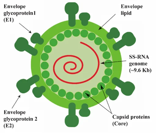
The physical structure of the HCV virion is perfectly adapted for entering human liver cells (hepatocytes).

  • Nucleocapsid: The virus features a tightly packed, Icosahedral nucleocapsid that houses the delicate viral RNA.
  • Lipid Envelope: This capsid is surrounded by a host-derived lipid envelope. The virus literally steals a piece of the human cell membrane as it exits, using it as a disguise.
  • Glycoproteins (E1 and E2): Embedded deep within this stolen lipid envelope are the heavily glycosylated viral envelope proteins E1 and E2. These proteins act as the "keys" that mediate host cell entry by binding to highly specific receptors on the surface of human hepatocytes (such as CD81, Scavenger Receptor B1, Claudin-1, and Occludin).

B. Viral Genome Architecture

The HCV genome is a masterclass in biological efficiency. It consists of a 9.6 kilobase (kb) positive-strand RNA genome (+ssRNA). Because it is "positive-sense," the viral RNA acts exactly like human messenger RNA (mRNA). As soon as it enters the cell, human ribosomes can read it immediately.

  • Untranslated Regions (UTRs): These are sequences at the ends of the genome that do not code for proteins but are vital for survival.
    • 5' UTR (The Hijacker): Contains an IRES (Internal Ribosome Entry Site). Physiology Expansion: Normal human mRNA requires a special "5' cap" for human ribosomes to recognize it and bind to it. The HCV IRES is a complex, 3D folded RNA structure that physically "hijacks" the human ribosome, forcing it to lock on and translate the viral RNA without needing a standard 5' cap!
    • 3' UTR: A highly structured noncoding region absolutely essential for the initiation of RNA replication.
  • Open Reading Frame (ORF): The vast majority of the genome is one continuous reading frame. It encodes a single, massive "polyprotein" consisting of approximately 3,000 amino acids.

C. The Polyprotein Cleavage Products (Crucial Exam Material)

A giant 3,000-amino-acid string is useless. It must be chopped up. This polyprotein is systematically cleaved by both host cellular proteases and the virus's own viral proteases into 10 distinct structural and non-structural (NS) proteins.

Protein Function / Characteristics
C (Core) Forms the physical viral nucleocapsid. Importantly, it is a primary weapon for immune evasion, actively targeting and blocking the host's JAK-STAT signaling pathway (preventing interferon response).
E1 & E2 The Envelope glycoproteins. E2 contains HVR-1 and HVR-2 (Hypervariable Regions). These regions mutate wildly and constantly, allowing the virus to rapidly change its surface "face" and completely evade neutralizing antibodies. E2 also actively targets and suppresses the host enzyme PKR.
p7 Thought to function as a viroporin—a tiny viral ion channel that punches holes in membranes, essential for efficient virus assembly and release from the cell.
NS2 A Zinc-dependent proteinase. Its only job is to cleave the junction between itself and NS3.
NS3 A multi-functional powerhouse and central therapeutic target. It acts as a Zinc-dependent proteinase, a serine protease, and an RNA helicase (unwinding RNA). (This is a major target for modern antiviral drugs!).
NS4A Acts as a vital, stabilizing cofactor for the NS3 protease, anchoring it to intracellular membranes.
NS4B Induces the massive rearrangement of the host's Endoplasmic Reticulum (ER), forcing the creation of a complex "membranous web" inside the host cell. This creates a hidden, secure factory strictly for viral replication, shielded from cellular immune sensors.
NS5A A heavily phosphorylated zinc-metalloprotein. Its exact function is heavily researched; it is an essential structural component of the replicase complex and heavily modulates host immune responses (specifically targeting and inhibiting PKR).
NS5B The RNA-dependent RNA polymerase (RdRp). This is the enzyme that physically copies the viral RNA to make new viral genomes.

The Discovery of Protein F (Frameshift Protein):
Advanced research has revealed a newly discovered protein produced by a ribosomal "frameshift" mutation (where the ribosome slips and reads the code out of phase) around codon 11 of the Core protein region. Its exact function remains a mystery, but we know it is produced during active infection because infected individuals reliably produce antibodies against it.


Genotypes & The Quasispecies Concept


Genotypes

HCV is extremely genetically diverse. It is divided into 6 major genotypes (1 through 6), which are distributed differently worldwide. Furthermore, these genotypes are broken down into numerous subtypes (e.g., 1a, 1b, 2a) and individual isolates based on nucleotide diversity. For example, Genotype 1 is the most common in the United States and Europe, while Genotype 4 dominates in the Middle East and Egypt.

The Quasispecies Concept

HCV possesses an astronomically high mutation rate. Why? Because its polymerase enzyme (NS5B) completely lacks "proofreading" ability (exonuclease activity). When a human cell copies DNA, it checks for typos and fixes them. When NS5B copies HCV RNA, it makes millions of random typos and leaves them there.

As a direct result, a single infected patient does not just carry one uniform virus. Instead, their blood contains a massive, swirling, complex swarm of millions of closely related, yet distinct mutant viruses. This swarm is called a Quasispecies.

Evolutionary Biology Application: This swarm functions as a unified evolutionary unit of selection. It brilliantly balances rapid mutation with high adaptability. If you give a patient a drug, or if their immune system generates a new antibody, 99% of the viral swarm might die. But because the swarm is so diverse, there is almost certainly a mutant in the remaining 1% that is perfectly immune to the drug or antibody. That mutant survives, replicates, and repopulates the liver.

Applied Clinical Question: Research Challenges

Case: A pharmaceutical company is struggling to develop new treatments and vaccines for Hepatitis C. According to clinical history, what were the major research roadblocks associated with studying HCV in the laboratory?

Answer: Historically, HCV research was severely hindered by three massive roadblocks:

  1. There was no reliable cell culture system (for decades, the virus outright refused to grow in laboratory petri dishes).
  2. There was no small animal model (HCV only naturally infects humans and chimpanzees, making ethical and affordable testing nearly impossible).
  3. The Quasispecies phenomenon. Its ability to rapidly mutate its Hypervariable Regions (HVR) makes finding stable, unchanging viral targets for vaccine development exceedingly difficult.

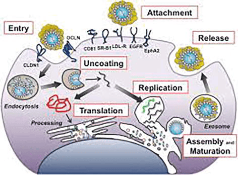
D. The Complete HCV Lifecycle (Cellular Infection Process)

To fully understand how Hepatitis C replicates and how direct-acting antivirals (DAAs) stop it, we must trace the exact chronological pathway the virus takes from the moment it encounters a human hepatocyte (liver cell) to the moment it bursts forth as a new, mature virion. The HCV lifecycle occurs entirely within the cell's cytoplasm and heavily relies on hijacking the host's lipid (fat) metabolism pathways.

Step 1

Attachment

The virus does not simply bump into a cell and enter; it requires a highly complex, multi-receptor "handshake" to ensure it is invading the correct target (a liver cell). The viral envelope glycoproteins (E1 and E2) bind to an array of specific host surface receptors and attachment factors.

Key Receptors Identified:

  • CD81: A critical tetraspanin cell-surface protein.
  • SR-B1 (Scavenger Receptor B1): A receptor normally used by the liver to uptake high-density lipoproteins (HDL). The virus disguises itself in lipids to bind here.
  • LDL-R: Low-Density Lipoprotein Receptor.
  • EGFR & EphA2: Epidermal Growth Factor Receptor and Ephrin receptor A2 act as essential co-factors that signal the cell membrane to prepare for entry.
Step 2

Entry (Endocytosis)

After initial attachment, the virus migrates along the cell surface to the tight junctions between hepatocytes. Here, it interacts with two more crucial tight-junction proteins: CLDN1 (Claudin-1) and OCLN (Occludin). Once this final lock is turned, the cell membrane invaginates (folds inward) and swallows the virus in a process called Clathrin-mediated Endocytosis, trapping the virus inside an intracellular bubble called an endosome.

Step 3

Uncoating

Inside the host cell, the endosome naturally becomes acidic (a drop in pH). The virus uses this acid as a trigger. The acidic environment causes a massive shape change in the viral envelope glycoproteins, forcing the viral lipid envelope to fuse with the endosomal membrane. The virus is "uncoated," and its naked positive-sense RNA genome (+ssRNA) is violently ejected directly into the host cell's cytoplasm.

Step 4 & 5

Translation and Processing

Because the viral RNA is "positive-sense," the human ribosomes immediately mistake it for human mRNA. The viral 5' IRES (Internal Ribosome Entry Site) hijacks the ribosomes located on the Rough Endoplasmic Reticulum (ER).

Translation: The ribosome translates the entire genome into one massive, continuous polyprotein.
Processing: This giant protein thread weaves in and out of the ER membrane. It is rapidly chopped into 10 active pieces (Core, E1, E2, p7, NS2, NS3, NS4A, NS4B, NS5A, NS5B) by both host signal peptidases and the viral NS3/4A protease.

Step 6

Replication

The viral protein NS4B causes the host's ER to physically warp and fold into a complex "membranous web." Inside this shielded web, the viral polymerase (NS5B) goes to work. First, it reads the original +ssRNA and creates a "negative-sense" intermediate template (the green strand). Then, it uses that negative template to continuously print millions of new, identical +ssRNA genomes (the black strands) to be packaged into new viruses.

Step 7 & 8

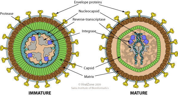
Assembly, Maturation, and Release

Assembly: HCV assembly is uniquely tied to the liver's fat metabolism. The newly minted viral RNA and the Core proteins gather around host Lipid Droplets located near the ER. The Core protein encases the RNA, forming the nucleocapsid.
Maturation: The capsid pushes into the ER lumen, wrapping itself in a piece of the ER membrane embedded with E1/E2 glycoproteins, forming a mature, enveloped virion.
Release (Exosome Pathway): The mature virus hitches a ride on the cell's Very-Low-Density Lipoprotein (VLDL) secretory pathway. It is transported to the cell surface inside an exosome (a secretory vesicle) and is expelled from the hepatocyte via exocytosis, ready to infect a neighboring cell or enter the bloodstream.


Clinical Presentation & Patterns of Viremia

HCV infection is notoriously stealthy. The clinical progression of the disease is broadly divided into an acute phase and a chronic phase.

1. Acute Phase Infection:

  • Asymptomatic Nature: The vast majority of patients are entirely asymptomatic and remain blissfully unaware they have been infected.
  • Symptomatic Cases: If symptoms do manifest (usually 2 to 12 weeks post-exposure), they are vague and mild: severe fatigue, nausea, abdominal discomfort, dark urine, and rarely, overt jaundice (yellowing of the skin and sclera due to bilirubin buildup from liver inflammation).
  • Spontaneous Clearance: Approximately 15% to 25% of individuals possess an immune system robust enough to successfully clear the virus on their own during this acute phase without ever requiring medical intervention.

2. Chronic Phase (The "Silent" Disease):

  • High Chronicity Rate: The vast majority of infected individuals—75% to 85%—fail to clear the virus, leading to a lifelong persistent HCV infection.
  • The Silent Progression: Patients can remain completely asymptomatic for 20 to 30 years while the virus slowly and methodically damages the liver. (Note: The liver itself contains no pain receptors; only the outer capsule does. Therefore, a liver can be 90% destroyed by fibrosis without the patient feeling any physical pain in the organ.)
  • Disease Trajectory: Chronic Hepatitis → Progressive liver scarring (Fibrosis) → Cirrhosis (irreversible architectural distortion of the liver) → End-stage liver failure or Hepatocellular Carcinoma (HCC).
  • Extraintestinal Manifestations: The virus doesn't just damage the liver. The persistent, massive immune response can trigger circulating immune complexes that deposit in other organs, causing severe autoimmune diseases such as Mixed Cryoglobulinemia (proteins clumping in cold blood leading to vasculitis), Glomerulonephritis (kidney damage), Porphyria Cutanea Tarda (skin blistering in sunlight), and Sjögren's syndrome.

3. Patterns of Viremia (Virus levels in the blood):

Monitoring the viral load in a patient's blood over time reveals three distinct patterns that determine their ultimate clinical outcome:

  1. Drop after peak: The viral load spikes, but then plummets to zero. Indicates successful, robust immune control and permanent viral clearance.
  2. Drop followed by rebound: The immune system initially mounts a strong defense, dropping the viral load. However, the viral Quasispecies rapidly mutates, evades the attack, and rebounds to high levels, establishing chronic infection.
  3. Consistent, unrelenting HCV levels: The immune system completely fails to mount an effective initial response, leading to immediate, rapid chronic infection and high, sustained viremia.

The Innate Immune Response & Viral Evasion Strategies

The innate immune system is the body's rapid-response first line of defense. It responds instantly (within hours to 2 days of infection), regardless of what the final outcome of the infection will be.

The Cellular Defense Mechanism (How the cell fights back):

As the HCV RNA genome replicates inside the cytoplasm, it must form dsRNA (double-stranded RNA) intermediates. dsRNA is highly unnatural in a human cell's cytoplasm. To the cell, dsRNA acts as a massive, blaring biochemical alarm bell!

  1. PKR Activation: The presence of this dsRNA activates an enzyme called Protein Kinase R (PKR). (Physiology Expansion: PKR is a cellular suicide switch. When activated, it phosphorylates translation initiation factors, effectively shutting down ALL protein translation in the cell to starve the virus of manufacturing parts).
  2. IRF Phosphorylation: Intracellular sensors (like RIG-I and MDA5) detect the viral RNA and trigger a cascade that phosphorylates Interferon Regulatory Factors (IRFs), specifically IRF-3.
  3. Gene Activation: These phosphorylated IRFs act as transcription factors. They travel directly into the cell's nucleus to upregulate and turn on massive arrays of antiviral gene products (specifically Type I Interferons and interferon-stimulated genes).
  4. Result: These gene products degrade the viral RNA, trigger apoptosis in infected cells, and warn neighboring cells to raise their shields.

HCV Viral Resistance & Targeting (How the virus wins):

HCV is incredibly successful at causing lifelong chronic infection because its viral proteins are exquisitely evolved to directly target, sabotage, and dismantle the human innate immune pathways!

  1. NS5A and E2 target PKR: These viral proteins physically bind to and completely inhibit Protein Kinase R. The cell is unable to shut down protein translation, allowing the virus to continuously print new viral parts.
  2. Core protein targets the JAK-STAT pathway: (Expansion: When Interferon is released, it binds to neighboring cells and activates the JAK-STAT pathway to arm them against incoming viruses). The HCV Core protein chemically blocks this exact pathway, making all neighboring cells "deaf" to the interferon alarm system.
  3. NS3/4A targets phosphorylated IRF-3: The viral protease NS3/4A acts as molecular scissors. It physically chops up the essential cellular adaptor proteins (like MAVS and TRIF) needed to activate IRF-3. This completely, irreparably shuts down the cell's ability to produce Type I Interferons!
High-Yield Concept

The Protease Weapon (NS3/4A)

The viral protein NS3/4A is a highly specialized protease. Not only does it cut the giant viral polyprotein into usable, individual pieces to build new viruses, but it literally snips the host cell's immune alarm wires (MAVS and TRIF proteins). By cutting these wires, the infected cell cannot alert the rest of the immune system. This dual-action sabotage is exactly why pharmaceutical NS3/4A protease inhibitors are one of the most powerful cures we have today—they stop viral assembly AND restore the cell's ability to call for help!


Adaptive Immune Response & Chronic Dysregulation

If the innate system fails (which it usually does with HCV), the adaptive immune system (T-cells, B-cells) steps in. The speed and vigor of this secondary response dictate whether the patient lives a virus-free life or suffers decades of chronic liver disease.

Characteristics of Individuals Who Successfully Control the Virus:

  • IFN-γ (Interferon-gamma): Preferentially and strongly expressed in the liver of recovering patients.
  • IFN-γ strongly induces the expression of genes encoding chemokines that attract armies of T-cells right into the inflamed liver tissues.
  • It also highly induces proteins associated with antigen processing and presentation (upregulating MHC Class I and II molecules on liver cells so they can "show" the immune system the hidden virus).
  • These patients mount highly vigorous, sustained, and multi-specific CD8+ (Cytotoxic) and CD4+ (Helper) T cell responses.

Why Chronic Infections Occur (Adaptive Failure):

The majority of patients develop chronic disease due to two primary failures:

  1. The patient's immune system is simply unable to mount a broad, HCV-specific T cell response from the beginning.
  2. There is a strong initial response resulting in apparent viral RNA clearance, but this is followed prematurely by a massive contraction (die-off or exhaustion) of the CD8+/CD4+ cells. With the army depleted, the surviving mutated Quasispecies virus mounts a massive rebound in viremia.

The State of Chronic HCV Immune Dysregulation:

  • CD8+ T-cells (Cytotoxic Killers): They experience abnormally low frequencies in the blood and a heavily reduced capacity to kill infected cells. This is a clinically recognized state known as T-cell exhaustion (often driven by the continuous upregulation of inhibitory receptors like PD-1).
  • CD4+ T-cells (Helpers): Suffer from severely reduced Interleukin-2 (IL-2) production and demonstrate poor proliferation. Without Helpers, the Killers fail.
  • Dendritic Cells (Antigen Presenters): They do not mature normally and exhibit severely impaired stimulatory activity. They fail to effectively capture the virus and "show" it to the T-cells to initiate an attack.
  • Natural Killer (NK) & NKT Cells: There is a severe impairment of NK cell cytotoxic (cell-killing) activity and a decreased frequency of NKT cells in the liver. (Note: Clinical trials show this profound impairment is actually fully reversible in patients who respond well to exogenous IFN-α drug therapy!)

The Role of Antibodies (B-Cell Response):

  • The role of humoral (antibody) immunity in HCV is surprisingly unclear, contradictory, and historically poorly studied.
  • Fascinatingly, the virus can actually be cleared completely by some patients in the absolute absence of detectable antibody responses (proving that CD8+ T-cells are the true heroes of viral clearance).
  • The body does produce neutralizing antibodies that attempt to target the E2 envelope glycoprotein. However, as noted earlier, E2 contains Hypervariable Regions (HVR1/HVR2). E2 mutates so astronomically fast that by the time the B-cell manufactures a perfect antibody, the virus has already changed its molecular "face" to evade it. The antibodies are always one step behind the Quasispecies swarm.

The Liver Environment & Hepatocellular Carcinoma (Oncogenesis)

The ultimate tragedy of long-term HCV infection is the development of Hepatocellular Carcinoma (HCC). To understand why this happens, we must look closely at the unique immune environment of the liver.

A. The Normal vs. Infected Liver Environment

The Normal Liver:

  • Maintains an intentionally "Immuno-silent" state. (Physiology Expansion: Because the liver receives all nutrient-rich blood straight from the intestines via the portal vein, it is constantly bombarded by harmless food antigens, plant proteins, and dead gut bacteria pieces. If the liver's immune system reacted to all of these foreign bodies, you would be in a constant, fatal state of anaphylactic shock!)
  • To maintain this vital silence, activated CD8+ T cells that wander into the normal liver are often trapped and forcibly pushed into apoptosis (programmed cell death) by local hepatic cells to prevent unnecessary immune reactions.

The HCV-Infected Liver:

  • The viral invasion breaks the silence. The stressed liver produces Type I IFN and releases chemokines that promote the rapid infiltration of NK cells.
  • This induces massive IFN-γ production in the NK cells and expresses chemokines that recruit tens of thousands of highly activated T-cells to the liver matrix.
  • Clinical Note: Experimental depletion of NK cells prior to a hepatotropic viral infection leads to the absolute inhibition of the virus-specific T-cell response, resulting in unabated viral replication and severe liver injury.

B. Immune-Mediated Liver Injury & "Bystander Killing"

The mechanisms responsible for liver injury were initially poorly understood. We now know that the Host immune response, NOT the direct viral replication itself, is what physically destroys the liver.

  • HCV is actually quite sparse; it typically infects only 1% to 10% of the total hepatocytes in the liver at any given time.
  • However, the massive influx of highly active, frustrated CD8+ T-cells into the liver results in the release of a fiery storm of toxic cytokines (like IFN-γ and TNF-α) and deadly granules (perforin and granzyme). These toxic compounds are indiscriminately sprayed into the tissue, destroying vast numbers of uninfected, innocent hepatocytes in a tragic, highly destructive process known as "Bystander Killing".

C. The Road to Hepatocellular Carcinoma (Indirect Oncogenesis)

Because HCV does NOT integrate into host DNA (unlike HBV), its mechanism of causing cancer is entirely indirect, stemming from the chronicity of the battle.

  1. The ongoing "Bystander Killing" by the immune system causes massive, unrelenting hepatocyte death and chronic Inflammation.
  2. To survive the destruction, the liver goes into overdrive, constantly attempting to regenerate. This extremely high turnover rate in hepatocytes pushes cells to divide faster than their DNA repair mechanisms can handle, leading to numerous erroneous genome replications.
  3. Furthermore, the chronic inflammation involves millions of immune cells generating massive amounts of Oxidative stress and Reactive Oxygen Species (ROS), which bathe the liver cells and physically shatter cellular DNA strands.
  4. This endless cycle of tissue damage → ROS generation → rapid cellular repair strongly activates Hepatic Stellate Cells. These cells lay down massive amounts of collagen, causing widespread Fibrosis (scarring) and eventually Cirrhosis (a hard, nodular, failing liver).
  5. The cirrhotic liver, full of rapidly dividing cells containing ROS-damaged DNA, creates a highly unstable "pre-cancerous niche" that ultimately promotes the transformation of these cells into Hepatocellular Carcinoma (HCC).
  6. Additional Factor: Viral proteins (like NS5A and Core) remaining in the few infected cells further disrupt normal cell cycle regulation and prevent apoptosis, sealing the malignant fate of the tissue.
Summary Analogy

HCV Oncogenesis: The Warzone

HCV does NOT directly mutate your DNA. Instead, think of the liver as a city and the infection as a Warzone:

  1. HCV hides in a few buildings (cells).
  2. The immune system (CD8+ cells) drops massive "bombs" (TNF-α, ROS) to kill the virus, blowing up thousands of innocent, healthy buildings alongside the infected ones (Bystander killing).
  3. The liver city tries to rebuild the destroyed buildings as fast as possible (High cell turnover/regeneration).
  4. Rebuilding quickly in a toxic, oxidative warzone causes the construction workers to make terrible mistakes (DNA replication errors).
  5. Over decades, the mistakes pile up, and the city becomes filled with chaotic, uncontrolled, collapsing structures. This is Hepatocellular Carcinoma.

Diagnostics, Vaccine Status, and Antiviral Therapeutics

IX. The Vaccine Challenge: Why is there no HCV Vaccine?

Unlike Hepatitis B (HBV) and Human Papillomavirus (HPV), for which we possess highly effective, globally distributed routine vaccines, there is currently NO approved vaccine for Hepatitis C.

The Biological Roadblocks preventing vaccine development include:

  1. Astronomical Genetic Variability: There are 6 major genotypes with dozens of highly distinct subtypes. A vaccine formulated to recognize Genotype 1 will likely offer zero protection against Genotype 4.
  2. The Quasispecies Moving Target: As discussed, the lack of proofreading by the NS5B polymerase creates a rapidly shifting swarm of viruses. The virus mutates its surface envelope proteins faster than the immune system—or a vaccine-induced antibody response—can lock onto them.
  3. Active Immune Evasion: The viral proteins (NS5A, E2, Core) actively suppress and disarm the host's innate and adaptive immune responses, making it hard for a vaccine to trigger strong memory cells.
  4. Lack of Animal Models: Since HCV only naturally infects humans and chimpanzees (and testing on chimpanzees is highly restricted/banned in most of the world), testing new, experimental vaccine candidates is incredibly difficult, expensive, and ethically complex.

X. Investigations & Clinical Diagnostics

Accurately diagnosing HCV requires a strict two-step process. You cannot rely on a single blood test to determine if a patient is currently harboring an active infection.

Step 1: Initial Screening (Indirect Serological Test)

  • The Test: HCV Antibody (anti-HCV) utilizing Enzyme-Linked Immunosorbent Assay (ELISA) or a Rapid Diagnostic Test (RDT).
  • Positive Result Meaning: It ONLY indicates that the patient was exposed to HCV at some point in their life and their immune system created antibodies. It does not mean they are currently sick. (Remember, 25% of people clear the virus naturally, but their antibody test will remain positive for life!).
  • Negative Result Meaning: Generally rules out infection entirely.
  • Exception 1 (The Window Period): It takes the human body 8 to 12 weeks after initial exposure to produce enough antibodies to be detected by a lab test. If a nurse suffers a contaminated needlestick injury yesterday, the antibody test today will be negative, even if the virus is currently ravaging their liver.
  • Exception 2 (Immunocompromised State): Patients with advanced, untreated HIV/AIDS or those on heavy immunosuppressants may fail to produce antibodies entirely, yielding a false-negative result.

Step 2: Confirmatory Testing (Direct Virological Tests)

If the screening antibody test is positive, the clinician MUST perform a "direct" test to look for the physical presence of the virus itself.

  • HCV RNA (Nucleic Acid PCR Testing): The absolute Gold Standard for diagnosis.
    • Qualitative RNA: A highly sensitive "Yes/No" test. It simply tells you if viral RNA is present in the blood.
    • Quantitative RNA (Viral Load): Measures the exact amount of viral copies per milliliter of blood. This is absolutely essential for establishing a baseline before treatment and monitoring therapeutic success.
  • HCV Core Antigen (HCVcAg): A test that detects the physical Core viral protein rather than the RNA. It is utilized heavily in resource-limited settings as a significantly cheaper, more accessible alternative to complex PCR machines; it correlates excellently with active, high viral loads.
Applied Clinical Question: Pediatric Testing

Case: A baby is born to an HCV-positive mother. The enthusiastic intern wants to run an HCV Antibody (ELISA) test on the newborn at 1 month of age to see if transmission occurred. Why is this a profound medical error, and what should be done instead?

Answer: For all children under 18 months of age, HCV antibody tests are completely unreliable. This is because the newborn passively carries the mother's maternal IgG antibodies, which crossed the placenta during the third trimester. The antibody test would be overwhelmingly positive even if the baby is perfectly healthy and virus-free. Instead, an HCV RNA PCR is the strictly preferred test to detect actual viral presence, typically performed when the infant is at least 2 months old to allow viral replication to reach detectable thresholds.


XI. Evolution of HCV Therapy

The treatment landscape for Hepatitis C has undergone a total, miraculous revolution over the last decade, transitioning from highly toxic, poorly effective "hit-or-miss" injections to nearly 100% effective, easily tolerated oral pills.

A. Historical Therapy (Standard/Current in older notes):

  • The Combination Regimen: Weekly injections of Pegylated Interferon-alpha (PEG-IFN) combined with daily oral doses of Ribavirin (a broad-spectrum nucleoside analog).
  • Mechanism of Action: Massive, systemic suppression of protein synthesis, enhancement of cellular apoptosis, and catastrophic degradation of the viral plus-strand RNA via induced mutagenesis.
  • Efficacy: Disappointing. Only 50% to 80% effective, heavily dependent on the specific viral genotype (Genotype 1 was notoriously resistant).
  • The "Nightmare" Side Effects: The treatment was often described by patients as worse than the disease itself.
    • Flu-like symptoms: Extreme, debilitating tiredness, high fevers, and severe myalgia after every injection.
    • Psychiatric: Profound trouble with concentration, severe mood instability, and the induction of deep clinical depression (frequently leading to suicidal ideation).
    • Hematologic (Blood) Toxicity: Severe bone marrow suppression leading to dangerous neutropenia (low white cells) and thrombocytopenia (low platelets).
    • Ribavirin Toxicity: Directly and fiercely toxic to red blood cells, causing severe Hemolytic Anemia in a large percentage of patients.

B. The Revolution: Direct-Acting Antivirals (DAAs):

Introduced to the global market in the mid-2010s, DAAs fundamentally changed HCV from an agonizing, chronic life sentence into a rapidly curable disease.

  • Cure Rate: Exceeds a phenomenal 95% to 99% across all known genotypes.
  • Treatment Course: Remarkably short duration, requiring only 8 to 12 weeks of strictly oral, once-daily pills, with near-zero side effects compared to Interferon.
  • Standard Global Regimens:
    • Sofosbuvir / Velpatasvir: Highly favored because it is suitable for ALL genotypes (1 through 6). It is considered "Pan-genotypic."
    • Glecaprevir / Pibrentasvir: Recommended for 8 or 12 weeks depending strictly on the presence or absence of advanced cirrhosis.
    • Sofosbuvir / Daclatasvir: Highly common and incredibly cost-effective in resource-limited and developing settings.
Memory Hack

DAA Naming Suffixes

DAAs are precision-engineered to target specific non-structural viral proteins. You can perfectly identify a drug's exact molecular target simply by looking at the suffix of its generic name:

  • -previr (e.g., Telaprevir, Glecaprevir): Targets the NS3/4A Protease (P for Protease).
  • -asvir (e.g., Daclatasvir, Velpatasvir): Targets the NS5A complex (A for 5A).
  • -buvir (e.g., Sofosbuvir): Targets the NS5B RNA-dependent RNA Polymerase (B for 5B). (Mechanism detail: Sofosbuvir acts as a defective nucleotide. When the NS5B polymerase tries to insert it into the growing RNA chain, it acts as a "chain terminator," permanently halting viral replication).

XII. Novel and Experimental Drug Therapies

While DAAs are highly successful, medical research continues deeply into allosteric inhibitors, host-targeting agents, and molecular "warheads" to ensure treatment options exist for the rare patients who develop DAA resistance or suffer from end-stage liver failure.

  • Non-nucleoside inhibitors (NNIs): These molecules target the RdRp (NS5B polymerase) at distinct, separate binding sites away from the active center. They work via allosteric inhibition (they bind to the side of the enzyme, physically changing the 3D shape of the enzyme so it can no longer function). Examples include Benzothiadiazine and Benzimidazole chemical derivatives.
  • Protease Inhibitors (BILN 2061): A highly specific peptidomimetic compound that physically blocks the NS3 active site. Early clinical trials showed a massive, rapid decline in viral load within 48 hours, though some patients experienced rebounds as the Quasispecies mutated.
  • Cyclosporin A (CsA): Traditionally an immunosuppressive drug used for transplant patients, CsA was discovered to strongly bind to cyclophilins (critical human host proteins that the HCV virus absolutely needs to fold its own proteins properly). By blocking the host's calcineurin pathway, it inhibits the activation of genes essential for T-cell activation. (Crucial Note: The highly related immunosuppressive drug FK506 does NOT suppress HCV replication, making Cyclosporin A's specific antiviral mechanism unique and fascinating).
  • Arsenic Trioxide: Historically a poison, modern research shows it surprisingly and potently inhibits HCV replication at submicromolar (extremely low) concentrations without displaying significant toxicity to the human host cells.

RNA-Based Genetic Treatments:

The bleeding edge of antiviral research involves manipulating RNA directly.

  • RNA Interference (RNAi): A technique that utilizes the cell's own natural defense machinery (the Dicer enzyme and the RISC complex) to seek out, recognize, and physically chop up specific viral RNA sequences. It is highly sequence-specific and lethal to the virus.
  • Small RNAs (Decoys): These engineered molecules act as biochemical "decoys." By overexpressing artificial viral RNA elements in the cell, they act like a sponge, binding up all the viral regulatory proteins and preventing them from binding to the real viral RNA. This effectively starves the virus of its machinery and halts gene expression.
  • siRNAs (Small Interfering RNAs): These are designed to target and silence the human cellular cofactors that HCV desperately needs to survive (such as the proteins La, PTB, and hVAP-33). If the virus cannot find its required "human helpers," it cannot replicate, regardless of how aggressively it mutates.

XIII. Conclusion & Prevention Strategies

Summary: Hepatitis C Virus remains a stealthy, highly adaptable oncogenic virus and a major global health concern. However, the advent of modern Direct-Acting Antivirals (DAAs) offers a realistic, achievable hope for the global elimination of the disease. Curing HCV is paramount because it doesn't merely clear a viral infection—it significantly and permanently halts the ongoing liver inflammation, thereby massively reducing the long-term risk of the patient developing fatal Hepatocellular Carcinoma.

Prevention Strategies (Since no vaccine exists):

  • Implementation of highly sensitive, routine nucleic acid blood screening protocols before all transfusions and organ transplants.
  • Aggressive public health Harm Reduction programs (such as sterile needle and syringe exchange programs for IV drug users).
  • Strict adherence to Universal Health Precautions (safe needle disposal, double-gloving, and proper sterilization of medical/dental/tattoo equipment in all clinical settings).

List of References

  • World Health Organization (WHO). (1999). Global surveillance and control of hepatitis C. Report of a WHO Consultation organized in collaboration with the Viral Hepatitis Prevention Board, Antwerp, Belgium.
  • Centers for Disease Control and Prevention (CDC). (2020). Hepatitis C Questions and Answers for Health Professionals. Atlanta, GA: US Department of Health and Human Services.
  • Moradpour, D., & Blum, H. E. (2004). A primer on the molecular virology of hepatitis C. European Journal of Gastroenterology & Hepatology, 16(11), 1297-1301.
  • Ahmad, A., et al. (2004). Natural killer cells and hepatitis C virus infection. Immunology, 112(1), 7-16.
  • Lindenbach, B. D., & Rice, C. M. (2005). Unravelling hepatitis C virus replication from cellular to molecular levels. Nature Reviews Microbiology, 3(8), 596-606.
  • Pawlotsky, J. M. (2014). New hepatitis C therapies: the toolbox, strategies, and challenges. Gastroenterology, 146(5), 1176-1192.
  • Guidotti, L. G., & Chisari, F. V. (2006). Immunobiology and pathogenesis of viral hepatitis. Annual Review of Pathology: Mechanisms of Disease, 1(1), 23-61.

Quick Quiz

Hepatitis C Virus Quiz

Microbiology - mobile-friendly and focused practice.

Privacy: Your details are used only for quiz tracking and certificates.

Hepatitis C Virus (HCV) Read More »

Epstein-Barr Virus (EBV)

Epstein-Barr Virus (EBV) 

The Epstein-Barr Virus (EBV)

Module Focus

The Epstein-Barr Virus (EBV) is a masterpiece of viral evolution. It is incredibly successful, infecting over 90% of humanity, yet it possesses a dark, deadly potential. By the end of this comprehensive guide, you will master:

  • The unique structural and virological features of EBV.
  • The exact molecular pathogenesis of how EBV enters cells, establishes lifelong latency, and drives immortalization.
  • The classic presentation, physical findings, and management of Infectious Mononucleosis (IM).
  • The profound oncogenic capability of EBV, specifically its role in Burkitt's Lymphoma, Nasopharyngeal Carcinoma, and Hodgkin's Lymphoma.
  • The interpretation of EBV laboratory diagnostics, including the Monospot test and specialized serology.

Part I: Introduction, Discovery & Classification

The Historical Discovery

The Epstein-Barr Virus (EBV) is one of the most ubiquitous (common) viruses in the human population. It is highly renowned for its lymphotropic properties (meaning it specifically hunts, targets, and thrives within B-lymphocytes) and its definite, proven association with human malignancies.

  • Discovery: It was definitively identified in 1964 by researchers Anthony Epstein, Yvonne Barr, and Bert Achong. They discovered it using an electron microscope while examining a cell line derived from an African patient with Burkitt's lymphoma.
  • Historical Significance: EBV holds the monumental historical distinction of being the very first isolated human tumor virus (oncogenic virus).
  • The "Kissing Disease": It is most commonly associated with the acute disease Infectious Mononucleosis (IM), colloquially and famously known as "the kissing disease" due to its primary mode of transmission via the exchange of saliva.
  • Oncogenic Potential: EBV does not just kill cells; it is able to "transform" infected B-cells. This transformation results in the immortalization of the cell, driving it into a state of uncontrolled, endless replication, which is the foundational step of cancer.

Taxonomy and Classification

  • Family: Herpesviridae (This family includes other famous viruses like Herpes Simplex, Varicella-Zoster, and Cytomegalovirus).
  • Subfamily: Gammaherpesvirinae. (Deeper Physiological Context: Gammaherpesviruses are uniquely characterized by their specific tissue tropism; they uniquely establish latent, lifelong infections specifically in lymphoid cells, unlike Alphaherpesviruses which hide in nerve ganglia).
  • Genus: Lymphocryptovirus (EBV is the prototype virus of this specific genus).
  • Nomenclature: Scientifically classified as Human Herpesvirus 4 (HHV-4). It is one of eight known human herpesviruses.
  • Viral Types: There are 2 distinct types of the virus (Type A and Type B). Interestingly, they can co-exist and simultaneously infect the same person without cross-immunity clearing the other.

Part II: Viral Morphology & Structure

EBV is a complex, large virus with a highly organized architecture designed to protect its massive DNA payload and ensure successful host cell hijacking.

  • Shape & Size: It is spherical in appearance, measuring approximately 100 to 180 nm in diameter (making it a relatively large virus).
  • Genome: The core of the virus is a linear, double-stranded DNA (dsDNA) molecule. It is massive, containing exactly 172 kbp (kilobase pairs) of genetic code. It possesses a toroid-shaped (doughnut-shaped) protein core that acts as a spool, around which the massive DNA is tightly wrapped.
  • Nucleocapsid: The shell protecting the DNA has perfect icosahedral symmetry, consisting of exactly 162 individual capsomers (protein building blocks).
  • The Tegument: A crucial, unstructured protein layer located directly between the nucleocapsid and the outer envelope.
    • Physiology Expansion: The tegument is a hallmark of all herpesviruses. It is essentially a "care package" of pre-formed viral proteins and enzymes. When the virus enters a human cell, it doesn't wait to synthesize proteins; it immediately dumps the tegument contents into the host cytoplasm to instantly hijack the cell's machinery and suppress early cellular alarms.
  • The Envelope: The outermost layer is a fragile lipid envelope. It is derived by the budding of immature viral particles through the host cell membrane (or nuclear membrane). Because it is made of lipids, the virus is easily destroyed by soap and alcohol.
  • Glycoprotein Spikes: Embedded in this lipid envelope are external virus-encoded glycoprotein spikes. These are absolutely required for infectivity, acting as the "keys" that lock onto the host cell's "keyholes" (receptors).

[ IMAGE PLACEHOLDER: 3D Structure of EBV highlighting the dsDNA genome, icosahedral nucleocapsid, protein tegument, and the lipid envelope studded with glycoprotein spikes. ]


Part III: Epidemiology & Transmission Dynamics

Epidemiology

EBV is a master of silent spread. It is found worldwide, in every country, infecting people of all socioeconomic statuses, races, and age groups.

  • According to the World Health Organization (WHO), global serologic tests (blood tests checking for past infection antibodies) show that approximately 90-95% of adults worldwide (including the United States) have been infected by EBV at some point in their lives.

The Two Epidemiological Patterns of Infection:

  1. Developing Countries: Infection occurs at a much earlier age due to crowded living conditions and shared resources. By the age of two, over 90% of children are already seropositive. Clinical Note: These early childhood infections are almost always mild, subclinical, or entirely silent (asymptomatic). The child gets a slight fever and recovers, never knowing they caught EBV.
  2. Developed Countries: Because of stricter hygiene, two distinct peaks of infection are seen.
    • The first peak is in very young preschool children (aged 1-6), often transmitted by parents kissing babies.
    • The second, more infamous peak occurs in adolescents and young adults (aged 14-20). Clinical Note: Infection during this adolescent/young adult window is much more aggressive and is highly likely to cause the full, symptomatic syndrome of Infectious Mononucleosis (IM).

Transmission

  • Saliva (The Primary Vector): The virus is spread primarily by contact with oral secretions (saliva). It is transmitted from asymptomatic, shedding adults to infants, and among young adults by the transfer of large amounts of saliva during deep kissing or sharing drinks/utensils.
  • Lifelong Viral Shedding: More than 90% of asymptomatic, completely healthy seropositive individuals intermittently shed live EBV virions in their oropharyngeal secretions for the rest of their lives. Clinical Note: Shedding is heavily increased in immunocompromised patients (like those with HIV or on chemotherapy).
  • Other Routes: Though far less common, EBV can also be transmitted via blood transfusions, solid organ transplants, and bone marrow transplantations.
  • Risk Factors: Close personal contact, crowded living conditions, immunosuppression, and poor personal hygiene practices.

Part IV: Viral Pathogenesis – Entry, Lytic Phase & Latency

EBV establishes a lifelong infection in the host. The pathogenesis involves a complex dance between lytic replication (destroying cells to make millions of new viruses) and latent infection (hiding quietly inside cells to evade the immune system).

1. Viral Entry & The Molecular Handshake

EBV can infect both B-lymphocytes and epithelial cells (specifically the oropharyngeal and salivary gland epithelial cells lining the throat).

  • Entering B-Cells: The viral envelope possesses a highly specific "key" called glycoprotein gp350. This gp350 binds precisely to the cellular receptor CD21 (also known as the Complement Receptor 2 / CR2 receptor) and the MHC Class II molecule on the surface of human B-cells. This lock-and-key fit is followed by membrane fusion.
  • Entering Epithelial Cells: To enter the throat lining, the virus utilizes completely different surface proteins, interacting with cellular integrins to facilitate entry.
🧠 Exam Mnemonic: EBV Receptor on B-Cells

How do you remember which receptor Epstein-Barr Virus uses to enter B-Cells?

"You must be 21 to drink at the Barr."

Epstein-Barr Virus uses the CD21 receptor!

2. Primary Infection & Lytic Replication

  1. After entering the body through the mouth, EBV first replicates actively in the epithelial cells of the pharynx. This massive viral replication destroys the throat cells, causing severe pharyngitis (sore throat).
  2. This lytic replication involves the viral DNA polymerase aggressively copying the viral genome. Lytic gene products are produced in three consecutive, highly regulated stages: immediate-early, early, and late.
  3. The newly formed virions burst out of the dying epithelial cells, cross the basement membrane, and invade the underlying lymphoid tissue (such as the tonsils and adenoids). Here, they find their true target: naive B-lymphocytes.

3. Establishing Latency & Oncogenesis

Once inside the B-cell, the virus changes its strategy. Unlike lytic replication, latency does not result in the production or shedding of virions. The virus goes into "stealth mode".

  • The Episome: Inside the resting memory B-cells, the linear viral DNA enters the nucleus and circularizes into a naked ring of DNA called an episome. This episome resides independently in the cell nucleus (it does not integrate into the human chromosomes like HIV does) and is copied quietly by the cellular DNA polymerase every time the host cell divides.

Viral Proteins & B-Cell Immortalization (Deep Dive)

Even though the virus is "latent" and hiding from the immune system, it is not entirely dormant. It continuously expresses a select few latent proteins to ensure the host B-cell survives forever. These include:

  • EBNA (Epstein-Barr Nuclear Antigens): Maintains the episome and regulates viral gene expression.
  • LMP (Latent Membrane Proteins): Sit in the B-cell membrane.
  • EBER RNAs: Small viral RNAs that prevent cell death.

The Mechanism of Oncogenesis: These latent proteins promote B-cell survival, prevent apoptosis (programmed cell death), stimulate uncontrolled cell proliferation, and help the virus evade immune destruction.


Excessive Detail (Pathology of LMP-1): LMP-1 is a highly oncogenic (cancer-causing) viral protein. It inserts itself into the B-cell membrane and acts exactly like a constitutively active (permanently switched on) CD40 receptor. Normally, a B-cell needs a Helper T-cell to physically bind to its CD40 receptor to give it permission to survive and multiply. The viral LMP-1 permanently turns on the cellular NF-KB signaling pathway, tricking the B-cell into thinking it has constant T-cell help. This directly drives the malignant transformation and "immortalization" of B-cells in vitro and in vivo!


Part V: The Host Immune Response & Reactivation

1. The Cell-Mediated Immune Attack (Cytokine Storm)

The human body does not ignore this immortalization process. It mounts a massive, aggressive cell-mediated immune response against the virus.

  • A few EBV-immortalized B-cells enter the circulation, but they are continually hunted, cleared, and destroyed by healthy immune surveillance mechanisms.
  • Cytotoxic CD8+ T-cells rapidly and massively multiply to attack the infected B-cells.
  • Clinical Relevance: The extreme clinical manifestations of Infectious Mononucleosis (high fever, profound extreme fatigue, massively swollen lymph nodes, hepatosplenomegaly) result largely from this massive, exhausting immune response and the resulting "cytokine storm", rather than direct damage caused by the virus itself.
💡 Board Exam Alert: Downey Cells

Note that activated T-cells appear as atypical lymphocytes (Downey cells) in peripheral blood films. This is a classic trick question on microbiology and pathology exams!

Students often mistakenly think that the weird-looking, large, stretched-out atypical lymphocytes seen on a blood smear in a patient with Mono are the infected B-cells. They are NOT. The Downey cells are actually the massive, highly reactive CD8+ Cytotoxic T-cells that are expanding and stretching around red blood cells to fight off the infected B-cells.

2. Persistence and Reactivation

  • Once infected, a lifelong carrier state develops whereby a low-grade infection is kept in check by the immune defenses. Some infected memory B-cells persist for life in a latent state.
  • Periodic Reactivation: The virus can switch back from latent to lytic replication when:
    • Immunity decreases naturally (stress, aging).
    • Immunosuppression develops (e.g., catching HIV/AIDS, or taking anti-rejection transplant medications).
  • Reactivated virus replicates back in the oral pharyngeal cells and is shed in the saliva, allowing transmission to new hosts.

Summary Flow of EBV Pathogenesis

  1. Infection of oropharyngeal epithelium via saliva.
  2. Infection of naive B-lymphocytes via the CD21 receptor.
  3. B-cell proliferation and latent infection (driven by viral LMP-1/EBNA).
  4. Massive CD8+ T-cell response (causing Infectious Mononucleosis symptoms and producing atypical Downey lymphocytes).
  5. Lifelong latency in memory B-cells.
  6. Periodic reactivation and viral shedding in saliva.
  7. Possible malignant transformation into cancer if T-cell immune surveillance fails.

Part VI: Clinical Syndromes & Cancers

1. Infectious Mononucleosis (IM)

Infectious Mononucleosis (also known as "glandular fever" or the "kissing disease") is the most common clinical manifestation of acute EBV infection. While primary EBV infection is usually subclinical (silent, nonspecific) in childhood, in adolescents and adults, there is a 50% chance that the full, debilitating syndrome of IM will develop.

Clinical Course & Symptoms:

  • Prodrome: A 1-2 week period of vague fatigue, headaches, and malaise before the fever hits.
  • Acute Phase (2-5 days): Worsening malaise, severe fatigue, and the sudden onset of high fever.
  • The Classic Triad / Main Symptoms:
    • Fever: Can be high and last for 4–5 weeks.
    • Lymphadenopathy: Massively swollen, tender lymph nodes (especially in the posterior cervical/neck region), lasting 2–4 weeks.
    • Sore throat & Tonsillopharyngitis: Often severe, with a thick white or gray exudate covering the tonsils.
  • Hepatosplenomegaly: Significant enlargement of the liver and spleen. Jaundice (yellowing of skin/eyes) may be seen in some patients due to EBV-induced hepatitis.
  • Other findings: Retro-orbital headache, myalgia (severe muscle pain), nausea, abdominal pain, and upper eyelid edema (Hoagland sign, seen in 15% of cases).
  • Resolution Phase: Organomegaly (enlarged liver/spleen) may safely persist for 1–3 months. While the fever and sore throat usually resolve in 2 to 4 weeks, profound fatigue may last for several months.

Complications of IM:

Complications occur rarely but can be sudden and life-threatening.

  • Splenic Rupture: The massively swollen, inflamed, and fragile spleen can rupture spontaneously or with mild abdominal trauma. This causes fatal internal hemorrhage.
  • Airway Obstruction: Severe tonsillar/pharyngeal swelling can literally close off the airway, suffocating the patient.
  • Neurological: Guillain-Barré syndrome, meningoencephalitis, cranial nerve palsies.
  • Chronic IM: In some rare patients, chronic IM may occur where eventually the patient succumbs to a lymphoproliferative disease or lymphoma.
High-Yield Clinical Pearl

The "Ampicillin Rash"

Your notes cite a specific complication published in the NEJM: "IM with rash after treatment with amoxicillin or ampicillin."

If a patient comes in with a severe sore throat and fever, a rushed doctor might misdiagnose them with bacterial "Strep Throat" and prescribe the antibiotics Ampicillin or Amoxicillin. If the patient actually has viral EBV (Mono), over 90% of them will develop a massive, full-body, extremely itchy, copper-colored maculopapular rash 7 to 10 days later! This is NOT a true IgE-mediated allergic reaction to penicillin, but rather a unique, immune-complex virus-drug interaction. Always check for a swollen spleen and atypical lymphocytes before blindly giving antibiotics for a sore throat!

Patient Advice Scenario

The Athlete and the Spleen

Case: A 19-year-old college football player is diagnosed with Infectious Mononucleosis. His fever breaks after 2 weeks, and he feels much better. He asks you when he can return to playing contact football.

Answer: He must wait at least 4 weeks (or until definitively cleared by a physician via abdominal ultrasound). Because of IM-induced splenomegaly, his spleen is massively enlarged, protruding dangerously below the protective rib cage, and its capsule is highly fragile. A simple tackle or fall could easily cause splenic rupture and fatal internal bleeding.


Part VII: Role of EBV in Cancers (Malignancies)

The CDC cites EBV as a major factor in oncogenesis, and the WHO lists EBV as one of only a handful of viruses definitively known to be causative agents in human cancer. Without robust T-cell surveillance, EBV-immortalized cells will mutate into tumors.

1. Burkitt's Lymphoma (BL)

  • Epidemiology: Occurs endemically in parts of Africa (where it is the absolute commonest childhood tumor, usually striking children aged 3-14 years) and Papua New Guinea. Sporadic cases of BL occur worldwide, especially highly aggressive forms in AIDS patients.
  • The Malaria Cofactor: Endemic BL in Africa is strictly restricted to geographic areas with holoendemic malaria. Malaria infection acts as a profound cofactor (chronic malaria continuously activates B-cells while simultaneously suppressing T-cell immunity, perfectly allowing EBV-infected cells to escape control and multiply).
  • Viral Presence: Multiple copies of the EBV genome and some EBV antigens can be found deeply embedded in BL tumor cells.

Molecular Pathophysiology of Burkitt's (High-Yield)

BL cells are defined by a catastrophic reciprocal chromosomal translocation.

  • It primarily occurs between the long arm of Chromosome 8 and Chromosome 14[t(8;14)], though variant translocations can also involve chromosomes 2 or 22.
  • The Result: The c-myc oncogene (a powerful gene located on Chromosome 8 that drives the cell cycle) is snapped off and transferred directly next to the highly active Immunoglobulin (Ig) heavy chain gene regions on Chromosome 14. B-cells always have their Ig genes turned "on" to make antibodies. By placing c-myc next to this "always-on" switch, it results in the complete deregulation and massive overexpression of the c-myc gene, driving endless, explosive cell replication.
  • Note: It is thought that this translocation is probably already present as a random error by the time of EBV infection, but EBV immortalizes the cell, preventing it from dying and allowing the mutation to thrive into a massive jaw or facial tumor.
  • Prognosis: Despite being one of the fastest-growing human tumors, it responds highly favorably to aggressive chemotherapy.

🧠 Mnemonic: Burkitt's Translocation
How do you remember the chromosomes involved in the classic Burkitt's translocation?
"Burkitt's ate (8) fourteen (14) c-myc (Mac) burgers."
Translocation t(8;14) moves the c-myc oncogene to the heavy chain Ig locus.

2. Nasopharyngeal Carcinoma (NPC)

  • Definition: A malignant, aggressive tumor of the squamous epithelium of the nasopharynx (the upper part of the throat behind the nose).
  • Epidemiology: Extremely prevalent in Southern China (where it is the commonest tumor in men and the second commonest in women, sometimes called "Cantonese Cancer"). It is rare in most parts of the world, though pockets occur in North/Central Africa, Malaysia, Alaska, and Iceland.
  • Pathology: Multiple copies of the EBV episome and the EBNA-1 antigen are universally found in the cells of undifferentiated NPC.
  • Cofactors: Besides EBV, there appears to be a strong link to environmental factors (e.g., traditional diets rich in salted, cured fish and nitrosamines) and specific genetic HLA haplotypes.
  • Prognosis: NPC usually presents late (often discovered only when a patient notices a painless lump in their neck from lymph node metastasis), and thus the prognosis is poor. In theory, it could be prevented by vaccination (if an EBV vaccine existed).

3. Hodgkin's Lymphoma (HL)

  • A specific type of lymphoma believed to result from mutated white blood cells of the lymphocyte kind, characterized by the presence of giant, multi-nucleated Reed-Sternberg cells (which look like "owl eyes" under a microscope).
  • Symptoms: Classic "B-symptoms" including unexplained fever, drenching night sweats, severe weight loss, and painless enlarged rubbery lymph nodes in the neck, under the arm, or in the groin.
  • Viral Association: About 50% of all cases of Hodgkin's lymphoma are definitively linked to the Epstein-Barr virus genome residing in the Reed-Sternberg cells.

Part VIII: Disease Association in Immunocompromised Patients

After primary infection, EBV maintains a steady low-grade latent infection. If the person's immune system collapses (specifically T-cell function), the virus will rapidly reactivate, developing into highly aggressive lymphoproliferative lesions and fatal lymphomas. These lesions tend to be extranodal and present in highly unusual sites such as the Gastrointestinal (GI) tract or the Central Nervous System (CNS).

1. Transplant Recipients

Patients receiving heavy immunosuppressants to prevent organ rejection (e.g., renal or heart transplant recipients) lose their T-cell surveillance. They are highly associated with the rapid development of Post-Transplant Lymphoproliferative Disease (PTLD) and aggressive EBV-driven B-cell lymphomas.

2. AIDS Patients

When HIV destroys the CD4+ T-cells, EBV runs rampant. It causes:

  • Oral Leukoplakia: Specifically "Hairy Oral Leukoplakia"—white, corrugated, hairy-looking patches on the lateral sides of the tongue. Clinical Note: Unlike oral thrush (Candida fungus), these patches are caused by massive EBV replication in the epithelium and cannot be scraped off with a tongue depressor.
  • Non-Hodgkin's Lymphoma (NHL): Highly aggressive, rapidly fatal B-cell lymphomas, often presenting as primary CNS lymphoma in the brain.
  • Chronic Interstitial Pneumonitis.
3. Duncan X-linked Lymphoproliferative (XLP) Syndrome

A devastating genetic tragedy. This condition occurs exclusively in young males who have inherited a defective, mutated gene on the X-chromosome (the SAP/SH2D1A gene).

  • When these seemingly healthy boys catch standard EBV (Mono), their specific T-cell/B-cell interaction system completely fails to control it.
  • It results in a massive, uncontrolled, fatal immune response (fulminant infectious mononucleosis), macrophage activation syndrome, or fatal lymphoma. This single genetic condition accounts for half of all fatal cases of Infectious Mononucleosis in the world.

Part IX: Laboratory Diagnosis of EBV

Diagnosis is based on identifying the classic clinical findings and correlating them with specialized laboratory tests.

  1. Complete Blood Count (CBC) with Peripheral Smear:
    • Findings include profound Leukocytosis (massively increased white blood cell count, usually 10,000 to 20,000/µL).
    • Presence of Atypical Lymphocytes (Downey cells) comprising >10% of total leukocytes. As mentioned, these are reactive CD8+ T-cells featuring an enlarged, abundant, frequently vacuolated cytoplasm and a folded nucleus.
  2. Heterophile Antibody Test (The "Monospot" Test):
    • The common, rapid, inexpensive screening test for IM.
    • Mechanism: During an acute EBV infection, the hyper-stimulated B-cells go haywire and produce random, non-specific "junk" antibodies called heterophile antibodies. These unique antibodies have the peculiar ability to agglutinate (clump together) red blood cells from sheep or horses. Placing the patient's serum on a card with horse RBCs—if it clumps, it's a positive result, rapidly suggesting acute EBV infection.
  3. EBV-Specific Serology Panel:

    Used when the Monospot is negative but EBV is highly suspected, or to track cancer.

    • Acute EBV infection: Diagnosed definitively by the presence of anti-EBV VCA IgM (Viral Capsid Antigen IgM antibody) which appears early and fades, alongside VCA IgG. At this stage, anti-EBNA (Epstein-Barr Nuclear Antigen) is NEGATIVE.
    • Past/Resolved Infection: VCA IgM is negative. VCA IgG is positive (lasts for life). Anti-EBNA IgG is POSITIVE (antibodies to the nuclear antigen take 2-3 months to develop, so their presence proves the infection happened months/years ago).
    • Cancer Screening: The determination of the titer of anti-EBV VCA IgA (an antibody found in mucosal secretions) is heavily used in screening for early lesions of Nasopharyngeal Carcinoma (NPC) and for monitoring its response to treatment. A patient with non-specific ENT (Ear, Nose, Throat) symptoms who has elevated EBV IgA must be given a thorough nasopharyngeal examination with a camera scope!
  4. Polymerase Chain Reaction (PCR):
    • Highly sensitive DNA testing. Detects circulating EBV DNA in the blood or CSF. Especially useful in diagnosing severe infections (like EBV encephalitis) and tracking viral load in immunocompromised patients (e.g., watching for the development of PTLD in transplant patients, allowing doctors to adjust immunosuppression before lymphoma develops).
  5. Histology & Tissue Biopsy:
    • Burkitt's Lymphoma: Must be definitively diagnosed by histology. The tumor features a classic "starry-sky" appearance under the microscope (macrophages eating dead cells among a sea of dark tumor cells). The tumor can be stained with antibodies to lambda light chains, revealing a monoclonal tumor of B-cell origin. In over 90% of cases, the cells express IgM at the cell surface.
    • NPC: Diagnosed strictly by histological biopsy of the nasopharynx mass.

Part X: Treatment, Management, and Prevention

A. Treatment of Infectious Mononucleosis

IM is a self-limited illness in healthy individuals and generally does not require specific antiviral therapy. The illness resolves autonomously without treatment, though debilitating fatigue symptoms may linger for weeks to months.

  • Supportive Care: The primary cornerstone of treatment is to manage symptoms. This includes strict bed rest, heavy oral hydration, fever control, and monitoring for complications.
  • Pharmacotherapy (Symptomatic): Patients are advised to take Acetaminophen (Paracetamol) or Non-Steroidal Anti-Inflammatory Drugs (NSAIDs like Ibuprofen) to help bring down the high fever, relieve myalgia (body aches), and ease overall discomfort. Gargling with warm salt water or viscous lidocaine can relieve the severe sore throat.
  • Corticosteroids: Routine use of steroids is NOT recommended. However, they can be used cautiously and briefly (e.g., Prednisone) to help rapidly alleviate severe swelling of the airway (massive tonsillar enlargement threatening suffocation) or to treat severe autoimmune complications triggered by the virus (like autoimmune hemolytic anemia or profound thrombocytopenia).
  • Inpatient Hospital Therapy: Strictly required for severe medical and surgical complications (e.g., ruptured spleen requiring emergency splenectomy, airway obstruction, or meningoencephalitis).

Antiviral Agents

According to the Merck Manual and infectious disease guidelines, standard antiviral agents (like acyclovir) have been proven not to be effective in significantly shortening the clinical course of routine IM (because the symptoms are caused by the immune system, not active viral replication, and the virus is largely latent in B-cells where antivirals can't reach it). However, in severe/life-threatening lytic cases (or in heavily immunocompromised patients), the following heroic measures may be used:

  • Acyclovir: 10 mg/kg/dose IV given every 8 hours (q8h) for 7-10 days to halt lytic replication.
  • IVIG (Intravenous Immunoglobulin): 400 mg/kg/day IV for 2-5 days to help modulate the immune storm in severe cases.

B. Prevention

  • Prevention of EBV infection on a population level is almost impossible, given that 90-95% of the world's adult population is already infected and shedding the virus intermittently without knowing it.
  • Vaccination: There is currently NO approved vaccine available for EBV, though several candidates (targeting the gp350 envelope protein to prevent B-cell entry) are in clinical trials. A successful vaccine could theoretically eradicate IM, Burkitt's Lymphoma, and Nasopharyngeal Carcinoma!
  • Personal Hygiene: If a seronegative individual is actively trying to avoid acute infection, they must avoid direct contact with an infected person's saliva (e.g., do not share drinks, water bottles, lip balm, utensils, or engage in deep kissing with someone actively sick with Mono). Overall, there are no broad, highly effective public health quarantine methods for EBV infection prevention.

List of References

The information detailed in this module is synthesized from standard microbiological and pathological principles found in the following core texts and institutional guidelines:

  • Jawetz, Melnick, & Adelberg's Medical Microbiology. (28th Edition). McGraw-Hill Education. (Core reference for viral structure, classification, and replication cycles).
  • Robbins & Cotran Pathologic Basis of Disease. (10th Edition). Elsevier. (Core reference for the molecular pathogenesis of Burkitt's Lymphoma, Nasopharyngeal Carcinoma, and the morphology of Infectious Mononucleosis).
  • Murray, P. R., Rosenthal, K. S., & Pfaller, M. A. (2020). Medical Microbiology. (9th Edition). Elsevier. (Reference for clinical syndromes, EBV serology, and diagnostics).
  • Centers for Disease Control and Prevention (CDC). Epstein-Barr Virus and Infectious Mononucleosis. (Epidemiological statistics, transmission dynamics, and public health guidelines).
  • The New England Journal of Medicine (NEJM). Literature regarding the "Ampicillin Rash" complication (Vol. 343:481-492) and EBV-associated malignancies.
  • The Merck Manual of Diagnosis and Therapy. (Clinical management, supportive care, and antiviral therapy protocols for severe IM).

Quick Quiz

Epstein-Barr Virus Quiz

Microbiology - mobile-friendly and focused practice.

Privacy: Your details are used only for quiz tracking and certificates.

Epstein-Barr Virus (EBV)  Read More »

Human T-Cell Lymphotropic Virus (HTLV-1 & HTLV-2)

Human T-Cell Lymphotropic Virus (HTLV-1 & HTLV-2)

Human T-Cell Lymphotropic Virus (HTLV)

Learning Objectives

By the end of this exhaustive guide, you will be deeply conversant with:

  • The unique epidemiology, taxonomy, and morphological structure of the HTLV retrovirus.
  • The complete molecular replication cycle, focusing heavily on how HTLV integrates into the human genome as a provirus.
  • The profound oncogenic mechanisms driven by viral proteins like Tax and HBZ.
  • The devastating clinical manifestations of HTLV, particularly Adult T-Cell Leukemia/Lymphoma (ATLL) and HAM/TSP.
  • The diagnostic modalities and current treatment limitations for HTLV-driven pathologies.

I. Introduction & Epidemiology of HTLV

Human T-Cell Leukemia Virus Type 1 (HTLV-1) holds a unique, monumental place in medical history: it was the very first retrovirus ever discovered to be directly involved in human cancer (discovered in 1980 by Robert Gallo and his team). It belongs to the exogenous type of retroviruses (meaning it spreads horizontally between host cells from the outside environment, unlike endogenous retroviruses which are ancient viral remnants passed down inherently in human DNA).

Epidemiology & Endemic Regions

Worldwide, approximately 15 to 20 million people are infected with HTLV-1. While it can be found sporadically in populations worldwide (including the United States and Europe), it is heavily and uniquely endemic in specific, isolated geographic regions. This distribution is believed to be linked to ancient human migration patterns:

  • Southwestern Japan: (The highest prevalence in the world; nearly 10% of the population in some islands are carriers).
  • The Caribbean basin: (Jamaica, Trinidad and Tobago).
  • South America: (Particularly Brazil, Peru, and Colombia).
  • Sub-Saharan Africa: (Endemic hotspots in central and western Africa).
  • Australo-Melanesia: (Indigenous populations of Australia and Papua New Guinea).

Clinical Latency & Transformation

HTLV-1 is a slow-transforming oncogenic retrovirus. Unlike acute-transforming viruses (which carry a stolen host oncogene and cause rapid, devastating cancer within weeks), HTLV-1 takes decades to slowly reprogram a cell.

  • Leukemia develops in only 3% to 5% of infected individuals.
  • When cancer does occur, it typically arises after a massive, prolonged latent period of 40 to 60 years! A person infected via breastmilk as an infant will likely not show signs of leukemia until they are in their 50s or 60s.

II. Taxonomy & Classification

HTLV is scientifically classified to define its precise viral lineage. Understanding its family helps predict its behavior.

  • Family: Retroviridae (RNA viruses that reverse-transcribe into DNA).
  • Subfamily: Orthoretrovirinae.
  • Genus: Deltaretrovirus.

Viral Species

  1. Human T-Lymphotropic Virus 1 (HTLV-1): The primary oncogenic driver and the most clinically significant. It is subdivided into four distinct geographical genotypes:
    • a. Cosmopolitan Group (Found worldwide, widely distributed).
    • b. Central African Group.
    • c. Melanesian Group.
    • d. New Central African Group.
  2. Human T-Lymphotropic Virus 2 (HTLV-2): A closely related but much less pathogenic retrovirus. Discovered shortly after HTLV-1, it is primarily found in intravenous drug users and indigenous populations of the Americas.
  3. HTLV-3 & HTLV-4: Extremely rare, recently discovered strains in Central Africa (usually transmitted from non-human primates, like hunters getting bitten by monkeys), with unproven clinical significance.
Advanced Concept: Deltaretroviruses

Why is it called a Deltaretrovirus? This genus includes complex retroviruses that infect mammalian species (like humans and bovines, e.g., Bovine Leukemia Virus). What makes them "complex" is that, in addition to the standard retroviral genes (gag, pol, env), they carry highly specialized accessory regulatory genes (like tax and rex/HBZ). These extra genes are the secret weapons that give them their unique, slow-acting oncogenic superpower, allowing them to evade the immune system and force the host cell to divide infinitely.


III. Morphology & Composition of the Virion

The physical structure of the HTLV particle (virion) is critical for its ability to survive, infect, and integrate into human cells.

Structural Characteristics

  • Virion Shape: Spherical to pleomorphic (meaning it can alter its shape slightly depending on the environment).
  • Size: 80 to 110 nm in diameter.
  • Capsid: Possesses an Icosahedral capsid. Inside this protective shell is a dense, helical nucleoprotein core holding the RNA.
  • Envelope: Present. Because it is an enveloped virus, it is highly sensitive to drying, heat, stomach acid, and standard detergents. Therefore, it cannot survive on dry surfaces (fomites) and requires direct, wet fluid contact (blood, breastmilk, semen) for transmission.

Chemical Composition

Protein (~60%): Includes the structural capsids, matrix proteins, and the pre-packaged internal enzymes (Reverse Transcriptase, Integrase, Protease) necessary to start an infection instantly upon entry.

Lipid (~35%): The viral envelope is actually stolen from the human host! When the virus exits the cell, it wraps itself in the host cell's lipid bilayer membrane.

Carbohydrate (3%): Primarily the glycosylation (sugar-coating) of the envelope spikes (like gp46 and gp21) used for receptor binding.

RNA (2%): The actual genetic payload of the virus.

The Genome

  • Type: Single-stranded RNA (ssRNA). It carries two identical copies of this RNA (it is diploid).
  • Format: Linear, positive-sense (+ssRNA).
  • Size: 7 to 11 kilobases (kb).
  • Special Feature: The virion contains Reverse Transcriptase pre-packaged inside, ready to act upon entry.

IV. Viral Genome Structure

The HTLV proviral DNA contains a highly specific arrangement of genes running from the 5' to the 3' end. This standard layout is the hallmark of all retroviruses.

The Standard Retroviral Genes

  • LTR (Long Terminal Repeat): Flanks the viral DNA at both the 5' and 3' ends. The LTR exerts absolute regulatory control on proviral gene function. It acts as the promoter/enhancer (the "on switch") and is the exact sequence that physically links to the human host DNA.
  • gag: Codes for group-specific antigens (the structural proteins that make up the viral matrix, p19, and the viral capsid, p24).
  • pol: Codes for the essential viral enzymes (Reverse Transcriptase, Integrase, and Protease).
  • env: Codes for the envelope glycoproteins (the spikes that allow the virus to dock onto a new host cell, primarily surface glycoprotein gp46 and transmembrane glycoprotein gp21).
Mnemonic: Retroviral Gene Order (5' to 3')
Think: "Grandpa Plays Excellently"
-> Grandpa = gag (Capsid/Structure)
-> Plays = pol (Polymerase/Enzymes)
-> Excellently = env (Envelope/Spikes)

The Oncogenic Additions (The pX Region)

At the 3' end of the genome, HTLV-1 possesses a unique region called the pX region. This region codes for the devastating accessory proteins Tax and HBZ, which are directly responsible for causing Leukemia. (Detailed in Section VIII).


V. Viral Replication, Morphogenesis & Maturation

Because HTLV is a retrovirus, it breaks the standard rules of biology. Instead of DNA making RNA, it must convert its RNA genome backward into DNA, and then permanently glue that DNA into the human chromosome. The cycle proceeds in the following strict order:

  1. Adsorption & Receptor Binding: The viral envelope glycoproteins (gp46) bind precisely to specific receptors on the surface of the target human T-cell.
    Extra Detail: HTLV-1 uses a triple-receptor complex to gain entry. It binds to Heparan sulfate proteoglycans (HSPG), then Neuropilin-1 (NRP-1), and finally GLUT1 (the standard glucose transporter).
  2. Penetration & Uncoating: The viral envelope fuses with the host cell membrane, releasing the viral capsid and RNA core deep into the host cytoplasm.
  3. Reverse Transcription: The pre-packaged viral enzyme Reverse Transcriptase reads the viral single-stranded RNA and synthesizes a double-stranded DNA copy (cDNA) of it right there in the cytoplasm. (Note: Reverse transcriptase is highly error-prone, which usually causes viruses to mutate rapidly, though HTLV mutates much slower than HIV).
  4. Integration (Forming the Provirus): The newly formed viral DNA is transported into the cell nucleus. Another viral enzyme, Integrase, snips the human host DNA and permanently inserts the viral DNA into the host chromosome.
    Crucial Concept: At this stage, the integrated viral DNA is officially called a Provirus. It is now a permanent part of the human genome.
  5. Transcription: Using the host's own RNA polymerase (tricked by the viral LTR promoter), the integrated Provirus is transcribed back into messenger RNA (mRNA) and full-length new genomic viral RNA.
  6. Translation: The host cell's ribosomes read the viral mRNA and synthesize viral proteins (capsid proteins, envelope proteins, and new enzymes).
  7. Capsid Assembly: The newly created viral RNA and viral proteins self-assemble near the inner surface of the cell membrane to form a new, immature viral core.
  8. Budding & Maturation: The assembled core pushes out against the host cell membrane. It wraps itself in a piece of the host lipid bilayer (which is now studded with viral env proteins) and pinches off. Viral protease then cleaves internal proteins, maturing the virion into a fully infectious particle.

Applied Virology Concept: The Virological Synapse & Mitotic Division

Question: Why is HTLV-1 completely immune to standard cell division (mitosis)? If a human T-cell infected with HTLV-1 divides, what happens to the virus?

Answer: Because of Step 4 (Integration)! Once HTLV-1 becomes a Provirus, it is literally a physical part of the human chromosome. When the human T-cell undergoes mitosis and copies its own DNA, it automatically copies the viral DNA with it, passing the infection to all daughter cells without ever needing to bud or create a new virion. This is called mitotic clonal expansion of the virus.

The Virological Synapse: Unlike HIV, which releases billions of free-floating virions into the blood, free HTLV virions are rarely found in blood plasma. To infect a new cell, an infected T-cell physically grabs a healthy T-cell, forms a tight bridge (a virological synapse), and shoots the virus directly into the healthy cell without exposing it to the outside environment!


VI. Pathogenesis & Cell Tropism

Tropism refers to the specific types of cells a virus is capable of infecting. While HTLV is similar to the Human Immunodeficiency Virus (HIV) in its targets, the ultimate fate of the infected cell is completely different.

Host & Tropism

  • Host: Humans (for both HTLV-1 and HTLV-2).
  • Cell Tropism:
    • HTLV-1: Exhibits a strict tropism for CD4+ T-cells (T-helper cells). Because of this, CD4+ T-cells are the major target for neoplastic (cancerous) transformation.
    • HTLV-2: Exhibits a tropism for CD8+ T-cells (Cytotoxic T-cells).
The Fate of the T-Cell: HTLV vs. HIV

HIV infects CD4+ T-cells, uses them as hyperactive viral factories, and ultimately destroys them via lysis or apoptosis (leading to immunodeficiency and AIDS). HTLV-1, however, infects CD4+ T-cells and immortalizes them. It refuses to let them die. It forces them to endlessly divide and live forever, which is the very definition of a cancer (Leukemia/Lymphoma)!


VII. Modes of Transmission

HTLV-1 does not survive well outside of a cell. Human infection strictly requires the transmission of intact, infected T-cells (cell-to-cell contact), rather than free-floating viral particles.

Mechanisms of Horizontal Spread (Between individuals)

  1. Sexual Intercourse: Spread through bodily fluids (semen and vaginal fluids containing infected lymphocytes). This is the most common route between sexual partners. Transmission is significantly more efficient from male-to-female than female-to-male.
  2. Blood Products: Transmission via blood transfusions containing infected white blood cells, or through the sharing of blood-contaminated needles among intravenous (IV) drug abusers.
    Clinical Note: Because HTLV is hidden inside cells, modern blood banks perform Leukoreduction (filtering out the white blood cells from donated blood before giving it to a patient). This massive safety step drastically reduces the risk of transmitting HTLV!

Vertical Spread (Mother to Child)

  1. Mother-to-Child Transmission (Breastfeeding): Transmitted primarily through infected lymphocytes present in breast milk. Unlike HIV, transmission across the placenta during pregnancy is actually quite rare. The major risk is prolonged breastfeeding (beyond 6 months).
    Clinical Note: In endemic areas like Japan, mothers who test positive for HTLV-1 are strongly advised to exclusively formula-feed their babies to completely break the transmission chain.

VIII. HTLV-1 Oncogenesis (The Molecular Mechanism)

While the complete mechanism of Adult T-cell Leukemia/Lymphoma (ATLL) leukemogenesis is still highly complex, we know it is a multifactorial process driven by specific viral Oncoproteins coded in the pX region.

1. The TAX Protein

The tax gene codes for the Tax oncoprotein. Its oncogenicity is "locked in" this gene. It functions as a master transcription activator essential for viral replication, but it aggressively modulates and hijacks host cell function:

  • Creates an Autocrine Proliferation Loop: Tax activates host genes coding for Interleukin-2 (IL-2) and the IL-2 receptor (CD25). It also activates myeloid growth factor and Granulocyte-Macrophage Colony Stimulating Factor (GM-CSF).
    Result: The T-cell secretes its own growth hormone (IL-2) and places extra receptors (CD25) on its own surface to catch it. The cell is essentially telling itself to rapidly and endlessly proliferate.
  • Inhibits the Cell Growth Cycle Checkpoints: Tax inactivates the crucial tumor-suppressor genes CDKN2A (p16) and TP53 (p53). Simultaneously, it activates Cyclin D (a cell cycle enhancer).
    Result: The brakes on cell division are destroyed, and the accelerator is jammed down.
  • Blocks Apoptosis (Cell Death): Tax activates Nuclear Factor kappa B (NF-kB), a powerful transcription factor that regulates host anti-apoptotic genes.
    Result: The cell refuses to undergo programmed cell death, even when it realizes its DNA is damaged.
  • Causes Genomic Instability: Interferes with host DNA repair pathways (Base excision repair and Nucleotide excision repair).
    Result: Leads to the sustained, rapid accumulation of DNA mutations in the host genome over decades.

2. The HBZ Protein (HTLV-1 Basic leucine Zipper factor)

This is a fascinating protein because it is encoded on the antisense strand of the provirus (it is read backward!).

  • Unlike Tax (which is highly immunogenic, meaning the immune system eventually recognizes and targets Tax-expressing cells), HBZ is constitutively expressed but evades the immune system, allowing the virus to hide.
  • While Tax causes the initial explosive growth, it is eventually silenced by the virus to hide from immune cells. HBZ takes over. It is crucial for the continuous, low-level proliferation of infected cells and promoting long-term viral persistence and latency.
The Multistep Timeline of HTLV Oncogenesis

Cancer from HTLV-1 is not an overnight event; it is a chronic, multistep process requiring 40 to 60 years!

  1. Initial Stage: Virus primarily infects CD4+ T-cells via direct cell-to-cell contact.
  2. Replication & Mitosis: Virus replicates by creating new infections and by multiplying the infected cells themselves (mitotic division).
  3. Polyclonal Expansion & Latency: Tax drives early proliferation, while HBZ promotes latency and survival despite the host immune response. Many different T-cell clones are expanding (Polyclonal).
  4. Monoclonal Malignancy: Due to Tax-based interference with DNA repair, several severe host mutations accumulate over decades. Eventually, one highly mutated cell goes completely rogue, outcompetes the others, and undergoes explosive monoclonal proliferation, resulting in mature ATLL!

IX. Diseases Caused by HTLV

The vast majority (about 95%) of HTLV infections are Asymptomatic (the most common outcome for both HTLV-1 and HTLV-2). However, when disease does manifest, it is devastating.

Diseases Caused by HTLV-1

1. Adult T-Cell Leukemia/Lymphoma (ATLL / ATL)

A highly aggressive, malignant neoplasm of CD4+ T-lymphocytes. ATLL presents in four distinct clinical subtypes: Acute (most common and aggressive), Lymphomatous, Chronic, and Smoldering. Symptoms include:

  • Swollen lymph nodes (lymphadenopathy) and hepatosplenomegaly (enlarged liver and spleen).
  • Skin lesions: Very common in ATLL, ranging from maculopapular rashes to massive nodules and tumors. The leukemic cells love to infiltrate the skin.
  • High Calcium Levels (Hypercalcemia):
    Physiology Expansion: The malignant T-lymphoblasts produce an osteoclast-activating factor—specifically PTHrP (Parathyroid Hormone-related Protein). This molecule aggressively destroys bone tissue, releasing massive amounts of calcium into the blood. This leads to bone lesions, confusion, severe constipation, abdominal pain, and fatal coma.
  • Immunosuppression: Patients frequently die from severe opportunistic infections (like Pneumocystis jirovecii, Cytomegalovirus, and aggressive fungal infections).

2. HTLV-1 Associated Myelopathy / Tropical Spastic Paraparesis (HAM/TSP)

A progressive, demyelinating neurological disease of the spinal cord (specifically the thoracic region).

  • Mechanism: It is an immune-mediated disease. The immune system tries to attack HTLV-infected T-cells that have infiltrated the spinal cord, causing massive collateral inflammatory damage to the myelin sheaths of motor neurons.
  • Symptoms: Causes severe weakness, stiffness, and spastic paralysis of the lower limbs. It is often accompanied by severe bladder dysfunction (urinary incontinence) and bowel dysfunction.

3. Inflammatory Conditions

The virus can cause opportunistic autoimmune-like inflammation throughout the body, including HTLV-uveitis (eye inflammation), infective dermatitis (skin), and bronchiectasis/bronchitis (lungs).

Diseases Caused by HTLV-2

Infections are mostly asymptomatic, but have been rarely linked to:

  • Atypical Hairy-Cell Leukemia.
  • Adult T-cell Leukemia (ALT) – though this is extremely RARE compared to HTLV-1.
  • Mild neurological abnormalities.
Mnemonic: HTLV-1 Pathologies
Remember "The 4 H's of HTLV-1"
1. HTLV-1 (The virus)
2. HAM/TSP (The neurological spine disease)
3. Hypercalcemia (The classic metabolic derangement in ATLL)
4. Hyper-proliferation of CD4 cells (The leukemia)

X. Laboratory Diagnosis

Because the virus hides inside cells and has a massive latent period, standard viral culture is impossible. Specialized virology and hematology techniques are required to confirm diagnosis.

  1. Peripheral Blood Smear (Morphology):
    • Examination of the blood under a microscope will show the pathognomonic ATLL cells.
    • Slide Image Note: These malignant T-cells have highly lobulated, deeply indented, multi-lobed nuclei. They are famously referred to as "Flower-like cells" or "Flower cells" by hematologists because the nucleus looks like the petals of a flower.
  2. Flow Cytometry (Immunophenotyping):
    • Used to analyze the cell surface markers of the leukemic cells. ATLL cells will typically test powerfully positive for CD4 and CD25 (the IL-2 receptor driven by Tax).
  3. Serology (Detecting Antibodies):
    • ELISA / CMIA / EIA: Enzyme Immunoassays are used as the primary, highly sensitive screening test to detect IgG antibodies against HTLV-1 and HTLV-2 in serum and Cerebrospinal Fluid (CSF).
    • Western Blot: Used as the strict confirmatory test if the ELISA is positive. It successfully distinguishes between HTLV-1 and HTLV-2 by identifying specific viral protein antibodies (e.g., bands for gag p24, env gp46).
  4. Detection of Proviral DNA:
    • PCR (Polymerase Chain Reaction): Used to directly detect and amplify the integrated proviral DNA of HTLV-1 hidden within the host cell genome. It can also quantify the proviral load (how many cells are infected), which is highly prognostic for disease progression and useful to confirm indeterminate Western blot results.
  5. Medical Imaging:
    • MRI of the Spinal Cord: Used to diagnose HAM/TSP. It detects demyelinating lesions and atrophy, separating to form multiple lesions along the spinal tracts.

XI. Treatment & Management

Unfortunately, because the viral DNA is permanently integrated into the human host chromosome (provirus), no curative treatment exists for the HTLV virus itself. Standard chemotherapy regimens (like CHOP) used for other lymphomas often fail in ATLL because the cells are highly chemo-resistant.

Management of ATL (Adult T-Cell Leukemia)

  • Antiviral Therapy: A combination of Interferon-alpha and Zidovudine (AZT) has been reported to be effective in treating and significantly prolonging survival in certain subtypes of ATL patients (particularly the leukemic subtypes).
  • Monoclonal Antibodies: Mogamulizumab (an antibody targeting CCR4, which is highly expressed on ATLL cells) is a modern, targeted therapy showing significant promise in relapsed ATLL.
  • Allogeneic Stem Cell Transplant: The only potentially curative approach for acute ATLL, replacing the patient's entire infected immune system with a healthy donor's system, though it carries a high mortality risk.
  • "Watch and Wait": For patients with the indolent (Smoldering or Chronic) subtypes of ATLL, aggressive chemo is often avoided, and patients are closely monitored for disease progression.

Management of TSP (Tropical Spastic Paraparesis)

  • A combination of Zidovudine (AZT), Danazol, and Vitamin C provides temporary, palliative relief for TSP patients. Corticosteroids (like pulse methylprednisolone) are often used to reduce the aggressive spinal inflammation and slow the demyelination process.

Applied Clinical Question: Blood Bank Screening

Case: A blood bank is updating its screening protocols. A technician suggests screening all donated plasma (the cell-free liquid portion of blood) for free-floating HTLV virions using an antigen test. Why is this protocol scientifically flawed based on HTLV transmission mechanisms?

Answer: Because HTLV requires cell-to-cell contact via the virological synapse to be transmitted! Free-floating virions in plasma are extremely rare, highly unstable, and rapidly destroyed. HTLV is transmitted via the intact infected white blood cells (lymphocytes) hidden in whole blood or cellular blood products. Blood banks must screen for HTLV antibodies (IgG) in the serum or proviral DNA inside the donor's cells, not free virions in the plasma.


XII. Academic References

The information comprehensively detailed in this guide is derived from the following established academic texts and virology resources:

  • Jawetz, Melnick, and Adelberg's Medical Microbiology, 26th Edition. (Authoritative text on viral taxonomy and replication).
  • White and Fenner: Medical Virology, 4th Edition.
  • eMedicine / Medscape: HTLV Overview and Clinical Guidelines for ATLL.
  • Mayo Medical Laboratories: Clinical & Interpretive testing for retroviral serology and PCR.
  • ViralZone (expasy.org): Swiss Institute of Bioinformatics (Detailed genomic maps of Deltaretroviruses).
  • ARUP Consult and Virology-online.com: Diagnostic algorithms for HTLV-1 and HTLV-2 differentiation.

Quick Quiz

HTLV Quiz

Microbiology - mobile-friendly and focused practice.

Privacy: Your details are used only for quiz tracking and certificates.

Human T-Cell Lymphotropic Virus (HTLV-1 & HTLV-2) Read More »

CEERVIX

HPV and Cervical Cancer

Microbiology: Oncogenic Viruses and Human Papillomavirus (HPV)

Module Overview

This comprehensive master guide details the clinically vital study of tumor-causing viruses. We will thoroughly explore the molecular virology, pathophysiology, clinical manifestations, diagnosis, and prevention of Human Papillomavirus (HPV). You will understand exactly how a microscopic pathogen hijacks cellular machinery to transform healthy tissue into fatal carcinomas.


1. An Overview of Oncogenic Viruses

Oncogenic viruses (tumor viruses) are specialized viruses that produce tumors in their natural hosts, in experimental animals, or induce malignant transformation of cells when placed in a laboratory culture.

The Concept of Transformation

Transformation represents the various molecular, genetic, and phenotypic changes that accompany the conversion of a normal, healthy cell into a malignant (cancerous) cell. It is important to note that a virus does not "want" to cause cancer; cancer is actually a biological accident. The virus simply wants to force the host cell to replicate its DNA, but in doing so, it permanently breaks the cell's normal growth controls.

Tumor viruses are broadly categorized into two distinct classes: DNA tumor viruses and RNA tumor viruses. Both classes possess immense oncogenic potential.

DNA Tumor Viruses

All known DNA tumor viruses either possess a DNA genome or generate a DNA provirus after the infection of host cells. In many cases, their DNA integrates directly into the host's chromosomes. These viruses are classified among the following families:

  • Papilloma: e.g., Human Papillomavirus (HPV).
  • Polyoma: e.g., Merkel cell polyomavirus, SV40.
  • Adeno: Certain Adenoviruses.
  • Herpes: e.g., Epstein-Barr Virus (causes Burkitt's Lymphoma) and Human Herpesvirus 8 (causes Kaposi's Sarcoma).
  • Hepadna: e.g., Hepatitis B Virus (causes Hepatocellular carcinoma).
  • Pox-virus group.
RNA Tumor Viruses

Most RNA tumor viruses belong to the Retrovirus family. Because human cells cannot read RNA to make DNA, these viruses bring their own special machinery.

  • Mechanism: Retroviruses carry an RNA-directed DNA polymerase known as Reverse Transcriptase. This enzyme constructs a DNA copy of the viral RNA genome.
  • Integration: The newly synthesized DNA copy (the provirus) becomes physically and permanently integrated into the host cell's DNA. From this integrated DNA copy, all viral proteins are translated.
  • Examples: Human T-cell Leukemia Viruses (HTLV-1 causes adult T-cell leukemia), Leukosis Sarcoma Viruses.

2. Introduction to Human Papillomavirus (HPV)

Human Papillomavirus (HPV) is the most common sexually transmitted infection globally. Most sexually active men and women will be exposed to the virus at some point during their lifetime.

General Characteristics

  • Nomenclature: They got their name because certain types cause benign tumors or warts known medically as papillomas.
  • Epitheliotropic Nature: They are widely distributed in nature and are strictly species-specific, host-specific, and tissue-specific. HPV only infects humans, specifically targeting and thriving in squamous epithelia and mucous membranes.
  • Diversity: There are more than 100 different kinds (strains/genotypes) of HPV. About 30 to 40 of these specifically cause genital infections.
  • Infectivity: The rate of transmission can be exceedingly high—up to 26% after a single unprotected sexual encounter.
  • Incubation: Genital warts typically appear 6 weeks to 8 months after contact with an HPV-infected person, though the virus can lie dormant for years.

The Cancer Link & Historical Context

The Danger: HPV is the single most important risk factor for developing cervical cancer, with over 99% of carcinoma of the cervix cases directly linked to HPV infection. Current epidemiological estimates state that up to 80% of sexually active women will be infected by age 50.

Historical Milestones:

  • Papillomaviruses were first identified in the early 20th century when it was shown that skin warts could be transmitted between individuals by a filterable infectious agent.
  • In 1935, Francis Peyton Rous demonstrated a cancer-causing sarcoma virus in chickens, and concurrent research showed that a papilloma virus could cause skin cancer in infected rabbits (the Shope papillomavirus). This was the first demonstration that a virus could cause cancer in mammals.
  • Professor Harald Zur Hausen: Received the Nobel Prize in Medicine in 2008 for being the first to conclusively demonstrate HPV-DNA sequences in human cervical cancer biopsies. He fought against the prevailing medical dogma of the time (which incorrectly blamed Herpes Simplex Virus for cervical cancer).

3. Morphology and Structure of HPV

Understanding the microscopic physical structure of HPV is critical because it directly explains its resilience in the outside environment and its pathogenesis inside the body.

  • Viral Architecture (Shape & Size): It is a small, spherical, icosahedral virus with a diameter of 52–55 nm.
  • The Envelope (Naked Virus): HPV is a non-enveloped (naked) virus. It completely lacks a lipid envelope.
    • Clinical Significance: Because it lacks a fragile lipid envelope (which is easily destroyed by alcohol or soap), HPV is incredibly hardy. It is highly resistant to drying, heat, and standard alcohol-based sanitizers. This allows it to survive on fomites (inanimate objects) for extended periods.
  • The Capsid: The viral shell is made of a rigid protein capsid composed of 72 pentameric capsomers. The capsid contains two virally encoded structural proteins:
    • L1 (Late 1) protein: The major capsid protein. It facilitates the primary attachment to host epithelial cells. (Note: This is the exact protein used to make the HPV vaccine!)
    • L2 (Late 2) protein: The minor capsid protein. It assists in viral entry and transport of the viral genome into the cell nucleus.
  • Genetic Material: Contains a single, double-stranded circular DNA molecule of about 7900 to 8000 base-pairs (bp). Interestingly, this viral DNA wraps itself around cellular histones stolen directly from the host cell to pack itself tightly!

4. Classification of HPV

The International Committee on the Taxonomy of Viruses (ICTV) classifies these viruses into two separate families: Papillomaviridae (which includes HPV) and Polyomaviridae. Clinically, HPV strains are rigidly divided based on their oncogenic (cancer-causing) potential.

1. Low-Risk HPV Types
  • Examples: HPV 6, 11, 42, 43, 44.
  • Clinical Presentation: These types tend to infect the skin and mucous membranes to cause benign genital lesions and warts (Condylomata acuminatum).
  • Appearance: Warts can appear as small, cauliflower-like growths on or around the genitals, hands, feet, and anus. They rarely, if ever, progress to cancer.
2. High-Risk HPV Types
  • Examples: HPV 16, 18, 31, 33, 34, 35, 39, 45, 51, 52, 56, 58, 59, 66, 68, 70.
  • Clinical Presentation: Found preferentially in precancerous and cancerous specimens. They cause persistent, silent infections leading to precancerous cellular changes (dysplasia) and full-blown invasive carcinoma.
  • The Deadliest: HPV 16 and 18 alone are the most dangerous, causing the vast majority (~70%) of all HPV-related cancers worldwide.
🧠 Memory Hook: High vs. Low Risk HPV
Low Risk: Think of a V6 engine or an 11-year-old child (HPV 6 & 11) — annoying (warts) but generally safe and not lethal.
High Risk: Think of the legal ages of adulthood and consent (HPV 16 & 18) — serious, grown-up consequences (Cancer).

5. Pathogenesis & Viral Life Cycle


Entry and Initial Infection

  1. The Breach: The virus cannot bind to dead, intact surface tissue. HPV strictly enters the body through micro-abrasions or micro-trauma in the skin or mucosa that exposes the deep basement membrane.
  2. The Target: It specifically infects the deep, dividing basal cells of stratified squamous epithelium (e.g., in the cervix, skin, and oropharynx).
  3. Receptor Binding: The HPV virion (via the L1 protein) associates with specific cellular receptors such as alpha-integrins, laminins, and annexin A2. It then enters the cell through endocytosis.

Viral Replication & Evasion

HPV replication is tightly, brilliantly linked to the natural differentiation (aging) of host epithelial cells. It uses the cell's natural life cycle to its advantage.

  1. Latent Phase: In the deep basal cells, the viral DNA exists as an episome (a free-floating circle of DNA). It is maintained in very low copy numbers. The virus is quiet here.
  2. Amplification Phase: As the infected host cells naturally mature, move upward, and differentiate into superficial skin cells, viral DNA replication rapidly increases.
  3. Assembly & Release: The "Late" structural genes (L1, L2) are finally expressed in the uppermost layers. Massive numbers of new virions are assembled. The virus is naturally released when the dead, superficial squamous cells are naturally sloughed/shed off during daily friction.

Immune Evasion Mechanisms ("The Stealth Virus")

HPV avoids immune detection for months or years by employing several stealth tactics:

  • No Viremia: It never enters the bloodstream; it stays completely localized in the avascular surface epithelium. Therefore, circulating white blood cells rarely encounter it.
  • Low Protein Expression: It hides in the basal cells by producing almost no viral proteins that the immune system could detect.
  • Non-lytic release: It doesn't burst live cells (which would cause inflammation). It simply rides dead cells off the surface, triggering absolutely no inflammatory immune response! Without inflammation, dendritic cells are not alerted.

6. Molecular Virology: How HPV Causes Cancer

The outcomes of HPV infection are usually either a transient infection (cleared by the immune system, mostly low-risk types) or a persistent infection (high-risk types). Persistent infection with high-risk HPV is what leads to malignant transformation via its major virulence factors: the Viral Oncoproteins.

The Engine of Cancer: E6 and E7 Proteins

High-risk HPV types (like 16 and 18) produce early oncoproteins, E6 and E7, which completely disrupt normal host cell cycle control by attacking the body's natural tumor suppressor proteins. (You MUST memorize these exact mechanisms for your exams).

1. The E6 Protein destroys p53

  • Normal Function: Normal p53 is the "Guardian of the Genome." It acts as a strict quality control inspector. If a cell's DNA is mutated, p53 halts the cell cycle to fix it, or induces apoptosis (programmed cell suicide) to prevent the mutation from spreading.
  • HPV Intervention: The HPV E6 protein binds to a host protein called E6-associated protein (E6AP), which acts as a ubiquitin ligase. Together, they attach ubiquitin tags to the normal p53. Ubiquitin is a "kiss of death" tag that marks p53 for complete destruction in the cell's proteasome (the cellular garbage disposal).
  • Result: The cell loses its p53 inspector. It completely loses its ability to undergo apoptosis, becoming essentially immortal and allowing massive DNA mutations to accumulate.

2. The E7 Protein destroys pRb

  • Normal Function: Normal Retinoblastoma protein (pRb) is the "Brakes" of the cell cycle. It physically hugs and traps a transcription factor called E2F, preventing the cell from transitioning from the G1 phase to the S phase (DNA synthesis).
  • HPV Intervention: The HPV E7 protein acts as the primary transforming protein. It aggressively binds to pRb with extremely high affinity, competing with and displacing the trapped E2F.
  • Result: The "brakes" are cut. E2F is set free to travel to the nucleus and transactivate its targets, pushing the cell cycle forward into unrestrained, uncontrollable cellular proliferation.

Other Important Viral Proteins

  • E4 Protein: Plays a crucial role in causing G2 arrest in HPV-infected cells (pausing the cell cycle at a specific point highly favorable for viral genome amplification) and helps break down the cellular cytoskeleton to aid virus release.
  • E5 Protein: Enhances growth factor signaling in the host cell (specifically the Epidermal Growth Factor Receptor), supporting the early stages of cellular transformation.

Conclusion: The combined, devastating actions of E5, E6, and E7 lead to massive cellular proliferation, allowing for the accumulation of host genetic mutations, and ultimately driving the progression from benign dysplasia to invasive carcinoma.


7. Transmission & Consequences of HPV Infection


Transmission Routes

  • Direct Skin-to-Skin Contact: The primary mode of transmission. In children, this easily transmits skin warts from hands or feet.
  • Sexual Transmission: Having vaginal, anal, or oral sex with someone who has the virus. It is most commonly spread during vaginal or anal sex. The virus can be passed even when an infected person has absolutely no visible signs or symptoms (asymptomatic shedding).
  • Fomites (Very Rare): Sharing of contaminated objects (e.g., gymnasium apparatus, damp towels, or swimming pool decks). Though the virus is hardy, fomite transmission for genital types is exceedingly rare compared to cutaneous types.
  • Vertical Transmission: From an infected mother to child during passage through an infected birth canal. This can lead to a dangerous condition in the infant called laryngeal papillomatosis.

The "Silent" Infection

A person can develop symptoms years or even decades after being infected. Because the immune system usually clears the virus (or forces it into a dormant latency) in most people, it is notoriously hard to know exactly when or from whom you first became infected.

Clinical Consequences of Infection

While most HPV goes away on its own within 1-2 years via cellular immunity, persistent infection causes:

  • Cutaneous warts (skin).
  • Genital warts.
  • Oral/laryngeal warts.
  • Precancerous changes of the cervix, vagina, rectum, or anus.
  • Frank malignancy (Cervical, penile, anal, or oropharyngeal cancer).

8. Cutaneous and Anogenital Warts


A. Cutaneous Warts (Skin)

These lesions are strictly benign. They involve hypertrophy (thickening) of all layers of the skin. They frequently affect the hands and feet, especially in children and adolescents.

  • Plantar warts: Found on the soles of feet and palms (HPV 1, 4). Described as hard, grainy, painful growths on the heels or balls of the feet.
  • Flat warts: Found on the face, neck, and hands (HPV 3, 10). Generally affect children/young adults; appear as flat-topped, slightly raised lesions darker than normal skin color. Often spread in a line by scratching (autoinoculation).
  • Common warts: Can occur anywhere on the skin, oral mucosa, hands, and around the mouth (HPV 2, 4, 7). Rough, raised bumps mostly on fingers and elbows.
  • Immunosuppression: There is an exponentially increased risk of severe, widespread cutaneous warts in persons with depressed cell-mediated immunity (e.g., patients with renal transplants receiving chronic steroid therapy, or HIV patients).
  • Epidermodysplasia verruciformis: Also known as "Treeman syndrome." This is a rare genetic immunodeficiency leading to massive susceptibility to normal cutaneous HPV types, causing tree-bark-like warts that carry a high risk of transforming into squamous cell skin cancers.

B. Anogenital Warts (Condylomata Acuminata)

  • Presentation: May appear as a small bump, a cluster of bumps, or stem-like protrusions. They can range in size and appearance (large, small, flat, or cauliflower-shaped) and may be white or flesh-toned.
  • Location: In women (vulva, cervix, vagina, anal, and perianal region). In men (shaft of the penis, scrotum, anal, and perianal region).
  • Causative Agents: HPV types 6 and 11 are the most common causes of these benign genital warts in both sexes.
🧠 Differential Diagnosis Mnemonic
Do not confuse Condylomata Acuminata (HPV Genital Warts) with Condylomata Lata!
Acuminata = "Accumulating" cauliflower-like warts caused by HPV.
Lata = "Flat", velvety, moist lesions caused by Secondary Syphilis (Treponema pallidum).

C. Orolaryngeal Lesions

  • Recurrent Respiratory Papillomatosis (RRP): Aerodigestive benign tumors, most commonly laryngeal papillomas (caused by HPV 6 and 11). These can grow rapidly on the vocal cords and obstruct the airway of children or adults, requiring repeated laser surgical removal.
  • Focal epithelial hyperplasia: Benign neoplastic condition in the mouth.
  • Oral papillomas: Benign epithelial tumors around the mouth and tongue.

9. HPV, Cervical Cancer, and HIV Co-infection

The Progression to Cervical Cancer

Malignant disease of the cervix is always preceded by a neoplastic change in the surface epithelium, a pre-cancerous warning condition known as Cervical Intraepithelial Neoplasia (CIN). This involves new, abnormal, disorganized growth of the cervix epithelium, marked by cells with large, dark nuclei (high nuclear-to-cytoplasmic ratio).

  • Natural History: Normal Cervix → HPV Infection → Viral Persistence → Progression to Pre-cancer (CIN) → Invasion (Cancer).
  • Causative Agents: Nearly all cervical cancer is due to HPV. HPV 16 and HPV 18 account for 70% of cases. (Other high-risk types include 45, 31, 33, 52, and 58).
  • Cofactors for Progression: HPV alone is absolutely necessary, but NOT sufficient to cause cervical cancer. A combination of HPV and one or more cofactors dramatically increases the risk of progression. These include:
    • Poor hygiene.
    • High parity (multiple full-term pregnancies).
    • Hormonal contraceptives (prolonged use of oral contraceptive pills).
    • Smoking (increases risk 4 times!).
    • Immunosuppression (HIV, Rheumatoid Arthritis, Cancer).
    • Multiple sexual partners & early age at first intercourse (< 16 years).
    • Other STIs (like Chlamydia).
    • Familial/genetic predisposition.
Histology of CIN

Staging Pre-Cancer

Pathologists stage the progression of this disease on a biopsy based on how deep the abnormal cells have penetrated:

  • CIN I (Mild): The number and depth of abnormal cells is low. Dysplasia is confined strictly to the bottom 1/3 of the epithelial thickness.
  • CIN II (Moderate): Abnormal cell growth penetrates about 1/2 to 2/3 the thickness of the cervical epithelium.
  • CIN III (Severe / Carcinoma In-Situ): Abnormal cell growth penetrates the entire thickness of the cervical epithelium. However, the critical basement membrane is completely intact.
  • Invasive Cervical Cancer: The malignant cells break through and penetrate beyond the basement membrane into the underlying connective tissue, accessing blood vessels to metastasize.
The HIV Connection

HPV and HIV Co-Infection

Individuals infected with both HPV and HIV have an exponentially increased risk of developing cervical or anal cancer.

  • Why? Immunosuppression (massive loss of CD4 T-cells) inhibits the body's ability to clear papillomaviruses and actively promotes HPV reactivation.
  • The prevalence of HPV is 5 times higher in HIV-infected individuals than in the general population.
  • These patients rapidly develop a greater number of precancerous lesions. In HIV-infected women undergoing colposcopy after an abnormal Pap smear, the detection of HPV is greater than 90%.

Epidemiology of Cervical Cancer

  • It is the 2nd most common cancer in women worldwide.
  • It is the most common cause of cancer death in females in developing countries.
  • Stats Highlight: In India, over 200 women die every day from cervical cancer (roughly 72,000 females per year; 1 woman dies every 7 minutes).
  • Symptoms: There may be absolutely no signs or symptoms until the cancer has progressed to a dangerous, invasive stage. When symptoms do occur, they include: abnormal vaginal bleeding (especially post-coital bleeding after intercourse, or bleeding other than during menstruation), abnormal foul-smelling vaginal discharge, and severe pelvic/back pain in advanced stages.

10. HPV and Head & Neck Cancer (OPSCC)

While HPV is famous for cervical cancer, the epidemiological landscape is changing. It is rapidly becoming the leading cause of Oropharyngeal Squamous Cell Carcinoma (OPSCC).

  • Epidemiology: 6th most common cancer worldwide (>600,000 new diagnoses annually). >95% are Squamous Cell Carcinomas.
  • In recent years, studies show that ~25% of all oropharyngeal carcinomas are associated with oncogenic high-risk HPV (specifically HPV-16, which is present in 94% of these specific cancers).
  • Commonest Sites: The virus prefers the deep, crypt-like lymphoid tissues of Waldeyer's ring. The most common sites are the Tonsils, Base of the tongue, Lingual tonsil, and the Lateral wall of the oropharynx.
  • The Shift: Patients with HPV-related SCCs often do NOT have the traditional risk factors (smoking, heavy alcohol consumption, or tobacco chewing). Instead, research links it to a higher number of sexual partners and a massive increase in oral sexual behavior.
  • Future Outlook: By 2030, OPSCC will likely constitute the absolute majority (47%) of all Head and Neck cancers, surpassing the annual number of cervical cancers, with the vast majority occurring among men.

❓ Advanced Molecular Pathology: Why do HPV+ throat cancers survive better?

Clinical Paradox: Clinical studies overwhelmingly show that patients with HPV-positive Head and Neck cancers have a much better prognosis and better survival rates than patients with traditional (smoking/alcohol-induced) HPV-negative cancers. Why?

Answer: In smoking-induced cancers, the chemical carcinogens physically mutate and destroy the critical tumor suppressor gene p53 genetically. The gene is gone forever. In HPV-induced cancer, the wildtype (normal, healthy) p53 gene is perfectly intact! The HPV E6 protein is simply holding it hostage via degradation. When radiation or chemotherapy damages the cancer cells, it disrupts the virus, releasing the normal p53 to flood the cell and trigger massive, highly successful apoptosis (cancer cell suicide). Furthermore, the expression of foreign viral proteins triggers a strong, targeted anti-tumor immune response from the patient's own T-cells!

Because of this high cure rate, clinical trials are actively testing "Deintensification" (reducing the dose of radiation/chemo) for HPV+ patients to save them from the long-term severe morbidities of heavy radiation (like permanent dry mouth and swallowing difficulties).


11. Diagnosis & Detection of HPV

Traditional viral diagnostic methods like electron microscopy and cell culture are useless for HPV detection because HPV cannot be cultured in standard laboratory cell cultures (it requires fully differentiating, intact, 3-dimensional human skin to complete its complex life cycle).

Important modern diagnostic methods include:

  1. The PAP Smear (Papanicolaou Test): Developed by Dr. George N. Papanicolaou in the 1940s. It is the most common and successful cancer screening test in history. A sample of mucus and cells is scraped from the cervix/endocervix using a wooden scraper or cervical brush, rinsed into a vial, and examined by a cytologist on a slide. It detects early precancerous changes (dysplasia) long before symptoms arise. It has vastly reduced cervical cancer mortality.
  2. Acetic Acid Test: A vinegar (3-5% acetic acid) solution is applied to the genital areas. HPV-infected, dysplastic tissue has highly dense nuclei and will rapidly turn white (acetowhite) because the acid dehydrates the cells. This helps clinicians identify difficult-to-see flat lesions.
  3. Colposcopy: A procedure that allows illuminated, stereoscopic, magnified viewing of the cervix to pinpoint exactly where to biopsy after an abnormal Pap smear or acetowhite test.
  4. Biopsy: Taking a physical tissue sample for histological grading by a pathologist (staging it as CIN I, II, or III). If pre-cancer or cancer is found, definitive treatment is initiated.
  5. Molecular DNA Testing (The Gold Standard for Virology):
    • PCR-based methods: HPV DNA is exponentially amplified selectively by a series of reactions. This detects the exact viral strain (e.g., typing for HPV 16 vs 11).
    • DNA In-Situ Hybridization / Southern Blot.
    • p16 Immunohistochemistry (IHC): A highly advanced stain. It detects the evidence of functioning Oncoprotein E7. (Because E7 destroys pRb, the cell massively overproduces a backup brake protein called p16 in a panic. Staining a slide brown for p16 essentially proves the virus is actively driving the cancer pathway).
    • mRNA of E6/E7 testing.

Note on Serology: Serological assays (ELISA/Western blot) detect antibodies in the blood. They are not useful for diagnosing current/active HPV infection because the virus evades the blood, but they are useful for epidemiological studies showing evidence of past exposure in populations.


12. Treatment and Management

There is currently NO specific antiviral treatment to cure an HPV infection. Management relies entirely on removing the problematic warts or precancerous/cancerous cells and letting the patient's own immune system handle the rest.

Medical/Topical Treatment of Warts:

  • Salicylic acid: Over-the-counter treatment that works by chemically peeling away layers of the wart a little at a time.
  • Imiquimod (Aldara, Zyclara): A prescription cream that acts as an immune response modifier, heavily enhancing the body's local immune system (interferon production) to attack the HPV. (Side effects: severe local redness/swelling).
  • Podofilox (Condylox): A topical cytotoxic prescription derived from plant extracts that directly arrests cell division and destroys genital wart tissue. (Side effects: pain/itching).
  • Trichloroacetic acid (TCA): A harsh chemical treatment applied by a clinician that literally burns off warts on palms, soles, and genitals through protein coagulation.
  • Interferons: Have been tried via injection, especially for stubborn CIN II and III lesions, though less common due to systemic side effects.

Surgical & Procedural Removal:

  • Cryotherapy: Freezing the wart/dysplasia with liquid nitrogen or dry ice, causing it to blister and fall off.
  • Electrocautery: Burning the tissue with a high-frequency electrical current.
  • Laser surgery & Surgical excision (scalpel): Used for large warts or high-grade CIN (e.g., LEEP procedure - Loop Electrosurgical Excision Procedure).
  • Management of Oropharyngeal Cancer: Standard single-modality treatment (surgery OR radiotherapy) is recommended for early (T1-T2, N0) disease.

13. Prevention & Vaccination


A. General Prevention

  • Abstinence / Mutual Monogamy: Avoid skin-to-skin contact by abstaining from sex or maintaining a mutually monogamous relationship with an uninfected partner.
  • Condoms and Dental Dams: Should be used every time. Crucial Note: Because HPV is transmitted via direct skin-to-skin contact (like rubbing against the base of the penis, scrotum, or vulva), condoms are not very good at completely preventing HPV (unlike HIV or Chlamydia, which require fluid exchange). Spermicide (nonoxynol-9) is also not protective against HPV. However, safer sex practices still significantly lower the chances of transmission and reduce viral load.
  • Good personal hygiene: Particularly for preventing cutaneous warts.
  • Secondary Prevention: Regular gynecologic exams and Pap smear screening to catch dysplasia before it turns into cancer.

B. Cervical Cancer Screening Guidelines

  • First screen 3 years after first sexual intercourse or by age 21.
  • Screen annually with regular Paps, or every 3 years with liquid-based tests.
  • After three completely normal tests in a row, women can transition to screening every 5 years (often co-tested with HPV DNA testing).
  • Stop screening at age 65-70 years if there is a consistent, documented history of negative tests.

C. The HPV Vaccines (A Major Public Health Triumph)

Three FDA-approved vaccines are available. They strictly prevent initial infection; they cannot be used to treat an infection, clear warts, or cure cancer after it has already developed.

Vaccine Mechanism of Action (MOA): They are non-infectious. They utilize Virus-Like Particles (VLPs) composed exclusively of the major L1 capsid protein. Because they contain absolutely no viral DNA, they cannot cause infection or replicate, but their perfectly icosahedral shape tricks the immune system into triggering a massive, highly protective neutralizing antibody response.

1. Cervarix

A Bivalent vaccine. Protects against HPV 16 and 18 (the two deadliest cancer-causing strains). Uses a proprietary ASO4 adjuvant to boost immune response.

2. Gardasil

A Quadrivalent vaccine. Protects against HPV 6, 11, 16, and 18. Prevents 90% of genital warts AND the major cancers. Uses an AAHS adjuvant.

3. Gardasil 9

A Nonavalent vaccine. Protects against 9 strains (6, 11, 16, 18, 31, 33, 45, 52, 58). Has the potential to prevent about 90% of all cervical, vulvar, vaginal, and anal cancers globally.

Vaccine Dosage & Administration

  • Should be administered intramuscularly (IM) as 3 separate 0.5-mL doses according to a schedule of 0, 2, and 6 months (though newer guidelines allow for a 2-dose schedule if started early enough).
  • Target age group: Routine vaccination in females and males starting at 11-12 years old (can start as early as 9 to ensure protection before sexual debut). Catch-up vaccination extends to age 26 (and in some guidelines, up to age 45 with clinical discretion).
  • Duration: Efficacy lasts 5 to 10 years at minimum, and currently, no booster dose has been formally recommended.
  • Clinical Warning: Care must be taken not to inject intravenously. The injection can lead to a syncopal (fainting) attack, so adolescents should be seated or lying down and observed for 15 minutes post-vaccination.

Special Populations

  • Should boys be vaccinated? Yes, absolutely. It prevents penile, anal, and massive numbers of rising oropharyngeal (throat) cancers, and generates herd immunity to stop transmission to females.
  • Can it be given to pregnant women? Generally avoided during pregnancy strictly due to a lack of robust safety data, but it is completely safe for lactating/breastfeeding mothers.

14. References

  • Brooks, G. F., Carroll, K. C., Butel, J. S., Morse, S. A., & Mietzner, T. A. (2013). Jawetz, Melnick & Adelberg's Medical Microbiology (26th ed.). McGraw-Hill Education.
  • Kumar, V., Abbas, A. K., & Aster, J. C. (2014). Robbins and Cotran Pathologic Basis of Disease (9th ed.). Elsevier Saunders.
  • Centers for Disease Control and Prevention (CDC). (2021). Human Papillomavirus (HPV) Infection. Retrieved from CDC Guidelines.
  • zur Hausen, H. (2002). Papillomaviruses and cancer: from basic studies to clinical application. Nature Reviews Cancer, 2(5), 342-350.
  • World Health Organization (WHO). (2020). Cervical Cancer. WHO Fact Sheets.

Quick Quiz

HPV Quiz

Microbiology - mobile-friendly and focused practice.

Privacy: Your details are used only for quiz tracking and certificates.

HPV and Cervical Cancer Read More »

Clinical HIV & AIDS in Children

HIV/AIDS IN MICROBIOLOGY

Comprehensive Master Guide: HIV Infection & AIDS


I. Introduction: What is HIV/AIDS?

To understand the clinical management of this disease, we must first master the strict definitions and the biological nature of the virus itself.

Definitions
  • HIV (Human Immunodeficiency Virus): A human retrovirus infection that specifically targets, invades, and destroys human T-helper lymphocytes (CD4+ cells). Because CD4+ cells act as the "generals" of the human immune system, their destruction critically compromises the host's entire immune defense network.
  • AIDS (Acquired Immunodeficiency Syndrome): The final, life-threatening, symptomatic stage of HIV disease. It occurs when the virus has severely depleted the CD4+ cell count (typically below 200 cells/mm³), leaving the host's immunity completely devastated and highly susceptible to opportunistic infections and infection-related cancers that a normal immune system would easily fight off.

The Nature of the Virus:

  • Continuous Replication: Once an individual is infected with HIV, the virus is never completely dormant in their system; it replicates continuously, producing billions of new viral copies every single day.
  • Immune Evasion: While the immune system can clear most other viruses out of the body (like the flu or common cold), it cannot get rid of HIV. Scientists attribute this to the virus hiding its DNA directly inside the host's own cellular genome, effectively creating a "reservoir" of hidden virus that the immune system cannot see or destroy.
  • Curability vs. Manageability: HIV infection is NOT curable, but it is highly PREVENTABLE and incredibly manageable. Antiretroviral Therapy (ART) can help patients live long, healthy lives by suppressing viral replication to undetectable levels, effectively halting disease progression.

II. Origin & Historical Timeline


Where did it come from?

Through extensive genomic sequencing, scientists identified a specific type of chimpanzee in West Africa as the original source of the HIV infection in humans. The virus found in these chimpanzees is called Simian Immunodeficiency Virus (SIVcpz).

The "Cut Hunter" Hypothesis: The virus most likely jumped to humans (a zoonotic spillover) when humans hunted these chimpanzees for bushmeat. During the butchering process, contact with the chimpanzee's infected blood allowed the virus to enter human cuts or wounds. Over several decades, the virus slowly mutated, adapted to the human host, and spread across Africa and later into other parts of the world via urbanization and global travel.

Crucial Historical Timeline:

Year Milestone Event
1981 First reported case of an unexplained, severe immune collapse occurred among clusters of homosexual men in New York and California, presenting with rare pneumonias and skin cancers.
1982 First official use of the "AIDS" acronym by the CDC.
1984 The causative agent (the HIV virus itself) was definitively isolated and identified by scientists.
1985 The first HIV Antibody Test (ELISA) was developed, allowing the blood supply to be screened.
1986 First global use of the "HIV" acronym to standardize the nomenclature.
1987 First medication available for HIV treatment: Zidovudine (AZT), an NRTI originally developed as a failed cancer drug.
1988 The First World AIDS Day was established (December 1st) to raise global awareness.
1992 The first combination HIV therapy (HAART - Highly Active Antiretroviral Therapy) was introduced, miraculously turning a universally fatal disease into a manageable chronic condition.
2002 First rapid diagnostic HIV test kit was approved by the FDA, revolutionizing point-of-care testing.

III. Epidemiology & Types of HIV

HIV/AIDS is a global pandemic. The statistical data highlights the massive, ongoing burden of the disease across the planet.

Global Statistics (UNAIDS Data Baseline):

  • 76.1 million people have become infected with HIV since the start of the epidemic.
  • 35.0 million people have died from AIDS-related illnesses since the start of the epidemic.
  • 36.7 million people globally are currently living with HIV (Comprising roughly 34.5 million adults, 17.8 million women aged 15+, and 2.1 million children <15 years).
  • 1.8 million people become newly infected annually.
  • 1.0 million people die from AIDS-related illnesses annually.

Regional Epidemiology Examples:

  • United States: Recent diagnoses by transmission category reveal a distinct demographic skew: Male-to-Male Sexual Contact (67%), Heterosexual Contact (24%), Injection Drug Use (6%), and Dual Risk (Male-to-Male + IDU: 3%).
  • Egypt (Middle East/North Africa): Egypt is historically a low-HIV-prevalence country. However, between 2006 and 2011, prevalence rates increased tenfold. New cases grew from roughly 400/year (pre-2011) to about 880/year in 2014. Currently, over 11,000 people live with HIV in Egypt. Unsafe behaviors among at-risk populations and limited condom use place it at severe risk for a broader, explosive epidemic.

Types of HIV:

HIV-1

The most common type globally. It is highly virulent, highly infectious, and is the primary agent responsible for the global AIDS epidemic. When we discuss "HIV" clinically, we are almost always referring to HIV-1.

HIV-2

A rare type, restricted mainly to West Africa (traced back to the sooty mangabey monkey reservoir). It is significantly less virulent and progresses much more slowly than HIV-1, but it still ultimately results in AIDS. Both types are transmitted via identical routes.


IV. Viral Classification & Structural Anatomy

Understanding the physical and genetic structure of the HIV virion is critical because every single structural component is a potential target for diagnostic laboratory tests or pharmacological antiretroviral drugs.

Classification:

  • Family: Retroviridae (Expansion: "Retro" means backwards. Normal biology dictates DNA makes RNA. Retroviruses violate this central dogma by using an enzyme to convert their RNA backwards into DNA).
  • Genus: Lentivirus (Expansion: "Lenti" means slow. This perfectly describes the long clinical latency period—often 10 years—between initial infection and the development of severe AIDS symptoms).

Viral Genome (Genetics):

The virus contains Diploid copies of positive-sense single-stranded RNA. This means the virus carries two identical, ready-to-read strands of RNA.

Structural Components of the Virion:

  • Size: Extremely small, approximately 100-120 nm in diameter.
  • Envelope (Spiked Lipid Bilayer): The outermost layer. Fascinatingly, the virus does not make this layer itself; it actually steals (derives) this lipid membrane from the human host cell's membrane as it buds out!
  • Envelope Glycoproteins: Embedded into the stolen lipid envelope are viral proteins:
    • gp120 (Docking Glycoprotein): A surface glycoprotein that acts as the "key," attaching directly to CD4 receptors on the host cell.
    • gp41 (Transmembrane Glycoprotein): Acts as the "hinge" or "harpoon" that mediates the actual physical fusion of the viral envelope with the host cell membrane, pulling the virus inside.
  • Matrix (p17): A supportive protein shell located just beneath the lipid envelope, providing structural integrity to the virion.
  • Capsid (p24): A bullet or cone-shaped core structure deep inside the virus containing the viral RNA and enzymes. Clinical Note: The p24 antigen is highly abundant and is a primary diagnostic marker used in early-stage HIV laboratory blood tests!

The Three Crucial Viral Enzymes:

  1. Reverse Transcriptase: Converts viral single-stranded RNA into double-stranded DNA. This conversion allows the viral genetic material to be compatible with the human host cell. (Note: This enzyme is highly error-prone, which is why HIV mutates so rapidly, easily developing drug resistance).
  2. Integrase: Integrates (splices) the newly formed viral DNA directly into the host cell's genome (the CD4 cell's own DNA), making the virus a permanent, lifelong part of the host cell.
  3. Protease: Cleaves (cuts) long, non-functional precursor polyproteins into mature, infectious, functional proteins. Without protease, new HIV particles that bud off remain immature, defective, and completely non-infectious!
Mnemonic: HIV Structural Proteins

To remember the glycoproteins and capsid numbers, think of a lock, a door, and a vault:

  • gp120: Is the "1-2-0" combination that unlocks the outside (attaches to CD4).
  • gp41: "For-ty one" forces the door open (fusion).
  • p24: Is the precious cargo protected "24/7" inside the capsule (Capsid protein / Early Diagnostic marker).

V. Pathogenesis: The 7 Steps of the HIV Life Cycle

The HIV Life Cycle involves seven distinct, sequential steps where the virus hijacks the machinery of the CD4 cells to multiply and spread throughout the body. Every step is a specific pharmacological target for Antiretroviral Therapy (ART).

  1. Binding (Attachment):
    • Mechanism: The viral gp120 binds to CD4 receptors on the surface of T-helper cells, macrophages, and dendritic cells. It MUST also bind to a co-receptor (either CCR5 or CXCR4) to enable entry.
    • Pharmacological Target: Blocked by CCR5 Antagonists (e.g., Maraviroc) and Post-attachment inhibitors.
  2. Fusion:
    • Mechanism: Mediated by gp41. The HIV envelope fuses with the CD4 cell membrane, allowing the viral capsid to plunge into the cytoplasm of the CD4 cell.
    • Pharmacological Target: Blocked by Fusion Inhibitors (e.g., Enfuvirtide).
  3. Reverse Transcription:
    • Mechanism: Inside the cytoplasm, HIV releases Reverse Transcriptase to convert its viral RNA into HIV DNA. This conversion is mandatory for the virus to enter the human nucleus.
    • Pharmacological Target: Blocked by Nucleoside Reverse Transcriptase Inhibitors (NRTIs) and Non-Nucleoside Reverse Transcriptase Inhibitors (NNRTIs).
  4. Integration:
    • Mechanism: The newly formed viral DNA enters the CD4 cell nucleus. HIV releases the enzyme Integrase to splice and physically insert its viral DNA directly into the host's cellular DNA.
    • Pharmacological Target: Blocked by Integrase Strand Transfer Inhibitors (INSTIs) (e.g., Dolutegravir).
  5. Replication:
    • Mechanism: Once integrated, HIV hijacks the CD4 cell's machinery to transcribe and translate its DNA, creating long, continuous chains of HIV proteins and thousands of copies of viral RNA.
  6. Assembly:
    • Mechanism: New HIV proteins and HIV RNA migrate to the inner surface of the cell membrane and assemble into an immature (non-infectious) HIV particle pushing against the cell wall.
  7. Budding & Maturation:
    • Mechanism: The newly formed immature HIV pushes itself out of the host cell (stealing some of the host's lipid membrane to form its envelope). Finally, the new HIV virus releases Protease, which acts as molecular scissors to break the long protein chains into smaller, mature, functional proteins. This creates a mature, highly infectious virus ready to attack other CD4 cells.
    • Pharmacological Target: Blocked by Protease Inhibitors (PIs) (e.g., Darunavir).

❓ Applied Pharmacology Question

Case: A patient with HIV is started on an antiretroviral regimen that includes a drug called Darunavir, which is a Protease Inhibitor. If this drug works perfectly, what will the patient's blood look like under an electron microscope?

Answer: The blood will contain numerous HIV particles, but they will all be immature, non-functional virions. Because the protease enzyme is blocked, the long polyproteins cannot be cleaved. This means the virus can successfully bud out of the cell, but it cannot mature into an infectious state to infect new CD4 cells! The infection is halted in its tracks.


VI. HIV Transmission Dynamics: Body Fluids & Viability

For HIV to be transmitted, a specific body fluid containing a sufficient, concentrated viral load must come into direct contact with a mucous membrane, damaged tissue, or be injected directly into the bloodstream.

Fluids with HIGH HIV Concentration

These fluids carry massive viral loads and have high chances of transmitting HIV infection:

  • Blood
  • Semen (cum)
  • Pre-seminal fluid (pre-cum)
  • Breast milk
  • Vaginal fluids
  • Rectal (anal) mucous
Fluids with LOW HIV Concentration

Transmission of the infection through these fluids is biologically highly unlikely/impossible:

  • Saliva, Tears, Sweat, Urine, Feces, Nasal fluid, Vomit.

Crucial Exception: If blood or open wounds are present and mixed with these fluids (e.g., visible blood in saliva from severely bleeding gums), the fluid immediately becomes an infectious hazard.

Occupational Exposure Fluids (Healthcare Workers):

Doctors, nurses, and lab technicians may be exposed to other specific body fluids that carry high concentrations of HIV. Standard precautions (gloves, goggles) must be strictly used when handling:

  • Amniotic fluid (surrounding the fetus during childbirth)
  • Cerebrospinal fluid (CSF) (surrounding the brain/spinal cord during lumbar punctures)
  • Synovial fluid (surrounding bone joints during aspirations)

Viability of the Virus Outside the Body:

  • HIV is an extremely fragile virus outside the human body.
  • Hot water, soap, bleach (1:10 concentration), and standard alcohol easily destroy the viral lipid envelope, instantly killing the virus.
  • The length of time the virus can survive outside the body depends directly on the amount of HIV present in the fluid and the environmental conditions (temperature, moisture).
  • The CDC reports that drying HIV fluids reduces the viral load by 90-99% within several hours, rendering it virtually non-infectious on surfaces like tables or clothing.

VII. Points of Entry & Transmission Routes


A. Points of Entry into the Host:

  • Percutaneously (Through the skin): Puncture/needle stick, or any break, cut, or abrasion in the skin.
  • Mucous Membranes: Eyes, Nose, Mouth, Genitals, and Anus.
Physiology Expansion: Why is Anal Sex the Highest Risk?

The most efficient way to transmit HIV sexually is through anal sex, followed by vaginal, and then oral. Why is anal sex so overwhelmingly risky? It comes down to histology.

The vagina is lined with tough, multi-layered stratified squamous epithelium designed by nature to withstand intense friction and trauma during childbirth and intercourse. Conversely, the rectum is lined with a delicate, single layer of simple columnar epithelium designed purely for gentle water absorption. It is highly vascular and tears incredibly easily during friction (micro-tears), creating immediate, wide-open direct pathways for the virus in the semen to enter the recipient's bloodstream.

B. Most Likely Routes of Transmission:

  1. Unprotected Sex: With an HIV infected person (Anal > Vaginal > Oral).
  2. Blood-to-Blood Contact: Mostly via sharing contaminated injection needles (IV drug abuse), tattoo needles, piercing needles, syringes, blades, or sharp equipment. (Note: HIV can live in a used needle/syringe for up to 42 days depending on temperature, because the blood trapped inside the vacuum of the barrel remains moist and protected from the air!).
  3. Mother-to-Child Transmission (MTCT): Also known as vertical transmission. Can occur during Pregnancy (in-utero crossing the placenta), during Delivery (exposure to maternal blood in the birth canal), or via Breastfeeding.
  4. Blood Transfusions / Organ Transplants: Receiving contaminated blood products or organs. However, there is now a very low risk of this route in developed nations because donors undergo extensive, mandatory nucleic acid testing (NAT).
  5. Sexual Abuse: Such as rape, which often involves violent tissue trauma, tearing, and bleeding, massively increasing transmission risk.

C. Less Likely Routes of Transmission:

  • Eating food pre-chewed by an HIV infected person (only possible if severe dental/gum wounds and blood are present in both the caregiver and the infant).
  • Being bitten by an HIV infected person (only if it results in a severe skin break with extensive blood exchange).
  • Contact of an open wound (broken skin) with HIV infected blood or fluids.
  • Open-mouth (French) kissing (only if sores or bleeding gums are actively present in both parties).

D. How HIV is NOT Transmitted (Debunking Myths):

  • Air or drinking water: Cannot be contracted from the same cooking pot or breathing the same air as an infected person.
  • Insects: Including mosquitoes, ticks, or bedbugs. (The virus cannot replicate inside a mosquito's gut and is instantly digested as food by the insect).
  • Saliva, tears, or sweat: There is NO documented case of HIV being transmitted by spitting or crying.
  • Casual Contact: Shaking hands, hugging, sharing dishes/food utensils, sharing swimming pools, or sharing toilet seats.
  • Social Kissing: Closed-mouth (cheek) kissing.

VIII. High Risk Populations

According to CDC data tracking new diagnoses, the highest risk groups are clearly defined by behavioral and demographic factors:

  1. Homosexual (Gay) and Bisexual Men: Accounting for 67% of new transmissions.
  2. Heterosexual Contact: Accounting for 24%.
  3. IV Drug Abusers: Accounting for 6%.
  4. Male-to-Male + IV Drug Use: Accounting for 3%.

Other Vulnerable / High-Risk Groups Include:

  • Young people (20–29 years old) engaging in exploratory behaviors.
  • HIV-negative spouses of HIV-positive persons (Serodiscordant couples).
  • Newborn babies of HIV-positive mothers lacking access to PMTCT care.
  • Women working in commercial sex (high partner turnover, trauma risk).
  • Healthcare workers (occupational risk of being accidentally wounded by contaminated needles/sharps).
  • Researchers working with concentrated HIV biosamples in laboratories.


IX. Stages of HIV Infection & Pathogenesis

Upon contracting HIV, the infected person often doesn't have obvious, immediate signs indicating a deadly infection. However, while signs may be absent, the virus continuously damages CD4 cells and the immune system. The disease progresses through 4 recognized clinical stages.

The Window Period

The Window Period is the amount of time it takes for an infected individual to seroconvert (produce detectable levels of HIV antibodies in response to the infection). This period usually lasts 2 to 6 weeks.

Clinical Significance: During this time, the patient's viral load is astronomically high, and they are highly contagious. However, standard HIV Antibody tests (like the rapid ELISA prick test) will come back NEGATIVE because the body hasn't built the antibodies yet! This leads to severe false-negative diagnoses.

STAGE 1: Primary / Acute HIV Infection

  • Timeline: Occurs 2 to 6 weeks after initial exposure.
  • Pathology (Acute Retroviral Syndrome): Characterized by wide dissemination of the virus, seeding of lymphoid organs, rapid viral replication, incredibly high viremia (millions of viral copies per mL in the blood), and a sudden, sharp drop in CD4 cells as the host immune system attempts to mount a massive antibody response.
  • Clinical Presentation: Highly non-specific "Flu-like" or "Mononucleosis-like" symptoms that last for 2 to 4 weeks. Because they are non-specific, it is frequently misdiagnosed as malaria, typhoid, or the common flu.
    • High fever
    • Sore throat (pharyngitis, mouth sores, thrush)
    • Headache
    • Persistent Generalized Lymphadenopathy (PGL) (Significantly swollen glands in the neck/armpits)
    • Stomach upset, Nausea, Vomiting & Diarrhea
    • Skin rashes (maculopapular rash on the trunk)
    • Joint and Muscle pain (Myalgia/Arthralgia)
    • Tiredness, Malaise, Fatigue, and mild Weight loss
    • Liver & Spleen enlargement (Hepatosplenomegaly)

❓ Applied Clinical Case

Case: A 24-year-old male presents to the clinic with a fever, severe sore throat, a rash on his chest, and swollen lymph nodes in his neck. He mentions having unprotected sex with a new partner 3 weeks ago. The doctor orders an HIV ELISA (Antibody) test, which comes back negative.

Does a negative ELISA test mean he does not have HIV? What stage is he likely in?

Answer: NO! He is exhibiting classic signs of Stage 1 (Acute Retroviral Syndrome). He is currently deep in the Window Period. His body has not yet produced enough antibodies to trigger a positive ELISA test. To secure a diagnosis, a Viral Load (PCR) test or a p24 Antigen test must be ordered immediately, which would likely show massive, active viremia.

STAGE 2: Chronic HIV Infection / Asymptomatic Stage

  • Timeline: Also known as Clinical Latency. Can last for a decade (approx. or >10 years) without treatment. Length depends heavily on the patient's baseline immunity and ART adherence.
  • Pathology: The virus retreats into the lymph nodes and is growing very slowly, reproducing at much lower levels. The CD4 count rebounds slightly but steadily, relentlessly declines over the years. HIV Antibodies are now fully detectable (Positive ELISA).
  • Clinical Presentation: The patient is largely free from symptoms (though there may be persistently swollen glands). Minor mucocutaneous infections may appear (e.g., Herpes Zoster / Shingles, and recurrent Upper Respiratory Tract Infections - URTI).
  • Weight Loss: Less than 10% of total body weight.
  • Transmission Risk: Although the viral level in the blood drops to low levels, the patient CAN STILL SPREAD HIV TO OTHERS! They feel perfectly fine, making this the stage where the disease is spread most widely in populations.

STAGE 3: Symptomatic HIV Disease

  • Pathology: The immune system heavily deteriorates. The CD4 count drops significantly (typically between 200–499 cells/mm³). The viral load begins to climb again.
  • Clinical Presentation: The patient begins showing moderate, persistent symptoms indicating systemic immune failure:
    • Loss of weight: Greater than 10% of total body weight.
    • Chronic diarrhea: Unexplained, lasting greater than 1 month.
    • Prolonged fever: Relentless, lasting greater than 1 month.
    • Oral Candidiasis (Thrush): Thick white plaques on the tongue, and Oral Hairy Leukoplakia (white, corrugated patches on the sides of the tongue caused by EBV).
    • Severe bacterial infections and Pulmonary Tuberculosis (TB).
    • Persistent Vulvovaginal candidiasis in women.

STAGE 4: AIDS (Acquired Immunodeficiency Syndrome)

  • Pathology: The final and most severe stage. Host immunity is badly damaged, making the patient highly susceptible to lethal Opportunistic Infections and cancers.
  • Diagnostic Criteria: Defined strictly by a CD4 Cell Count below 200 cells/mm³ OR the emergence of specific AIDS-defining illnesses (regardless of CD4 count).
  • Clinical Presentation: A life-threatening condition presenting with:
    • HIV Wasting Syndrome: Extreme, rapid weight loss with severe muscle atrophy.
    • Severe respiratory signs: Shortness of breath (SOB) and chronic cough.
    • Skin lesions, poor wound healing, and extreme night sweats soaking the bedsheets.
    • Central Nervous System (CNS) Complications: HIV actively crosses the blood-brain barrier causing HIV Encephalopathy, AIDS Dementia Complex, Confusion, Personality changes, Visual changes, Seizures, and HIV-associated Progressive Encephalopathy (HPE). (Occurs in ~70% of end-stage AIDS clients).

X. Opportunistic Infections (OIs)

Opportunistic infections are caused by normal, everyday pathogens that take advantage of a severely weakened host immunity. They Do Not usually cause disease in a healthy immune system. However, they occur far more often and are far more severe in HIV/AIDS patients. Without ART, this phase is generally fatal.

1. Fungal Infections:

  • Candidiasis: Can severely affect the stomatitis (mouth/oral thrush), esophagus (causing severe pain when swallowing), trachea, bronchi, lungs, or vagina.
  • Cryptococcosis: Causes deadly fungal meningitis in the brain.
  • Pneumocystis carinii pneumoniae (PCP): A classic, deadly AIDS-defining fungal pneumonia (recently renamed Pneumocystis jirovecii). Causes severe "ground-glass" opacities in the lungs on X-ray.

2. Bacterial Infections:

  • Tuberculosis (TB): Both Pulmonary (usually Stage 3) and Extrapulmonary (spreading to bones/organs in Stage 4). TB is the leading cause of death among people living with HIV globally.
  • Mycobacterium Avium Complex (MAC): A severe systemic bacterial infection resembling TB but disseminated throughout the blood and bone marrow.
  • Severe recurrent bacterial pneumonias.

3. Viral Infections:

  • Cytomegalovirus (CMV): Can cause permanent blindness (CMV retinitis) and severe painful GI ulcers.
  • Herpes Simplex Virus (HSV): Causes chronic, non-healing ulcers lasting more than a month.
  • Varicella-Zoster Virus (VZV): Presents as severe, multi-dermatomal Shingles.

4. Parasitic (Protozoal) Infections:

  • Toxoplasmosis: A parasitic infection of the brain (often acquired from cat feces or undercooked meat) causing severe ring-enhancing lesions, seizures, and encephalitis.
  • Cryptosporidiosis: Causes massive, relentless, watery gastrointestinal diarrhea leading to extreme dehydration.

5. Malignancies (AIDS-Defining Cancers):

  • Kaposi's Sarcoma (KS): A cancer caused by Human Herpesvirus 8 (HHV-8) that causes dark purple/red patches of abnormal tissue to grow under the skin, in the lining of the mouth, nose, throat, and internal organs.
  • Non-Hodgkin's Lymphoma and Hodgkin's Lymphoma.
  • Invasive Cervical Carcinoma: Driven by Human Papillomavirus (HPV) taking advantage of the depressed immune system to rapidly cause cervical cancer in women.
Mnemonic: AIDS-Defining Illnesses
Remember that when CD4 drops below 200, the patient is at risk for "The Big C's":
- Candidiasis (Esophageal/Lung)
- Cryptococcus (Meningitis)
- Cryptosporidium (Diarrhea)
- CMV (Cytomegalovirus Retinitis)
- Carcinomas (Kaposi's & Cervical)

XI. HIV Diagnosis: Laboratory Tests

Because the acute symptoms of HIV are so non-specific, laboratory diagnosis is the only definitive way to confirm infection. Tests are divided into those that look for the antibodies and those that look for the virus itself.

A. Antibody Tests (Detect Host Response):

  • ELISA / EIA (Enzyme-Linked Immunosorbent Assay):
    • Tests for HIV Antibodies, NOT the virus itself!
    • Samples used: Blood, Serum, Plasma, Saliva, Urine.
    • Usually tests positive 2 to 8 weeks after infection.
    • Clinical Pitfall: Because it relies on antibodies, an infected person can test Negative during the "Window Period" (few weeks to few months after infection).
  • Western Blot: If an ELISA test shows HIV-Positive results, a Western Blot cross-check MUST be done to confirm the results (it is highly specific and checks for specific viral protein antibodies). If the Western Blot is positive, HIV infection is definitively confirmed.

B. Quick / Rapid HIV Diagnosis Tests:

  • Rapid HIV Antibodies Test: Rapid & easy to perform. Uses a finger-prick blood sample. Detection occurs within 30 minutes. (Common in resource-limited settings).
  • OraQuick: An FDA-approved oral swab in-home test for HIV-1 and HIV-2. Results in 20 minutes.
  • Crucial Rule: If ANY of these rapid/home tests show a positive result, it must be confirmed by a standard blood test (Western Blot or Geenius assay).

C. Tests for the Virus (Detect Viral Particles):

  • Qualitative PCR (Polymerase Chain Reaction): Tests if the virus DNA/RNA is present. Highly useful in newborns where maternal antibodies crossing the placenta would cause a false positive on an ELISA test.
  • Antigen p24 Test: Detects the p24 capsid protein of the virus itself. Can detect HIV much earlier than antibody tests (typically 18 – 45 days post-exposure).

XII. Monitoring Therapy: Viral Load & CD4 Counts

1. Viral Load Test (Quantitative PCR):

  • Measures the exact amount of HIV-RNA in the blood (Number of copies of HIV-RNA in a milliliter of blood).
  • Can be tested very early: 1 to 4 weeks from HIV exposure.
  • Clinical Use: It is the primary test used in the evaluation of the efficacy of Antiretroviral Therapy (ART). A successful ART regimen should cause the Viral Load to fall dramatically within 3 to 6 months of treatment.
  • What does a HIGH (↑↑) Viral Load indicate?
    • High HIV amount in the blood.
    • Fast damage to CD4 cells (CD4 count will fall rapidly).
    • High risk of developing AIDS and death.
    • Treatment failure: The patient may not be taking their ART medications (non-compliance), or the virus has mutated and is not responding well to the ART.

2. CD4 Cell Counts:

  • Normal blood values for CD4 cells are 500 – 1500 cells/mm³.
  • Since HIV specifically targets and destroys CD4 cells, this count is a vital biomarker to assess:
    • The state of the patient's immune system.
    • HIV disease progression.
    • Efficacy of HIV Therapy.
  • Should be measured every 3 to 6 months in the first 2 years of HIV infection.
  • What does a LOW (↓↓) CD4 count indicate? Severe damage to CD4 cells, imminent risk of developing AIDS (count < 200) and opportunistic infections, or ART failure/non-compliance.

XIII. Management of HIV: Antiretroviral Therapy (ART)

HIV is NOT curable. However, significant advances have been made since the introduction of the first drug, Zidovudine (AZT), in 1987. With the advent of HAART (Highly Active Antiretroviral Therapy), HIV is now manageable as a chronic disease for patients who have access to medication and achieve durable virologic suppression.

Goals of HIV Therapy:

  • Improving the patient's quality of life and reducing morbidity/mortality.
  • Restoring the patient's CD4 cell counts.
  • Preventing HIV replication and reducing Viral Load to an undetectable level (< 50 copies/mL).
  • Preventing and treating opportunistic infections.
  • Preventing HIV transmission to others (U=U: Undetectable = Untransmittable).
Physiology Link: How ART Classes Block the HIV Life Cycle

ART is evolving quickly with over 30 drugs available. Modern ART consists of a combination of three drugs from two different pharmacological classes to create a synergistic effect and prevent the virus from mutating into drug-resistant strains. Each class blocks a specific step of the life cycle:

  1. Entry / Fusion Inhibitors (Enfuvirtide): Blocks Step 2. Stops the viral envelope from fusing with the CD4 membrane.
  2. CCR5 Co-receptor Antagonists (Maraviroc): Blocks Step 1. Plugs the host's CCR5 receptor so gp120 cannot dock.
  3. NRTIs & NNRTIs: Blocks Step 3. Inhibits Reverse Transcriptase so viral RNA cannot become DNA. (NRTIs act as broken building blocks causing DNA chain termination).
    • Examples (NRTIs): Tenofovir, Emtricitabine, Zidovudine, Lamivudine, Abacavir. (Truvada® = Tenofovir + Emtricitabine).
    • Examples (NNRTIs): Efavirenz, Nevirapine, Etravirine.
  4. INSTIs (Integrase Strand Transfer Inhibitors): Blocks Step 4. Prevents viral DNA from splicing into host DNA.
    • Examples: Raltegravir, Dolutegravir.
  5. Protease Inhibitors (PIs): Blocks Step 7. Prevents the new virus from maturing, leaving it non-infectious.
    • Examples: Darunavir, Ritonavir, Atazanavir, Saquinavir.

Examples of ART Combinations:

  • Preferred Combinations:
    • HIV-PI Based: Darunavir + Ritonavir + Tenofovir + Emtricitabine.
    • INSTI Based: Raltegravir + Tenofovir + Emtricitabine.
  • Alternative Combinations:
    • NNRTI Based: Efavirenz + Tenofovir + Emtricitabine.
    • Alternative PI Based: Atazanavir + Ritonavir + Tenofovir + Emtricitabine.

When to Initiate ART:

  • DHHS 2017 Guidelines (And Current Standard): ART is recommended for ALL individuals with HIV, regardless of CD4 cell counts, to reduce morbidity, mortality, and comprehensively prevent transmission to partners!
  • Older / Specific Priorities (WHO Guidelines): Historically, initiated as a priority if CD4 ≤ 500 cells/mm³. High priority for: Severe/advanced HIV (WHO stage 3 or 4), CD4 ≤ 350, Active TB disease, HBV coinfection with severe liver disease, Pregnant/breastfeeding women, HIV-positive individuals in serodiscordant partnerships, and ALL Infants < 1 year old (regardless of stage/CD4).

Common Side Effects of ART (Drug Toxicity):

  • NRTIs: Bone-marrow suppression (anemia), myopathy, peripheral neuropathy, diarrhea, life-threatening lactic acidosis, pancreatitis.
  • NNRTIs: Severe skin rash, central nervous system effects (dizziness, vivid nightmares with Efavirenz), teratogenicity (Efavirenz causes birth defects in the first trimester), hepatic enzyme induction.
  • Protease Inhibitors (PIs): Nephrolithiasis (kidney stones), massive diarrhea, elevated triglycerides, insulin resistance, and fat redistribution (lipodystrophy/buffalo hump).
  • Integrase Inhibitors: Mild hepatotoxicity, insomnia, weight gain.
  • Fusion Inhibitors: Injection site reactions, headache, increased risk of bacterial pneumonia.

XIV. Management of HIV in Pregnancy & PMTCT

A pregnant woman living with HIV can pass the virus to her baby in-utero, during childbirth, and through breastfeeding (Mother-To-Child-Transmission / MTCT). However, taking treatment correctly can virtually eliminate this risk down to less than 1%.

  • Pregnancy Rules: HIV-infected women should be treated regardless of pregnancy status. ART is NOT contraindicated in pregnancy. Safety, efficacy, and pharmacokinetic data must be carefully considered (e.g., avoiding Efavirenz in the first trimester due to teratogenicity risk).
  • Preferred Pregnancy ART Combination: Two NRTIs (Tenofovir + Emtricitabine) + Two PIs (Darunavir + Ritonavir).

Breastfeeding Guidelines:

Breast milk contains a high viral load of HIV. Guidelines vary globally purely based on available resources and clean water supply.

  • If formula is always accessible and safe (Developed Nations): You should NOT breastfeed. Give formula exclusively instead to completely eliminate transmission risk.
  • If formula is NOT accessible (Developing Nations): You are advised to breastfeed while BOTH mother and baby take ART. You must exclusively breastfeed for at least 6 months. Mixing breast milk and other foods (mixed feeding) before 6 months damages the infant's delicate gut lining, heavily increasing the baby's risk of contracting HIV!

Testing the Baby:

  • The baby should be tested for HIV (using early PCR, not antibody ELISA) at birth and again 4 to 6 weeks later.
  • If negative, test again at 18 months and/or when breastfeeding is finished to determine final status.
  • If ANY of these tests come back positive, the baby must start pediatric ART treatment straight away.

Study Evidence (Malawi & Mozambique):

A retrospective cohort of 3,273 HIV+ women received triple antiviral therapy during pregnancy until 6 months postpartum. Result: Regardless of maternal CD4 count, ART provided a massive protective effect against mortality, fetal demise, and premature birth compared to historical controls.


XV. Broad HIV Prevention Strategies

There is NO vaccination for preventing HIV. (Initial hopes were dashed, though a Thai study using 4 priming injections of canarypox vector vaccine [ALVAC-HIV] plus 2 booster shots [AIDSVAX B/E] in 16,402 participants showed a modest 31.2% efficacy, especially in those who maintained lower-risk sexual behavior. However, this is not clinically viable for global rollout).

  1. Prevent Sexual Transmission:
    • Abstinence and marital fidelity are the absolute best ways to prevent transmission.
    • Ensure pre-marital HIV testing (ensure mutual non-infected marriage).
    • Condoms (Male & Female): A critical element. Without a prescription, if used consistently and correctly, they reduce transmission risk by up to 96% (approx 80% on average).
  2. Voluntary Medical Male Circumcision (VMMC):
    • One of the most powerful, cost-effective prevention tools. Studies (2006) proved it reduces a man's risk of acquiring HIV from a female partner by up to 60% in high-risk areas like Sub-Saharan Africa. The inner foreskin is highly susceptible to HIV due to dense target cells.
    • PrePex Device: A newly available non-surgical circumcision kit. Prequalified by WHO/UNAIDS in 2007. It claims: No injected anesthesia, No surgery, No sutures, and No sterile settings required. (Laser circumcision is also a modern new method).
  3. Blood-Borne & Healthcare Prevention:
    • Avoid IV drug abuse and never share needles/syringes. Ensure proper NAT testing of blood products and organs.
    • Standard Precautions (Healthcare Workers): Wash hands, wear protective barriers (gloves, mask, eye shield, gown). Consider ALL body fluids contaminated. DO NOT recap needles. Clean blood spills immediately using germicidal solution (1:10 concentration of household bleach is highly effective).
  4. Chemical Prophylaxis:
    • Postexposure Prophylaxis (PEP): Taken after an accidental exposure (e.g., needlestick, rape).
      • Basic 2-drug regimen: Zidovudine + Lamivudine, OR Tenofovir + Emtricitabine.
      • Expanded regimen: Basic PEP + Lopinavir-ritonavir (for high-risk exposures).
    • Pre-exposure Prophylaxis (PrEP): Taken before exposure.
      • Indicated for: Non-HIV-infected people at high risk: those in serodiscordant relationships, those who don't consistently use condoms with high-risk partners, or those who have injected illicit drugs/shared equipment in the last 6 months.
      • Regimen: Daily oral fixed-dose combination of Tenofovir (300mg) and Emtricitabine (200mg) known as Truvada.
  5. Non-Pharmacological Treatment (For the Patient):
    • Patient education, providing social support, regular exercising, adequate rest, avoiding alcohol, smoking cessation, and eating a healthy nutritious diet to boost baseline immunity.

❓ Applied Clinical Question: Prevention Protocols

Case: A nurse accidentally sticks herself with a hollow-bore needle after drawing blood from a known HIV-positive patient with a high viral load. What is the immediate pharmacological protocol?

Answer: She requires immediate Expanded Postexposure Prophylaxis (PEP). Because it is a high-risk exposure (deep wound with a hollow needle containing fresh blood, high patient viral load), she should be started on a robust 3-drug regimen (e.g., Tenofovir + Emtricitabine + Lopinavir/ritonavir) as soon as possible, ideally within 1-2 hours, to prevent the virus from establishing integration into her CD4 cells.


Part II: Uganda Consolidated HIV Guidelines (2022) - Clinical Application

6.1 The Goal of Antiretroviral Therapy (ART)

The primary aim of antiretroviral therapy in Uganda is not to cure, but to achieve complete virological control. The specific goals are to:

  • Suppress viral load levels amongst People Living with HIV (PLHIV) to strictly undetectable levels.
  • Reduce the risk of morbidity and mortality associated with HIV (preventing opportunistic infections).
  • Reduce the transmission of HIV (Undetectable = Untransmittable).

6.2 Composition of ART

Standard ART requires a synergistic pharmacological approach to prevent the virus from mutating and developing resistance.

  • The Triple Therapy Rule: Standard ART consists of a combination of at least 3 antiretroviral (ARV) drugs to maximally suppress the HIV and stop the spread of HIV/AIDS disease.
  • The "Backbone": Usually comprises 2 Nucleoside Reverse Transcriptase Inhibitors (NRTIs).
    • Pharmacology Expansion: NRTIs act as "faulty building blocks." When the viral Reverse Transcriptase enzyme tries to build viral DNA, it grabs the NRTI, which lacks a 3'-OH group on its chemical structure, causing immediate, irreversible DNA chain termination.
  • The "Anchor": A 3rd ARV drug from an entirely different pharmacological class to attack the virus from a second angle. This includes:
    • Integrase Strand Transfer Inhibitors (INSTIs) - The preferred anchor in modern Uganda guidelines (e.g., Dolutegravir).
    • Protease Inhibitors (PIs).
    • Non-Nucleoside Reverse Transcriptase Inhibitors (NNRTIs).

6.3 When to Start ART

  • The "Treat All" Policy: ART should be initiated at the earliest opportunity in all people with confirmed HIV infection, regardless of their clinical stage or CD4 cell count.
  • Rationale: Since 2013, massive global evidence and programmatic experience have continued to favor early initiation of ART because it directly results in drastically reduced mortality, morbidity, and significantly limits HIV transmission outcomes in the community by slashing the viral load of the population.

💡 Clinical Exception: When do we DEFER starting ART?

While the goal is to "Treat All" immediately, starting ART the same day is exceptionally dangerous if the patient has a severe, active central nervous system or systemic infection (especially TB or fungal meningitis). Why?

Because rapidly restoring the immune system with ART will cause the newly awakened white blood cells to aggressively attack the infection all at once, causing massive, fatal inflammation known as Immune Reconstitution Inflammatory Syndrome (IRIS). In the brain, this sudden inflammation causes deadly swelling.

6.4 The Process of Starting ART

Although the Uganda program recommends starting all PLHIV on ART, health workers must systematically follow these specific steps to ensure safety:

  1. Assess for Advanced Disease & Opportunistic Infections (OIs):
    • Use the Symptom Screen for Advanced Disease Pathway.
    • Look for any evidence of OIs, especially Tuberculosis (TB) and Cryptococcal Meningitis.
    • If the patient has TB or cryptococcal meningitis, ART MUST be deferred. It should be initiated only after starting targeted treatment for these specific OIs.
    • Note: Treatment for other minor OIs (like oral thrush) and ART can be initiated concurrently without delay.
  2. The Opt-Out Approach (Same-Day Initiation):
    • For patients without TB or cryptococcal meningitis, offer ART on the same day through an opt-out approach.
    • Patients should be comprehensively prepared for ART on the same day and assessed for psychological and social readiness using the readiness checklist.
    • If a client is ready, ART should be initiated immediately on the same day.
  3. Delayed Initiation Protocol:
    • If a client is not ready or opts out of same-day initiation, a timely ART preparation plan must be agreed upon.
    • Target timelines for delayed initiation: Within 7 days for children and pregnant women, and within 1 month for adults.
  4. Post-Trial Access (For Research Institutions):
    • For institutions starting patients on experimental/new ARTs for research purposes, there must be a clear post-ART access plan for drugs that are approved but not yet accessible in standard public facilities.
    • Research institutions must collaboratively work with partners to ensure continuous post-trial access to efficacious/safe study regimens for enrolled clients until the drugs become available through the national supply chain.
    • If patients can afford prescribed ART regimens that are not on the essential drugs list or accessible via the national system, the institution must facilitate access to options of these new generation drugs.
Mnemonic: Deferring ART
Remember "T.C." takes Time and Care before starting ART!
- T - Tuberculosis (Treat TB first, wait 2-8 weeks before ART to prevent IRIS)
- C - Cryptococcal Meningitis (Treat with antifungals first, wait 4-6 weeks before ART to prevent fatal brain swelling)

6.5 First-Line ART Regimens (Treatment Optimization)

The first-line ART regimens for treating HIV infection in Uganda were selected based on universal optimization principles:

  • Toxicity: Regimens with fewer side effects are preferred.
  • Palatability and Pill Burden: Better taste (for children) and lower pill burden (e.g., fixed-dose combination, one pill a day) preferred to heavily boost patient adherence.
  • Increased Durability and Efficacy: The drugs must work powerfully for a long time.
  • Sequencing: The first-line choice must safely spare other available drug formulations so they can be used later as a 2nd-line regimen if the patient fails 1st-line therapy.
  • Harmonization: Keeping regimens standard across different ages and populations to avoid supply chain confusion.
  • Lower Cost: Essential for maintaining sustainable public health supply chains in Uganda.

Dolutegravir (DTG) - The Anchor of Choice:

DTG is an Integrase Inhibitor (INSTI). It is currently recommended for use as the anchor ARV in the preferred first, second, and third-line treatment regimens for all recipients of care (children, adolescents, men, women, including pregnant women, breastfeeding women, adolescent girls, and women of childbearing potential).

Rationale for Using Dolutegravir (DTG) in Uganda:

  1. High Circulating Levels of NNRTI Resistance: NNRTI-containing combinations (like Efavirenz or Nevirapine) have been used as first-line regimens in Uganda since 2005. There are massive growing concerns about transmitted drug resistance. A 2016/2017 study by the Uganda Virus Research Institute (UVRI) revealed extremely high levels of pre-treatment drug resistance (PDR) estimated at 15.9% to NNRTIs, far exceeding the 10.0% safety threshold set by the WHO!
  2. Superior Efficacy over Standard of Care: DTG is vastly superior to alternative options. Patients experience rapid viral suppression, heavily reducing the risk of transmitting HIV while prolonging the time they can safely stay on first-line treatment. Patients on DTG achieve viral suppression much faster compared to those on Efavirenz (EFV).
  3. Better Tolerability: DTG shows improved tolerability with substantial reductions in treatment-limiting adverse drug reactions. Specifically, patients avoid the severe psychiatric adverse events associated with EFV (e.g., severe depression, vivid nightmares, and suicidal tendencies). Less toxicity equals fewer patients stopping their meds.
  4. Higher Genetic Barrier to Resistance: The virus has a very hard time mutating against DTG. This high genetic barrier means patients are far less likely to develop resistance even with minor adherence lapses, postponing the need for expensive and complex second-line treatments.

❓ Applied Pharmacology Question

Case: A newly diagnosed HIV patient in Kampala asks why they are being given Dolutegravir (an Integrase Inhibitor) instead of Efavirenz (an NNRTI), which their friend was given 10 years ago. How do you explain the epidemiological reason to them?

Answer: You explain that because Efavirenz has been used in Uganda for so long, the virus has mutated in the general population. The UVRI found that nearly 16% of new patients have a virus that is already immune to Efavirenz before they even take their first pill! Dolutegravir is stronger, safer, and the virus in the community is not currently resistant to it, ensuring their treatment will work successfully.

6.6 Screening for Risk Factors Prior to Initiating DTG

While DTG is very well-tolerated, it has one major metabolic side effect: Hyperglycemia (High Blood Sugar) and weight gain. This has been reported among previously non-diabetic adults, and it heavily worsens hyperglycemia and insulin resistance among existing diabetics. (This occurs more commonly in clients transitioning to DTG from other regimens than in newly initiated clients).

Adults being initiated on DTG MUST be screened for these 3 Hyperglycemia Risk Factors:

  1. Age ≥ 40 years
  2. BMI ≥ 24 kg/m² (Overweight/Obese)
  3. History of hypertension

The DTG Initiation Algorithm based on Risk Factors:

  • 1. Known Diabetics: Should NOT be initiated or transitioned to DTG. Give them an EFV400 or an ATV/r-based regimen instead.
  • 2. Patients with 2 or more risk factors AND a High Baseline RBS/FBS: Should NOT be initiated/transitioned to DTG. Give an EFV400 or ATZ/r-based regimen.
  • 3. Patients with 2 or more risk factors BUT a Normal Baseline RBS/FBS: Initiate or transition to DTG, but closely monitor their Random Blood Sugar (RBS) or Fasting Blood Sugar (FBS) every 3 months for the first 6 months to ensure they are not developing diabetes.

6.6.1 Rationale for Using EFV400 (Efavirenz 400mg):

  • When DTG is contraindicated (like in severe diabetics), EFV400 is the highly preferred alternative anchor.
  • Studies have shown that a 400mg dose of Efavirenz is virologically non-inferior (works just as well) to the old 600mg dose, but has significantly fewer psychiatric adverse events (which was the major limiting factor of EFV use).
  • Furthermore, EFV 400mg can be safely co-administered with Rifampicin-containing anti-TB treatment, maintaining effective plasma concentrations without dangerous drug-drug interactions.

6.7 - 6.11 First-Line Regimen Guidelines & Alternatives

A. Adults & Adolescents (Weighing ≥ 30kg)

  • Preferred Regimen: Tenofovir (TDF) or Tenofovir Alafenamide (TAF) + Lamivudine (3TC) + Dolutegravir (DTG).
    (Commonly known as TLD: TDF + 3TC + DTG in a single pill).
  • When to use EFV400: If ineligible for DTG (e.g., diabetics, weight under 50mg formulation limits, or needing concurrent TB treatment where doubling the DTG dose is not a viable option).
  • When to use ATV/r (Atazanavir/ritonavir): Only if they are ineligible for BOTH DTG and EFV.
  • When to use Abacavir (ABC) backbone: Used only if TDF is contraindicated. Contraindications for TDF include: Severe kidney disease (GFR below 60 ml/min) or adolescents below 30kg where TDF harms bone density.

B. Pregnant & Breastfeeding Women

  • Newly Diagnosed Preferred: TDF (or TAF) + 3TC + DTG.
  • Alternative: Use EFV400 only if DTG is contraindicated. Use ATV/r if both EFV and DTG are contraindicated.
  • Managing women ALREADY on ART at 1st ANC/PNC Visit:
    • If on TDF/TAF + 3TC + EFV (and VL is suppressed): Maintain the EFV until 6-9 months postpartum, then transition safely to DTG.
    • If already on TDF/TAF + 3TC + DTG (and VL is suppressed): Maintain this regimen throughout.
    • If on NVP, ABC, or AZT (and VL is suppressed): Maintain the same regimen during pregnancy to avoid disrupting stability, then switch to TLD at 6-9 months postpartum. Note: Carefully screen women on Abacavir (ABC) to see if they were originally put on it because of a kidney contraindication to Tenofovir before attempting to switch them to TLD!

C. Children (≥20Kg to <30Kg)

  • Preferred Regimen: Abacavir (ABC) or TAF + 3TC + DTG.
  • Rationale for ABC: Using ABC in first-line pediatric regimens safely spares AZT (Zidovudine) for use in 2nd-line therapy. Additionally, ABC+3TC+DTG can be given as a once-a-day dose, highly improving adherence in children compared to twice-daily syrups.
  • Alternatives: If DTG is contraindicated, use LPV/r (Lopinavir/ritonavir tablets). If ABC is contraindicated, use AZT or TAF (TAF only if >6 years and ≥25kg).

D. Infants & Small Children (< 20Kg)

  • Preferred Regimen: ABC + 3TC + DTG.
  • Alternatives: If DTG is intolerant or unavailable in correct pediatric formulations, initiate on ABC + 3TC + LPV/r (Ritonavir-boosted Lopinavir).
  • Formulation Note: LPV/r syrup, pellets, or tablets must be prescribed strictly on the individual child's ABILITY to correctly take them. As soon as a child can take pellets, stop the awful-tasting syrup. As soon as they can swallow tablets without chewing/crushing, stop the pellets.
  • Transitioning: ALL PLHIV on Raltegravir should be immediately transitioned to DTG irrespective of their Viral Load status. Use AZT only if the child experiences a severe hypersensitivity reaction to Abacavir.

Table Summary: Recommended First-Line ARV Regimens (Uganda 2022)

Patient Category Preferred Regimens Alternative Regimens
Adults & Adolescents (≥30Kg) TAF+FTC+DTG
or TDF+3TC+DTG
If DTG Contraindicated: TDF+3TC+EFV400
If TDF Contraindicated: ABC+3TC+DTG
If Both Contraindicated: ABC+3TC+EFV400
Pregnant & Breastfeeding Women TAF+FTC+DTG
or TDF+3TC+DTG
TDF+3TC+EFV400
If EFV/DTG Contraindicated: TDF+3TC+ATV/r
Children (≥20Kg to <30Kg) ABC+3TC+DTG If DTG Contraindicated: ABC+3TC+LPV/r (tablets)
If ABC Contraindicated: AZT+3TC+DTG
Children (<20Kg) ABC+3TC+DTG If DTG unavailable: ABC+3TC+LPV/r (granules/syrup)
If LPV/r intolerant: ABC+3TC+EFV (if >3 yrs and >10kg)

XVII. References

  • UNAIDS. (2017). Global HIV & AIDS statistics — Fact sheet. Joint United Nations Programme on HIV/AIDS.
  • Centers for Disease Control and Prevention (CDC). (2016-2020). HIV Surveillance Report: Diagnoses of HIV Infection in the United States and Dependent Areas.
  • Ministry of Health Uganda. (2022). Consolidated Guidelines for the Prevention and Treatment of HIV and AIDS in Uganda. Kampala, Uganda.
  • World Health Organization (WHO). (2016). Consolidated guidelines on the use of antiretroviral drugs for treating and preventing HIV infection. Geneva: WHO.
  • Uganda Virus Research Institute (UVRI). (2017). National HIV Drug Resistance Surveillance Report.
  • Department of Health and Human Services (DHHS). (2017). Guidelines for the Use of Antiretroviral Agents in Adults and Adolescents with HIV.

Quick Quiz

HIV Quiz

Microbiology - mobile-friendly and focused practice.

Privacy: Your details are used only for quiz tracking and certificates.

HIV/AIDS IN MICROBIOLOGY Read More »

Want notes in PDF? Join our classes!!

Send us a message on WhatsApp
0726113908

Scroll to Top
Enable Notifications OK No thanks