
Chickenpox is a very common childhood illness caused by a virus called Varicella Zoster Virus (VZV). It is one of the most contagious diseases in children. Once a child has had chickenpox, they usually develop lifelong immunity (protection), meaning they will not get it again.
Varicella Zoster is a herpesvirus. When it enters the body through the respiratory tract, it replicates in the regional lymph nodes and causes a primary viremia (virus in the blood). It travels to the liver and spleen, replicates massively, and causes a secondary viremia, which is what delivers the virus to the skin, causing the classic rash. Note: After the infection clears, the virus travels up the sensory nerves and lies dormant in the dorsal root ganglia of the spinal cord forever. If the immune system weakens decades later, it can reactivate and travel back down the nerve to cause Shingles (Herpes Zoster).
- Most common in children under 10 years old.
- Can also affect teenagers and adults (usually more severe in adults, with a higher risk of varicella pneumonia).
- Very rare in babies under 3 months if the mother had chickenpox before pregnancy (due to passive immunity from transplacental maternal IgG antibodies).
- Airborne droplets: When an infected person coughs or sneezes, tiny droplets containing the virus float in the air.
- Direct contact: Touching the fluid from the blisters.
- Contaminated objects: Touching toys, clothes, or bedding used by an infected person.
- A person is contagious from 1-2 days BEFORE the rash appears until all blisters have crusted over (usually 5-7 days after the rash starts).
Chickenpox rash appears in crops (groups), meaning new spots appear while old ones are healing. This is why you see spots at different stages on the same child:
| Stage | Appearance | Duration |
|---|---|---|
| Stage 1: Red spots (macules) | Small, flat, pink-red spots on face, scalp, chest, back | 1-2 days |
| Stage 2: Fluid-filled blisters (vesicles) | Raised bumps with clear fluid, look like "dew drops on rose petals" | 2-3 days |
| Stage 3: Scabs/Crusts | Blisters burst, dry out, and form brown crusts | 5-7 days |
Mnemonic: The Chickenpox Progression
"Red, Wet, Dry" — Red spots (macules) ➔ Wet blisters (vesicles) ➔ Dry crusts (scabs).
- Itching (pruritus): This is the most bothersome symptom and drives the risk of secondary infections.
- Fever: Usually mild (37.5°C – 38.5°C).
- Feeling tired and unwell (malaise).
- Loss of appetite and headache.
- The rash starts on the face, scalp, and trunk (chest/back), then spreads to arms and legs.
- The rash is usually more on the trunk and fewer on the limbs (central distribution).
| Complication | Signs to Watch For | Pathophysiological Reason |
|---|---|---|
| Secondary bacterial skin infection | Blisters become very red, swollen, painful, or have yellow pus. | Scratching breaks the skin barrier, allowing Staphylococcus aureus or Streptococcus pyogenes to enter. |
| Pneumonia | Fast breathing, chest pain, difficulty breathing. | The virus directly attacks the lung parenchyma (Varicella pneumonia, mostly in adults). |
| Encephalitis (brain swelling) | Severe headache, confusion, seizures, stiff neck. | Virus crosses the blood-brain barrier causing CNS inflammation. |
| Dehydration | Dry mouth, no tears when crying, sunken eyes, not passing urine. | Fever increases insensible fluid loss; oral lesions make drinking painful. |
| Reye's syndrome | Vomiting, confusion, seizures. | Linked to aspirin use during viral illness. Causes acute mitochondrial failure in the liver, leading to cerebral edema. NEVER give aspirin! |
Since chickenpox is caused by a virus, antibiotics do NOT work. The body fights it off naturally. We treat the symptoms:
- Calamine lotion: Apply gently to itchy spots using cotton wool.
- Cool baths: Add oatmeal or baking soda to lukewarm bath water.
- Keep nails short: To prevent scratching and skin infection.
- Wear loose, soft cotton clothes: Avoid wool or synthetic fabrics.
- Antihistamines: (like chlorpheniramine or cetirizine) Can be given to reduce itching, especially at night.
- Fever Management
- Paracetamol (acetaminophen): Safe for fever and pain.
- NEVER give aspirin to children with chickenpox — it can cause Reye's syndrome, a life-threatening brain and liver condition.
- Avoid ibuprofen during chickenpox — some clinical studies suggest it may worsen skin infections and increase the risk of invasive Group A Strep infections.
- Skin Care
- Keep skin clean and dry.
- Do NOT burst blisters — this causes severe scarring and opens the door to infection.
- Pat skin dry gently after bathing — do not rub.
- Hydration
- Encourage plenty of fluids: water, oral rehydration solution (ORS), diluted fruit juice, soup.
- Offer small, frequent feeds if appetite is poor.
Acyclovir (an antiviral medicine) interferes with viral DNA polymerase. It may be given to:
- Children with weakened immune systems.
- Newborns whose mothers had chickenpox near delivery.
- Children with severe chickenpox.
- Adults with chickenpox.
- Best given within 24 hours of the rash appearing to be effective.
- Keep child at home until ALL blisters have crusted over (usually 5-7 days).
- Keep away from pregnant women, newborn babies, and people with weak immune systems.
- Do not send child to school or daycare.
- Wash hands frequently and do not share towels, clothes, or bedding.
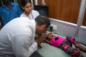
- Varicella vaccine: Given in some countries as part of routine immunization (not yet universal in Uganda, but available in private clinics).
- One dose gives about 85% protection; two doses give about 98% protection.
"Keep your child comfortable, prevent scratching, give paracetamol for fever, and watch for signs of skin infection like redness, swelling, or pus. Bring the child back immediately if they have trouble breathing, severe headache, are very drowsy, or the rash looks infected."
Case: A 5-year-old child presents with a low-grade fever and a rash. Upon examination, you notice a mixture of red macules, clear vesicles, and a few crusted scabs on the child's trunk and face. The mother asks if she can give the child Aspirin for the fever and when the child can return to school.
Answer: The diagnosis is Chickenpox (classic "crop" presentation of all 3 stages). You must emphatically tell the mother NO ASPIRIN due to the risk of fatal Reye's Syndrome; she should use Paracetamol instead. The child can return to school only when all the lesions have completely crusted over (usually 5-7 days).
Whooping cough is a serious bacterial infection of the lungs and breathing tubes caused by the bacterium Bordetella pertussis. It is called "whooping cough" because of the high-pitched "whoop" sound children make when they try to breathe in after a severe coughing fit.
Bordetella pertussis attaches to the cilia (tiny hair-like sweepers) of the respiratory epithelial cells. The bacteria release Pertussis Toxin and Tracheal Cytotoxin, which paralyze and kill the cilia. Without cilia to sweep away mucus, thick secretions build up massively in the lungs. The body attempts to forcefully expel this mucus, resulting in the violent, unending "paroxysmal" coughing fits because the normal clearing mechanism is destroyed.
- It is most dangerous for babies under 6 months old.
- It can cause apnoea (pauses in breathing), pneumonia, seizures, and even death.
- Babies may not "whoop" — they may just stop breathing or turn blue.
- Spread through respiratory droplets when an infected person coughs or sneezes.
- Very contagious — one infected person can infect up to 15 others.
- Incubation period: 7-10 days (range 4-21 days).
| Phase | Duration | Symptoms |
|---|---|---|
| Catarrhal Phase | 1-2 weeks | Runny nose, mild cough, low fever, sneezing — looks like a common cold. (Highly infectious phase!) |
| Paroxysmal Phase | 2-6 weeks (up to 10 weeks) | Severe coughing fits, "whooping" sound on breathing in, vomiting after coughing, face turns red or blue. |
| Convalescent Phase | 2-6 weeks | Coughing gradually decreases but can return with other respiratory infections due to damaged cilia. |
Mnemonic: The Pertussis Phases
"Cold, Whoop, Better" — Catarrhal (cold-like) ➔ Paroxysmal (whooping) ➔ Convalescent (getting better).
- Paroxysms: Sudden, violent bursts of rapid coughing (5-10 coughs in a row without breathing).
- Whoop: High-pitched sound when inhaling forcefully through a narrowed glottis after a coughing fit.
- Post-tussive vomiting: Vomiting after coughing (due to severe vagal nerve stimulation).
- Cyanosis: Lips and face turn blue during coughing due to oxygen deprivation.
- Apnoea: Breathing stops entirely, especially in fragile infants.
- Symptoms are notably worse at night.
- Pneumonia: Most common complication; bacteria or secondary virus infects the lung parenchyma.
- Apnoea and bradycardia: Breathing stops and heart rate slows — life-threatening in infants.
- Seizures & Encephalopathy: Brain damage resulting directly from hypoxia (lack of oxygen) during coughing fits or from bacterial toxins.
- Weight loss and malnutrition: Due to continuous post-tussive vomiting preventing nutrient absorption.
- Rib fractures & Subconjunctival haemorrhage: Mechanical trauma from the extreme physical pressure of violent coughing.
- Assessment: Ask about duration of cough, contact with infected persons, and vaccination history. Observe the color of the child (cyanosis?), breathing pattern, feeding ability. Listen for the "whoop" sound or lung crackles.
- Investigations:
- Nasopharyngeal swab/aspirate for PCR: Best test, most accurate in first 2-3 weeks.
- Culture: Takes longer but checks antibiotic resistance.
- Full Blood Count (FBC): Very unique for a bacterial infection, pertussis presents with a high white cell count with massive lymphocytosis (normally bacteria cause elevated neutrophils, but pertussis toxin specifically blocks lymphocytes from leaving the blood, causing them to pool).
Antibiotics do not cure the cough once the paroxysmal phase has started (because the ciliary damage is already done), but they: 1) Reduce severity if given in the catarrhal phase, 2) Stop the child from being infectious after 5 days, and 3) Prevent spread to others.
| Antibiotic | Age Group / Dose | Route | Duration |
|---|---|---|---|
| Azithromycin (Drug of Choice) | <6 months: 10 mg/kg once daily ≥6 months: 10 mg/kg day 1, then 5 mg/kg days 2-5 |
Oral | 5 days |
| Clarithromycin | ≥1 month: 7.5 mg/kg twice daily | Oral | 7 days |
| Co-trimoxazole | >2 months (if macrolides contraindicated) | Oral | 14 days |
Important Note: Erythromycin is NOT recommended for infants due to a high risk of pyloric stenosis (hypertrophy and narrowing of the stomach outlet).
- Oxygen therapy: If child is cyanosed or has low oxygen levels.
- Suctioning: Clear thick secretions from nose and throat, especially in infants who cannot clear it themselves.
- Feeding: Nasogastric feeding if child cannot feed. Offer small, frequent feeds immediately after a coughing bout.
- IV fluids: If child is dehydrated. Restrict fluids slightly to 2 mL/kg/hour to prevent pulmonary fluid overload (SIADH is a potential complication).
- Isolation: Droplet precautions.
- Give antibiotics to close contacts (especially infants <6 months, pregnant women in 3rd trimester, unvaccinated children).
- Exclusion: Child is infectious until 21 days after symptoms start, OR 14 days after paroxysmal cough starts, OR 5 days after starting antibiotics. Notify public health authorities.
- DTaP/DTP vaccine: Given at 6, 10, and 14 weeks, with a booster at 18 months (Uganda EPI schedule).
- Cocooning strategy: Vaccinate pregnant women in the third trimester to pass maternal antibodies to the newborn, protecting them until they are old enough to be vaccinated at 6 weeks.
"Keep your baby away from anyone with a cough. If your baby stops breathing, turns blue, or has a coughing fit with vomiting, come to hospital immediately. Complete all vaccinations on time — this is the best protection."
Impetigo is a highly contagious bacterial skin infection very common in young children, especially in hot, humid climates like Uganda. It is the third most common skin disease in children worldwide.
- Causes: Streptococcus pyogenes (Group A strep), Staphylococcus aureus (including MRSA), or a mixed infection of both.
- Spread: Direct skin-to-skin contact, sharing towels/clothes, auto-inoculation (scratching one sore and touching another part of the body). Insect bites or minor cuts provide the entry point.
| Type | Pathophysiology & Description | Appearance |
|---|---|---|
| Non-bullous impetigo (70% of cases) | Bacteria enter traumatized skin. Host response forms a pustule which ruptures. | Red sores with thick, honey-coloured (golden-yellow) crusts; usually on face (around nose and mouth), arms, legs. |
| Bullous impetigo | Caused exclusively by Staph aureus. It releases Exfoliative Toxins which dissolve the protein connections (desmoglein 1) holding epidermal skin cells together, causing the skin to separate and blister. | Large, thin-walled, fluid-filled blisters that burst easily; more common on the trunk and buttocks. |
- Cellulitis: Deeper skin infection — red, hot, swollen, painful skin.
- Post-streptococcal glomerulonephritis: Kidney disease 1-3 weeks after skin infection (immune complexes clog the kidney filters). Watch for blood in urine, swollen face, high blood pressure.
- Sepsis: Bacteria enter bloodstream — life-threatening.
- Crust Removal (Crucial Nursing Action!)
Topical antibiotics cannot penetrate the thick crusts. You MUST soak crusts in warm water/saline for 10-15 minutes, gently wash with soap, and pat dry before applying medicine. - Topical Antibiotics (Mild Cases)
Mupirocin ointment or Fusidic acid applied 3 times daily for 5-7 days. - Oral Antibiotics (Widespread/Severe/Bullous)
Cephalexin or Cloxacillin for 7 days. If MRSA is suspected, use Co-trimoxazole (contraindicated in infants under 2 months or those with G6PD deficiency). - School Exclusion: Exclude until 24 hours after starting antibiotics OR until sores are completely healed/crusted.
Dental caries (cavities) is the destruction of the hard tissues of the tooth. It is the most common chronic disease in children worldwide.
The tooth has three layers: 1) The hard outer Enamel, 2) The softer middle Dentine, and 3) The deep Pulp containing nerves and blood vessels.
Bacteria (Streptococcus mutans) in plaque feed on sugars and excrete lactic acid. This acid drops the pH of the mouth below 5.5, which dissolves (demineralizes) the calcium in the enamel. Once the hole breaches the enamel and reaches the nerve-rich pulp, the child experiences agonizing pain and is at risk for a dental abscess.
- Baby bottle tooth decay: Sleeping with a bottle of milk or juice pools sugar around the teeth all night.
- Frequent sugar intake and poor oral hygiene.
- Complications: Dental abscess, facial cellulitis, osteomyelitis (bone infection of the jaw), malnutrition (due to inability to chew painlessly), and spread to developing permanent adult teeth below the gums.
- Oral Hygiene: Brush twice daily with a pea-sized amount of fluoride toothpaste as soon as the first tooth erupts. Parents must help brush until age 7-8.
- Dietary Advice: Limit fizzy drinks, cakes, and sweets. Give water between meals. Absolutely NO bottle feeding at bedtime!
- Fluoride: Ensure fluoride toothpaste is used (1000-1500 ppm). Fluoride physically incorporates into the tooth structure (fluorapatite), making it highly resistant to acid attacks.
- Nursing Role: Screen children for caries during routine health visits, educate on the dangers of nocturnal bottle feeding, and refer to dentists for fillings or extractions.
Diarrhoea is the passing of loose or watery stools, usually three or more times in 24 hours. It is one of the leading causes of death in children under 5 years old worldwide, especially in low-resource settings like Uganda. The main danger is dehydration (loss of too much water and vital electrolytes from the body).
Normally, the intestines absorb massive amounts of water from digested food. In diarrhoea, pathogens (like Rotavirus or Vibrio cholerae) produce enterotoxins that destroy the absorptive cells (enterocytes) on the intestinal villi, or force chloride channels to open. When chloride pumps into the gut lumen, sodium follows it, and water follows the sodium (osmosis). The result is a massive outpouring of fluid into the gut, overwhelming absorption and causing severe watery stools.
- Children have a smaller body size and higher metabolic rate — they lose fluids much faster relative to their body weight.
- Their immune systems are still developing.
- They may not be able to tell you they are thirsty.
- Malnutrition makes diarrhoea worse (vicious cycle: diarrhoea causes malnutrition, malnutrition worsens diarrhoea).
- Poor sanitation and unsafe water increase exposure to germs.
| Type | Duration | Causes | Key Features |
|---|---|---|---|
| Acute watery diarrhoea | <14 days | Viruses (Rotavirus, Norovirus, Adenovirus), Bacteria (E. coli), Parasites | Most common; major risk is rapid dehydration. |
| Persistent diarrhoea | 14 days or more | Malnutrition, chronic infections, food intolerance | Leads to severe weight loss and malnutrition. Intestinal lining fails to heal. |
| Dysentery (bloody) | Variable | Shigella, Campylobacter, Entamoeba histolytica | Blood and mucus in stool; implies mucosal invasion; needs antibiotics. |
| Cholera | Variable | Vibrio cholerae | Severe watery "rice water stools"; causes profoundly rapid, lethal dehydration. |
WHO classifies dehydration into three levels. Accurate assessment dictates the treatment plan.
| Sign | No Dehydration | Some Dehydration | Severe Dehydration |
|---|---|---|---|
| General condition | Well, alert | Restless, irritable | Lethargic, unconscious |
| Eyes | Normal | Sunken | Very sunken |
| Tears | Present | Absent | Absent |
| Mouth and tongue | Moist | Dry | Very dry |
| Thirst | Drinks normally | Thirsty, drinks eagerly | Drinks poorly or unable to drink |
| Skin pinch | Goes back quickly | Goes back slowly (>2 seconds) | Goes back very slowly (>2 seconds) |
| Urine | Normal | Reduced | Very reduced or absent |
Mnemonic: Severe Dehydration
"SHOCK"
- Sunken eyes
- Hypotension (low blood pressure from low volume)
- Oliguria (little or no urine)
- Cold skin (poor perfusion)
- Ketones (in urine due to starvation/metabolic stress)
Four Rules for Home Treatment:
- Rule 1: Give Extra Fluid
Breastfeed more often and longer. Give ORS after every loose stool: Under 2 years (50-100 mL); 2 years+ (100-200 mL). Also give food-based fluids: soup, rice water, porridge. Give small, frequent sips. - Rule 2: Give Zinc Supplements
Under 6 months: 10 mg (½ tablet) daily for 10-14 days. 6 months+: 20 mg (1 tablet) daily for 10-14 days.
Why Zinc? Zinc physically regenerates the destroyed intestinal epithelium, restores intestinal enzyme function, and boosts local immunity. It reduces the duration of the current episode and prevents future episodes! - Rule 3: Continue Feeding
Do not starve the child! Continue breastfeeding and regular food. Offer an extra meal daily after recovery. Avoid very fatty or sugary foods (osmotic load worsens diarrhoea). - Rule 4: Know When to Return
Many watery stools, very thirsty, sunken eyes, fever, blood in stool, or not improving after 3 days.
- Give ORS in the clinic: 75 mL per kg over 4 hours. (e.g., 10 kg child = 750 mL ORS over 4 hours).
- Give frequent small sips. If child vomits, wait 10 minutes, then continue more slowly.
- Reassess after 4 hours. Start zinc and continue breastfeeding.
- This is a medical emergency. Start IV fluids immediately with Ringer's Lactate or Normal Saline.
- If no IV access within 30 minutes, use a nasogastric tube.
- IV Fluid Volumes (First 4 hours for children > 12 months, or over 6 hours for infants): 30 mL/kg rapidly, followed by 70 mL/kg more slowly.
- Give ORS by mouth as soon as child can drink. Monitor pulse, breathing, urine output, and LOC closely.
- For Dysentery: Needs antibiotics (Ciprofloxacin 15 mg/kg BID x 3 days, or Azithromycin).
- For Cholera: Aggressive rehydration and Azithromycin/Erythromycin. Monitor for severe hypokalaemia.
- For Persistent Diarrhoea: Assess for malnutrition/HIV, give multivitamins (Vitamin A is crucial for gut healing), consider lactose-free diet.
What NOT to Give:
- ✗ Anti-diarrhoeal drugs (loperamide): They paralyze the gut, trapping the infectious bacteria inside, which can lead to toxic megacolon.
- ✗ Anti-emetics routinely: Vomiting helps clear the infection naturally.
- ✗ Stop breastfeeding or food: Starvation worsens malnutrition and delays gut healing.
Promote exclusive breastfeeding for 6 months, safe water boiling, handwashing, latrine use, and Rotavirus/Measles vaccination. Tell parents: "Diarrhoea kills through dehydration. Give ORS and zinc immediately. Do not stop feeding your child!"
Atopic eczema is a chronic inflammatory skin condition causing itchy, dry, cracked, and red skin. It is the most common eczema in children. "Atopic" refers to a genetic tendency to develop allergic conditions.
Children with eczema often follow a predictable allergic progression called the Atopic March: Eczema → Food Allergy → Allergic Rhinitis (hay fever) → Asthma.
Pathophysiology: Many of these children have a genetic mutation in the filaggrin gene. Filaggrin is a protein that binds skin cells tightly together. Without it, the skin barrier is "leaky". Water escapes easily (causing profound dryness), and allergens/bacteria enter easily (triggering massive immune overreactions and inflammation).
| Trigger | Examples |
|---|---|
| Irritants | Soaps, detergents, bubble bath, wool clothing, perfumes |
| Allergens | Dust mites, pollen, pet dander, mould |
| Foods | Cow's milk, eggs, peanuts, wheat, soy (in some children) |
| Environmental | Heat, sweating, cold dry weather, low humidity |
| Infections & Stress | Staph bacteria, viruses, emotional stress, hormonal changes (puberty) |
- Intense itching (pruritus): Worse at night, causing severe sleep disturbance.
- Thickened skin (lichenification): Caused by long-term scratching.
| Age | Common Sites |
|---|---|
| Infants | Face (cheeks, forehead), scalp, extensor surfaces (outer arms and legs). |
| Children (2-12 yrs) | Flexural areas — inside elbows, behind knees, around neck, wrists, ankles. |
| Adolescents/Adults | Hands, feet, face, neck, skin folds. |
Mnemonic: Flexural Eczema Sites in Children
"Eyes Behind, Elbows Inside, Knees Behind" (Around eyes/behind ears, inside elbows, behind knees).
- Secondary bacterial infection: Staph or strep entering scratched skin (yellow crusts, pus).
- Eczema herpeticum (EMERGENCY): Herpes simplex virus infecting eczema. Causes rapid, painful, punched-out blisters, fever, and severe illness. Can be fatal.
- Psychosocial: Sleep disturbance affecting growth, low self-esteem, anxiety from visible skin.
- Emollients (Moisturisers) — The Foundation
Must be used generously, at least twice daily, even when skin looks clear! Apply within 3 minutes of bathing to lock in moisture. Use thick, greasy ointments (Vaseline, emulsifying ointment) rather than watery lotions. Apply in the direction of hair growth to prevent folliculitis. - Bathing & Triggers
Use lukewarm water (short baths 5-10 mins). Pat dry, never rub. Avoid soaps/perfumes. Wear soft cotton. Keep nails very short to minimize scratch damage. - Topical Corticosteroids (For Flare-ups)
Many parents have "steroid phobia." Reassure them that when used correctly (thin layer, only on inflamed red areas, lowest effective strength), they are incredibly safe. Mild: Hydrocortisone 1% (face/folds). Moderate: Betamethasone valerate 0.1% (body). - Other Therapies
Topical Calcineurin Inhibitors (Tacrolimus) for face/folds. Sedating antihistamines at night to break the itch-scratch cycle and promote sleep. Wet wrap therapy for severe flare-ups. Antibiotics/Antivirals if infected.
Measles is a highly contagious viral disease. It is one of the most infectious diseases known — 9 out of 10 unvaccinated contacts will catch it. It spreads via airborne droplets and can survive in the air for up to 2 hours after the patient has left the room!
Why do so many children die from pneumonia or severe diarrhea weeks after the measles rash fades? The measles virus directly infects and destroys memory T-cells and B-cells. This causes a phenomenon called "immune amnesia," wiping out the child's immune memory of previous diseases. For months after recovering from measles, the child is dangerously vulnerable to secondary bacterial and viral infections.
- Prodromal Phase (First 3-4 Days)
- High fever (up to 40.5°C).
- The 3 C's: Cough (dry, hacking), Coryza (watery runny nose), Conjunctivitis (red, watering, photophobic eyes).
- Koplik's spots (Pathognomonic sign!): Tiny white spots like "grains of salt on a red carpet" on the buccal mucosa inside the cheeks. They appear 1-2 days BEFORE the rash and confirm the diagnosis.
- Rash Phase (Days 3-7)
- Maculopapular rash starting behind the ears and hairline.
- It spreads strictly downward: face → neck → trunk → arms → legs.
- Fades in the same order it appeared, leaving a fine peeling skin (desquamation).
| Complication | Frequency | Description |
|---|---|---|
| Pneumonia | 1 in 20 cases | Leading cause of measles death; can be direct viral or secondary bacterial. |
| Diarrhoea | Very common | Can lead to severe dehydration and exacerbation of malnutrition. |
| Encephalitis | 1 in 1000 | Brain inflammation causing seizures, coma, or permanent brain damage. |
| SSPE (Subacute sclerosing panencephalitis) | Rare | A 100% fatal, degenerative brain disease appearing 7-10 years after the initial measles infection. |
| Corneal ulceration / Blindness | Common | Measles rapidly depletes the body's Vitamin A stores, destroying the cornea. |
- Supportive Care: Paracetamol for fever (NO ASPIRIN). Frequent fluids. Keep feeding. Darken the room if photophobia is severe. Clean eyes gently.
- Vitamin A Supplementation (Critical!): All children with measles MUST receive high-dose Vitamin A, regardless of nutritional status. It prevents blindness and reduces mortality by 50% by regenerating damaged epithelial linings in the gut, eyes, and lungs.
Doses (given once daily for 2 days): < 6 mos (50,000 IU), 6-11 mos (100,000 IU), 12 mos+ (200,000 IU) - Isolation & Antibiotics: Airborne precautions (N95). Isolate for 4 days after the rash appears. Give antibiotics ONLY if secondary pneumonia/otitis media occurs.
- Prevention: Measles vaccine (MR or MMR) at 9 months and 15-18 months.
Mumps is a viral infection (paramyxovirus) that specifically targets glandular tissue. It is best known for causing painful swelling of the salivary glands (parotid glands) on one or both sides of the face, giving a "hamster face" or "chipmunk cheeks" appearance.
- Early signs: Fever, headache, muscle aches, fatigue.
- Parotid Swelling: Painful swelling pushing the earlobe outward/upward. Pain significantly worsens when chewing or swallowing sour foods (because sour foods heavily stimulate saliva production, forcing fluid into an already swollen, blocked gland).
- Complications:
- Orchitis: Inflammation of the testicles in post-pubertal males (painful, can rarely cause infertility).
- Oophoritis: Ovary inflammation in females.
- Meningitis/Encephalitis: Virus crosses into the meninges (headache, stiff neck).
- Pancreatitis: Severe abdominal pain.
- Deafness: Rare but usually permanent in one ear.
- Supportive: Soft, easy-to-chew foods. Avoid citrus/vinegar. Paracetamol for pain. Scrotal support (jockstrap) and cold packs for orchitis.
- Isolation: Exclude from school for 5 days from the onset of parotid swelling.
- Prevention: MMR vaccine (Measles, Mumps, Rubella).
| Age | Vaccine | Diseases Prevented |
|---|---|---|
| Birth | BCG, OPV0 | Tuberculosis, Polio |
| 6 weeks | DTP-HepB-Hib, OPV1, PCV1, Rotavirus1 | Diphtheria, Tetanus, Pertussis, Hepatitis B, Hib, Polio, Pneumococcal, Rotavirus |
| 10 weeks | DTP-HepB-Hib, OPV2, PCV2, Rotavirus2 | (Same as above) |
| 14 weeks | DTP-HepB-Hib, OPV3, PCV3, IPV | (Same as above) + Inactivated Polio |
| 9 months | Measles-Rubella (MR), Yellow Fever | Measles, Rubella, Yellow Fever |
| 15-18 months | MR2, DTP booster | (Second doses) |
| Condition | Key Exam Point |
|---|---|
| Chickenpox | All spots at different stages (crops); NEVER give aspirin (Reye's); contagious until all crusted. |
| Whooping Cough | "Whoop" on inspiration; most dangerous <6 months; severe lymphocytosis; azithromycin first choice. |
| Impetigo | Honey-coloured crusts; highly contagious; MUST soak crusts before applying ointment. |
| Dental Caries | Most common chronic childhood disease; no bottles at bedtime. |
| Diarrhoea | Assess dehydration (SHOCK); ORS + Zinc for all; never stop feeding or breastfeeding. |
| Atopic Eczema | "Atopic march"; flexural distribution; daily thick emollients; topical steroids safe if used right. |
| Measles | Koplik's spots; 3 C's prodrome; rash spreads downward; high-dose Vitamin A is mandatory. |
| Mumps | Parotid swelling; "hamster face"; watch for orchitis in teenage boys; 5-day school exclusion. |
Case: A 3-year-old child comes to your clinic with a rash. You see red spots, fluid-filled blisters, and crusts all on the same child. The mother says the child had a fever 2 days ago. What is your diagnosis? What advice do you give about school?
Answer: The diagnosis is Chickenpox (Varicella), evidenced by the classic "crops" of lesions in multiple stages at once. The child must be completely excluded from school until every single blister has dried up and formed a crust (usually 5-7 days).
Case: A 2-month-old baby is brought in with coughing fits, turning blue, and vomiting after coughing. The mother says the cough is worse at night. What is your immediate concern? What antibiotic will you give? What precautions must you take?
Answer: The diagnosis is severe Whooping Cough (Pertussis). The immediate concern is apnoea, hypoxia, and respiratory failure, as infants this young often stop breathing instead of "whooping". You will give an oral macrolide, specifically Azithromycin (Erythromycin is avoided due to pyloric stenosis risk). You must institute strict Droplet precautions.
Case: A mother brings her 18-month-old with watery diarrhoea for 2 days. The child is irritable, has sunken eyes, and drinks eagerly when offered water. How do you classify the dehydration? What is your treatment plan?
Answer: This child has "Some Dehydration" (irritable, sunken eyes, drinks eagerly). You will implement Plan B: Administer ORS in the clinic (75 mL/kg over 4 hours), observe, continue breastfeeding, and start a 10-14 day course of Zinc supplements.
Case: During a community outreach, you see several children with golden-yellow crusts around their mouths and noses. Some have sores on their arms. What is this? How do you manage it in the community setting?
Answer: This is classic non-bullous Impetigo. Management involves educating parents to soak off the crusts with warm water and soap (vital so the medicine can penetrate), applying topical antibiotics (like Mupirocin), keeping nails short to stop auto-inoculation, and ensuring children do not share towels or bedding.
Case: A 10-month-old baby presents with high fever, cough, runny nose, red eyes, and a rash starting behind the ears. You notice white spots inside the cheeks. What are these spots called? What vitamin must you give? What is the most dangerous complication?
Answer: The diagnosis is Measles. The spots inside the cheeks are Koplik's Spots. You MUST give two daily doses of high-dose Vitamin A (100,000 IU for a 10-month-old) to prevent blindness and epithelial damage. The most dangerous, leading cause of death from measles is secondary Pneumonia.
Quick Quiz
Common Health Problems Quiz
Paediatrics - mobile-friendly and focused practice.
Privacy: Your details are used only for quiz tracking and certificates.
Common Health Problems Quiz
Paediatrics
Preparing questions...
Choose your answer and keep your streak alive.
Great effort.
Here is your quick performance summary.