
Pathology of Hypertension: Systemic & Pulmonary
I. Introduction to Blood Pressure Regulation
Before understanding the pathology of hypertension, we must first understand how the body normally regulates blood pressure. Blood pressure is essentially a function of two variables: Cardiac Output (CO) and Peripheral Vascular Resistance (PVR). The equation is BP = CO × PVR. Both variables are constantly adjusted by a complex web of genetic, neural, humoral, and environmental factors.
Determined by Heart Rate and Contractility (Cardiac Factors), and Blood Volume. Blood volume is strictly regulated by:
- Sodium (Na+) Balance: Where sodium goes, water follows.
- Mineralocorticoids: Aldosterone promotes sodium and water retention in the kidneys.
- Atrial Natriuretic Peptide (ANP): Released when the heart atria are stretched by high blood volume. It forces the kidneys to excrete sodium (natriuresis) to lower blood pressure.
Determined by the constriction or dilation of the arterioles. It is influenced by:
- Humoral Constrictors: Angiotensin II, Catecholamines (Epinephrine/Norepinephrine), Thromboxane, Leukotrienes, and Endothelin.
- Humoral Dilators: Prostaglandins, Kinins, and Nitric Oxide (NO).
- Neural Factors: α-adrenergic (constricts) and β-adrenergic (dilates) systems.
- Local Factors: Tissue pH, hypoxia, and local autoregulation.

The Renin-Angiotensin-Aldosterone System (RAAS)
The RAAS is the body's primary long-term blood pressure control mechanism. Factors released from the kidneys, adrenals, and myocardium interact to influence vascular tone and regulate blood volume by adjusting sodium balance.
- When the kidneys sense low blood volume or low sodium, the juxtaglomerular cells secrete an enzyme called Renin.
- Renin cleaves Angiotensinogen (produced by the liver) into Angiotensin I.
- As blood passes through the lungs, Angiotensin Converting Enzyme (ACE) converts Angiotensin I into Angiotensin II.
- Angiotensin II is a potent vasoconstrictor (raises PVR) and immediately signals the adrenal cortex to release Aldosterone.
- Aldosterone travels to the kidneys, forcing them to resorb Na+ and water, restoring blood volume (raises CO).
II. Systemic Hypertension
Hypertension is clinically defined as a sustained elevation of blood pressure above a systolic of 140 mmHg and/or a diastolic of 90 mmHg.
Unfortunately, hypertension typically remains completely asymptomatic until late in its course. Even severely elevated pressures can be clinically silent for years. During this time, it causes immense end-organ damage.
- 50% of hypertensive patients die due to Ischemic Heart Disease (IHD) or Congestive Heart Failure (CHF).
- 33% die from cerebrovascular strokes.
- Treatment: Utilizing blood-pressure-lowering drugs dramatically reduces the incidence and death rates attributable to all forms of hypertension-related pathology.
1. Malignant Hypertension
While most hypertension progresses slowly, about 5% of patients develop a rapidly rising blood pressure that, if untreated, leads to death within 1 to 2 years.
- Vitals: Characterized by severe pressure elevations (Systolic > 200 mmHg, Diastolic > 120 mmHg).
- Clinical Presentation: Rapid onset of acute renal failure, and severe eye damage including retinal hemorrhages and exudates (often with papilledema - swelling of the optic disc).
2. Causes & Pathogenesis of Hypertension
A. Essential (Primary) Hypertension (90-95%)
The vast majority of cases are idiopathic. Essential hypertension is the result of a highly complex interaction between multiple genetic polymorphisms and environmental factors (diet, stress, obesity, smoking).
B. Secondary Hypertension (5-10%)
Secondary hypertension has a specific, identifiable, and often curable underlying anatomic or biochemical cause.
| System | Specific Pathological Causes |
|---|---|
| Renal | Acute glomerulonephritis, Chronic renal disease, Polycystic kidney disease. Renovascular Hypertension: Renal artery stenosis causes decreased glomerular flow and pressure in the afferent arteriole. The kidney thinks the body is bleeding to death, inducing massive Renin secretion leading to increased blood volume and vascular tone via Angiotensin/Aldosterone pathways. |
| Endocrine | Primary Hyperaldosteronism (Conn's Syndrome): May be idiopathic or caused by aldosterone-secreting adrenal adenomas. Adrenocortical hyperfunction: Cushing syndrome (excess cortisol), Congenital Adrenal Hyperplasia. Others: Pheochromocytoma (massive catecholamine release), Acromegaly, Thyrotoxicosis, Pregnancy-induced (Preeclampsia), Exogenous hormones (OCPs, glucocorticoids, licorice ingestion). |
| Cardiovascular | Coarctation of the aorta, Polyarteritis nodosa, Increased intravascular volume, Increased cardiac output, Rigidity of the aorta. |
| Neurologic | Psychogenic, Increased intracranial pressure, Sleep apnea, Acute stress (including surgery). |
C. Single-Gene Disorders (Rare Causes)
Specific genetic mutations can cause severe, rare forms of secondary hypertension:
- Aldosterone Metabolism Enzyme Defects: Genetic defects in enzymes like aldosterone synthase, 11β-hydroxylase, or 17α-hydroxylase lead to an overproduction of mineralocorticoids. This causes downstream increases in salt/water resorption and massive plasma volume expansion.
- Liddle Syndrome: A moderately severe form of salt-sensitive hypertension. It is caused by mutations in the epithelial Na+ channel protein (ENaC) that cause the channel to remain permanently open, constantly increasing distal tubular reabsorption of sodium in response to aldosterone.
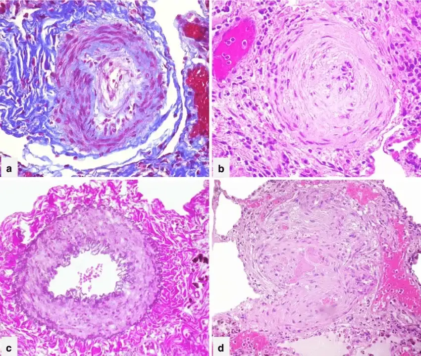
III. Vascular Morphology of Hypertension
Chronic hypertension physically destroys the microscopic blood vessels (arterioles), resulting in two distinct anatomical variants of Arteriolosclerosis.
Seen in chronic, essential hypertension and diabetes.
- Morphology: Arterioles show homogeneous, pink hyaline thickening with associated luminal narrowing.
- Pathogenesis: The high mechanical pressure injures the endothelial cells. Plasma proteins leak across the injured endothelium into the vessel wall. Simultaneously, the smooth muscle cells (SMCs) produce excess extracellular matrix in response to the chronic hemodynamic stress.
- Clinical Impact: In the kidneys, this arteriolar narrowing causes diffuse impairment of renal blood supply and glomerular scarring, known as Nephrosclerosis.
Seen specifically in severe, Malignant Hypertension.
- Morphology: Vessels exhibit concentric, laminated ("Onion-Skin") thickening of the walls with extreme luminal narrowing.
- Pathogenesis: The laminations consist of rapidly proliferating Smooth Muscle Cells with thickened, reduplicated basement membranes attempting to contain the explosive blood pressure.
- Clinical Impact: It is frequently accompanied by fibrinoid deposits and frank vessel wall necrosis (Necrotizing Arteriolitis), particularly destroying the kidney tissue.
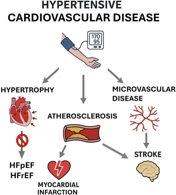
IV. Hypertensive Heart Disease (HHD)
Hypertensive Heart Disease is the direct morphological consequence of the increased demands placed on the heart by hypertension. The heart is a muscle; when forced to pump against extremely high pressure (afterload), it undergoes pressure overload ventricular hypertrophy.
1. Systemic (Left-Sided) Hypertensive Heart Disease
Hypertrophy of the heart is initially an adaptive response to chronic hypertension. However, these compensatory changes are ultimately maladaptive. The thick muscle demands more oxygen than the coronary arteries can supply (because capillaries do not multiply at the same rate the muscle grows). This leads to myocardial dysfunction, demand ischemia, cardiac dilation, Congestive Heart Failure (CHF), and sudden death.
- Diagnostic Criteria:
- Left ventricular hypertrophy (usually concentric—the wall thickens inward, drastically reducing lumen size) in the absence of other cardiovascular pathology (like aortic stenosis).
- A clinical history or pathologic evidence of hypertension in other organs (e.g., the kidney).
- Gross Morphology: Hypertension induces LV pressure overload hypertrophy, initially without ventricular dilation. The LV wall thickening increases the weight of the heart disproportionately to its overall size. The LV wall thickness may exceed 2.0 cm, and heart weight may exceed 500 g.
- Diastolic Dysfunction: Over time, the immensely thick muscle and increased interstitial connective tissue impart a stiffness that severely impairs diastolic filling. Because the LV won't stretch to accept blood, pressure backs up into the left atrium, leading to Left Atrial Enlargement.
- Microscopic Morphology: The earliest change is an increase in the transverse diameter of individual myocytes. At advanced stages, extreme cellular and nuclear enlargement (often called "boxcar nuclei") become apparent, accompanied by perivascular and interstitial fibrosis.
2. Pulmonary (Right-Sided) Hypertensive Heart Disease (Cor Pulmonale)
Cor Pulmonale stems from right ventricular pressure overload specifically caused by disorders of the pulmonary circulation. (Note: While left-sided heart failure is the most common cause of pulmonary hypertension overall, true isolated Cor Pulmonale is due strictly to intrinsic lung/pulmonary vessel issues).
- Acute Cor Pulmonale: Usually follows a massive Pulmonary Embolism. Because the pressure spike is sudden, there is marked dilation of the right ventricle without hypertrophy. On cross-section, the normal crescent shape of the RV is transformed into a dilated ovoid.
- Chronic Cor Pulmonale: The RV has time to adapt to chronic lung disease. The RV wall thickens dramatically (sometimes up to 1.0 cm or more). Subtle hypertrophy can be seen in the outflow tract muscle bundles or the thickening of the moderator band (the muscle bundle connecting the ventricular septum to the anterior right ventricular papillary muscle).
- Complications: The massively hypertrophied right ventricle can bulge into the septum, physically compressing the left ventricular chamber, or dilate so much that it pulls the tricuspid valve apart, leading to tricuspid regurgitation.
Disorders Predisposing to Cor Pulmonale
| Category | Specific Diseases |
|---|---|
| Diseases of Pulmonary Parenchyma | Chronic Obstructive Pulmonary Disease (COPD), Diffuse pulmonary interstitial fibrosis, Pneumoconioses, Cystic fibrosis, Bronchiectasis. |
| Diseases of Pulmonary Vessels | Recurrent pulmonary thromboembolism, Primary pulmonary hypertension, Extensive pulmonary arteritis (e.g., granulomatosis with polyangiitis), Drug/toxin/radiation-induced vascular obstruction, Microembolism from tumors. |
| Disorders Affecting Chest Movement | Kyphoscoliosis, Marked obesity (Sleep apnea, Pickwickian syndrome), Neuromuscular diseases. |
| Disorders Inducing Pulmonary Arterial Constriction | Metabolic acidosis, Hypoxemia, Chronic altitude sickness, Obstruction of major airways, Idiopathic alveolar hypoventilation. |
References & Further Reading
- Kumar, V., Abbas, A. K., & Aster, J. C. (2021). Robbins & Cotran Pathologic Basis of Disease (10th ed.). Elsevier. (Chapters on Blood Vessels and the Heart).
- Lilly, L. S. (2015). Pathophysiology of Heart Disease (6th ed.). Wolters Kluwer.