
Table of Contents
ToggleThe urinary bladder is a hollow, muscular organ that stores urine and empties it through the urethra. Bladder conditions may inflame its lining, obstruct emptying, disturb storage, form stones, injure the wall, or produce abnormal growths. The symptoms often overlap, so assessment must identify the cause rather than treating every urinary complaint as simple cystitis.
These notes give an overview of important bladder conditions for nursing learners. The linked Cystitis and Urinary Tract Infections lessons discuss infection in greater depth. Management of an individual patient must follow the responsible clinician’s plan, current Uganda guidance and local facility protocols.
Seek urgent clinical assessment for sudden inability to urinate; a painful, distended lower abdomen; fever or rigors with urinary symptoms; visible blood with clots or reduced urine output; faintness, confusion or signs of sepsis; or blood in the urine after pelvic trauma. Visible haematuria, even if it stops, requires evaluation. With pelvic fracture and visible haematuria, bladder injury must be actively investigated. Do not force a urinary catheter through resistance or instrument the urethra before assessment when urethral injury is suspected.
Learning objectives
After studying this topic, the learner should be able to:
- Describe bladder anatomy and explain normal storage and voiding.
- Classify common inflammatory, obstructive, storage, structural, traumatic and neoplastic bladder conditions.
- Recognise common symptoms and red flags, including acute retention, sepsis, haematuria and bladder trauma.
- Explain the purpose and limitations of urinalysis, culture, post-void residual measurement, imaging, cystoscopy and urodynamic tests.
- Outline condition-specific treatment and the nursing care required before, during and after interventions.
- Teach patients safe catheter care, prevention, follow-up and when to seek urgent help.
I. Relevant anatomy and normal physiology
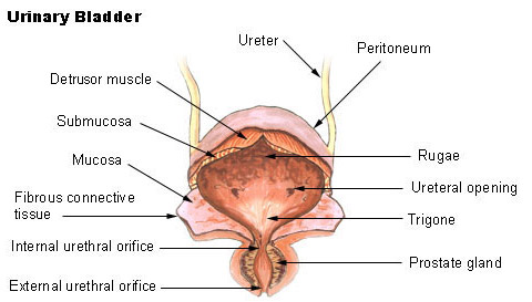
The bladder lies in the pelvis behind the pubic symphysis. When it fills, it expands upward into the lower abdomen. Its position, capacity and support can be affected by age, pregnancy, pelvic floor changes, surgery and neurologic disease.
- Detrusor muscle: Smooth muscle in the bladder wall. It relaxes during filling and contracts during voiding.
- Mucosa and urothelium: The inner lining forms a protective barrier between urine and the bladder wall. Irritation, infection, stones, chemicals, radiation and tumours can affect it.
- Trigone: A smooth triangular area between the two ureteric openings and the internal urethral opening. It is sensitive to stretching and is clinically important in cystoscopy and bladder surgery.
- Bladder neck and urethral sphincters: These help maintain continence and coordinate urine release.
- Nerve control: Brain, spinal cord and peripheral nerves coordinate bladder sensation, detrusor activity and sphincter relaxation. Neurologic injury can impair storage, emptying or both.
During normal filling, the bladder accommodates increasing urine volume at low pressure while the outlet remains closed. When it is appropriate to void, the detrusor contracts and the outlet relaxes in a coordinated way. A problem with the bladder muscle, nerves, outlet, mucosal lining or surrounding support can produce frequency, urgency, pain, leakage, weak flow, incomplete emptying or retention.
II. Classification and overview
| Group | Examples | Typical problem or clue |
|---|---|---|
| Inflammatory or infectious | Acute cystitis, recurrent infection, chemical or radiation cystitis, bladder pain syndrome | Dysuria, urgency, frequency, suprapubic discomfort; fever or flank pain suggests systemic infection or upper tract involvement. |
| Calculus | Bladder stones | Intermittent stream, painful voiding, recurrent infection or haematuria, often with urinary stasis. |
| Emptying dysfunction | Acute or chronic urinary retention, outlet obstruction, underactive detrusor | Hesitancy, weak stream, straining, incomplete emptying, overflow leakage or inability to pass urine. |
| Storage dysfunction | Urge, stress, mixed or overflow incontinence; overactive bladder | Leakage, urgency, frequency, nocturia or leakage with coughing and exertion. |
| Neurologic | Neurogenic lower urinary tract dysfunction | Abnormal sensation, high residual urine, incontinence, retention, recurrent symptomatic infection or autonomic dysreflexia in susceptible patients. |
| Structural | Bladder diverticulum, congenital abnormalities, fistula | Urinary stasis, recurrent infections, stones, leakage or persistent symptoms. |
| Traumatic or iatrogenic | Bladder contusion, extraperitoneal or intraperitoneal rupture, operative injury | Haematuria, pelvic trauma, abdominal pain or distension, reduced urine output or urine leakage. |
| Neoplastic | Bladder cancer, most often urothelial carcinoma in many regions | Often painless, intermittent visible haematuria; irritative symptoms may also occur. |
- Infection: Bacteria may ascend through the urethra. Risk rises with urinary stasis, incomplete emptying, pregnancy, some sexual exposures, diabetes, catheterisation, urinary tract abnormalities and immunosuppression.
- Obstruction or poor emptying: Prostate enlargement, urethral stricture, bladder neck obstruction, pelvic organ prolapse, stones, constipation, postoperative effects and weak detrusor contraction may leave residual urine.
- Medicines and anaesthesia: Some medicines, including anticholinergic agents, opioids, sympathomimetics and some anaesthetic or postoperative medicines, can worsen retention. Patients should not stop prescribed medicines without clinical advice; the medication list should be reviewed.
- Neurologic disease: Spinal cord injury, multiple sclerosis, stroke, Parkinson disease, diabetic neuropathy and other nerve disorders can disrupt sensation or coordination.
- Stones or foreign bodies: Bladder stones may form when urine stagnates or a foreign body acts as a focus for mineral deposition. Stones can also migrate from the upper urinary tract.
- Chronic irritation and cancer risk: Tobacco smoking and some occupational chemical exposures are recognised risks for bladder cancer. In parts of sub-Saharan Africa, chronic urinary schistosomiasis caused by Schistosoma haematobium is associated with bladder inflammation and squamous cell carcinoma. Ask respectfully about exposure and previous treatment where clinically relevant.
- Pelvic or abdominal trauma and procedures: A blow to a distended bladder, pelvic fracture, penetrating injury, pelvic surgery or endoscopic procedure can injure the bladder wall.
| Symptom pattern | Examples | Clinical significance |
|---|---|---|
| Storage symptoms | Urgency, frequency, nocturia, urge leakage | May occur with cystitis, overactive bladder, stones or neurologic dysfunction. |
| Voiding symptoms | Hesitancy, weak or interrupted stream, straining, prolonged voiding | Consider outlet obstruction, retention or impaired detrusor contraction. |
| Pain or discomfort | Dysuria, suprapubic pain, pressure relieved or worsened by voiding | May occur with infection, stones, inflammation or bladder pain syndrome. |
| Abnormal urine | Visible or microscopic blood, cloudy urine, clots | Haematuria may be due to infection, stones, trauma, schistosomiasis or malignancy; it needs assessment. |
| Systemic or traumatic features | Fever, rigors, confusion, hypotension, pelvic fracture, abdominal distension, low output | Escalate for systemic UTI, sepsis, obstruction, renal impairment or bladder injury. |
- Sudden inability to pass urine, especially with suprapubic pain or a distended bladder.
- Fever, rigors, fast pulse, low blood pressure, confusion or marked weakness with urinary symptoms.
- Visible haematuria with clots, heavy bleeding, dizziness or reduced urine output.
- Pelvic fracture or significant lower abdominal trauma with visible blood in the urine, inability to void or abdominal distension.
- Persistent or recurrent haematuria, even if the patient has no pain and the bleeding stops.
- New severe pain, vomiting, flank pain, worsening renal function or suspected urinary leak after surgery.
III. Assessment and diagnosis
Assessment is guided by the symptoms and level of urgency. A focused history and examination help determine whether the problem is infection, obstruction, storage dysfunction, trauma, stone disease or another cause. No single symptom or urine test identifies every bladder condition.
- Ask onset, duration, progression and severity of dysuria, frequency, urgency, nocturia, stream changes, leakage, pain and visible blood. Clarify the last time the patient passed urine and the approximate amount.
- Ask about fever, rigors, flank pain, abdominal distension, clots, recurrent infection, previous stones, catheter use, urologic procedures, pelvic surgery, radiation and trauma.
- Review medical conditions, neurologic symptoms, diabetes, pregnancy possibility where relevant, bowel habits, hydration instructions and all prescribed, over-the-counter and traditional medicines.
- For haematuria, ask about smoking, work exposures, urinary schistosomiasis or freshwater contact where relevant, and previous treatment. Do not assume that blood is caused by infection without appropriate follow-up.
- Assess vital signs, general appearance, hydration, lower abdominal tenderness or bladder fullness, urine output and pain. Examination of the pelvis, prostate, perineum or nervous system is performed by an appropriately trained clinician when indicated and with consent.
- Use a bladder diary for selected patients with urgency, frequency or incontinence. Record voiding times, approximate volumes, fluid intake, urgency and leakage to clarify patterns and response to treatment.
| Investigation | What it can show | Nursing responsibilities and limitations |
|---|---|---|
| Urinalysis | Blood, leukocytes, nitrites, glucose, protein or other clues to infection, bleeding or systemic disease. | Collect a correctly labelled clean-catch specimen when possible. A dipstick alone does not prove the cause of haematuria and a negative result does not exclude every bladder disorder. |
| Urine microscopy and culture | Identifies bacteria and may guide antibiotic choice when symptomatic infection is suspected. | Collect before antibiotics when ordered and when doing so will not delay urgent treatment. Do not treat a laboratory result in isolation; asymptomatic bacteriuria is not routinely treated except in selected settings such as pregnancy or before a mucosa-breaching urologic procedure, according to local guidance. |
| Blood tests | Renal function, electrolytes, full blood count and inflammatory markers may help assess retention, bleeding or systemic infection. | Check identification and follow-up of results. Escalate reduced renal function, significant anaemia or deterioration. |
| Post-void residual (PVR) | Ultrasound or catheter measurement estimates urine left after voiding. | Measure promptly after voiding and record the value. A high residual supports incomplete emptying but does not identify its cause by itself. |
| Ultrasound | May show bladder volume, residual urine, stones, wall changes, masses or hydronephrosis. | Follow preparation instructions and report significant retention or upper tract changes. A normal ultrasound does not rule out every small bladder lesion. |
| CT or other cross-sectional imaging | Can assess stones, masses, surrounding structures, upper urinary tract and selected traumatic injuries. | Check contrast history, renal function and pregnancy status when relevant; follow radiation safety and the ordered protocol. |
| Cystoscopy and biopsy | Directly views the bladder lining. Tissue sampling provides histology when cancer is suspected; stones or other abnormalities may also be seen. | Explain preparation and aftercare. Biopsy or tumour resection is performed by a trained clinician; follow the plan for bleeding, catheter drainage and pathology review. |
| Uroflowmetry and urodynamic tests | Assess urine flow and bladder storage, pressure, sensation and emptying in selected patients. | These tests assess function, not every structural cause. Explain the procedure and protect privacy; results need interpretation alongside symptoms and examination. |
| Cystography / CT cystography | Evaluates suspected bladder rupture. In trauma, the bladder is actively filled retrogradely with contrast under the imaging protocol. | Visible haematuria with pelvic fracture is a strong indication. Passive bladder filling during a routine CT is not sufficient to exclude rupture. |
Key interpretation: Urinalysis and culture look for infection or blood; PVR and flow studies evaluate emptying; imaging assesses anatomy; cystoscopy with biopsy can diagnose a bladder lesion; cystography is selected for suspected injury. These tests answer different questions and are not interchangeable.
IV. Common conditions of the urinary bladder
Cystitis means inflammation of the bladder, commonly from bacterial infection. Typical lower urinary symptoms include dysuria, frequency, urgency and suprapubic discomfort. Some patients notice visible blood. Fever, rigors, flank pain, vomiting, hypotension or confusion suggest systemic illness or possible upper urinary tract involvement and require urgent assessment.
Diagnosis is based on the history and clinical assessment, with urinalysis or culture used according to the patient’s risks and local protocol. Recurrent symptoms, pregnancy, male sex, childhood, catheter use, structural abnormality, immunosuppression, treatment failure or suspected systemic infection usually need more careful assessment and culture-directed management. Treatment is selected by a clinician using patient factors, culture where indicated, allergy history and current local antimicrobial guidance. The nurse monitors response, adverse reactions, hydration as appropriate and warning signs.
Non-infectious cystitis may follow radiation, some medicines or chemical exposure. Bladder pain syndrome causes persistent or recurrent bladder-related pain, pressure or discomfort with urgency or frequency when infection and other causes have been excluded. Repeated antibiotic courses without evidence of infection are not appropriate; management is individualised and may include bladder training, pain management, lifestyle adjustment and specialist review.
A positive urine culture in a person without urinary symptoms does not automatically mean the person has a symptomatic infection. Routine screening or antibiotic treatment is not recommended for many groups, including most non-pregnant adults and people with long-term catheters or neurogenic lower urinary tract dysfunction. Important exceptions include pregnancy and selected urologic procedures that breach the mucosa; follow national and facility guidance.
Bladder stones are mineral deposits that form in the bladder or migrate from the kidney or ureter. In adults, urinary stasis and bladder outlet obstruction are common contributing factors. Other causes include neurogenic bladder, chronic retention, foreign bodies, recurrent infection, bladder augmentation or urinary diversion. In children, primary stones may be associated with dehydration, diarrhoeal illness and nutritional factors in some settings.
- Suprapubic pain or discomfort, especially near the end of voiding.
- Frequency, urgency, painful urination or an interrupted stream that may improve when the patient changes position.
- Visible or microscopic haematuria, recurrent infection, cloudy urine or acute retention.
- Some stones cause few symptoms and are found during imaging or cystoscopy for another reason.
Assessment may include urinalysis, urine culture, ultrasound, CT or cystoscopy. The stone should be analysed when available, and the clinician should investigate the reason it formed, including outlet obstruction, poor bladder emptying or a foreign body.
Treatment depends on stone size, cause, anatomy and available expertise. Endoscopic transurethral cystolithotripsy is commonly preferred when it can be performed safely; a percutaneous approach or open cystolithotomy may be selected for particular patients or very large stones. Treating the underlying obstruction or dysfunction is important because stone removal alone may not prevent recurrence. Nursing care includes pain assessment, urine monitoring, specimen collection as ordered, preparation for the procedure, catheter and wound care when indicated, and teaching follow-up and recurrence prevention.
Urinary retention is incomplete or failed bladder emptying. It may be acute and painful, or chronic and relatively painless. Causes include prostate enlargement, urethral stricture, bladder neck obstruction, pelvic organ prolapse, constipation, medications, infection, postoperative effects, stones, neurologic disease or weak detrusor contraction. Chronic retention may lead to overflow leakage, recurrent infection, bladder damage, hydronephrosis or renal impairment.
| Acute retention | Chronic retention |
|---|---|
| Sudden inability to pass urine, often with pain, distress and a full bladder. Treat as an urgent problem. | Persistent incomplete emptying; may present with weak stream, hesitancy, nocturia, overflow leakage, recurrent infection or renal effects. It can be painless. |
Assessment includes last void, symptoms, abdominal examination, medication review, PVR, urinalysis, renal tests and imaging or cystoscopy when indicated. Acute retention requires prompt bladder drainage by a trained clinician, usually with a catheter when urethral access is safe. The cause should then be evaluated and treated. Chronic retention management is individualised and may involve intermittent or indwelling catheterisation, treatment of obstruction, medication review or a procedure.
Safety point: Blood at the urethral opening, perineal bruising, pelvic fracture or suspected urethral injury changes the approach. Do not make blind or repeated catheter attempts. Escalate for specialist assessment and appropriate imaging; a suprapubic route may be considered by a qualified clinician if urethral drainage is unsafe or unsuccessful.
Urinary incontinence is involuntary urine leakage. It is a symptom with different mechanisms, not a normal or inevitable consequence of ageing. Common patterns include:
- Stress incontinence: Leakage with coughing, laughing, lifting or exertion when pressure exceeds urethral support.
- Urgency incontinence: Leakage associated with a sudden compelling urge to void, often with frequency and nocturia.
- Mixed incontinence: Both stress and urgency leakage.
- Overflow incontinence: Dribbling or frequent small leaks when the bladder does not empty adequately; consider retention or obstruction.
- Functional incontinence: Leakage because mobility, cognition, clothing, environment or access to a toilet prevents timely voiding.
Assessment includes a respectful history, medication review, examination when indicated, urinalysis, a bladder diary and PVR if retention is possible. Management depends on the type and may include bladder training, scheduled toileting, pelvic floor muscle training, practical changes to toilet access, skin care, medicines, devices or surgery. Encourage normal, appropriate fluid intake; excessive restriction can cause dehydration and concentrated urine. Medication or procedures require clinician assessment.
Neurologic disease or injury can disrupt bladder sensation, storage, detrusor contraction or sphincter coordination. A patient may have urgency and leakage, incomplete emptying, retention, a high-pressure bladder or a combination. The risk is not limited to discomfort: unsafe bladder pressures or chronic retention may threaten the kidneys.
Assessment is guided by the neurologic diagnosis and may include renal and bladder ultrasound, PVR, urodynamics, urinalysis and a review of the current bladder-management method. Treatment aims to protect renal function, achieve safe storage and emptying, reduce symptomatic infections and support the patient’s independence. Options may include timed voiding, clean intermittent catheterisation, an indwelling catheter in selected cases, medicines or specialist procedures. The plan is individualised; catheter technique and frequency must be taught and reviewed by a trained clinician.
Symptoms of infection can be atypical in people with spinal cord injury or neurogenic bladder. New or worsening leakage, increased spasticity, malaise, autonomic dysreflexia, new bladder discomfort or systemic signs may be more useful than classic dysuria. Do not routinely screen for or treat asymptomatic bacteriuria in neurologic lower urinary tract dysfunction. Recurrent symptomatic infection should prompt review of bladder function, stones, foreign bodies and catheter need.
A bladder diverticulum is an outpouching of the bladder wall. It may be congenital or develop with long-standing outlet obstruction. Urine can remain in the pouch after voiding, increasing the risk of infection, stone formation and incomplete emptying. Some diverticula are discovered incidentally; others contribute to recurrent urinary infection, haematuria, retention or voiding difficulty. Ultrasound, cystoscopy or other imaging may be used to assess the bladder and outlet. Management depends on symptoms and complications and may include treatment of the obstruction, observation or surgical repair. Persistent leakage through an abnormal connection, such as a fistula, requires specialist assessment.
Bladder injury may follow blunt trauma to a full bladder, pelvic fracture, penetrating injury or pelvic/urologic surgery. It is classified as extraperitoneal, intraperitoneal or combined. Suspect injury after significant pelvic trauma when there is visible haematuria, inability to void, suprapubic pain, abdominal distension, reduced urine output, peritonism or unexplained deterioration. Visible haematuria with a pelvic fracture is a strong indication for cystography.
In a stable patient, retrograde cystography or CT cystography is used to demonstrate leakage. A routine CT with passive bladder filling does not reliably exclude a rupture. Initial care follows trauma priorities: assess airway, breathing and circulation; control haemorrhage; monitor observations and output; provide analgesia as prescribed; and urgently involve the trauma/urology team. Avoid urethral catheterisation until associated urethral injury has been considered.
- Uncomplicated extraperitoneal rupture: Often managed with continuous bladder drainage and observation under the specialist plan.
- Complex extraperitoneal injury: Bladder-neck involvement, bone fragments, bladder-wall entrapment or associated rectal/vaginal injury may require operative repair.
- Intraperitoneal rupture: Usually requires surgical exploration and repair because urine leakage can cause peritonitis and sepsis. Selected small iatrogenic injuries may be managed conservatively only under a specialist plan when there is no peritonitis or ileus.
Nursing priorities include serial observations, pain and abdominal assessment, accurate input/output, catheter drainage care as prescribed, monitoring for fever, ileus, haematuria or urine leakage, and ensuring follow-up imaging or cystography is completed when ordered. Do not remove, clamp, irrigate or replace a post-repair catheter unless directed by the surgical team.
Bladder cancer is an abnormal growth of cells in the bladder lining or wall. Urothelial carcinoma is common in many parts of the world. Tobacco exposure and certain workplace chemicals are important risks. In some African populations, chronic urinary schistosomiasis and long-standing bladder irritation are associated with squamous cell carcinoma. This regional association does not mean every person with haematuria has cancer; it means persistent or visible blood must not be dismissed without assessment.
- Painless visible haematuria is a common presentation and may appear intermittently. A normal-looking urine sample later does not remove the need for evaluation.
- Microscopic haematuria, frequency, urgency or dysuria can also occur.
- Advanced disease may cause pelvic or lower abdominal pain, urinary obstruction, back pain, weight loss, fatigue or swelling.
Assessment may include urinalysis, renal function tests, imaging of the urinary tract, cystoscopy and biopsy or transurethral resection of a suspicious lesion. Histopathology establishes the diagnosis and helps determine tumour type, grade and depth. Further imaging is selected to stage disease. Urine cytology or tumour-marker tests may be used in selected cases but do not replace cystoscopy and tissue diagnosis when indicated.
Treatment depends on the tumour’s stage, grade, location, patient fitness and preferences. Non-muscle-invasive disease may be treated by transurethral resection with surveillance and selected intravesical therapy. Muscle-invasive or extensive disease may require radical cystectomy with urinary diversion, systemic treatment, radiation-based approaches or combinations. Palliative treatment may focus on bleeding, pain and urinary obstruction. Decisions are made by a specialist oncology/urology team.
Nursing care includes explaining investigations in plain language, maintaining dignity, monitoring haematuria and catheter drainage after resection, providing stoma and urinary-diversion education when needed, supporting nutrition and symptom control, and addressing fear, sexual health, body image and family concerns. Teach the patient to attend surveillance appointments because recurrence monitoring is an important part of care.
V. General management principles
Management aims to relieve symptoms, protect the bladder and kidneys, treat infection or bleeding when present, correct obstruction where possible and preserve the patient’s dignity and function. Treatment is selected for the specific diagnosis rather than for “bladder symptoms” alone.
- Stabilise and triage: Identify sepsis, acute retention, heavy haematuria, trauma, renal impairment or severe pain. Escalate immediately when the patient is unstable or the bladder cannot empty.
- Relieve urgent obstruction safely: Acute retention usually needs prompt bladder drainage by a trained clinician. If urethral injury is suspected, obtain specialist assessment before urethral instrumentation.
- Confirm the cause: Use history, examination, urine tests, PVR, imaging, cystoscopy or urodynamics as indicated. Investigate persistent haematuria, recurrent infection or treatment failure.
- Treat the underlying disorder: Examples include culture-guided antimicrobial treatment for symptomatic infection, stone removal plus correction of urinary stasis, treatment of outlet obstruction, continence therapy, bladder-protective neuro-urologic care, repair of rupture or stage-directed cancer therapy.
- Review medicines and comorbidities: Identify medicines that may impair emptying, assess constipation and diabetes control, and review renal function. Changes to prescribed treatment are made by the responsible clinician.
- Plan follow-up: Reassess symptoms, urine output, PVR, infection results, pathology, imaging, catheter plan and recurrence risk. Document who is responsible for communicating outstanding results.
VI. Nursing management
- Assess airway, breathing, circulation, level of consciousness and vital signs when the patient is unwell. Observe for fever, rigors, hypotension, tachycardia, confusion, dehydration or shock.
- Ask when the patient last passed urine, estimate output, assess suprapubic fullness and pain, check for visible blood or clots, and use a bladder scanner when available and appropriate.
- Assess pain using a consistent scale; note location, onset, severity, relation to voiding and response to prescribed measures.
- Collect urine and blood specimens as ordered, label promptly and record the time. Obtain culture before antimicrobial treatment when indicated and when it will not delay emergency care.
- Escalate inability to void, no catheter drainage with pain, significant haematuria, fever with deterioration, suspected pelvic injury or reduced urine output.
- Provide privacy, a chaperone where indicated, respectful language and an opportunity for the patient to ask questions. Urinary and sexual symptoms can be embarrassing; do not shame or blame the patient.
- Use a urinary catheter only for an appropriate indication and according to institutional policy. Catheter insertion is performed by trained, authorised staff using hand hygiene, aseptic technique and suitable equipment.
- Maintain a closed drainage system; secure the catheter to prevent traction; keep tubing unkinked and the bag below bladder level and off the floor.
- Observe and document urine volume, colour, clarity, clots, leakage, catheter comfort and the condition of surrounding skin. Report absent drainage, new pain, bladder distension or heavy bleeding.
- Do not disconnect the system unnecessarily or collect a specimen from the drainage bag. Obtain a specimen from the designated sampling port using aseptic technique when ordered.
- Do not irrigate, clamp, remove, advance or replace a catheter unless this is prescribed, within scope and consistent with the urology plan. After bladder or urethral repair, catheter manipulation can disrupt healing.
- Review the continuing need daily and arrange removal as soon as clinically appropriate and ordered. Teach the patient or caregiver catheter care using demonstration and teach-back if long-term drainage is required.
- Before cystoscopy, stone treatment or tumour resection: Confirm identity, procedure and consent status; check allergies, relevant medicines, required investigations and fasting instructions; explain expected preparation and recovery.
- After endoscopic procedures: Monitor observations, pain, urine colour and output, catheter patency and clots. Mild transient haematuria may occur after some procedures, but increasing bleeding, clot retention, fever or worsening pain requires review.
- After bladder repair: Maintain the prescribed drainage system without traction, monitor abdominal findings and output, report fever, ileus, urine leakage or deterioration, and follow the surgeon’s plan for cystography and catheter removal.
- After cystectomy and urinary diversion: Assess stoma colour and perfusion, appliance fit, output, skin, hydration, pain and the patient’s confidence. Teach pouch emptying, skin protection, warning signs and follow-up with the specialist team.
- Document procedure-related findings, fluid balance, urine output, medicines, education, escalation and handover clearly.
- Provide prescribed analgesia and non-pharmacologic comfort measures. Reassess and document the effect rather than assuming pain has resolved.
- For leakage, provide prompt hygiene, barrier skin protection and an appropriate continence aid. Treat the patient respectfully and maintain privacy.
- Support timed voiding, bladder training or pelvic floor exercises only when appropriate for the diagnosed problem and included in the care plan.
- Encourage the patient to follow a balanced fluid plan. Avoid blanket advice to drink excessive amounts or to severely restrict fluids; individualise advice for cardiac, renal or other restrictions.
- Explain medicines, possible adverse effects, catheter or wound care, follow-up and red flags in simple language. Ask the patient to repeat the plan in their own words.
VII. Nursing care plan examples
| No. | Problem, goal and nursing actions | Rationale and evaluation |
|---|---|---|
| 1 | Impaired urinary elimination related to infection, obstruction, impaired bladder contraction or altered neurologic control. Goal: The patient will maintain a safe voiding or drainage plan, with urine output documented and symptoms escalated promptly. Assess voiding pattern and last void; monitor intake/output; assess bladder fullness and PVR as ordered; maintain prescribed drainage and report inability to void, rising pain, distension or falling output. | Trend information helps detect retention, obstruction, infection or renal compromise. Evaluate urine flow, residual, discomfort and observations after intervention; document changes and hand them over. |
| 2 | Acute pain related to bladder inflammation, distension, stone or instrumentation. Goal: The patient will report tolerable pain and demonstrate improved comfort. Assess pain location and severity; provide privacy and positioning; administer prescribed analgesia; support ordered bladder drainage; reassess within the expected response time. | Suprapubic pain may reflect distension or inflammation; sudden severe pain or failure to improve may signal obstruction or injury. Evaluate the patient’s reported response and escalate worsening pain. |
| 3 | Risk for infection or sepsis related to urinary stasis, catheterisation or urinary tract intervention. Goal: The patient will remain free from systemic infection or receive prompt treatment. Use aseptic technique; obtain specimens as ordered; monitor temperature and observations; maintain closed catheter drainage; administer prescribed treatment and review catheter need. | Stasis and instrumentation may increase infection risk. Review culture results and clinical response; promptly escalate fever, rigors, hypotension, confusion or worsening pain. |
| 4 | Urinary incontinence and risk of impaired skin integrity related to urgency, weak pelvic support, retention or functional limitation. Goal: The patient will maintain skin integrity and participate in an individualised continence plan. Assess leakage pattern and skin; provide timely toileting, hygiene and barrier protection; use suitable continence aids; support bladder diary, scheduled voiding or prescribed pelvic floor plan. | Identifying the leakage pattern guides appropriate care; clean, dry skin reduces irritation. Evaluate frequency of leakage, skin condition, comfort and the patient’s confidence with the plan. |
| 5 | Anxiety related to haematuria, suspected cancer, catheterisation or unfamiliar procedures. Goal: The patient will describe the immediate plan and have questions addressed. Explain each step in plain language; listen without interruption; protect privacy; invite a support person if the patient wishes; refer concerns to the responsible clinician. | Clear information and respectful communication reduce avoidable fear and improve participation. Evaluate understanding by teach-back and document concerns requiring follow-up. |
| 6 | Knowledge gap about catheter care, medication, procedure aftercare or recurrence prevention. Goal: Before discharge, the patient or caregiver will explain daily care, warning signs and follow-up arrangements. Teach only the care included in the treatment plan; demonstrate catheter, stoma or skin care when indicated; provide written instructions; confirm how to seek help. | Teach-back identifies misunderstandings before discharge. Evaluate whether the patient can describe what to do for fever, blood with clots, stopped drainage, inability to void or worsening symptoms. |
VIII. Complications
- Acute retention and bladder overdistension: May cause severe pain, infection, haematuria, detrusor injury or renal effects.
- Recurrent urinary infection and urosepsis: Risk rises with obstruction, residual urine, stones, catheters and impaired host defences.
- Hydronephrosis and renal impairment: Chronic or high-pressure obstruction can impair urine drainage from the kidneys.
- Bladder stones and diverticula: Stasis and structural change can perpetuate recurrent symptoms and infection.
- Haemorrhage and clot retention: Bleeding after trauma, tumour or instrumentation can obstruct catheter drainage or voiding.
- Peritonitis, urine leak and sepsis: Missed intraperitoneal rupture or postoperative injury can rapidly become serious.
- Skin breakdown and psychosocial effects: Leakage, odour, pain, cancer treatment or urinary diversion can affect self-esteem, sleep, relationships and participation in daily life.
IX. Prevention and health promotion
- Promote timely assessment of new haematuria, recurrent infection, worsening stream, retention or persistent bladder pain.
- Support adequate, appropriate hydration and regular voiding according to the patient’s condition; treat constipation and address mobility or toilet-access barriers.
- Use urinary catheters only for clear indications, with aseptic insertion, closed drainage and daily review for removal.
- Identify and manage causes of urinary stasis such as outlet obstruction, stones, prolapse, neurologic dysfunction or a foreign body.
- Encourage smoking cessation and use of occupational safety measures to reduce avoidable bladder-cancer risk.
- In schistosomiasis-endemic communities, follow public-health prevention and treatment guidance, including safe water, sanitation and treatment of confirmed infection. Persistent haematuria still needs clinical evaluation.
- Teach patients with recurrent symptomatic UTI, neurogenic bladder, stones or cancer to attend follow-up and report new symptoms early.
X. Case-based application
A 67-year-old man presents with a weak stream, suprapubic discomfort and inability to pass urine for several hours. His lower abdomen is distended. He is anxious and has not passed urine since the previous evening.
- What is the immediate concern? Acute urinary retention. Assess observations, pain, last void, bladder fullness and urine output; notify the responsible clinician urgently.
- What assessment helps confirm the problem? A bladder scan and PVR may support the assessment if available, alongside history, examination, medication review and urinalysis or blood tests as ordered.
- What is the priority treatment? Prompt bladder drainage by a trained clinician, provided urethral injury is not suspected. If catheterisation meets resistance or there are signs of urethral trauma, stop and escalate; do not force the catheter.
- What causes should be investigated? Prostate enlargement, urethral stricture, medicines, infection, constipation, neurologic dysfunction or another outlet problem may contribute. A single symptom does not establish the diagnosis.
- What should nursing follow-up include? Record drainage volume and urine appearance, monitor pain and observations, report haematuria or ongoing high output, review renal results, and confirm that the patient understands the catheter and follow-up plan.
XI. Revision questions
- Describe the main structures of the urinary bladder and explain how storage and voiding are coordinated.
- List four storage symptoms and four voiding symptoms associated with bladder conditions.
- Differentiate acute from chronic urinary retention and give three possible causes of each.
- What does a post-void residual measurement assess, and why does it not identify the cause of retention on its own?
- State four possible symptoms of bladder stones and explain why the underlying cause should be assessed after stone removal.
- Explain why asymptomatic bacteriuria should not automatically be treated with antibiotics.
- Name three signs that may suggest systemic UTI rather than localised cystitis.
- What finding after pelvic trauma strongly raises suspicion of bladder injury, and which imaging test is used to investigate it?
- Why should visible haematuria be evaluated even when it stops or is painless?
- State six nursing responsibilities for a patient with a urinary catheter.
XII. Suggested answers
- The bladder includes the detrusor muscle, mucosal lining, trigone, bladder neck and ureteric openings. Nerve pathways coordinate detrusor relaxation and outlet closure during storage, then detrusor contraction and outlet relaxation during voiding.
- Storage symptoms include urgency, frequency, nocturia and urge leakage. Voiding symptoms include hesitancy, weak stream, interrupted flow and straining.
- Acute retention is sudden inability to void, often painful; chronic retention is persistent incomplete emptying and may be painless or cause overflow leakage. Causes include prostate enlargement, stricture, prolapse, medicines, constipation, neurologic dysfunction or weak detrusor contraction.
- PVR estimates urine remaining after voiding. It shows incomplete emptying but does not distinguish outlet obstruction from weak bladder contraction or another cause.
- Symptoms include suprapubic pain, dysuria, interrupted stream, urgency, recurrent infection or haematuria. Persistent obstruction, poor emptying, neurogenic bladder or a foreign body can cause recurrence if not addressed.
- Bacteria may be present without symptoms and unnecessary antibiotics can cause adverse effects and antimicrobial resistance. Treatment is reserved for selected evidence-based situations, such as pregnancy or certain mucosa-breaching urologic procedures, according to local guidance.
- Fever or hypothermia, rigors, hypotension, tachycardia, confusion or flank pain/tenderness may indicate systemic infection and require urgent assessment.
- Visible haematuria with a pelvic fracture strongly suggests bladder injury. Cystography or CT cystography with active retrograde filling is used according to the trauma protocol.
- Haematuria can be intermittent and may reflect infection, stones, schistosomiasis, trauma or cancer. Disappearance of visible blood does not establish the cause or exclude important disease.
- Use aseptic technique, maintain a closed system, secure the catheter, keep the bag below bladder level, prevent kinks, monitor output and urine appearance, use the sampling port for specimens, review the indication daily and remove only as ordered.
XIII. Key points to remember
- Bladder symptoms overlap; assessment must distinguish infection, obstruction, storage dysfunction, stones, trauma and malignancy.
- Sudden inability to urinate is urgent. Drainage must be arranged promptly by a trained clinician.
- Never force a catheter through resistance or blindly instrument a patient with suspected urethral injury.
- Visible haematuria requires evaluation, especially after pelvic trauma or when persistent, recurrent or painless.
- Asymptomatic bacteriuria does not automatically need antibiotics; follow evidence-based and Uganda-specific guidance.
- Bladder stones, retention and neurogenic dysfunction require attention to the underlying cause to reduce recurrence and protect the kidneys.
- Safe nursing care includes accurate urine monitoring, aseptic catheter practice, privacy, clear teaching and reliable follow-up.
XIV. References and further reading
- European Association of Urology (EAU). Guidelines on Urological Infections, 2026. Read the guideline.
- European Association of Urology (EAU). Guidelines on Urolithiasis: Bladder Stones, 2026. Read the bladder-stones chapter.
- European Association of Urology (EAU). Guidelines on Urological Trauma: Bladder Injury, 2026. Read the trauma guideline.
- European Association of Urology (EAU). Guidelines on Neuro-urology, 2026. Read the neuro-urology guideline.
- National Institute of Diabetes and Digestive and Kidney Diseases (NIDDK). Diagnosis and Treatment of Urinary Retention. Diagnosis and treatment.
- National Institute of Diabetes and Digestive and Kidney Diseases (NIDDK). Bladder Control Problems. Diagnosis and treatment.
- National Cancer Institute (NCI). Bladder Cancer: Symptoms, Diagnosis and Treatment. Symptoms, diagnosis and treatment.
- International Agency for Research on Cancer (IARC). Bladder Cancer Epidemiology and Early Detection in Africa. Read the background and aims.
- Nurses Revision Uganda. Cystitis lecture notes; Urinary tract infections; Urethral stricture; and Kidney stones / renal calculi.
Apply current Uganda Ministry of Health guidance, referral pathways, antimicrobial policy, catheterisation standards and institutional protocols when caring for an individual patient. Prescribing, catheterisation, cystoscopy, cystography and surgery require appropriate assessment, training and authorisation.