
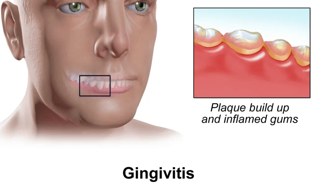
GINGIVITIS
On this page
Gingivitis is defined as an inflammation of the gingiva (gums)—the soft mucosal tissue that surrounds the necks of the teeth and covers the alveolar ridges of the jaws—without loss of the underlying periodontal attachment or alveolar bone. It is the earliest and most common form of periodontal disease.
A key clinical feature of uncomplicated plaque-induced gingivitis is that it is completely reversible when dental plaque biofilm is effectively removed and good oral hygiene is restored. However, if left untreated, chronic gingival inflammation can progress to periodontitis (irreversible destruction of the periodontal ligament and supporting bone) or, in immunocompromised and malnourished individuals, to severe necrotizing infections.
Classification of Gingivitis
Gingivitis is broadly classified according to its etiology, clinical course, and severity. Distinguishing between routine chronic gingivitis and acute necrotizing disease is essential for appropriate clinical management and antibiotic stewardship.
1. Plaque-Induced Gingivitis (Chronic Gingivitis)
- Dental Plaque Biofilm–Induced Gingivitis: The most prevalent form globally, caused by persistent accumulation of bacterial dental plaque along the gingival margin. It typically presents with redness, swelling, and bleeding on tooth brushing or probing, usually with minimal or no pain.
- Systemically Modified Gingivitis: Gingival inflammation exaggerated by host hormonal, metabolic, or nutritional factors even with relatively small amounts of plaque:
- Hormone-Related (Pregnancy / Puberty Gingivitis): Elevated estrogen and progesterone levels increase gingival vascular permeability and amplify the inflammatory response to plaque.
- Metabolic / Immunological: Uncontrolled diabetes mellitus, HIV infection, and hematological disorders (such as leukemia, which can cause marked gingival swelling and spontaneous bleeding).
- Nutritional Deficiency: Severe vitamin C (ascorbic acid) deficiency (scurvy) impairs collagen synthesis and capillary integrity, causing spongy, swollen, easily bleeding gums.
- Drug-Influenced Gingival Enlargement: Certain medications induce fibrous gingival overgrowth that traps plaque and worsens inflammation, most notably phenytoin (anticonvulsant), nifedipine/amlodipine (calcium channel blockers), and cyclosporine (immunosuppressant).
2. Non-Plaque-Induced Gingival Lesions
- Specific Infections: Primary herpetic gingivostomatitis (herpes simplex virus), oral candidiasis (Candida albicans), or specific bacterial infections (e.g., streptococcal or gonococcal gingivitis).
- Traumatic, Allergic, and Mucocutaneous Lesions: Mechanical injury (aggressive brushing, sharp foods), chemical/thermal burns, allergic reactions to toothpaste flavorings or dental materials, and autoimmune conditions (e.g., lichen planus, pemphigus vulgaris).
3. Acute Necrotizing Ulcerative Gingivitis (ANUG)
- Overview: Also known as necrotizing gingivitis, trench mouth, or Vincent's infection, ANUG is an acute, painful, destructive anaerobic infection of the gingiva caused by a fusospirochetal bacterial complex (primarily Fusobacterium species, Prevotella intermedia, and spirochetes such as Treponema).
- Relevance in Uganda: In Uganda and similar resource-limited settings, ANUG is strongly associated with HIV infection, severe childhood malnutrition, poor oral hygiene, smoking, and severe physiological or psychological stress.
- Hallmark Clinical Features: Rapid onset of severe gingival pain, "punched-out" ulcerated and necrotic interdental papillae covered by a grayish-white pseudomembrane, profuse spontaneous bleeding, intense foul breath (fetor oris), fever, malaise, and submandibular lymphadenopathy. Without prompt treatment, it can spread to the periodontium (necrotizing periodontitis), oral mucosa (necrotizing stomatitis), or cheek tissues (noma / cancrum oris).
Etiology, Pathophysiology, and Risk Factors
Healthy gingiva is pale pink (or physiologically pigmented with melanin), firm, stippled like an orange peel, knife-edged at the tooth margin, and does not bleed on gentle brushing or probing. Gingivitis develops when microbial challenge overwhelms local host gingival defenses.
Pathophysiology of Plaque-Induced Gingivitis
- Plaque Biofilm Accumulation: Within hours of brushing, salivary glycoproteins form a pellicle on tooth surfaces, colonized by oral bacteria to create a sticky dental plaque biofilm along the gingival sulcus.
- Calculus Formation: If plaque is not mechanically disrupted, minerals from saliva precipitate into the biofilm within days to form hard, mineralized calculus (tartar), which cannot be removed by a toothbrush and provides a rough surface that traps more bacteria.
- Host Inflammatory Response: Bacterial endotoxins and enzymes diffuse into the gingival epithelium, triggering vasodilation, increased capillary permeability, and infiltration by neutrophils and macrophages.
- Clinical Tissue Changes: The gingiva becomes erythematous, edematous (losing its stippling), and engorged with fragile blood vessels that bleed easily upon minor mechanical trauma (such as brushing or eating).
Predisposing and Risk Factors
- Local Factors:
- Poor Oral Hygiene: Infrequent or improper brushing technique and failure to clean interdental spaces.
- Plaque-Retentive Factors: Dental calculus, crowded or malaligned teeth, overhanging dental fillings, poorly fitting dentures, and orthodontic appliances.
- Xerostomia (Dry Mouth) and Mouth Breathing: Reduced salivary flow diminishes natural mechanical cleansing and antimicrobial buffering (e.g., lysozyme, IgA).
- Systemic and Lifestyle Factors:
- Tobacco Use (Smoking): Impairs gingival blood flow, suppresses neutrophil function, and masks early bleeding while accelerating tissue breakdown and ANUG risk.
- Immunosuppression: HIV/AIDS, chemotherapy, and prolonged corticosteroid therapy increase susceptibility to severe gingivitis and ANUG.
- Malnutrition: Protein-energy malnutrition and deficiencies in vitamin C, vitamin B complex, or zinc impair mucosal repair and host immunity.
- Endocrine Changes and Systemic Illness: Pregnancy, puberty, poorly controlled diabetes mellitus, and leukemias.
- Medications: Phenytoin, nifedipine, cyclosporine, and anticholinergic drugs that cause dry mouth.
Comparison: Chronic Gingivitis vs. ANUG vs. Periodontitis
Differentiating reversible chronic gingivitis from acute necrotizing ulcerative gingivitis (ANUG) and irreversible periodontitis guides urgent triage, antibiotic decisions, and dental referral.
| Clinical Feature | Chronic Plaque-Induced Gingivitis | Acute Necrotizing Ulcerative Gingivitis (ANUG) | Periodontitis |
|---|---|---|---|
| Onset & Course | Slow, insidious, chronic course. | Sudden, rapid, acute onset. | Usually chronic progression from untreated gingivitis. |
| Primary Cause | Dental plaque biofilm and calculus. | Anaerobic fusospirochetal complex in an immunocompromised, malnourished, stressed, or smoking host. | Subgingival plaque pathogens plus destructive host immune response. |
| Pain | Typically painless or mild soreness. | Severe, intense gingival pain interfering with eating and brushing. | Often painless until late stages (abscess or tooth mobility). |
| Gingival Appearance | Red, swollen, shiny/smooth margins, loss of stippling. | "Punched-out" cratered necrotic interdental papillae with grayish pseudomembrane. | Inflamed or fibrotic gums, gingival recession, deep periodontal pockets (≥ 4 mm). |
| Bleeding | Bleeding on brushing or gentle probing. | Profuse spontaneous bleeding or bleeding on minimal contact. | Bleeding on probing and sometimes pus drainage from pockets. |
| Halitosis & Systemic Signs | Mild bad breath; no fever or lymphadenopathy. | Severe metallic/fetid halitosis (fetor oris), fever, malaise, tender lymph nodes. | Persistent halitosis; systemic signs only if acute periodontal abscess develops. |
| Attachment & Bone Loss | Absent (no alveolar bone loss; teeth remain firm). | Usually absent initially, but rapid progression causes necrotizing periodontitis. | Present (irreversible periodontal attachment loss, bone loss, loose teeth). |
| Reversibility | Completely reversible with plaque removal and oral hygiene. | Heals with debridement, rinses, and antibiotics, though papillary blunting may remain. | Irreversible bone loss (progression can be halted, not fully regenerated without surgery). |
| Systemic Antibiotics | Not indicated. | Indicated (Metronidazole ± Amoxicillin). | Usually not routine; reserved for severe/aggressive forms or periodontal abscess. |
Advertisements
Clinical Manifestations of Gingivitis
- Gingival Erythema and Edema: Gums appear bright red or purplish-red (or darker hyperpigmented red in dark-skinned individuals), swollen, puffy, and rounded at the tooth margins rather than sharp and stippled.
- Bleeding Gums: Bleeding during tooth brushing, flossing, biting into firm foods (e.g., apples or maize), or gentle probing; blood-tinged saliva on the pillow in the morning.
- Halitosis (Bad Breath): Persistent foul mouth odor caused by volatile sulfur compounds produced by anaerobic plaque bacteria and decomposing blood/debris.
- Gingival Tenderness or Discomfort: Mild soreness during brushing in chronic gingivitis; severe, throbbing pain in ANUG or herpetic gingivostomatitis.
- Visible Plaque and Calculus Deposits: Soft whitish-yellow debris or hard yellowish-brown tartar along the gumline, particularly on the lingual surfaces of lower incisors and buccal surfaces of upper molars.
- Red-Flag Features Suggesting ANUG or Systemic Disease:
- Necrotic, ulcerated, "punched-out" interdental papillae with a grayish slough.
- Fever, regional submandibular lymphadenopathy, and difficulty swallowing or chewing.
- Marked, boggy, spontaneous gingival bleeding or severe generalized hyperplasia (raises suspicion for hematological disorders such as acute leukemia, severe thrombocytopenia, or drug toxicity).
Investigations and Diagnostic Assessment
Gingivitis is primarily diagnosed through a careful oral and systemic clinical examination; bedside findings alone must be interpreted alongside periodontal and medical evaluation to rule out deeper bone destruction or systemic illness.
- Comprehensive Oral Inspection: Assess gingival color, contour, swelling, presence of plaque/calculus, ulceration, pseudomembrane, and spontaneous bleeding using good lighting and a tongue depressor or mouth mirror.
- Periodontal Probing (Dental Assessment): Gentle measurement of gingival sulcus depth and bleeding on probing. Absence of true pocket deepening (> 3 mm with attachment loss) helps distinguish gingivitis from periodontitis.
- Dental Radiographs (Periapical / Orthopantomogram): Ordered by a dental officer when periodontitis is suspected to evaluate alveolar bone levels (normal bone crest in gingivitis).
- HIV Counseling and Testing: Strongly recommended in patients presenting with ANUG, severe refractory gingivitis, or concurrent oral candidiasis/hairy leukoplakia.
- Random or Fasting Blood Glucose: Indicated in recurrent, severe, or poorly healing gingivitis to screen for undiagnosed or poorly controlled diabetes mellitus.
- Complete Blood Count (CBC) and Peripheral Film: Indicated when there is unexplained spontaneous gingival bleeding, severe gingival hypertrophy, pallor, petechiae, or persistent fever to rule out leukemia, severe anemia, or thrombocytopenia.
- Nutritional and Medication History: Screen children and adults for malnutrition, vitamin C deficiency, and drugs causing gingival hyperplasia (phenytoin, nifedipine, cyclosporine).
Complications of Untreated Gingivitis
- Progression to Periodontitis: Extension of inflammation into the periodontal ligament and alveolar bone, causing deep pockets, gum recession, tooth mobility, and eventual tooth loss.
- Periodontal Abscess: Localized collection of pus within the gingival wall of a periodontal pocket, causing acute throbbing pain and facial swelling.
- Acute Necrotizing Ulcerative Gingivitis / Stomatitis: Rapidly destructive necrosis of gums and adjacent oral mucosa in malnourished or immunocompromised patients.
- Noma (Cancrum Oris): A devastating, life-threatening gangrenous infection spreading from necrotizing gingivitis through the cheeks, lips, and facial bones—seen primarily in severely malnourished young children in resource-limited settings, carrying high mortality and severe facial disfigurement if not treated emergently.
- Systemic Bacteremia: Ulcerated gingival epithelium allows oral bacteria to enter the bloodstream during chewing or dental procedures, posing a risk of infective endocarditis in patients with prosthetic heart valves or congenital/rheumatic heart disease, as well as poorer glycemic control in diabetes.
Management of Gingivitis
Management follows a logical sequence: controlling bacterial plaque mechanically, soothing and disinfecting inflamed tissues, treating acute necrotizing infections with targeted antimicrobials, correcting underlying systemic/drug factors, and preventing recurrence through patient education.
1. Aims of Management
- To eliminate dental plaque biofilm and calculus deposits.
- To resolve gingival inflammation, bleeding, and pain.
- To eradicate anaerobic infection promptly in ANUG and prevent tissue necrosis or noma.
- To identify and manage systemic predisposing conditions (HIV, diabetes, malnutrition, offending drugs).
- To prevent progression to irreversible periodontitis and tooth loss.
2. Non-Pharmacological and Preventive Management
- Meticulous Mechanical Plaque Control:
- Tooth Brushing: Brush at least twice daily (morning and before bedtime) using a soft-bristled toothbrush and fluoridated toothpaste. Teach the Modified Bass technique (angling bristles at 45° toward the gumline with gentle circular/vibratory strokes) so plaque at the gingival margin is cleared without traumatizing inflamed gums.
- Interdental Cleaning: Daily use of dental floss or interdental brushes to remove plaque between teeth where brush bristles cannot reach.
- Toothbrush Care: Replace toothbrushes every 3 months or after acute oral infections.
- Professional Dental Therapy:
- Scaling and Polishing: Referral to a dental officer or public health dental officer (HC IV / hospital dental clinic) for mechanical removal of supra- and subgingival calculus and polishing of tooth surfaces.
- Gentle Debridement (in ANUG): Careful removal of necrotic slough and superficial debris under local comfort measures alongside antiseptic rinses.
- Lifestyle, Nutritional, and Systemic Corrections:
- Smoking Cessation: Strongly counsel patients to stop smoking or chewing tobacco.
- Balanced Diet: Encourage fresh fruits and vegetables rich in vitamin C, adequate protein, and reduced sugary/sticky snacks between meals.
- Underlying Medical Review: Optimize glycemic control in diabetes, initiate or maintain antiretroviral therapy (ART) in HIV, and collaborate with the prescriber to consider alternative drugs if phenytoin or nifedipine causes severe gingival hyperplasia.
Advertisements
3. Pharmacological Management (Aligned with Uganda Clinical Guidelines 2023)
- Antiseptic and Soothing Mouth Rinses (Essential Adjuncts):
- Warm Salt Water Rinses: Dissolve ½ teaspoon of table salt in a glass (approx. 200 mL) of warm water and rinse gently three times daily (TID) to soothe inflamed gums and flush debris.
- Chlorhexidine Gluconate 0.12% or 0.2% Mouthwash: Swish 10–15 mL for 30–60 seconds twice daily (BID) for short-term plaque control (typically 7–14 days), especially when pain prevents effective brushing.
- Hydrogen Peroxide Mouth Rinse (Short Term, especially in ANUG): Dilute 3% hydrogen peroxide 1:1 with water (or 6% diluted 1:4 with water) and rinse three times daily (TID) for a few days to liberate oxygen, inhibit anaerobic bacteria, and loosen necrotic debris. Do not swallow and avoid prolonged use to prevent mucosal irritation.
- Analgesics (For Painful Gingivitis / ANUG):
- Paracetamol: Adults 500 mg–1 g PO every 8 hours (max 4 g/day); Children 10–15 mg/kg PO every 6–8 hours as needed for pain and fever.
- Ibuprofen: Adults 400 mg PO every 8 hours with food (avoid in active peptic ulcer disease, renal impairment, or severe bleeding tendency).
- Systemic Antibiotics (Reserved for ANUG or Systemic Spread):
- Antimicrobial Stewardship Rule: Systemic antibiotics are NOT indicated for routine chronic plaque-induced gingivitis.
- ANUG First-Line Regimen: Metronidazole 400 mg PO every 8 hours for 5–7 days (Children: 10–12.5 mg/kg every 8 hours) to target the anaerobic fusospirochetal complex.
- Combined Therapy (Deep Involvement / Pronounced Systemic Signs): Add Amoxicillin 500 mg PO every 8 hours for 5–7 days (Children: 25 mg/kg every 8 hours) alongside metronidazole if lymphadenopathy, fever, or deeper tissue spread is present.
Key Clinical Nursing Pearls
1. Chlorhexidine and Toothpaste Timing: Anionic surfactants in standard toothpaste (such as sodium lauryl sulfate) inactivate chlorhexidine. Instruct patients to rinse thoroughly with water and wait at least 30 minutes after brushing before using chlorhexidine mouthwash.
2. Antibiotic Stewardship: Routine chronic gingivitis resolves with mechanical plaque removal and hygiene—giving antibiotics alone without scaling and brushing fails to cure the condition and drives antimicrobial resistance.
Pharmacokinetics and Pharmacodynamics of Gingivitis Medications
Understanding the pharmacodynamics (mechanism of action) and pharmacokinetics (absorption, distribution, metabolism, and excretion) of agents used in gingivitis and ANUG ensures safe patient counseling and prevents adverse drug interactions.
| Drug / Agent | Pharmacodynamics (Mechanism & Action) | Pharmacokinetics (ADME) | Key Nursing & Patient Counseling Points |
|---|---|---|---|
| Chlorhexidine Gluconate (0.12% or 0.2% mouthwash) | Cationic bisbiguanide antiseptic; binds electrostatically to negatively charged bacterial cell walls, disrupting cell membrane integrity (bacteriostatic at low concentrations, bactericidal at higher concentrations). Also binds to oral mucosa and pellicle for prolonged substantivity (anti-plaque action lasting 8–12 hours). |
| Instruct patient to spit out, do not swallow, and avoid rinsing with water or eating/drinking for 30 minutes after use. Wait ≥ 30 minutes after toothpaste brushing. Warn about reversible brown staining of teeth/tongue and altered taste with prolonged (> 2 weeks) use. |
| Hydrogen Peroxide (3% diluted 1:1 or 6% diluted 1:4) | Oxidizing agent; reacts with tissue and bacterial catalase enzymes to release nascent oxygen bubbles, producing mechanical effervescent debridement of necrotic slough and creating an aerobic microenvironment toxic to obligate anaerobic bacteria (such as fusospirochetes in ANUG). |
| Always ensure proper dilution before rinsing to prevent chemical burns to the oral mucosa. Instruct patient not to swallow. Limit to short-term use (3–5 days in ANUG) because prolonged use can irritate mucosa and promote black hairy tongue. |
| Metronidazole (400 mg PO Q8H in ANUG) | Bactericidal nitroimidazole; enters anaerobic bacteria passively and is reduced by microbial ferredoxin-like proteins into reactive nitro radicals that disrupt bacterial DNA helices and inhibit nucleic acid synthesis. |
| Strictly advise against all alcohol during treatment and for 48–72 hours afterward due to a severe disulfiram-like reaction (flushing, nausea, vomiting, palpitations). Warn about metallic taste, dark urine, and GI upset (take with food). Reduce dose in severe hepatic impairment. |
| Amoxicillin (500 mg PO Q8H if deep spread in ANUG) | Bactericidal aminopenicillin (beta-lactam); binds penicillin-binding proteins (PBPs) to inhibit bacterial cell-wall peptidoglycan cross-linking, leading to osmotic lysis of susceptible gram-positive and some gram-negative oral bacteria. |
| Screen for penicillin allergy before administration (anaphylaxis risk). Emphasize completing the full 5–7 day course even after pain subsides. Adjust dosing interval in severe renal impairment. Monitor for diarrhea or secondary oral/vaginal candidiasis. |
| Paracetamol (Acetaminophen) (500 mg–1 g PO Q8H) | Central inhibitor of prostaglandin synthesis (via COX pathways in the CNS) and modulator of serotonergic descending pain pathways, producing analgesic and antipyretic effects without significant peripheral anti-inflammatory or antiplatelet action. |
| Safe first-line analgesic in patients with bleeding gums or peptic ulcer risk because it does not impair platelet aggregation or irritate gastric mucosa. Do not exceed 4 g/day in adults (lower in malnutrition, liver disease, or chronic alcohol use) to avoid hepatotoxicity. |
| Ibuprofen (400 mg PO Q8H) | Non-selective reversible cyclooxygenase (COX-1 and COX-2) inhibitor; reduces peripheral prostaglandin synthesis at inflamed gingival tissues, providing analgesic, antipyretic, and anti-inflammatory relief. |
| Administer with food or milk to minimize gastric irritation. Avoid in patients with active peptic ulcer disease, dehydration, renal impairment, severe bleeding tendencies (e.g., suspected leukemia/thrombocytopenia), or third-trimester pregnancy. |
4. Emergency and Referral Management (Severe ANUG, Noma, or Suspected Systemic Disease)
While uncomplicated chronic gingivitis is managed on an outpatient basis, severe necrotizing disease or underlying systemic pathology requires urgent hospital care:
- Indications for Urgent Admission / Referral: Progression to necrotizing stomatitis or noma (cancrum oris), inability to swallow fluids (dehydration), high fever or sepsis, airway compromise from floor-of-mouth/neck swelling, or severe spontaneous gingival bleeding with pallor/petechiae suggesting acute leukemia.
- Initial Stabilization: Assess airway, breathing, and circulation (ABCs); establish IV access for rehydration; obtain urgent blood samples (CBC, blood glucose, HIV test, electrolytes); administer IV broad-spectrum antibiotics covering anaerobes and gram-positives/gram-negatives per hospital severe oral sepsis/noma protocols; provide gentle local wound care, high-protein/high-calorie nutritional rehabilitation, and urgent dental/maxillofacial surgical review.
Advertisements
Nursing Management of Gingivitis
Nursing care addresses local oral tissue healing, pain relief, prevention of bleeding and secondary necrotizing infection, nutritional support when mastication is painful, and empowerment of the patient with sustainable daily oral hygiene practices.
Nursing Care Plan for a Patient with Gingivitis / ANUG (NANDA-I 2024–2026)
This care plan summarizes priority actual and risk nursing diagnoses using NANDA-I 2024–2026 terminology (Impaired oral mucous membrane integrity, Acute pain, Risk for excessive bleeding, Inadequate nutritional intake, and Inadequate health knowledge) prior to the detailed nursing interventions below.
| Assessment | Diagnosis (NANDA-I 2024–2026) | Planning (Goals or Expected Outcomes) | Implementation / Interventions | Rationale | Evaluation |
|---|---|---|---|---|---|
| Impaired oral mucous membrane integrity related to bacterial dental plaque biofilm accumulation, calculus, and microbial infection as evidenced by gingival erythema, edema, bleeding on brushing, and halitosis. | Restore healthy gingival tissue integrity within 1–2 weeks of hygiene and medical/dental therapy. Expected outcomes:
|
|
| After 7 days, gingival edema and redness subsided, necrotic slough resolved, and gums appeared firmer with minimal halitosis. |
| Acute pain related to acute gingival inflammation, papillary ulceration, and exposure of subepithelial nerve endings as evidenced by pain score of 7/10 and avoidance of chewing. | Reduce gingival pain to a tolerable level within 24–48 hours of initiating therapy. Expected outcomes:
|
|
| Within 48 hours, patient reported gum pain decreased from 7/10 to 2/10 and tolerated warm salt water rinses and soft meals. |
| Risk for excessive bleeding related to severe gingival vascular engorgement, mucosal ulceration, and potential underlying coagulopathy or thrombocytopenia. | Prevent significant gingival hemorrhage and identify systemic bleeding disorders promptly. Expected outcomes:
|
|
| By day 4, spontaneous gingival bleeding ceased, vital signs remained stable, and no systemic bleeding signs were observed. |
| Inadequate nutritional intake related to painful oral mucosal lesions, chewing difficulty, and altered taste as evidenced by reduced meal consumption and weight loss. | Achieve adequate daily caloric, protein, vitamin C, and fluid intake throughout recovery. Expected outcomes:
|
|
| Patient consumed 90% of soft, high-protein meals daily, maintained normal hydration, and began gaining weight on follow-up. |
| Inadequate health knowledge related to misinformation about bleeding gums, brushing technique, mouthwash use, and antibiotic stewardship as evidenced by stopping brushing and requesting unnecessary antibiotics. | Demonstrate correct daily oral hygiene practices and accurate understanding of treatment before discharge. Expected outcomes:
|
|
| Prior to discharge, patient accurately demonstrated gentle brushing technique and explained chlorhexidine timing, metronidazole precautions, and dental follow-up. |
Advertisements
Detailed Nursing Diagnoses, Assessments, and Interventions
1. Impaired Oral Mucous Membrane Integrity
- Definition: Injury to the lips, soft tissues, buccal cavity, and/or oropharynx.
- Related to: Bacterial plaque biofilm, mineralized calculus, anaerobic fusospirochetal infection (ANUG), poor oral hygiene, immunosuppression, malnutrition, or drug-induced gingival overgrowth.
- Assessment:
- Inspect the gingiva, interdental papillae, tongue, palate, and buccal mucosa daily for redness, swelling, loss of stippling, ulceration, necrotic slough, or white plaques (candidiasis).
- Note degree of halitosis, plaque/calculus buildup, and tooth mobility.
- Assess for systemic risk factors (HIV status, diabetes, malnutrition, phenytoin/nifedipine use).
- Interventions:
- Administer or supervise warm salt water rinses TID and chlorhexidine 0.12%/0.2% mouthwash BID (separated by at least 30 minutes from toothpaste brushing).
- In ANUG, assist with diluted hydrogen peroxide rinses (3% diluted 1:1 or 6% diluted 1:4) TID for short-term debridement and administer prescribed metronidazole (± amoxicillin).
- Teach gentle, twice-daily brushing with a soft-bristled toothbrush and fluoride toothpaste (Modified Bass technique) and daily flossing as tenderness resolves.
- Coordinate referral to a dental clinic for professional scaling and polishing to remove calculus.
2. Acute Pain
- Definition: Unpleasant sensory and emotional experience associated with actual or potential tissue damage, or described in terms of such damage; sudden or slow onset of any intensity from mild to severe with an anticipated or predictable end.
- Related to: Acute gingival inflammation, necrotic papillary ulceration (ANUG), and thermal/mechanical stimulation of exposed oral mucosa.
- Assessment:
- Assess pain location, severity (0–10 numerical or faces pain scale), quality, and impact on eating, speaking, and sleeping.
- Observe non-verbal pain behaviors (grimacing, drooling in children, guarding of the jaw, refusal to open mouth).
- Interventions:
- Administer prescribed analgesics (paracetamol or ibuprofen if not contraindicated) 30–45 minutes prior to meals and oral hygiene care.
- Provide soothing lukewarm saline rinses after meals and before sleep.
- Advise avoiding hot, spicy, sour/citrus, alcohol-containing, or sharp/crunchy foods until mucosa heals.
- Monitor pain response after analgesic and antimicrobial therapy; escalating facial pain or trismus should be reported immediately to rule out abscess or necrotizing stomatitis.
3. Risk for Excessive Bleeding
- Definition: Susceptible to a decrease in blood volume or localized hemorrhage that may compromise health.
- Related to: Fragile, hyperemic gingival capillaries, necrotic mucosal ulceration, mechanical trauma, or underlying systemic coagulopathy/hematologic disease.
- Assessment:
- Observe whether bleeding occurs only on brushing/probing or spontaneously at rest.
- Assess vital signs (pulse, BP), conjunctival pallor, skin petechiae, bruising, or lymphadenopathy to screen for systemic causes.
- Review CBC, platelet count, and coagulation profile when ordered.
- Interventions:
- Avoid hard toothbrushes, wooden toothpicks, and vigorous flossing during acute bleeding; use gentle rinses or an ultra-soft brush.
- Apply gentle pressure with sterile gauze soaked in normal saline if localized gingival oozing persists.
- Avoid NSAIDs/aspirin in patients with spontaneous unexplained bleeding or suspected thrombocytopenia.
- Report profuse, uncontrollable bleeding or signs of anemia/leukemia urgently to the clinician.
4. Inadequate Nutritional Intake
- Definition: Intake of nutrients insufficient to meet metabolic needs.
- Related to: Oral pain during mastication, dysgeusia (metallic taste), anorexia, fever, or underlying food insecurity/malnutrition.
- Assessment:
- Monitor daily food and fluid intake, weight, and mid-upper arm circumference (MUAC) in children.
- Assess for signs of dehydration (dry mucous membranes, reduced urine output, sunken eyes) and micronutrient deficiencies (scurvy, protein-energy malnutrition).
- Interventions:
- Provide soft, cool or lukewarm, nutrient-dense meals (milk, eggs, mashed beans, porridge, minced fish/meat, avocado, mashed bananas/pawpaw) rich in protein and vitamin C.
- Offer frequent small meals and ample oral fluids (water, oral rehydration, nutritious soups) rather than large hard meals.
- Perform gentle mouth rinsing before meals to clear necrotic debris and improve taste.
- Refer malnourished children or immunocompromised patients for therapeutic nutritional supplementation.
5. Inadequate Health Knowledge
- Definition: Absence or deficiency of cognitive information related to a specific health topic.
- Related to: Lack of prior oral health education, misconceptions regarding bleeding gums, and unfamiliarity with antimicrobial stewardship and mouthwash precautions.
- Assessment:
- Assess current oral hygiene habits (brushing frequency, toothbrush hardness, toothpaste use, flossing, traditional chewing stick/miswak technique).
- Identify misconceptions (e.g., stopping brushing when gums bleed, self-medicating with antibiotics for simple gum swelling).
- Interventions:
- Teach and visually demonstrate proper twice-daily soft brushing, interdental cleaning, and gentle tongue cleaning using the teach-back method.
- Educate on safe mouthwash use (diluting hydrogen peroxide properly, waiting 30 minutes after toothpaste before chlorhexidine, not swallowing rinses).
- Counsel on completing prescribed antibiotics for ANUG, avoiding alcohol with metronidazole, stopping tobacco use, and avoiding unnecessary antibiotics for uncomplicated chronic gingivitis.
- Emphasize routine 6-monthly dental check-ups and scaling.
Ongoing Ward Nursing Care Plan (For Hospitalized Severe Gingivitis / ANUG)
For patients admitted with severe ANUG, dehydration, noma risk, or underlying systemic illness, ongoing ward nursing management covers the following structured priorities:
Admission
Admit the patient to the medical or pediatric ward (or isolation/oral care bay if severe sepsis), record particulars in the ward admission book, assess airway/hydration immediately, and perform baseline anthropometry (weight, MUAC in children) and oral examination.
Psychological Care
Establish a supportive rapport with the patient and caregivers. Reassure them that uncomplicated gingivitis and timely treated ANUG heal well, address embarrassment related to halitosis, and counsel on any newly diagnosed underlying conditions (e.g., HIV or diabetes).
Position
Nurse the patient in an upright or semi-Fowler's position, especially during oral rinses, debridement, and feeding, to facilitate comfortable swallowing, prevent pooling of oral secretions, and protect against aspiration.
Observations
- Vital Observations: Monitor and chart temperature, pulse, respiration, blood pressure, and oxygen saturation every 4 hours (more frequently if febrile or septic).
- Specific / Oral Observations: Inspect the mouth every shift for:
- Reduction or progression of gingival erythema, necrotic slough, and bleeding.
- Any facial/cheek swelling, skin discoloration, or foul discharge (early warning signs of noma).
- Trismus (difficulty opening mouth), dysphagia, or cervical lymphadenopathy.
Investigations
Collect and follow up ordered investigations promptly: Complete Blood Count (CBC), HIV serology (with counseling), fasting/random blood glucose, serum electrolytes if dehydrated, and dental X-rays/dental officer review once stabilized.
Medications / Drugs
Administer and chart all prescribed medications accurately: systemic antibiotics (oral or IV metronidazole ± amoxicillin/penicillin for ANUG), scheduled analgesics (paracetamol), antiseptic mouthwashes (chlorhexidine 0.12%/0.2%, diluted hydrogen peroxide, warm saline), vitamin C/multivitamin supplementation, and IV fluids if unable to tolerate oral intake.
Oral Care and Hygiene
Provide supervised oral rinses after every meal and at bedtime, assist with gentle removal of loose debris using saline-moistened gauze as directed, ensure daily bed baths or showers, keep lips lubricated (e.g., petroleum jelly) to prevent cracking, and maintain clean linen and personal hygiene.
Diet and Hydration
Maintain a fluid balance chart if dehydrated. Transition from cool clear fluids and soft high-protein, vitamin C–rich feeds (porridge, milk, soup, mashed legumes, soft fruits) to a normal balanced diet as oral pain resolves. Encourage generous water intake.
Elimination
Monitor urine output and bowel habits (especially monitoring for antibiotic-associated diarrhea from amoxicillin or dark urine from metronidazole); maintain fluid balance charting until oral intake is fully established.
Rest and Sleep
Schedule analgesia before bedtime, provide a quiet restful ward environment, and group nursing procedures so the patient can sleep undisturbed and conserve energy for tissue healing.
Exercises and Ambulation
Encourage early gentle ambulation as fever and malaise subside to maintain circulation and prevent immobility complications; provide passive range-of-motion exercises for severely debilitated or malnourished patients.
Advice on Discharge
- Daily Oral Hygiene: Brush twice daily with a soft toothbrush and fluoride toothpaste, clean between teeth daily, and replace the toothbrush after recovery.
- Medication Completion: Complete the full prescribed antibiotic course (if treated for ANUG), avoid alcohol during and for 72 hours after metronidazole, and observe the 30-minute gap between toothpaste and chlorhexidine.
- Nutrition and Lifestyle: Eat a balanced diet rich in fruits, vegetables, and protein; limit sugary snacks; stop smoking completely.
- Dental and Medical Follow-Up: Attend the dental clinic for professional scaling and polishing, continue care for any underlying condition (HIV, diabetes), and return immediately if fever, severe pain, facial swelling, or spontaneous bleeding recurs.
Quick Quiz
Gingivitis Quiz
Medical Nursing
Privacy: Your details are used only for quiz tracking and certificates.
Gingivitis Quiz
Medical Nursing
Preparing questions...
Choose your answer and keep your streak alive.
Great effort.
Here is your quick performance summary.