
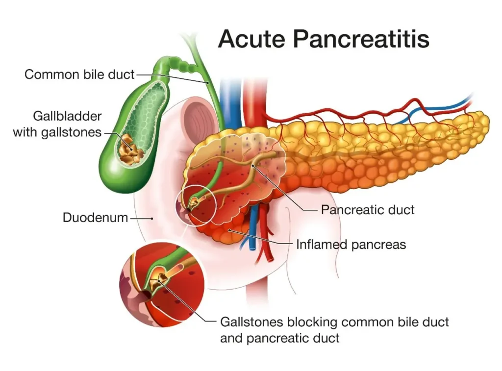
PANCREATITIS (ACUTE AND CHRONIC)
On this page
Pancreatitis is an inflammatory condition of the pancreas, a vital retroperitoneal gland located posterior to the stomach that performs both exocrine functions (secreting digestive enzymes such as amylase, lipase, and inactive proteases like trypsinogen into the duodenum) and endocrine functions (secreting insulin, glucagon, and somatostatin from the islets of Langerhans into the bloodstream).
Depending on the onset, clinical course, and reversibility of tissue injury, pancreatitis is broadly divided into acute pancreatitis (sudden inflammation that can heal completely or progress to life-threatening necrosis and organ failure) and chronic pancreatitis (progressive, irreversible inflammation resulting in pancreatic fibrosis, calcification, and permanent loss of digestive and hormonal function).
Pathophysiology of Pancreatitis
Pathophysiology of Acute Pancreatitis
Under normal physiological conditions, pancreatic proteolytic enzymes are synthesized and stored inside acinar cells as inactive precursors (zymogens) and are activated only after reaching the duodenal lumen via enterokinase. In acute pancreatitis, an initiating insult (such as biliary reflux from an impacted gallstone or direct ethanol toxicity) triggers premature intra-acinar activation of trypsinogen into active trypsin.
- Enzymatic Cascade and Autodigestion: Active trypsin converts other zymogens (proelastase, prophospholipase A2, prekallikrein, and chymotrypsinogen) into destructive active enzymes that digest pancreatic parenchyma and surrounding peripancreatic fat (autodigestion).
- Vascular Injury and Third-Spacing: Activated kallikrein and inflammatory cytokines (TNF-α, IL-1, IL-6) cause severe vasodilation, endothelial damage, and increased capillary permeability. Massive volumes of fluid shift from the intravascular compartment into the retroperitoneum and peritoneal cavity (third-spacing), precipitating hypovolemia, hemoconcentration, and shock.
- Systemic Inflammatory Response (SIRS): Spillover of pancreatic enzymes and pro-inflammatory mediators into the systemic circulation can trigger acute respiratory distress syndrome (ARDS), acute kidney injury (AKI), disseminated intravascular coagulation (DIC), and multi-organ failure.
Pathophysiology of Chronic Pancreatitis
Chronic pancreatitis develops through repeated episodes of acute or subclinical pancreatic injury (often described by the necrosis–fibrosis sequence) combined with chronic metabolic and oxidative stress.
- Fibrogenesis and Ductal Distortion: Persistent inflammation activates pancreatic stellate cells, depositing dense collagenous scar tissue (fibrosis) that replaces normal acinar parenchyma. Protein plugs precipitate within pancreatic ducts and calcify into intraductal calculi, creating ductal strictures and upstream hypertension.
- Exocrine Pancreatic Insufficiency (EPI): When more than 90% of acinar function is lost, inadequate secretion of lipase, amylase, and proteases causes maldigestion and malabsorption of fats, proteins, and fat-soluble vitamins (A, D, E, K), clinically manifesting as steatorrhea.
- Endocrine Insufficiency (Pancreatogenic Diabetes): Progressive destruction of the islets of Langerhans impairs both insulin and glucagon secretion, causing Type 3c (pancreatogenic) diabetes mellitus, which is characteristically prone to severe hypoglycemia.
Classification and Types of Pancreatitis
Understanding every clinical, morphologic, and etiologic subtype of pancreatitis is essential for guiding triage, imaging, antibiotic stewardship, and long-term rehabilitation.
Types of Acute Pancreatitis (Revised Atlanta Classification)
International consensus (Revised Atlanta Classification, 2012) classifies acute pancreatitis by morphology, clinical phase, and disease severity.
- Morphologic Subtypes:
- Acute Interstitial Edematous Pancreatitis: Accounts for approximately 80–85% of cases. Characterized by diffuse or localized enlargement of the pancreas due to inflammatory edema without parenchymal or peripancreatic necrosis. Typically resolves within the first week.
- Acute Necrotizing Pancreatitis: Accounts for 15–20% of cases. Characterized by areas of non-viable pancreatic parenchyma, peripancreatic tissue necrosis, or both (seen as non-enhancing zones on contrast-enhanced CT). Necrosis may remain sterile or become infected (usually after 7–14 days due to bacterial translocation from the gut), carrying high mortality.
- Temporal Disease Phases:
- Early Phase (First 1 Week): Driven by the host cytokine cascade and systemic inflammatory response syndrome (SIRS). Management and prognosis are determined by clinical parameters and the presence/duration of organ failure rather than CT morphology.
- Late Phase (After 1 Week): Occurs predominantly in moderately severe or severe disease. Characterized by persistent systemic inflammation, ongoing organ dysfunction, and evolving local morphologic complications (pseudocysts or walled-off necrosis).
- Severity Categories (Modified Marshall Organ Failure Criteria):
- Mild Acute Pancreatitis: No organ failure and no local or systemic complications. Usually resolves rapidly within 1 week with supportive fluids and analgesia.
- Moderately Severe Acute Pancreatitis: Characterized by transient organ failure (resolving within 48 hours) and/or local complications (peripancreatic fluid collections or sterile necrosis) or exacerbation of comorbid illness without persistent organ failure.
- Severe Acute Pancreatitis: Defined by persistent organ failure lasting longer than 48 hours involving one or more organ systems (respiratory, cardiovascular, or renal). Requires high-dependency or intensive care monitoring.
Types of Chronic Pancreatitis
- Alcoholic (Toxic-Metabolic) Chronic Pancreatitis: The most common cause worldwide in adults with prolonged heavy alcohol intake. Concurrent tobacco smoking acts synergistically to accelerate calcification, fibrosis, and progression to pancreatic insufficiency.
- Idiopathic and Tropical Chronic Pancreatitis: Accounts for cases with no identifiable alcohol or biliary cause. Tropical calcific pancreatitis is an important regional variant seen in tropical East Africa (including Uganda) and South Asia, typically affecting younger, often non-alcoholic individuals and presenting with severe epigastric pain, large pancreatic duct calculi, and early-onset fibrocalculous diabetes.
- Hereditary / Genetic Chronic Pancreatitis: Caused by inherited mutations—most notably cationic trypsinogen (PRSS1), serine protease inhibitor Kazal-type 1 (SPINK1), or cystic fibrosis transmembrane conductance regulator (CFTR)—leading to recurrent pancreatitis starting in childhood or early adulthood and an elevated lifetime risk of pancreatic adenocarcinoma.
- Autoimmune Pancreatitis (AIP): A steroid-responsive immune-mediated subtype often presenting with painless obstructive jaundice or a focal pancreatic mass mimicking malignancy:
- Type 1 AIP: Part of systemic IgG4-related disease, affecting older males with elevated serum IgG4 levels and extra-pancreatic organ involvement (bile ducts, salivary glands, kidneys).
- Type 2 AIP: Idiopathic duct-centric pancreatitis affecting younger adults equally across sexes, with normal serum IgG4 and frequent association with inflammatory bowel disease (especially ulcerative colitis).
- Obstructive Chronic Pancreatitis: Results from long-standing mechanical blockage of the main pancreatic duct due to benign strictures, pancreatic stones, periampullary tumours, post-traumatic scarring, or congenital anomalies such as pancreas divisum.
- Recurrent Acute Pancreatitis (RAP) and Acute-on-Chronic Pancreatitis: Represents the clinical continuum between acute and chronic disease. Repeated attacks of acute pancreatitis progressively scar the gland into chronic pancreatitis, while patients with established chronic pancreatitis frequently suffer acute painful inflammatory flares ("acute-on-chronic" episodes).
Comparison: Acute vs. Chronic Pancreatitis
| Feature | Acute Pancreatitis | Chronic Pancreatitis |
|---|---|---|
| Pathological Change | Sudden, potentially reversible interstitial edema or acute necrosis. | Progressive, irreversible parenchymal fibrosis, atrophy, ductal strictures, and calcifications. |
| Leading Causes | Gallstones (most common biliary cause) and acute/heavy alcohol intake. | Chronic alcohol abuse, smoking, recurrent acute attacks, idiopathic/tropical, genetic, autoimmune. |
| Pain Pattern | Sudden, severe, constant boring epigastric pain radiating to the back; relieved by leaning forward. | Chronic or recurrent epigastric/back pain, often worsened by meals (especially fatty foods) and alcohol. |
| Serum Amylase / Lipase | Markedly elevated (≥ 3× upper limit of normal). | Often normal or only mildly elevated due to loss of acinar tissue (may rise slightly during acute flares). |
| Exocrine & Endocrine Function | Transient hyperglycemia or ileus may occur during the acute attack, usually recovering afterward. | Permanent exocrine insufficiency (steatorrhea, vitamin A, D, E, K deficiency) and endocrine insufficiency (Type 3c diabetes). |
| Primary Management Focus | Aggressive/monitored IV fluid resuscitation, analgesia, antiemetics, early enteral/oral feeding, treating biliary cause. | Alcohol/smoking cessation, stepwise pain control, Pancreatic Enzyme Replacement Therapy (PERT), insulin, ductal drainage. |
Advertisements
Etiology and Risk Factors
Causes and Risk Factors of Acute Pancreatitis
- Gallstones and Biliary Tract Disease: Accounts for 40–70% of acute cases in many settings. Small stones migrate from the gallbladder and transiently or persistently obstruct the ampulla of Vater, raising pancreatic ductal pressure and triggering enzyme activation.
- Excessive Alcohol Consumption: Accounts for 25–35% of cases. Ethanol metabolites directly injure acinar cells, increase pancreatic juice viscosity, and sensitize zymogens to premature activation.
- Metabolic Causes:
- Hypertriglyceridemia: Markedly elevated serum triglycerides (typically > 1,000 mg/dL or > 11.3 mmol/L) generate toxic free fatty acids within pancreatic capillaries.
- Hypercalcemia: Excess calcium (e.g., from hyperparathyroidism or malignancy) stimulates premature trypsinogen activation and calcified ductal plugs.
- Drugs and Toxins: Notable medications include sulphonamides, furosemide, thiazides, lamivudine (and other antiretrovirals), azathioprine/6-mercaptopurine, sodium valproate, corticosteroids, and organophosphate poisoning.
- Infections: Viral (Mumps, HIV, Hepatitis A/B, CMV, EBV), bacterial (Mycoplasma, Salmonella), and parasitic (migration of Ascaris lumbricoides into the biliary/pancreatic duct).
- Mechanical, Iatrogenic, and Structural Causes: Blunt or penetrating abdominal trauma, post-ERCP (Endoscopic Retrograde Cholangiopancreatography) ductal injury, penetrating posterior duodenal/gastric peptic ulcers, and pancreatic or ampullary tumours.
Mnemonic: Causes of Acute Pancreatitis ("I GET SMASHED")
- I – Idiopathic
- G – Gallstones (biliary obstruction)
- E – Ethanol (alcohol)
- T – Trauma (blunt/penetrating abdominal injury)
- S – Steroids
- M – Mumps (and other infections such as HIV, Hepatitis A, Ascaris) & Malignancy
- A – Autoimmune
- S – Scorpion sting (and organophosphate poisoning)
- H – Hypertriglyceridemia and Hypercalcemia
- E – ERCP (post-procedure)
- D – Drugs (sulphonamides, furosemide, lamivudine, azathioprine, valproate)
Causes and Risk Factors of Chronic Pancreatitis
- Chronic Alcohol Abuse and Tobacco Smoking: Long-term alcohol intake is the primary driver; smoking independently and synergistically hastens ductal calcification and exocrine/endocrine failure.
- Recurrent Acute Pancreatitis: Repeated acute inflammatory episodes gradually replace parenchyma with scar tissue.
- Idiopathic and Tropical Pancreatitis: Unexplained or region-specific calcific pancreatitis seen in young adults across tropical regions.
- Genetic, Autoimmune, and Obstructive Factors: Inherited mutations (PRSS1, SPINK1, CFTR), IgG4-related autoimmune disease, chronic ductal strictures/stones, and persistent metabolic disturbances (hypercalcemia, hyperlipidemia).
Clinical Manifestations
Clinical Features of Acute Pancreatitis
- Characteristic Abdominal Pain: Sudden, severe, constant, "boring" pain located in the epigastrium or left upper quadrant, classically radiating straight through to the back. The pain is worsened by eating or lying supine and partially relieved by sitting upright and leaning forward (or assuming the knee-chest position).
- Nausea, Vomiting, and Ileus: Persistent nausea and forceful vomiting (often bilious) that does not relieve the abdominal pain; abdominal distension and hypoactive or absent bowel sounds due to paralytic ileus.
- Systemic and Hemodynamic Signs: Low-grade fever, tachycardia, tachypnea, dry mucous membranes, hypotension, and oliguria from third-space fluid loss and SIRS.
- Abdominal Findings and Jaundice: Marked epigastric tenderness and voluntary guarding; board-like rigidity and rebound tenderness indicate peritonitis or perforation. Scleral icterus (jaundice) suggests biliary obstruction from gallstones or edema of the pancreatic head.
- Retroperitoneal Hemorrhage Signs (Late, Severe Necrotizing Disease):
- Cullen's Sign: Bluish-purple ecchymosis (bruising) around the umbilicus.
- Grey Turner's Sign: Bluish-purple ecchymosis along the flanks.
- Signs of Hypocalcemia: Perioral numbness, carpopedal spasm, tetany, positive Chvostek's sign (facial twitch on tapping facial nerve), and positive Trousseau's sign (carpal spasm on BP cuff inflation) due to calcium binding by saponified peripancreatic fat.
Clinical Features of Chronic Pancreatitis
- Chronic Abdominal Pain: Deep, persistent or episodic epigastric pain radiating to the back, frequently triggered 15–30 minutes after eating (especially high-fat meals) or drinking alcohol, leading to fear of eating (sitophobia).
- Exocrine Pancreatic Insufficiency (Malabsorption): Passage of bulky, pale, greasy, foul-smelling stools that float or leave an oily film in the toilet (steatorrhea), chronic diarrhea, progressive weight loss, muscle wasting, and clinical deficiencies of fat-soluble vitamins (A – night blindness; D – osteomalacia/osteopenia; E – neuropathy; K – coagulopathy/bruising).
- Endocrine Insufficiency (Pancreatogenic Diabetes): Late presentation with polyuria, polydipsia, unexplained weight loss, and labile blood glucose levels.
- Obstructive Symptoms: Persistent jaundice or recurrent post-prandial vomiting if fibrosis, calcifications, or a pseudocyst compresses the common bile duct or duodenum.
Diagnosis, Severity Assessment, and Investigations
Diagnostic Criteria and Differential Diagnosis
Under the Revised Atlanta Classification, diagnosing acute pancreatitis requires at least two of the following three criteria:
- Characteristic abdominal pain (acute-onset, severe, persistent epigastric pain radiating to the back).
- Serum lipase and/or amylase ≥ 3 times the upper limit of normal (ULN).
- Characteristic findings of acute pancreatitis on cross-sectional imaging (contrast-enhanced CT, MRI, or transabdominal ultrasound).
Diagnosing chronic pancreatitis combines compatible clinical features (chronic epigastric pain, steatorrhea, weight loss, or diabetes) with supportive structural imaging (pancreatic calcifications, ductal dilatation/beading, or parenchymal atrophy) and/or functional evidence of exocrine/endocrine insufficiency.
Key Differential Diagnoses: Perforated peptic ulcer, acute cholecystitis or ascending cholangitis, intestinal obstruction or mesenteric ischemia, acute myocardial infarction (inferior MI), ruptured aortic aneurysm/dissection, diabetic ketoacidosis (DKA), and vaso-occlusive sickle cell crisis.
Laboratory and Functional Investigations
- Serum Lipase and Amylase: Both rise within hours of acute injury; serum lipase is preferred because it is more specific to the pancreas and remains elevated longer (7–14 days vs. 3–5 days for amylase). Important: Enzyme levels do not correlate with disease severity once the diagnosis is made, and they are frequently normal or only minimally elevated in chronic pancreatitis.
- Liver Function Tests (LFTs): Serum bilirubin, ALP, GGT, AST, and ALT. An elevated ALT > 3× ULN in acute pancreatitis strongly predicts a gallstone (biliary) etiology.
- Complete Blood Count (CBC) and Hematocrit: Leukocytosis indicates acute inflammation or secondary infection; a rising hematocrit (> 44–47%) reflects hemoconcentration from third-space fluid loss and predicts severe disease.
- Renal Function and Electrolytes (BUN, Creatinine, Na+, K+, Ca2+): Elevated or rising BUN (> 20 mg/dL or 7.1 mmol/L) indicates intravascular volume depletion and poor pancreatic/renal perfusion; hypocalcemia (< 2.0 mmol/L) indicates fat saponification and severe disease.
- Metabolic Panel and Blood Gases: Fasting triglycerides (to rule out hypertriglyceridemic pancreatitis), blood glucose and HbA1c (hyperglycemia/diabetes), and arterial blood gas (ABG) in severe cases to detect hypoxemia (PaO2 < 60 mmHg) or metabolic acidosis.
- Pancreatic Function Tests (Chronic Pancreatitis): Faecal Elastase-1 (non-invasive stool assay; low levels < 200 μg/g confirm exocrine insufficiency) and 72-hour faecal fat quantification (gold standard for steatorrhea, though cumbersome).
Advertisements
Imaging Studies
- Transabdominal Ultrasound: First-line bedside imaging on admission in acute pancreatitis to detect gallstones, biliary sludge, or common bile duct dilatation; may show pancreatic edema or pseudocysts, though bowel gas can obscure the pancreas.
- Contrast-Enhanced CT (CECT) of the Abdomen: The gold standard for grading pancreatic necrosis, peripancreatic fluid collections, pseudocysts, and chronic calcifications/atrophy. In acute pancreatitis, CECT should be reserved for diagnostic uncertainty or failure to improve, and is best performed after 48–72 hours when necrosis becomes radiographically apparent.
- Plain Abdominal and Chest X-rays: Abdominal X-ray may show a localized "sentinel loop" of dilated bowel in acute pancreatitis, free air under the diaphragm (to exclude perforated peptic ulcer), or radio-opaque pancreatic calcifications (pathognomonic for chronic pancreatitis). Chest X-ray detects pleural effusions or pulmonary infiltrates/ARDS.
- MRI / MRCP and Endoscopic Ultrasound (EUS): MRCP visualizes pancreatic ductal strictures, stones, and side-branch ectasia non-invasively; EUS offers high sensitivity for early chronic parenchymal changes and occult microlithiasis.
- ERCP (Endoscopic Retrograde Cholangiopancreatography): Reserved primarily as a therapeutic procedure (stone extraction, sphincterotomy, duct stenting) rather than a diagnostic test, because ERCP itself carries a risk of inducing pancreatitis.
Severity Scoring and Referral Criteria (Uganda Context)
- Clinical Severity Scores:
- BISAP Score (Bedside Index for Severity in Acute Pancreatitis): Evaluates BUN > 25 mg/dL, Impaired mental status, SIRS (≥ 2 criteria), Age > 60 years, and Pleural effusion within 24 hours. A score ≥ 3 indicates high risk of organ failure and mortality.
- Ranson's Criteria & APACHE II: Assess metabolic, hematologic, and physiologic parameters at admission and 48 hours (Ranson's ≥ 3 or APACHE II ≥ 8 predicts severe disease).
- Modified Marshall Scoring System: Quantifies organ failure across respiratory (PaO2/FiO2), renal (serum creatinine), and cardiovascular (systolic BP/fluid responsiveness) systems; a score ≥ 2 in any system defines organ failure.
- When to Admit and Refer (Uganda Clinical Guidelines 2023):
- Acute Pancreatitis: All suspected cases require hospital admission (Health Centre IV or General Hospital). Refer urgently to a Regional Referral Hospital (RRH), specialist surgical/gastroenterology unit, or ICU for persistent organ failure (> 48 h), moderately severe/severe disease, extensive or infected necrosis requiring drainage/necrosectomy, urgent ERCP for cholangitis, or symptomatic pseudocysts.
- Chronic Pancreatitis: Refer to Gastroenterology or Pancreatobiliary Surgery for diagnostic confirmation, intractable pain, severe exocrine/endocrine failure, biliary/duodenal obstruction, or suspicion of pancreatic malignancy (new mass, rapid weight loss, painless jaundice).
- Clinical Red Flags Requiring Immediate Escalation: Persistent hypotension/shock despite fluids, respiratory distress/hypoxia, oliguria or rising creatinine/BUN, rising hematocrit despite resuscitation, rigid board-like abdomen (peritonitis), hypocalcemia, high BISAP (≥ 3), or pancreatic necrosis on CT.
Complications of Pancreatitis
Local Complications (Including Revised Atlanta Fluid Collections)
- Acute Peripancreatic Fluid Collection (APFC): Homogeneous fluid collection occurring within the first 4 weeks of interstitial edematous pancreatitis without a well-defined wall; most resolve spontaneously.
- Pancreatic Pseudocyst: An encapsulated fluid collection with a well-defined fibrous/inflammatory wall (lacking an epithelial lining) that persists more than 4 weeks after acute interstitial pancreatitis or in chronic pancreatitis. Can compress adjacent structures, rupture, bleed, or become infected.
- Acute Necrotic Collection (ANC) and Walled-Off Necrosis (WON): Within the first 4 weeks of necrotizing pancreatitis, mixed fluid and necrotic debris form an ANC; after 4 weeks, this matures into an encapsulated Walled-Off Necrosis (WON). Either collection may remain sterile or progress to infected pancreatic necrosis (suspect if fever, leukocytosis, clinical deterioration, or gas bubbles within necrosis on CT appear after 7–14 days).
- Vascular Complications: Splenic or portal vein thrombosis (causing segmental portal hypertension and gastric varices) and pseudoaneurysm of the splenic or gastroduodenal artery (risk of catastrophic GI or retroperitoneal hemorrhage).
- Mechanical and Neoplastic Complications: Common bile duct stricture (obstructive jaundice), duodenal/gastric outlet obstruction, pancreatic duct strictures/fistulae, and an increased risk of pancreatic adenocarcinoma in long-standing chronic pancreatitis.
Systemic and Metabolic Complications
- Cardiopulmonary and Renal Failure: Systemic Inflammatory Response Syndrome (SIRS), pleural effusions, ARDS (severe hypoxemia), hypovolemic or distributive shock, Acute Kidney Injury (AKI), Disseminated Intravascular Coagulation (DIC), and Multi-Organ Dysfunction Syndrome (MODS).
- Metabolic and Nutritional Derangements: Severe hypocalcemia, acute hyperglycemia progressing to chronic pancreatogenic (Type 3c) diabetes mellitus, subcutaneous fat necrosis, protein-energy malnutrition, and fat-soluble vitamin deficiencies (A, D, E, K).
Advertisements
Medical, Pharmacological, and Surgical Management
Aims of Management
- To restore and maintain intravascular volume, organ perfusion, and oxygenation.
- To relieve severe abdominal pain and control nausea/vomiting.
- To minimize pancreatic autodigestion while supporting early, safe nutrition.
- To identify and treat the underlying etiology (e.g., gallstones, alcohol, hypertriglyceridemia) and prevent or manage local and systemic complications.
Initial Resuscitation and Non-Pharmacological Management (Acute Pancreatitis)
Supportive care is the cornerstone of acute pancreatitis management, especially during the critical first 24–48 hours.
- Monitored IV Fluid Resuscitation: Begin early, moderately aggressive isotonic crystalloid resuscitation—Ringer's Lactate (Lactated Ringer's solution) is preferred over 0.9% Normal Saline (ACG 2024 guideline) because it lowers systemic inflammation (SIRS) and avoids hyperchloremic metabolic acidosis.
- Typical initial regimen: bolus if hypovolemic followed by 5–10 mL/kg/hr (or ~1.5 mL/kg/hr moderate rate after stabilization), titrated every 4–6 hours to clinical perfusion targets: urine output ≥ 0.5 mL/kg/hr, normalizing heart rate and blood pressure, and falling BUN and hematocrit.
- Caution: Reassess lung bases, oxygen saturation, and jugular venous pressure frequently to avoid fluid overload and pulmonary edema.
- Nil By Mouth (NPO) vs. Early Feeding:
- Mild Acute Pancreatitis: Keep NPO only briefly while severe pain, nausea, or vomiting persist; initiate early oral low-fat solid feeding within 24–48 hours as tolerated (prolonged fasting and stepwise clear-liquid-only delays are no longer recommended).
- Moderately Severe / Severe Acute Pancreatitis: If oral intake cannot be resumed within 48–72 hours, start early enteral nutrition via a nasogastric (NG) or nasojejunal (NJ) tube to maintain gut mucosal integrity and reduce bacterial translocation and infected necrosis. Reserve parenteral nutrition (TPN) only if enteral feeding fails or is contraindicated (e.g., severe ileus).
- Nasogastric Decompression: Routine NG tube insertion is not required for uncomplicated acute pancreatitis; insert only for intractable vomiting or severe paralytic ileus.
- Cause-Specific Measures: Discontinue offending drugs or alcohol immediately; treat hypertriglyceridemia (insulin infusion if severe); arrange timely biliary intervention when indicated.
Non-Pharmacological and Lifestyle Management (Chronic Pancreatitis)
- Strict Alcohol Abstinence and Smoking Cessation: Absolutely essential to halt progressive parenchymal destruction, reduce pain episodes, and lower pancreatic cancer risk.
- Dietary Modification and Nutritional Support: Encourage small, frequent, low-fat meals (avoiding large fatty meals that stimulate pancreatic enzyme secretion and trigger pain) alongside high-protein, nutrient-dense foods and supplementation of fat-soluble vitamins (A, D, E, K) when deficient.
Pharmacological Management
- Analgesia in Acute Pancreatitis: Pain control is urgent because severe pain worsens sympathetic stress and splinting of respiration.
- Opioid Analgesics: IV Morphine (1–3 mg titrated increments every 4 hours PRN, or weight-based dosing per ward protocol) is preferred. Pethidine (Meperidine) (25–100 mg IM/SC or 25–50 mg slow IV every 4–6 hours PRN) is an alternative, though less preferred due to accumulation of its neurotoxic metabolite (normeperidine) causing seizures in repeated dosing or renal impairment.
- Adjuncts and Drugs to Avoid: IV or rectal Paracetamol may be used as a multimodal adjunct. Avoid NSAIDs in acute pancreatitis because dehydration and third-spacing heighten the risk of acute kidney injury and GI bleeding.
- Antiemetics: Metoclopramide 10 mg IV/IM every 8 hours PRN (or ondansetron where available) to relieve nausea and vomiting.
- Antibiotic Stewardship (Acute Pancreatitis): Routine prophylactic antibiotics are NOT recommended for mild acute pancreatitis or sterile pancreatic necrosis. Administer broad-spectrum antibiotics with good pancreatic tissue penetration (such as carbapenems [meropenem/imipenem], piperacillin-tazobactam, or a fluoroquinolone/cephalosporin plus metronidazole) only when specific infection is suspected or confirmed (e.g., infected necrosis, acute cholangitis, pneumonia, or UTI).
- Glycemic and Electrolyte Therapy (Acute Pancreatitis): Administer Insulin for persistent hyperglycemia (target blood glucose 6–9 mmol/L) and replace depleted electrolytes (especially IV calcium gluconate for symptomatic hypocalcemia and magnesium sulfate).
- Pharmacotherapy for Chronic Pancreatitis:
- Stepwise Pain Control (WHO Ladder): Begin with Paracetamol (use NSAIDs cautiously if at all), progress to weak opioids (Tramadol), and reserve strong opioids for severe intractable pain with close monitoring for dependence. Add neuropathic adjuvants such as amitriptyline (TCA) or gabapentin/pregabalin to reduce central pain sensitization.
- Pancreatic Enzyme Replacement Therapy (PERT): Enteric-coated microsphere preparations (e.g., pancrelipase containing lipase, protease, and amylase; typical starting dose 25,000–75,000 units of lipase per main meal and half-dose with snacks). Must be taken WITH meals and snacks so enzymes mix directly with food; a PPI or H2RA may be added to prevent gastric acid from destroying the enzymes before reaching the duodenum.
- Endocrine and Autoimmune Therapy: Insulin therapy is usually required for pancreatogenic diabetes (oral hypoglycemics are often ineffective and caution is needed against hypoglycemia). Corticosteroids (e.g., prednisolone) under specialist supervision induce remission in Autoimmune Pancreatitis.
Pharmacokinetics and Pharmacodynamics of Pancreatitis Medications
| Drug / Class | Pharmacodynamics (Mechanism & Effect) | Pharmacokinetics (ADME) | Key Nursing & Safety Implications |
|---|---|---|---|
| Ringer's Lactate (Isotonic Crystalloid) | Expands intravascular volume directly to restore cardiac preload and pancreatic/renal microcirculation; lactate is converted to bicarbonate, helping prevent hyperchloremic acidosis and blunting SIRS. | Given IV (100% immediate intravascular distribution); rapidly equilibrates across extracellular fluid compartment (~25% remains intravascular); lactate metabolized in liver to bicarbonate; electrolytes cleared renally. | Monitor urine output (≥ 0.5 mL/kg/hr), HR, BP, lung sounds, and BUN/Hct trends; avoid fluid overload in cardiac/renal disease. |
| Morphine / Pethidine (Opioid Analgesics) | Potent mu-opioid receptor agonists in the brain and spinal cord; inhibit ascending pain transmission and reduce anxiety/pain perception. | IV/IM administration bypasses oral first-pass metabolism (rapid onset 5–15 min); hepatically glucuronidated (morphine → M3G/M6G; pethidine → normeperidine); excreted renally. | Monitor respiratory rate, SpO2, sedation score, and BP before and after each dose; avoid repeated pethidine in renal impairment due to seizure risk from normeperidine. |
| Paracetamol (Acetaminophen) | Inhibits central prostaglandin synthesis (COX pathways in the CNS), providing analgesia and antipyresis without peripheral antiplatelet or renal prostaglandin inhibition. | Rapid IV/rectal/oral bioavailability (IV gives immediate peak); uniform tissue distribution; metabolized in liver (glucuronidation/sulfation; minor CYP2E1 pathway); metabolites excreted in urine. | Safe opioid-sparing adjunct when NSAIDs are contraindicated; do not exceed 4 g/24 h (reduce dose in severe alcoholic liver disease or malnutrition). |
| Metoclopramide | Central dopamine D2-receptor antagonist in the chemoreceptor trigger zone (antiemetic) and peripheral cholinomimetic/prokinetic increasing upper GI motility. | Rapid IV/IM/oral absorption; crosses blood–brain barrier; half-life 5–6 hours; minimal hepatic metabolism, excreted primarily unchanged in urine. | Monitor for extrapyramidal symptoms (acute dystonia, oculogyric crisis); avoid prokinetics if mechanical GI obstruction or perforation is suspected. |
| Pancreatic Enzyme Replacement Therapy (PERT — Pancrelipase) | Delivers exogenous lipase, amylase, and protease directly into the duodenum to hydrolyze dietary fats, starches, and proteins, reversing steatorrhea and malnutrition. | Enteric-coated microspheres resist gastric acid (< pH 5.5) and dissolve rapidly in the alkaline duodenum; acts entirely locally within the GI lumen without systemic absorption; excreted in feces. | Administer WITH meals and snacks (never on an empty stomach); swallow whole without crushing/chewing (prevents oral mucosal ulceration); add PPI if acid degradation persists. |
| Broad-Spectrum Antibiotics (Meropenem, Piperacillin-Tazobactam, Ciprofloxacin + Metronidazole) | Bactericidal inhibition of bacterial cell-wall synthesis (beta-lactams) or DNA replication/integrity (fluoroquinolones, metronidazole) against enteric Gram-negative rods and anaerobes. | IV administration achieves therapeutic concentrations inside pancreatic necrotic tissue; carbapenems and piperacillin-tazobactam are cleared primarily renally; metronidazole undergoes hepatic metabolism + renal/fecal excretion. | Use ONLY for suspected/confirmed infected necrosis or cholangitis (not sterile necrosis); adjust doses for AKI; counsel against alcohol with metronidazole. |
| Insulin & Neuropathic Adjuvants (Gabapentin / Amitriptyline) | Insulin: Promotes cellular glucose uptake and suppresses lipolysis/ketogenesis. Gabapentin: Binds α2δ calcium channel subunits to dampen neuropathic pain. Amitriptyline: Inhibits serotonin/norepinephrine reuptake in descending pain pathways. | Insulin: Rapid SC/IV action, short half-life, degraded in liver/kidneys. Gabapentin: Non-metabolized, excreted 100% unchanged renally. Amitriptyline: Hepatic CYP metabolism, long half-life. | Monitor capillary blood glucose closely in Type 3c diabetes (high risk of sudden hypoglycemia because glucagon is also deficient). Adjust gabapentin in renal impairment; watch for sedation/anticholinergic effects with amitriptyline. |
Endoscopic and Surgical Management
- Biliary Pancreatitis Interventions:
- Urgent ERCP (≤ 24 hours): Indicated only when acute gallstone pancreatitis is complicated by acute cholangitis or persistent common bile duct obstruction; routine early ERCP without cholangitis is not beneficial.
- Cholecystectomy: In mild acute biliary pancreatitis, laparoscopic cholecystectomy is recommended before hospital discharge (same admission) to prevent recurrent attacks.
- Infected Necrosis and Pseudocyst Interventions: Use a "step-up" approach—delay invasive drainage for 4 weeks if the patient is stable to allow wall maturation (WON), starting with endoscopic or percutaneous catheter drainage and reserving minimally invasive or open surgical necrosectomy for persistent sepsis or failure to improve. Symptomatic or enlarging pseudocysts are drained endoscopically (cystogastrostomy) or surgically.
- Surgery for Chronic Pancreatitis: Indicated for intractable pain with dilated pancreatic ducts or biliary/duodenal obstruction—includes endoscopic ductal stenting/stone extraction, lateral pancreaticojejunostomy (Puestow procedure) for duct decompression, or pancreatic head resection (Frey or Whipple procedure) when malignancy cannot be excluded.
Advertisements
Nursing Management of Pancreatitis
Nursing care focuses on rapid hemodynamic stabilization, aggressive pain relief, respiratory and metabolic surveillance, careful nutritional transition, and patient empowerment to prevent relapse.
Sample Nursing Care Plan for a Patient with Pancreatitis (NANDA-I 2024–2026)
This care plan applies current NANDA-I 2024–2026 diagnostic terminology (Inadequate fluid volume / Risk for shock, Inadequate protein energy nutritional intake, Inadequate health knowledge, Acute pain, and Risk for ineffective breathing pattern) prior to the detailed nursing interventions.
| Assessment | Diagnosis (NANDA-I 2024–2026) | Planning (Goals & Expected Outcomes) | Implementation / Interventions | Rationale | Evaluation |
|---|---|---|---|---|---|
| Acute pain related to pancreatic autodigestion, edema, capsular distension, and peritoneal irritation as evidenced by pain score of 8/10, guarding, and flexed posture. | Achieve significant pain reduction within 1–2 hours of analgesia. Expected outcomes:
|
|
| Within 2 hours, patient rested in semi-Fowler's position and reported pain decreased from 8/10 to 2/10. |
| Inadequate fluid volume (and Risk for shock) related to massive retroperitoneal third-spacing, persistent vomiting, and NPO status as evidenced by hypotension, tachycardia, dry mucosa, and low urine output. | Restore intravascular volume and organ perfusion within 6–12 hours. Expected outcomes:
|
|
| After 8 hours of Ringer's Lactate therapy, BP stabilized at 112/70 mmHg, HR fell to 88 bpm, and urine output reached 45 mL/hr. |
| Inadequate protein energy nutritional intake related to acute NPO status, vomiting, fear of meal-triggered pain, and pancreatic exocrine enzyme insufficiency as evidenced by weight loss and steatorrhea. | Meet daily metabolic caloric and protein needs safely without provoking pain. Expected outcomes:
|
|
| Patient tolerated low-fat meals (or enteral feeds) without pain or vomiting; stool consistency improved and weight stabilized. |
| Risk for ineffective breathing pattern related to severe epigastric pain splinting, abdominal distension/ascites, and systemic inflammatory lung injury (pleural effusion/ARDS). | Maintain effective ventilation and adequate oxygenation throughout admission. Expected outcomes:
|
|
| Patient maintained RR 18 breaths/min and SpO2 97% with clear bilateral breath sounds and no respiratory distress. |
| Inadequate health knowledge related to unfamiliarity with pancreatitis triggers, PERT administration, diabetes monitoring, and complication warning signs as evidenced by patient questions and recurrent flares. | Demonstrate accurate self-care knowledge prior to hospital discharge. Expected outcomes:
|
|
| Prior to discharge, patient accurately demonstrated teach-back of alcohol/smoking cessation, low-fat diet, PERT timing, and red-flag symptoms. |
Advertisements
Detailed Nursing Diagnoses, Assessments, and Interventions
1. Acute Pain
- Assessment: Assess pain severity (0–10), constant boring epigastric location, back radiation, effect of posture/food, vital sign surges, and abdominal guarding or rigidity.
- Interventions: Administer prescribed IV morphine (or pethidine where ordered) and IV paracetamol; maintain NPO status during acute nausea/vomiting; insert NG tube only if persistent vomiting or ileus occurs; position in semi-Fowler's with knees flexed or leaning forward; avoid NSAIDs.
2. Inadequate Fluid Volume and Risk for Shock
- Assessment: Monitor HR, BP, temperature, capillary refill, skin turgor, hourly urine output, hematocrit, BUN/creatinine, serum calcium/magnesium/potassium, and inspect flanks/periumbilical skin for Grey Turner's and Cullen's signs.
- Interventions: Maintain large-bore IV access; infuse Ringer's Lactate as prescribed while watching for lung crackles; administer IV antiemetics and electrolyte replacements; maintain an accurate 24-hour fluid balance chart; prepare for ICU escalation if persistent organ failure develops.
3. Inadequate Protein Energy Nutritional Intake
- Assessment: Monitor weight, bowel sounds, abdominal distension, stool frequency/greasiness (steatorrhea), and serial capillary blood glucose levels.
- Interventions: Transition mild acute patients to early low-fat oral feeding within 24–48 hours once pain/vomiting subside; coordinate NG/NJ enteral feeding within 48–72 hours in severe acute cases; administer PERT with meals in chronic insufficiency; give fat-soluble vitamins and insulin as ordered.
4. Risk for Ineffective Breathing Pattern
- Assessment: Monitor respiratory rate, depth, SpO2, bilateral air entry, cough, and ABG results for pleural effusion, atelectasis, or early ARDS.
- Interventions: Keep head of bed elevated 30–45°; encourage coughing and deep-breathing exercises after pain relief; administer humidified oxygen as ordered; report sudden dyspnea or desaturation immediately.
5. Inadequate Health Knowledge
- Assessment: Evaluate understanding of disease triggers (alcohol, gallstones, smoking, offending drugs), nutrition, enzyme replacement, diabetes self-care, and complication warning signs.
- Interventions: Deliver structured verbal and written counseling involving family; verify teach-back; link to alcohol cessation counseling and surgical follow-up for cholecystectomy.
Ongoing Ward Nursing Care and Discharge Plan
- Admission and Ward Environment: Admit acute cases to an acute medical/surgical bay or HDU; record baseline vitals, weight, and allergy status; maintain a calm, quiet environment to reduce metabolic demand.
- Psychological Care: Allay fear and anxiety caused by severe pain and ICU/HDU monitoring; provide non-judgmental support for alcohol cessation.
- Positioning: Nurse in semi-Fowler's or upright leaning-forward position with knees flexed to ease diaphragmatic excursion and relieve retroperitoneal pain.
- Observations: Record vital signs, SpO2, pain score, Glasgow Coma Scale, abdominal girth/tenderness, and capillary blood glucose regularly; alert the clinician immediately for fever spikes (possible infected necrosis), rigidity, tetany, or oliguria.
- Hygiene and Elimination: Provide frequent oral care while NPO (to prevent parotitis and dryness); perform daily bed baths and pressure-area care; maintain strict urine output charts and inspect stools for steatorrhea or melena.
- Exercises, Rest, and Discharge Advice: Encourage passive/active leg exercises and early ambulation as pain resolves to prevent DVT; ensure uninterrupted rest periods; on discharge, reinforce complete alcohol and smoking abstinence, small low-fat meals, correct PERT/insulin adherence, scheduled cholecystectomy/clinic review, and immediate return for severe pain, fever, jaundice, or persistent vomiting.
Prevention, Patient Education, and Uganda Clinical Context
- Primary and Secondary Prevention: Prompt surgical treatment of symptomatic gallstones (cholecystectomy), abstaining from or strictly moderating alcohol, smoking cessation, managing severe hypertriglyceridemia/hypercalcemia, and avoiding causative drugs where alternatives exist.
- Uganda-Specific Clinical Considerations: Alcohol consumption and gallstones are leading causes of acute pancreatitis in Uganda, while chronic alcoholic and idiopathic/tropical calcific pancreatitis are important chronic presentations. Because access to serum lipase assays, contrast-enhanced CT scans, MRCP, ERCP, and commercial PERT capsules is often limited outside tertiary centres, clinical vigilance, careful bedside severity assessment, early Ringer's Lactate resuscitation, judicious analgesia, and timely referral to a Regional Referral Hospital or specialist unit are critical to saving lives.
References
- Ministry of Health, Republic of Uganda. Uganda Clinical Guidelines (UCG) 2023. Chapter 6: Gastrointestinal and Hepatic Diseases — Section 6.1.2 (Acute Pancreatitis) and Section 6.3.6 (Chronic Pancreatitis).
- Tenner S, Vege SS, Sheth SG, et al. American College of Gastroenterology Guidelines: Management of Acute Pancreatitis. Am J Gastroenterol. 2024;119(3):419–437.
- Banks PA, Bollen TL, Dervenis C, et al. Classification of acute pancreatitis—2012: revision of the Atlanta classification and definitions by international consensus. Gut. 2013;62(1):102–111.
- Herdman TH, Kamitsuru S, Lopes CT (eds). NANDA International Nursing Diagnoses: Definitions and Classification, 2024–2026 (13th ed.). Thieme; 2024.