
Table of Contents
TogglePancreatic trauma is injury to the pancreas after blunt force or a penetrating wound. It is uncommon, lies deep in the retroperitoneum, and can be difficult to recognise early. Injury to the main pancreatic duct is especially important because leaked pancreatic enzymes can cause severe inflammation, fistula, collections and sepsis. A patient may have few early signs, so repeated assessment and timely escalation are essential.
Emergency reminder: Suspected significant abdominal trauma is an emergency. Activate the local trauma response, assess and resuscitate using the facility protocol, and arrange urgent review or transfer to a centre able to provide appropriate imaging, surgery and critical care. These notes support nursing education; they do not replace bedside assessment, senior clinical decisions or current local guidance.
Learning objectives
After studying this lesson, the learner should be able to:
- Explain the position, structure and functions of the pancreas that affect injury assessment.
- Describe common blunt and penetrating mechanisms of pancreatic injury.
- Recognise why pancreatic trauma may be missed initially and identify warning signs.
- Outline the role and limitations of E-FAST, laboratory tests, CT, MRCP and ERCP.
- Summarise how haemodynamic status, associated injuries and pancreatic duct involvement guide management.
- Describe nursing priorities during resuscitation, observation, procedures, surgery and recovery.
- Identify major complications and give appropriate follow-up and discharge advice.
Definition and overview
Pancreatic trauma is damage to pancreatic tissue, blood vessels or the pancreatic duct caused by injury. It may occur alone, but associated injuries are common because the pancreas lies close to the duodenum, stomach, spleen, liver, major vessels, kidneys and spine. The mechanism may be blunt, such as a steering-wheel or bicycle-handlebar impact, or penetrating, such as a knife or gunshot wound.
The central management questions are whether the patient is haemodynamically stable, whether another injury requires urgent surgery, how much pancreatic tissue is damaged, and whether the main pancreatic duct is disrupted. A superficial contusion with an intact duct may be managed very differently from a ductal transection. Delayed recognition of a significant injury can lead to pancreatic leakage, collections, infection and prolonged illness.
Relevant anatomy and functions
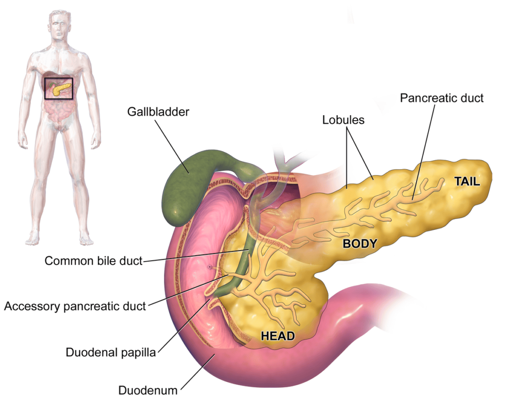
The pancreas is an elongated gland in the upper abdomen. Most of it lies behind the stomach in the retroperitoneum. The head sits in the curve of the duodenum; the neck crosses in front of major vessels; the body extends toward the left; and the tail approaches the spleen. The organ is close to the posterior abdominal wall and vertebral column. A strong blow can compress it against the spine, while penetrating wounds can cross the pancreas and damage nearby organs.
The exocrine pancreas produces digestive enzymes and bicarbonate. Pancreatic juice travels through small ducts into the main pancreatic duct and then to the duodenum, usually near the common bile duct at the major papilla. The endocrine islets release hormones, including insulin and glucagon, that help regulate blood glucose. A duct injury permits enzyme-rich fluid to escape into surrounding tissue; tissue digestion and inflammation can then cause a leak, fistula, pseudocyst, abscess or sepsis. Extensive tissue loss may also affect digestion or glucose control.
The splenic artery and vein run close to the upper border and posterior aspect of the pancreas, while the superior mesenteric vessels and portal venous structures lie near the neck and head. Trauma may therefore cause major bleeding or combined vascular, bowel, duodenal or biliary injury. In the operating theatre, identifying the whole duodeno-pancreatic region and associated injuries is important.
Causes and mechanisms of injury
Blunt abdominal trauma
Blunt injury is a common mechanism in adults and children. The pancreas may be crushed between the abdominal wall and spine or stretched during rapid deceleration. Mechanisms include road traffic collisions, a steering-wheel or seat-belt impact, bicycle handlebar injury, falls, assault, sports injury and industrial crush injury. An upper abdominal bruise or seat-belt mark, lower rib injury or upper lumbar vertebral fracture should increase suspicion.
Penetrating trauma
Knives, bullets or other penetrating objects may pass through the upper abdomen or back and injure the pancreas, duodenum, stomach, liver, spleen, bowel or major vessels. The external wound may look small even when the internal track is serious. Do not estimate depth or severity from the skin opening alone.
Procedure-related injury and associated trauma
Pancreatic injury may rarely occur during abdominal surgery or an endoscopic procedure. In trauma, identify the full injury pattern rather than focusing only on the pancreas. Bleeding, bowel perforation, duodenal injury, splenic injury, liver injury, renal injury and spinal injury can determine the patient's immediate risk and treatment.
Pathophysiology
Injury can produce bruising, a haematoma, a laceration, tissue loss, vessel damage or disruption of the main pancreatic duct. With a duct leak, digestive enzymes escape into surrounding tissue and the peritoneal or retroperitoneal space. This may trigger inflammation, fat necrosis and a local fluid collection. If the leak persists, a pancreatic fistula or pseudocyst may form; infection can progress to an abscess or sepsis.
Bleeding may occur from the pancreas or nearby vessels. As blood loss increases, compensatory tachycardia and peripheral vasoconstriction may initially maintain blood pressure. Restlessness, pallor, cool clammy skin, weak pulses, reduced urine output and altered consciousness can precede hypotension. Associated bowel or duodenal injury may cause peritonitis. Delayed swelling, ongoing enzyme leakage or infection can present after the initial trauma assessment, which is why observation and follow-up matter.
Classification and severity
The AAST organ injury scale describes anatomical pancreatic injury. The WSES-AAST approach also considers the patient's haemodynamic status. Duct involvement and the location of injury have major management implications, but a grade should be assigned by the responsible trauma or surgical team using the best available imaging and operative findings.
| AAST grade | Simplified anatomical description | Key point for care |
|---|---|---|
| I | Minor contusion or superficial laceration without pancreatic duct injury. | Selected stable patients may be treated without operation under a monitored specialist plan. |
| II | More substantial contusion or laceration without duct injury or major tissue loss. | Confirm the duct status as far as possible; observe closely if the team chooses non-operative care. |
| III | Distal transection or parenchymal injury with duct injury, generally to the body or tail. | Main duct disruption raises the risk of fistula and other complications; specialist operative review is usually required. |
| IV | Proximal injury or transection involving the head region or ampulla. | Often complex because of nearby duodenum, bile duct and major vessels; management is individualised by an experienced team. |
| V | Massive disruption of the pancreatic head. | Life-threatening, often with associated injuries; damage-control priorities and specialist input are central. |
In the WSES-AAST system, an unstable patient with a pancreatic injury is classified in the unstable category regardless of anatomical grade. This highlights an important clinical principle: physiology and associated injuries can be more urgent than the anatomical label. The classification table is an educational summary, not a bedside grading tool.
Clinical features and warning signs
Early clinical features are often non-specific. Abdominal pain or tenderness may be mild at first and can worsen over several hours. A patient can initially appear relatively well despite a significant injury. Ask about the mechanism and repeat assessment when concern remains.
Possible local findings
- Epigastric or upper abdominal pain, sometimes radiating to the back.
- Abdominal tenderness, guarding, distension or increasing pain.
- Bruising, abrasions or a seat-belt or handlebar mark across the upper abdomen.
- Penetrating wounds to the upper abdomen or back.
- Nausea or vomiting; reduced bowel sounds may occur but are not diagnostic.
- Later fever, persistent pain, a palpable collection or increasing abdominal fullness.
Features of blood loss or associated injury
- Increasing pulse, weak peripheral pulses, pallor, cool clammy skin or delayed capillary refill.
- Restlessness, dizziness, fainting, confusion or decreasing responsiveness.
- Rapid breathing, falling blood pressure, reduced urine output or other evidence of poor perfusion.
- Worsening abdominal signs, peritonitis, gastrointestinal bleeding or respiratory distress.
- New or worsening symptoms after an initially reassuring assessment.
Escalate urgently if observations deteriorate, the patient develops shock, peritonitis, increasing abdominal distension, new confusion, persistent vomiting or uncontrolled pain. A normal early amylase, lipase, blood pressure or ultrasound does not safely exclude pancreatic injury when the mechanism and examination remain concerning.
Immediate first aid and pre-hospital priorities
- Ensure scene safety and call for trained emergency help or arrange urgent transfer to an appropriate facility.
- Check responsiveness, airway and breathing. Give basic life support within training and local protocol when indicated.
- Minimise unnecessary movement if a spinal or major limb injury is possible, unless movement is needed to protect life or maintain the airway.
- For external bleeding, apply direct pressure around the wound with a clean dressing when safe. Do not remove an embedded object or push exposed tissue back inside.
- Keep the patient warm, reassure them and monitor breathing and responsiveness while awaiting transfer.
- Do not give food, drink or oral medicines before urgent clinical assessment. Do not delay transfer for traditional substances or unproven treatment.
- Share the mechanism, time of injury, changes observed, first aid and any treatment already given with the receiving team.
Emergency assessment: the ABCDE approach
Trauma care is multidisciplinary. The nurse should work within scope, delegated responsibilities and local protocol, and should communicate changes early. The primary survey identifies immediate threats; assessment and treatment happen together, with reassessment after each intervention.
| Step | What the team assesses | Nursing priorities and rationale |
|---|---|---|
| A — Airway with cervical-spine protection | Airway patency, speech, obstruction, facial injury and possible neck injury. | Call for airway support when needed, maintain indicated spinal precautions, prepare suction and prescribed equipment, and report any change in voice or consciousness. An obstructed airway rapidly threatens oxygen delivery. |
| B — Breathing | Respiratory rate and effort, chest movement, oxygenation and associated chest injury. | Observe breathing, attach pulse oximetry when available, give oxygen as prescribed or per protocol, and alert the team to respiratory distress or asymmetry. |
| C — Circulation and haemorrhage control | Pulse, blood pressure, skin perfusion, external bleeding, suspected internal bleeding and response to resuscitation. | Obtain help early, prepare appropriate vascular access and blood samples as directed, control visible bleeding, keep the patient warm and document response. Do not wait for hypotension if other signs suggest shock. |
| D — Disability | Consciousness, pupils, neurological deficit and glucose if indicated. | Record a recognised consciousness score and changes. Consider head injury or hypoxia as well as shock when mental state changes. |
| E — Exposure and environment | Full examination for injury while preventing heat loss and preserving dignity. | Expose only as needed, inspect the back during a safe log-roll when indicated, document wounds, then cover with warm blankets. Hypothermia can worsen trauma-related coagulopathy. |
After immediate threats are managed, repeat the primary survey and continue monitoring. Deterioration can occur after an initial response to treatment. Use the facility's structured trauma handover or SBAR process when communicating with the next team.
Focused history and secondary survey
Obtain a focused history without delaying resuscitation. Use AMPLE when appropriate: allergies; medicines including anticoagulants or traditional medicines; past history including bleeding disorder, prior abdominal surgery and pregnancy possibility; last oral intake; and events, including force, direction, time, loss of consciousness, symptoms and care already given.
During the secondary survey, look for other injuries to the chest, abdomen, pelvis, back and limbs. Document the location and appearance of wounds, bruising and tenderness. Do not repeatedly or forcefully palpate an unstable abdomen. Record who was informed, when escalation occurred, the response and any barriers to safe transfer.
Investigations
The diagnostic pathway depends on haemodynamic status, clinical suspicion and available services. Tests support clinical judgement and must not delay urgent haemorrhage control in a deteriorating patient.
| Investigation | Role and limitations | Nursing considerations |
|---|---|---|
| Serial clinical assessment | Repeated examination and observation trends help identify evolving injury or shock. | Record vital signs, consciousness, pain, abdominal findings, urine output and response to interventions at the prescribed frequency. Escalate a trend, not only a single abnormal value. |
| E-FAST | Rapid bedside test that can detect free fluid or selected associated injuries. It is not a reliable stand-alone test to exclude pancreatic or retroperitoneal injury. | Prepare the patient and equipment, assist the trained clinician and report that a negative scan does not rule out injury when suspicion remains. |
| Serum amylase and lipase | Repeated measurements, often beginning several hours after injury, can support evaluation. Early values may be normal; an elevated result is not specific to pancreatic trauma. | Collect ordered samples at the requested time and trend results alongside symptoms, examination and imaging. Do not use a single enzyme result to reassure or diagnose by itself. |
| Blood tests | Full blood count, group and crossmatch, coagulation, renal function, electrolytes, glucose and other tests assess bleeding, transfusion needs and organ function. | Label samples correctly, communicate urgent results and monitor for clinical change while awaiting results. |
| Contrast-enhanced CT | Key imaging for a haemodynamically stable or stabilised patient. It may show pancreatic injury, fluid, associated injury or signs of duct disruption; early CT can be non-specific. | Confirm stability and readiness for transfer. Do not send a persistently unstable patient to CT if this delays definitive haemorrhage control. |
| Repeat CT | May be considered within 12–24 hours when suspicion for pancreatic or ductal injury remains high despite negative or non-specific initial imaging, according to specialist direction and local resources. | Continue observation and escalate persistent pain, rising enzymes or deterioration rather than waiting passively for a scheduled scan. |
| MRCP or ERCP | MRCP can further evaluate the duct without an invasive endoscopic procedure. ERCP may diagnose and treat a duct injury in selected stable or stabilised patients where expertise is available. | Prepare and monitor the patient according to procedure protocol; explain that these are specialist tests and availability varies. |
CT with intravenous contrast is generally used for a stable or stabilised patient. Routine oral contrast does not improve detection of duodeno-pancreatic trauma. A negative early scan, a normal early enzyme level or a negative E-FAST should not override a concerning mechanism, changing examination or ongoing clinical suspicion.
Differential diagnosis
Upper abdominal trauma can cause overlapping symptoms. Consider associated liver, spleen, stomach, duodenal, small bowel, mesenteric, renal, vascular, biliary, chest-wall and spinal injuries. Other causes of acute epigastric pain include perforated peptic ulcer, acute pancreatitis unrelated to trauma, bowel perforation and retroperitoneal bleeding. The trauma team determines the differential and investigations; the nurse should report new signs that suggest an injury beyond the known diagnosis.
Initial medical management and resuscitation
The first goals are to support oxygenation and perfusion, control life-threatening bleeding, recognise associated injuries and prevent avoidable deterioration. The trauma, emergency, surgical, anaesthesia, radiology and transfusion teams coordinate management according to the patient's condition and facility resources.
- Maintain airway and breathing support; administer oxygen or assisted ventilation when clinically indicated.
- Establish appropriate vascular access, collect ordered samples and begin resuscitation as directed.
- Activate the facility's major haemorrhage or transfusion protocol when its criteria are met. Use blood components and warming measures according to protocol.
- Prevent heat loss during examination, transport, infusion and procedures.
- Use fluids, blood products and other resuscitation measures under clinical direction; do not allow repeated non-definitive measures to delay necessary source control.
- Provide prescribed analgesia and antiemetics, then reassess comfort, alertness and breathing.
- Keep the patient nil by mouth when ordered or while urgent surgical assessment is proceeding. Provide mouth care and explain the reason.
- Assess open wounds and operative needs for tetanus protection or antimicrobials as prescribed and per local protocol.
- Check medicines that may increase bleeding, including anticoagulants, and promptly inform the senior team. Reversal decisions require clinical review.
Medication doses, transfusion decisions and operative plans depend on the injury, age, weight, laboratory results, available products and local protocols. Nurses should verify prescriptions, patient identity, blood-product checks and required monitoring before administration.
Non-operative management
Some patients with low-grade pancreatic injury and no confirmed main duct disruption may be managed without an operation when they remain stable and have no associated injury requiring surgery. This is active hospital care, not discharge to wait at home. It requires a clear specialist plan, reliable serial examination, monitoring and the ability to intervene quickly if the patient's condition changes.
Depending on the patient's injury, the plan may include monitored admission, serial vital signs and abdominal examinations, repeat laboratory tests, pain control, fluid balance, nutritional planning and repeat imaging when clinically indicated. Any persistent or worsening pain, fever, rising enzymes, abdominal distension, new shock or other deterioration needs immediate reassessment.
Non-operative management of a suspected duct injury requires particular caution and specialist decision-making. WSES-AAST guidance limits such observation to centres capable of diagnosing injury severity and providing close monitoring, serial tests and prompt access to surgery, endoscopy, interventional radiology and blood products. Where these services are not available, early senior consultation and timely referral or transfer are important. An observation plan is unsafe if necessary monitoring and rescue treatment cannot realistically be provided.
Operative, endoscopic and interventional management
Urgent operative assessment is required for haemodynamic instability, peritonitis, uncontrolled haemorrhage, deterioration, or an associated injury needing surgery. The surgeon chooses treatment based on anatomy, duct status, associated injuries, physiology and available expertise. Imaging must not delay life-saving haemorrhage control in a patient who is unstable.
Surgical priorities
- Control life-threatening bleeding and treat other injuries that threaten life.
- Identify the duodenum, pancreas, bile duct and nearby vessels during exploration when the injury pattern warrants it.
- Use damage-control principles for a severely unstable patient: prioritise rapid haemorrhage and contamination control, resuscitation and planned reassessment.
- For minor injuries without duct disruption, drainage or limited repair may be considered by the surgeon.
- For selected distal injuries involving the duct, distal pancreatic resection may be considered. The decision depends on injury location, physiology, splenic involvement and specialist judgement.
- Complex head injuries require careful multidisciplinary planning. A major reconstruction is not a routine first step in an unstable patient.
- Place and manage drains or feeding access only as directed by the operating team.
Endoscopy and interventional radiology
In selected stable or stabilised patients, ERCP may help define a duct injury and may allow stenting. Percutaneous drainage, endoscopic procedures or image-guided interventions may help manage a later collection, pseudocyst, fistula or bleeding complication. Availability and suitability differ between facilities; specialist review and referral are needed where appropriate.
Nursing management
Nursing care continues from first contact through recovery and follow-up. It combines repeated assessment, early escalation, safe delivery of prescribed care, clear documentation and respectful communication.
| Nursing action | Rationale | What to document or escalate |
|---|---|---|
| Use a structured trauma handover and establish the mechanism and time of injury. | Mechanism and timing guide the search for pancreatic and associated injuries. | Mechanism, symptoms, pre-hospital care, arrival time and team members notified. |
| Repeat and trend observations at the prescribed frequency; increase observation or request review when the patient changes. | Trends may reveal blood loss or deterioration before one isolated value becomes abnormal. | Pulse, blood pressure, respiratory rate, oxygen saturation, temperature, consciousness and response to interventions. |
| Assess pain, abdominal distension, tenderness, guarding, skin perfusion and mental state. | Pancreatic injury can evolve and may initially have subtle signs. | New or worsening findings, time observed, escalation time and response. |
| Maintain accurate intake and output; measure urine output as prescribed. | Urine output contributes to assessment of perfusion and response to resuscitation. | Urine volume and interval, catheter output if present, fluid balance and significant decline. |
| Prepare and assist with ordered blood tests, crossmatch and imaging. | Timely results support diagnosis, resuscitation and treatment planning. | Specimen times, blood-bank communication, imaging readiness, delays and urgent results. |
| Keep the patient warm and minimise unnecessary exposure. | Hypothermia can worsen coagulopathy and complicate trauma care. | Temperature trends, warming interventions and response. |
| Administer prescribed fluids, blood products, analgesia and other treatment using safety checks. | Timely, accurate treatment supports perfusion and comfort while reducing preventable errors. | Medicine or product, checks, start and finish times, observations and reactions. |
| Maintain nil-by-mouth status when ordered and provide mouth care. | A procedure, anaesthesia or surgery may be needed. | Last oral intake, status, mouth care and any exception authorised by the team. |
| Support monitored observation with reliable serial assessment. | Observation is safe only if deterioration is recognised and acted on quickly. | Findings, prescribed frequency, missed observations and immediate escalation. |
| Explain care in simple language and support the patient and family. | Trauma and emergency procedures are frightening; communication supports dignity and cooperation. | Information given, questions, interpreter or support needs and referrals. |
Nursing priorities during resuscitation
- Recognise a high-risk mechanism and suspect internal injury even when the wound is small or early pain is limited.
- Call the appropriate emergency, trauma or surgical team early; do not wait for every test result before escalating shock or peritonitis.
- Assist with airway support, oxygenation, vascular access, ordered blood sampling and resuscitation within scope and protocol.
- Prepare equipment, documentation and a safe transfer plan for imaging, theatre, higher-acuity care or referral.
- Reassess after each intervention and communicate changes using the local structured handover.
Nursing care during observation
For a patient managed without surgery, carry out serial assessments exactly as prescribed and record them promptly. Check for increasing pain, abdominal distension, guarding, fever, dizziness, pallor, sweating, vomiting, reduced urine output or a change in consciousness. Keep the call system accessible and ensure the patient is observed at the planned level. Report a missed observation, a deteriorating trend or difficulty obtaining reliable readings. Do not assume that temporary pain relief or one normal test means the injury has resolved.
Post-operative and recovery care
After laparotomy, drainage, resection, ERCP or another procedure, follow the specific postoperative plan and local recovery-unit standards. The level of monitoring depends on the patient's physiology and associated injuries.
- Assess airway, breathing, circulation, oxygenation, consciousness, pain and temperature at the prescribed frequency.
- Inspect the incision, dressing, drains and access sites for bleeding, leakage, swelling or infection.
- Record drain amount and character as directed. Report sudden increases, blood, cloudy fluid or fluid that the team identifies as suspicious for pancreatic leakage.
- Maintain fluid balance and monitor urine output. Review ordered blood count, coagulation, renal function, glucose, pancreatic enzymes and other results with the clinical team.
- Observe for increasing abdominal distension, persistent vomiting, fever, worsening pain, jaundice, respiratory difficulty or signs of sepsis.
- Provide wound care and device care using aseptic technique. Support respiratory exercises, pressure-area prevention and early mobility when allowed.
- Administer prescribed analgesia, antiemetics, antibiotics, nutrition and thrombosis-prevention measures; monitor their effect and adverse reactions.
- Support nutrition according to the surgeon's plan. Some patients need gradual oral intake, enteral feeding or specialist dietetic advice.
- Explain the plan in language the patient understands and include a chosen support person when appropriate.
Before transfer or discharge, hand over the mechanism, injury grade if known, procedures, blood products, latest observations and results, drains, access devices, allergies, nutrition plan and outstanding concerns.
Complications
| Complication | Possible clues | Nursing response |
|---|---|---|
| Pancreatic leak or fistula | Persistent fluid from a drain or wound, abdominal pain, fever or a collection identified by the team. | Observe and measure drainage as directed, maintain aseptic care and report changes promptly. |
| Pseudocyst or fluid collection | Persistent or recurrent pain, abdominal fullness, nausea, vomiting or a palpable mass later in recovery. | Escalate symptoms and support ordered imaging and follow-up. |
| Pancreatitis and tissue inflammation | Increasing epigastric pain radiating to the back, nausea, vomiting or worsening clinical status. | Reassess, report urgently and administer prescribed treatment safely. |
| Abscess, infection or sepsis | Fever, rigors, increasing pain, tachycardia, confusion, abnormal observations or purulent drainage. | Escalate promptly, collect ordered samples and administer prescribed antimicrobial treatment. |
| Haemorrhage or vascular complication | New shock, falling haemoglobin with clinical change, bleeding from a wound or drain, or sudden severe pain. | Activate urgent review, repeat ABCDE assessment and prepare for the team's haemorrhage-control plan. |
| Delayed recognition or missed associated injury | New peritonitis, worsening pain, vomiting, fever, blood in urine or unexplained deterioration. | Repeat a complete assessment and escalate; do not assume the known pancreatic injury explains every new sign. |
| Endocrine or exocrine dysfunction after extensive injury or resection | Changes in blood glucose, poor digestion, diarrhoea, weight loss or nutrition problems. | Monitor ordered tests, reinforce follow-up and refer concerns to the treating team. |
| Respiratory, renal or other organ complications | Breathlessness, reduced urine output, confusion or worsening perfusion. | Continue close observation and urgent multidisciplinary escalation. |
Some complications appear days or weeks after the original injury. Worsening abdominal or back pain, fever, vomiting, fainting, abdominal swelling, wound or drain changes, jaundice or sudden weakness after discharge requires urgent clinical assessment.
Special considerations
Children
Children may sustain pancreatic injury from bicycle handlebars, falls or road traffic collisions. A small upper abdominal bruise can conceal significant internal injury. Use age-appropriate observations, equipment and escalation pathways, involve paediatric and trauma expertise early, and follow local radiation and imaging guidance. A negative early test should not overrule persistent clinical concern.
Pregnancy
Prioritise maternal stabilisation while involving obstetric and trauma teams according to the facility pathway. Pregnancy may alter baseline observations and creates additional assessment needs. Do not delay urgent life-saving evaluation or treatment because the patient is pregnant.
Older adults and patients taking anticoagulants
Older adults, people with frailty, bleeding disorders or chronic illness, and patients using anticoagulants may have atypical signs or increased risk of serious bleeding. Obtain a careful medicine history, monitor trends closely and escalate early.
Limited-resource settings and referral
Where CT, blood products, interventional radiology, endoscopy or specialist surgery are not immediately available, early recognition, stabilisation within facility capability, senior consultation and timely referral are essential. Do not attempt prolonged observation if the required monitoring and rescue treatment cannot be provided. Discuss the safest feasible transfer early and send clear documentation of the mechanism, serial findings, tests and treatment already given.
Health education and discharge planning
Discharge is planned by the treating team after the patient is clinically suitable. Explain the injury and treatment in simple language, check understanding and provide written instructions when available. The patient should know who to contact, where to attend follow-up and how to return for urgent care.
- Take medicines only as prescribed; clarify any medicine that was stopped or adjusted during admission.
- Attend the surgical or trauma follow-up appointment and complete tests or imaging that were planned.
- Resume work, sport, heavy lifting and strenuous activity only according to the treating team. Restrictions depend on injury, symptoms and recovery.
- Keep wounds and drain sites clean and follow the specific wound-care plan.
- Seek urgent help for increasing abdominal or back pain, fainting, weakness, fever, vomiting, jaundice, abdominal swelling, shortness of breath, wound discharge or new bleeding.
- Discuss transport, costs, caregiver support and how to reach the facility if symptoms recur.
Before discharge, ask the patient to explain the warning signs and follow-up plan in their own words. Provide an interpreter or other communication support when needed.
Sample nursing care plan
These examples are a learning guide. Individualise nursing diagnoses, goals and interventions to the patient's assessment, facility policy and current orders.
| Problem or nursing diagnosis | Expected outcome | Key nursing interventions |
|---|---|---|
| Risk of shock related to suspected internal haemorrhage. | Perfusion is maintained or improves while definitive assessment and management are arranged. | Trend observations and mental state; assess perfusion and abdomen; maintain prescribed access and monitoring; prepare ordered samples or products; keep warm; escalate deterioration immediately. |
| Acute pain related to pancreatic or associated tissue injury. | The patient reports acceptable pain relief and remains appropriately monitored. | Assess pain using an appropriate scale; administer prescribed analgesia; reassess pain, sedation and breathing; report sudden or changing pain. |
| Risk of infection related to an open injury, operation, drain or invasive device. | The patient remains free of preventable wound or device infection. | Use aseptic technique; inspect wounds and devices; provide ordered care; monitor temperature and clinical signs; administer prescribed treatment safely. |
| Anxiety related to trauma, emergency procedures and uncertainty. | The patient and family demonstrate improved understanding of immediate care. | Give calm, brief explanations; protect privacy; facilitate questions and communication with the team; include a chosen support person when appropriate. |
| Knowledge need related to recovery and warning signs. | Before discharge, the patient describes follow-up, activity advice and symptoms that require urgent review. | Teach in plain language using teach-back; provide written instructions; address access barriers; document education and referrals. |
Revision questions and answers
Question 1
Why may pancreatic trauma be missed on the first assessment?
Early symptoms can be non-specific, the pancreas is retroperitoneal, bleeding or duct leakage may evolve, and early imaging or enzyme levels can be inconclusive. Repeated clinical assessment is important when the mechanism or symptoms remain concerning.
Question 2
Does a normal early amylase or lipase rule out pancreatic injury?
No. Enzyme levels may be normal early and are not specific to pancreatic trauma. Interpret trends alongside the mechanism, examination and imaging.
Question 3
Which patient is usually considered for CT with intravenous contrast?
A haemodynamically stable or stabilised patient, when the trauma team considers imaging appropriate. A persistently unstable patient should not be sent to CT if this delays urgent haemorrhage control.
Question 4
Why is main pancreatic duct injury important?
Duct disruption permits pancreatic secretions to leak and increases the risk of fistula, pseudocyst, abscess and other complications. It can change the treatment plan and requires specialist assessment.
Question 5
What should a nurse do if an observed patient develops increasing abdominal pain, tachycardia and fever?
Repeat the immediate assessment, recognise possible deterioration or a developing complication, escalate urgently to the responsible team, maintain monitoring and prepare for the investigations or intervention requested.
Key points to remember
Think PANCREAS in suspected pancreatic trauma
- P — Position: remember the deep retroperitoneal location and nearby organs.
- A — Assess: repeat ABCDE and abdominal assessment; trend observations.
- N — Note: record mechanism, timing, pain, urine output and changes.
- C — Consider: associated bowel, duodenal, spleen, liver, vascular and spinal injury.
- R — Recognise: an early normal test does not always exclude injury.
- E — Escalate: shock, peritonitis or deterioration needs urgent team review.
- A — Assess the duct: specialist imaging and management may be needed.
- S — Support: maintain safe monitoring, clear handover and recovery follow-up.
Pancreatic trauma can be subtle at first and serious when the main duct or nearby structures are injured. Safe care depends on early suspicion, repeated assessment, prompt escalation, appropriate imaging in stable patients and timely specialist management. Non-operative care requires reliable monitoring and the ability to respond to deterioration.
References and further reading
- Coccolini F, et al. Duodeno-pancreatic and extrahepatic biliary tree trauma: WSES-AAST guidelines. World Journal of Emergency Surgery. 2019;14:56.
- Eastern Association for the Surgery of Trauma (EAST). Pancreatic Injuries: Practice Management Guideline. 2017.
- Moore EE, et al. Organ injury scaling: pancreas, duodenum, small bowel, colon, and rectum. Journal of Trauma. 1990;30(11):1427–1429.
- Ministry of Health, Uganda. Uganda Clinical Guidelines 2023. Use current facility and Ministry of Health protocols for emergency assessment, transfusion, referral, surgery and follow-up.
These notes are for nursing revision and should be taught alongside supervised clinical practice and local trauma pathways.