Applied anatomy and Physiology of the nervous system

Applied anatomy and Physiology of the nervous system

REVIEW OF THE ANATOMY AND PHYSIOLOGY OF NERVOUS SYSTEM

ANATOMY OF THE NERVOUS SYSTEM

The nervous system can be separated into parts based on structure and on function:
Structurally, it can be divided into the central nervous system (CNS) and the peripheral nervous system (PNS).
The CNS is composed of the brain and spinal cord, both of in the embryo
The PNS is composed of all nervous structures outside the CNS that connect the CNS to the body. Elements of this system develop from neural crest cells and as outgrowths of the CNS. It consists of the spinal and cranial nerves, visceral nerves and plexuses, and the enteric system
Functionally, it can be divided into somatic and visceral parts.

 The somatic part “soma” in Greek for body) innervates structures (skin and most skeletal muscle) derived from somites and is mainly involved with receiving and responding to information from the external environment;
The visceral part (‘viscera’ from the Greek for guts) innervates organ systems in the body and other visceral elements, such as smooth muscle and glands, in peripheral regions of the body it is concerned mainly with detecting and responding to information from the internal environment
The neuron is the functional unit of the entire nervous system; its cell body and axon terminates
in the synapse

Structure of a neuron

applied neuron structure


FUNCTIONS OF NEURON STRUCTURES
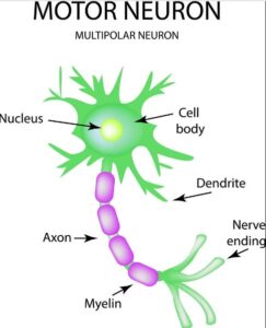
  • Nucleus – controls the entire neuron.
  • Dendrite – receive stimulus and carries its impulses toward the cell body
  • Cell Body (soma) – has a nucleus & cytoplasm. It acts as a factory of the neuron. It produces all protein for the dendrites and neural transmitters
  • Axon – fiber which carries impulses away from cell body ie it forms conduction region for the neuron
  • Schwann Cells/ neurolemmocyte – cells which produce myelin or fat layer in the Peripheral Nervous System (axon maintenance and regeneration) It’s a glial cell that wrap the nerve fibre in PNS.
  • Myelin sheath – dense lipid layer which insulates the axon ( makes the axon look gray) It speeds-up nerve transmission.
  • Node of Ranvier – gaps or nodes in the myelin sheath. They speed up nerve transmission.
  • Axon terminals – form junctions with other cells
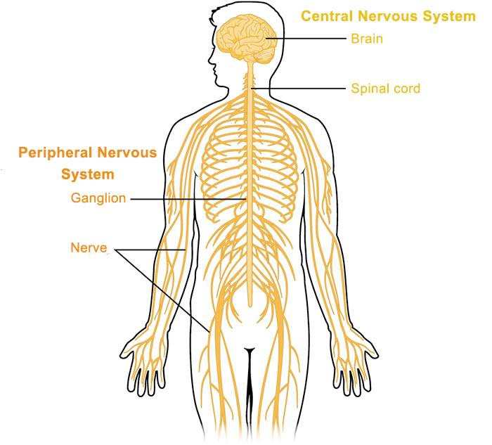
There are three types of Neurons
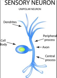
  1. Sensory neurons – bring messages to CNS
  2. Motor neurons – carry messages from CNS
  3. Interneurons – between sensory & motor neurons in the CNS
applied sensory neuron
applied motor neuron


Other cells of the nervous system
  1.  Satellite cells – surround cell bodies of neurons in ganglia. Their role is to maintain the micro-environment and provide insulation of the ganglion.
  2. Ependymal cells: these cells line cavities in CNS i.e spinal canal and ventricles in the brain. They secrete cerebral spinal fluid and form choroid plexuses.
  3. Oligodendrocytes: These cells wrap around CNS neurons to produce the myelin sheath. They function as Schwann Cells in CNS.
  4. Astrocyte: these are neuroglia/ glial cell of the CNS they anchor the neuron to the blood vessels and for blood brain barrier. They are star shaped.
  5. Microglia: These are monocytes in the nervous system they move to damaged brain tissue for phagocytosis.


CENTRAL NERVOUS SYSTEM

The Brain Anatomy

applied anatomy of the brain

It consists of four main parts i.e

1) Cerebrum– largest brain structure with four lobes i.e frontal, temporal, parietal and occipital.
It is divided into hemispheres by a fissure called longitudinal cerebral fissure with corpus callosum (mass of white matter) deep connecting the. Its outer layer is the cerebral cortex made of gray
mater and area below is made of white matter.

2) Cerebellum– located behind the ponds below the occipital lobe of cerebrum occupying the posterior cranial fossa.
Its oval shaped with two hemisphere separated by vermis. It has grey matter and white mater as in cerebrum.

3) Brain stem (mid brain and hind brain{Pons & medulla oblongata})
Mid brain is located around cerebral aqueduct with cerebrum above and Pons below. It has nuclei and group of nerve fibers with lower parts of the brain and with the spinal cord.
Pons is located in front of the Cerebellum nuclei and nerve fibers. Medulla oblongata extents from the Pons and is continuous with the spinal cord. It has central  grey matter and white matter which passes from the spinal cord and brain on outer aspect 

4) Diencephalon (Thalamus, Hypothalamus) – part of the brain that connect cerebrum and mid brain made of structure located around the third ventricle. Peneal gland and optic chiasma is also located here. Thalamus has two masses of grey and white mater just below corpus callosum, one on either side of the ventricle and hypothalamus is below in front of the thalamus connected to pituitary gland with number of nuclei.


Physiology of the Brain

Cerebral cortex:

  •  For mental processes like memory,
  •  For sensory perception i.e pain, temperature, touch sight, hearing, taste and smell
  •  Initiation and control of skeletal muscle contractions

Cerebellum:

  •  Coordination of voluntary muscle movement
  •  Posture and balance
  •  Learning and language processing


Thalamus:

  •  Relays and distributes impulses from the most parts of the brain to the cerebral cortex
  •  Play role in memory


Hypothalamus
 

  • Control autonomic nervous system
  •  Controls appetite, thirst, body temperature, water balance, emotional reaction and sexual
    behavior
  •  Sleeping and waking cycles due to melatonin hormone from pineal gland in the epithalamus
  •  Secrete ADH and oxytocin


Brain stem

  • Mid brain has nuclei that act as relay station for ascending and descending nerve fibers
    (connects cerebrum with lower brain fibers and spinal cord)
  •  Pons works with medulla to control respiration and acts as relay station with some cranial nerves

Medulla oblongata

  • Control respiration center, cardiovascular center, and reflex center for vomiting, coughing, sneezing and swallowing. It is also a site of decussation pyramidal tracts (corticospinal) from motor area in the cerebrum to spinal cord crossing to opposite site

Anatomy of the Spinal Cord

It is roughly cylindrical in shape, and is circular to oval in cross-section with a central canal.

 Made up of 31 pairs of spinal nerves
• Each spinal nerve has two components or roots
Sensory fiber (dorsal root) — Brings information into the CNS from the periphery
Motor fiber (ventral root) — Causes movement or reaction
The spinal cord extends from the foramen magnum to approximately the level of the disc between vertebrae LI and LII in adults, although it can end as high as vertebra T12 or as low as the disc between vertebrae LII and LIII. In neonates, the spinal cord extends approximately to vertebra LIII, but can reach as low as vertebra LIV

The distal end of the cord (the conus medullaris) is cone shaped. A fine filament of connective tissue (filum terminale) continues inferiorly from the apex of the conus medullaris

It has two major swellings or enlargements in regions associated with the origin of spinal nerves that innervate the upper and lower limbs. A cervical enlargement occurs in the region  associated with the origins of spinal nerves C5 to T1, which innervate the upper limbs. A lumbosacral enlargement occurs in the region associated with the origins of spinal nerves L1 to
S3, which innervate the lower limbs.

Internally, the cord has a small central canal surrounded by gray and white matter: the gray
matter is rich in nerve cell bodies and white matter surrounds the gray matter and is rich in nerve cell processes, which form large bundles or tracts that ascend and descend in the cord to other spinal cord levels or carry information to and from the brain.

cross section of the spinal cord
spinal cord

Physiology of the Spinal Cord

Spinal cord provides communication between brain and the peripheral nerves. Tracts of white matter of the spinal cord carry sensory impulses to the brain and motor impulses from the brain to the skeletal muscles.

The grey matter of the spinal cord is a site of integration of reflexes which is rapid involuntary action in relation to a particular stimulus.

The meninges

They are three connective tissue coverings that surround, protect, and suspend the brain and spinal cord within the cranial cavity and vertebral canal, respectively:

  1.  The dura mater is the thickest and most external of the coverings;
    The spinal dura mater is continuous with the cranial dura mater at the foramen magnum of the skull and is the outermost meningeal membrane. In the cranial cavity, one layer of the dura mater is fused to the bone and represents the periosteum, but the spinal dura mater is separated from the bones of the vertebral canal by an extradural space. Inferiorly, the Dural sac dramatically narrows at the level of the lower border of vertebra SII and forms an investing sheath for the pial part of the filum terminale of the spinal cord. The dural part of the filum terminale attaches to the posterior surface of the vertebral bodies of the coccyx.
  2. The arachnoid mater is against the internal surface of the dura mater;
    This is a thin delicate membrane against, but not adherent to, the deep surface of the dura mater. It is separated from the pia mater by the subarachnoid space. The arachnoid mater ends at the
    level of vertebra SII. The sub-arachnoid space contain CSF.
  3.  The pia mater is adherent to the brain and spinal cord. It extends into the anterior median fissure and reflects as sleeve-like coating onto posterior and anterior rootlets and roots as they cross the subarachnoid space. As the roots exit the space, the sleeve-like coatings reflect onto the arachnoid mater. On each side of the spinal cord, a longitudinally oriented sheet of pia mater (the denticulate ligament) extends laterally from the cord toward the arachnoid and dura mater. Because the subarachnoid space can be accessed in the lower lumbar region without endangering the spinal cord, it is important to be able to identify the position of the lumbar vertebral spinous processes. The LIV vertebral spinous process is level with a horizontal line between the highest points on the iliac crests. In the lumbar region, the palpable ends of the vertebral spinous processes lie opposite their corresponding vertebral bodies. The subarachnoid space can be accessed between vertebral levels LIII and LIV and between LIV and LV without endangering the spinal cord

PERIPHERAL NERVOUS SYSTEM

CRANIAL NERVES

In a clinical practice, it’s very important for the nurse to know the basic cranial nerves, there location and function. Below are the major cranial nerves in the body

  1. Olfactory nerve (CN I): this nerve arises from the olfactory (smell) receptors within the nasal mucosa. Loss of smell (Anosmia) is caused by head injury and tumors of the olfactory groove
    e.g. meningioma, frontalglioma). The nerve is assessed by applying different odious substances in each nose.   FIND THE REST OF THE NERVES BY CLICKING HERE

SPINAL NERVES

Spinal nerves contain both sensory and motor fibers, as do most nerves. Spinal nerves are given numbers which indicate the portion of the vertebral column in which they arise. There are 8 cervical nerves (C1-C8), 12 thoracic nerves (T1 – T12), 5 lumbar nerves (L1 – L5), 5 sacral nerves (S1-S5), and 1 coccygeal nerve. Nerve C1 arises between the cranium and atlas. All the others arise below the respective vertebra or former vertebra in the case of the sacrum.
A plexus is an interconnection of fibers which form new combinations as the “named” or
peripheral nerves.
Dermatomes are somatic or musculocutaneous areas served by fibers from specific spinal
nerves.

The map of the dermatomes is shown by Figure below . The map is useful in diagnosing origin of certain somatic pain, numbness, tingling, when symptoms are caused by pressure or inflammation of the spinal cord or nerve roots.


Referred pain is caused when the sensory fibers from an internal organ enter the spinal cord in the same root as fibers from a dermatome. The brain is poor at interpreting visceral pain and instead interprets it as pain from the somatic area of the dermatome.

So pain in the heart is often interpreted as pain in the left arm or shoulder, pain in the diaphragm is interpreted as along the left clavicle and neck, and the “stitch in your side” you sometimes feel when running is pain in the liver as its vessels constrict.

Myotome is that region of skeletal muscle innervated by a single nerve or spinal cord level.
Most individual muscles of the body are innervated by more than one spinal cord level so the evaluation of myotomes is usually accomplished by testing movements of joints or muscle groups.

Spread the love

5 thoughts on “Applied anatomy and Physiology of the nervous system”

  1. Thanks for the good work.
    I asking whether unmeb sets questions and review of anatomy and physiology or we just need to remind our selves so that we can understand the medical conditions under the system?

Leave a Comment

Your email address will not be published. Required fields are marked *

Contact us to get permission to Copy

We encourage getting a pen and taking notes,

that way, the website will be useful.

Scroll to Top