Nurses Revision

Applied anatomy and Physiology of the nervous system

Applied anatomy and Physiology of the nervous system

REVIEW OF THE ANATOMY AND PHYSIOLOGY OF NERVOUS SYSTEM

Anatomy of the Nervous System

The nervous system can be separated into parts based on structure and on function:

Structurally, it’s organized into the central nervous system (CNS) and the peripheral nervous system (PNS)

The CNS consists of the brain and spinal cord, both of which originate in the embryo. The PNS comprises all neural structures outside the CNS, connecting it to the rest of the body. These structures develop from neural crest cells and as extensions of the CNS itself. The PNS includes spinal and cranial nerves, visceral nerves and plexuses, and the enteric nervous system.

Functionally, the nervous system is divided into somatic and visceral components. The somatic nervous system (from the Greek ‘soma,’ meaning body) innervates structures derived from somites, such as skin and most skeletal muscle. 

It’s primarily responsible for receiving and responding to external environmental information. The visceral nervous system (from the Greek ‘viscera,’ meaning guts) innervates organ systems and other visceral elements like smooth muscle and glands throughout the body. It mainly detects and responds to information about the body’s internal environment. The neuron, with its cell body, axon, and synapse, is the functional unit of the entire nervous system.

Structural Division:
  • Central Nervous System (CNS): Composing the brain and spinal cord.
  • Peripheral Nervous System (PNS): Includes structures outside the CNS connecting it to the body.
Functional Division:
  • Somatic Part: Innervates structures from somites (skin, skeletal muscles). Responds to external environmental stimuli.
  • Visceral Part: Innervates organ systems, smooth muscles, and glands. Detects and responds to internal environmental stimuli.
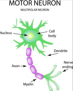
Structure of a neuron

applied neuron structure

FUNCTIONS OF NEURON STRUCTURES
  • Nucleus controls the entire neuron.
  • Dendrite – receives stimulus and carries its impulses toward the cell body.
  • Cell Body (soma) – has a nucleus & cytoplasm. It acts as a factory of the neuron. It produces all protein for the dendrites and neurotransmitters.
  • Axon – fiber which carries impulses away from the cell body i.e it forms a conduction region for the neuron.
  • Schwann Cells/ neurolemmocytecells which produce myelin or fat layer in the Peripheral Nervous System (axon maintenance and regeneration) It’s a glial cell that wraps the nerve fibre in PNS.
  • Myelin sheath – dense lipid layer which insulates the axon ( makes the axon look gray) It speeds-up nerve transmission.
  • Node of Ranvier – gaps or nodes in the myelin sheath. They speed up nerve transmission.
  • Axon terminalsform junctions with other cells.
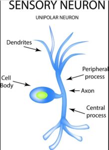
There are three types of Neurons
  • Sensory neurons – bring messages to CNS.
  • Motor neurons – carry messages from CNS.
  • Interneurons – between sensory & motor neurons in the CNS.
applied sensory neuron
applied motor neuron

Neuron – Functional Unit:

  • Composed of nucleus, dendrites, cell body (soma), axon, Schwann cells, myelin sheath, Node of Ranvier, and axon terminals.
  • Three types: sensory neurons (to CNS), motor neurons (from CNS), and interneurons (between sensory and motor neurons in CNS).

Other Nervous System Cells:

  • Satellite Cells: Surround neuron cell bodies in ganglia. Maintain a micro-environment and provide insulation.
  • Ependymal Cells: Line CNS cavities, secrete cerebrospinal fluid, and form choroid plexuses.
  • Oligodendrocytes: Wrap around CNS neurons to produce myelin sheath.
  • Astrocytes: Glial cells in the CNS. Anchor neurons to blood vessels and form the blood-brain barrier.
  • Microglia: Monocytes in the nervous system. Move to damaged tissue for phagocytosis.

CENTRAL NERVOUS SYSTEM

Brain Anatomy & Physiology.

applied anatomy of the brain
Cerebrum:
  • Largest brain structure with frontal, temporal, parietal, and occipital lobes.
  • Divided into hemispheres by the longitudinal cerebral fissure.
  • Cerebral cortex (gray matter) and subcortical white matter.
  • Responsible for memory, sensory perception (pain, temperature, touch, sight, hearing, taste, smell), and control of skeletal muscle contractions.
Cerebellum:
  • Located behind the pons, below the occipital lobe.
  • Oval-shaped with hemispheres separated by vermis.
  • Contains gray and white matter.
  • Coordinates voluntary muscle movement, maintains posture and balance, and contributes to learning and language processing.
Brain Stem (Midbrain and Hindbrain – Pons & Medulla Oblongata):
  • Midbrain surrounds the cerebral aqueduct, connecting cerebrum and pons.
  • Pons, in front of the cerebellum, have nuclei and nerve fibers.
  • Medulla oblongata extends from the pons, continuous with the spinal cord, containing gray and white matter.
  • Midbrain acts as a relay station for ascending and descending nerve fibers, connecting cerebrum with lower brain fibers and spinal cord.
  • Pons collaborates with the medulla to control respiration.
  • Medulla oblongata controls respiration, cardiovascular function, and reflexes (vomiting, coughing, sneezing, swallowing). 
Diencephalon (Thalamus, Hypothalamus):
  • Connects cerebrum and midbrain.
  • Houses thalamus (gray and white matter masses) and hypothalamus (below thalamus, connected to pituitary gland).

Thalamus

  • Relays and distributes impulses from various brain parts to the cerebral cortex.
  • Plays a role in memory processing.

Hypothalamus

  • Controls the autonomic nervous system.
  • Regulates appetite, thirst, body temperature, water balance, emotional reactions, and sexual behavior.
  • Influences sleeping and waking cycles through melatonin from the pineal gland.
  • Secretes ADH (antidiuretic hormone) and oxytocin.

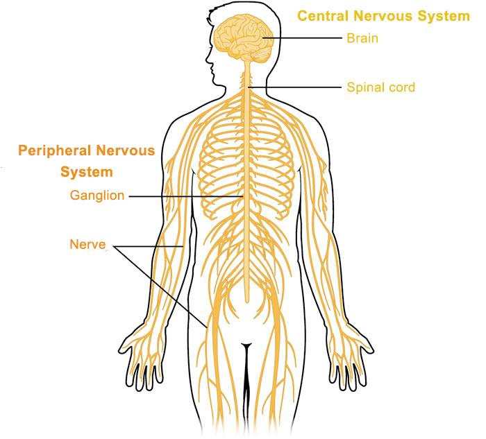
I. Introduction to Cerebrospinal Fluid (CSF)

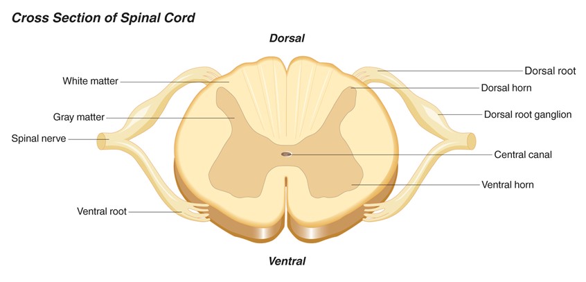
The Cerebrospinal Fluid (CSF) is a clear, colorless, ultrafiltrate of blood plasma that fills the ventricles of the brain, the central canal of the spinal cord, and the subarachnoid space surrounding the entire Central Nervous System (CNS). It serves as the vital “lifeblood” and shock absorber for the brain and spinal cord.

Volume and Production Rate:

  • Total Volume: The average adult CNS contains about 130 to 150 mL of CSF at any given time.
  • Rate of Production: It is produced at a rate of roughly 20 mL per hour (or about 500 mL per day).
  • Physiological Implication: Because 500 mL is produced daily but the system only holds 150 mL, the entire volume of CSF is completely flushed and turned over 3 to 4 times a day! If absorption is blocked, this rapid production quickly leads to hydrocephalus.

Formation of CSF:

  • The Choroid Plexus: Approximately 70-80% of CSF is actively secreted by the choroid plexuses (networks of blood capillaries lined by highly specialized ependymal cells) located in the roofs of the lateral, third, and fourth ventricles.
  • Blood-CSF Barrier: Unlike normal leaky capillaries, the ependymal cells of the choroid plexus are joined by tight junctions. This forms the Blood-CSF barrier, strictly controlling what substances from the blood are allowed to be actively transported into the CSF.
  • The remaining 20-30% of CSF is produced by the ependymal lining of the ventricles and cerebral capillaries.

II. Functions of the CSF

The CSF is not just “water in the brain.” It has four highly specific, life-sustaining functions:

Mechanical Protection (Buoyancy):

  • The brain is essentially “floating” in a bath of CSF. According to Archimedes’ principle, this buoyancy reduces the effective weight of the human brain from ~1,400 grams to a mere 50 grams.
  • Without CSF, the heavy brain would sink and crush the vital centers in the lower brainstem against the base of the skull, cutting off its own blood supply.

Shock Absorption:

  • It acts as a liquid hydraulic cushion. When the head takes a blow, the CSF dissipates the physical force, preventing the delicate brain tissue from violently smashing against the hard inner skull.

Chemical Protection & Homeostasis:

  • Optimal neuronal signaling (action potentials) requires a highly stable ionic environment. The CSF provides a strictly regulated, optimized chemical bath for the neurons, free from the wild hormonal and chemical fluctuations of normal blood plasma.

Circulation & Waste Removal (The “Glymphatic” System):

  • Because the brain lacks a traditional lymphatic system, the CSF acts as the brain’s waste clearance pathway. It washes away toxic metabolic byproducts (like amyloid-beta plaques) that accumulate during the day. (Interestingly, this flushing mechanism is highly active while we sleep!)

III. The Circulation Pathway of CSF

CSF flows in a strict, one-way path driven by its own continuous production, the pulsating of nearby blood vessels, and the beating of cilia on ependymal cells.

🧠 Mnemonic: The CSF Flow Pathway

“Love In The Air, For Lovers & Maidens, So Sweet”

  • Lateral ventricles
  • Interventricular foramina (of Monro)
  • Third ventricle
  • Aqueduct (Cerebral Aqueduct of Sylvius)
  • Fourth ventricle
  • Luschka (Lateral foramina) & Magendie (Median foramen)
  • Subarachnoid space
  • Superior sagittal sinus (Absorption)

Absorption of CSF:

  • After circulating through the subarachnoid space, the CSF must be returned to the venous blood to prevent pressure buildup.
  • This occurs at the Arachnoid Villi (which clump together to form Arachnoid Granulations).
  • These granulations protrude into the dural venous sinuses (primarily the Superior Sagittal Sinus). They act as one-way pressure valves: when CSF pressure is higher than venous pressure, CSF empties into the blood. If venous pressure rises, the valves snap shut to prevent blood from flowing backward into the brain.

IV. Composition of CSF vs. Blood Plasma

A crucial topic for board exams and clinical practice. Normal CSF is crystal clear and looks like water. Because of the Blood-CSF barrier, its composition is very different from blood plasma.

Component

Normal CSF Values

Comparison to Blood Plasma

Appearance

Clear and Colorless

Plasma is yellowish. (Cloudy CSF indicates infection; Red/Pink indicates bleeding).

White Blood Cells (WBCs)

0 – 5 cells/mm³ (Lymphocytes only)

Drastically lower. (Normal blood has 4,000-11,000 WBCs).

Red Blood Cells (RBCs)

ZERO (0)

There should NEVER be RBCs in normal CSF.

Protein

15 – 45 mg/dL

Massively Lower. Plasma has vast amounts of protein (~7,000 mg/dL). High CSF protein indicates barrier breakdown.

Glucose

50 – 80 mg/dL

About 60-70% (two-thirds) of the patient’s blood glucose.

Chlorides (Cl-) & Magnesium (Mg2+)

Higher than plasma

Actively transported into CSF to maintain electrical neutrality.

Potassium (K+) & Calcium (Ca2+)

Lower than plasma

Kept strictly low to prevent neurons from becoming hyper-excitable.

💡 Points for Attention: Lumbar Puncture (Spinal Tap)

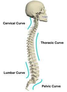
To analyze the composition of CSF, doctors perform a Lumbar Puncture. Because the solid spinal cord ends at the L1/L2 vertebral level in adults, the needle is safely inserted between L3 and L4 (or L4 and L5) into the subarachnoid space (the lumbar cistern). This area contains floating nerve roots (cauda equina) that easily move out of the needle’s way, making it the safest place to draw CSF.

❓ Applied Clinical Question: Meningitis

Case: A 19-year-old student presents with a stiff neck, severe headache, high fever, and photophobia (light sensitivity). A lumbar puncture is performed. The CSF drawn is cloudy and turbid. Laboratory analysis reveals a heavily elevated WBC count (mostly neutrophils), massively elevated protein (250 mg/dL), and drastically reduced glucose (15 mg/dL). What is the diagnosis?


Answer: Acute Bacterial Meningitis.
Why? The bacteria are literally “eating” the glucose for energy (causing low CSF glucose). The immune system sends neutrophils to fight the infection (high WBCs), and the inflammation destroys the Blood-Brain Barrier, allowing large blood proteins to leak into the CSF (high protein), turning the fluid cloudy.

Anatomy of the Spinal Cord

  • Cylindrical shape with circular to oval cross-section and a central canal.
  • Comprises 31 pairs of spinal nerves, each with sensory (dorsal root) and motor (ventral root) fibers.

Physiology of the Spinal Cord:

Spinal cord provides communication between brain and the peripheral nerves. Tracts of white matter of the spinal cord carry sensory impulses to the brain and motor impulses from the brain to the skeletal muscles.

The grey matter of the spinal cord is a site of integration of reflexes which is rapid involuntary action in relation to a particular stimulus.

  • Facilitates communication between the brain and peripheral nerves.
  • White matter tracts carry sensory impulses to the brain and motor impulses from the brain to skeletal muscles.
  • Grey matter serves as the site for reflex integration, rapid involuntary actions in response to stimuli.
cross section of the spinal cord
spinal cord

Meninges:

Three connective tissue coverings surrounding and protecting the brain and spinal cord.

  • Dura Mater: Thickest and outermost layer, continuous with cranial dura mater. The spinal dura mater is continuous with the cranial dura mater at the foramen magnum of the skull and is the outermost meningeal membrane. In the cranial cavity, one layer of the dura mater is fused to the bone and represents the periosteum, but the spinal dura mater is separated from the bones of the vertebral canal by an extradural space. Inferiorly, the Dural sac dramatically narrows at the level of the lower border of vertebra SII and forms an investing sheath for the pial part of the filum terminale of the spinal cord. The dural part of the filum terminale attaches to the posterior surface of the vertebral bodies of the coccyx.
  • Arachnoid Mater: Thin, delicate membrane against the internal surface of the dura mater. This is a thin delicate membrane against, but not adherent to, the deep surface of the dura mater. It is separated from the pia mater by the subarachnoid space. The arachnoid mater ends at the level of vertebra SII. The sub-arachnoid space contain CSF.
  • Pia Mater: Adherent to the brain and spinal cord, extends into the anterior median fissure, and forms the denticulate ligament. It extends into the anterior median fissure and reflects as sleeve-like coating onto posterior and anterior rootlets and roots as they cross the subarachnoid space. As the roots exit the space, the sleeve-like coatings reflect onto the arachnoid mater. On each side of the spinal cord, a longitudinally oriented sheet of pia mater (the denticulate ligament) extends laterally from the cord toward the arachnoid and dura mater. Because the subarachnoid space can be accessed in the lower lumbar region without endangering the spinal cord, it is important to be able to identify the position of the lumbar vertebral spinous processes. The LIV vertebral spinous process is level with a horizontal line between the highest points on the iliac crests. In the lumbar region, the palpable ends of the vertebral spinous processes lie opposite their corresponding vertebral bodies. The subarachnoid space can be accessed between vertebral levels LIII and LIV and between LIV and LV without endangering the spinal cord.
cranial nerves

PERIPHERAL NERVOUS SYSTEM

CRANIAL NERVES and ASSESSMENT

In a clinical practice, it’s very important for the nurse to know the basic cranial nerves, there location and function. Below are the major cranial nerves in the body.

Olfactory Nerve (I):

  • Function: Smell.
  • Assessment: Identify different smells with eyes closed.

Optic Nerve (II):

  • Function: Vision.
  • Assessment: Visual test and examination with a special light.

Oculomotor Nerve (III):

  • Function: Pupil size and certain eye movements.
  • Assessment: Pupil examination with light, eye movement in various directions.

Trochlear Nerve (IV):

  • Function: Eye movement.
  • Assessment: Eye movement evaluation.

Trigeminal Nerve (V):

  • Function: Face sensation, inside mouth sensation, and chewing.
  • Assessment: Touch face, observe biting down.

Abducens Nerve (VI):

  • Function: Eye movement.
  • Assessment: Follow light or finger for eye movement.

Facial Nerve (VII):

  • Function: Face muscle movement and taste.
  • Assessment: Identify tastes, smile, move cheeks, show teeth.

Acoustic Nerve (VIII):

  • Function: Hearing.
  • Assessment: Hearing test.

Glossopharyngeal Nerve (IX):

  • Function: Taste and swallowing.
  • Assessment: Identify tastes on the back of the tongue, test gag reflex.

Vagus Nerve (X):

  • Function: Swallowing, gag reflex, taste, and part of speech.
  • Assessment: Swallowing, elicit gag response with a tongue blade.

Accessory Nerve (XI):

  • Function: Shoulder and neck movement.
  • Assessment: Turn head side to side against resistance, shrug shoulders.

Hypoglossal Nerve (XII):

  • Function: Tongue movement.
  • Assessment: Stick out tongue, speak.

 FIND THE REST OF THE ASSESSMENT BY CLICKING HERE

Spinal Nerves

Spinal nerves, like most nerves, contain both sensory and motor fibers. They are named and numbered according to the region of the vertebral column from which they originate: 

  • 8 cervical nerves (C1-C8), 12 thoracic nerves (T1-T12), 
  • 5 lumbar nerves (L1-L5), 
  • 5 sacral nerves (S1-S5), and 
  • 1 coccygeal nerve. 

Nerve C1 emerges between the cranium and the atlas (first cervical vertebra). All other spinal nerves emerge below the vertebra (or former vertebra, in the case of the sacrum) corresponding to their number.

A plexus is a network of interconnected nerve fibers that recombine to form new, named peripheral nerves.

Dermatomes are areas of skin and muscle innervated by specific spinal nerves. A dermatome map (as shown in the figure) is a valuable diagnostic tool. It helps determine the origin of somatic pain, numbness, or tingling, especially when these symptoms result from pressure or inflammation of the spinal cord or nerve roots.

  • Dermatomes are somatic or musculocutaneous areas served by fibers from specific spinal nerves.
  • Dermatome map aids in diagnosing somatic pain, numbness, tingling caused by spinal cord or nerve root pressure or inflammation.

Myotome:

  • Region of skeletal muscle innervated by a single nerve or spinal cord level.
  • Most muscles receive input from multiple spinal cord levels.
Autonomic Nervous System (ANS)

The Autonomic Nervous System (ANS) is the portion of the peripheral nervous system that operates independently of conscious control (involuntarily). It is responsible for regulating visceral functions to maintain homeostasis, such as heart rate, digestion, respiratory rate, pupillary response, urination, and sexual arousal.

I. Divisions of the Autonomic Nervous System

The ANS is classically divided into two primary, often antagonistic, branches. A third branch, the enteric nervous system, governs the gastrointestinal tract.

1. The Sympathetic Nervous System (SNS)
  • Role: Responsible for the "Fight or Flight" response. It mobilizes body resources in response to stress, danger, or physical exertion.
  • Primary Actions: Increases heart rate, dilates bronchioles, shunts blood away from the digestive tract and towards skeletal muscles, mobilizes energy reserves (glycogenolysis), and dilates pupils.
2. The Parasympathetic Nervous System (PNS)
  • Role: Responsible for the "Rest and Digest" or "Feed and Breed" response. It conserves energy and promotes housekeeping functions during rest.
  • Primary Actions: Decreases heart rate, constricts bronchioles, increases gastrointestinal motility and secretions, promotes urination and defecation, and constricts pupils.

The "SLUDD" Mnemonic for Parasympathetic Responses

To remember the primary functions of the Parasympathetic Nervous System, think of the acronym SLUDD:

  • Salivation
  • Lacrimation (tearing)
  • Urination
  • Digestion
  • Defecation

Plus: 3 Decreases (Decreased Heart Rate, Decreased Airway Diameter, Decreased Pupil Diameter).

II. Anatomical Differences: SNS vs. PNS

The two divisions differ significantly in their anatomical origin, the length of their nerve fibers, and the location of their ganglia.

Feature Sympathetic (SNS) Parasympathetic (PNS)
Origin in CNS Thoracolumbar: Spinal cord segments T1 to L2. Craniosacral: Brainstem (Cranial Nerves III, VII, IX, X) and sacral spinal cord (S2-S4).
Ganglia Location Paravertebral (sympathetic chain) or Prevertebral ganglia, close to the spinal cord. Terminal or Intramural ganglia, close to or within the target effector organ.
Preganglionic Fiber Length Short (because ganglia are close to the spinal cord). Long (because ganglia are near the target organ).
Postganglionic Fiber Length Long (from sympathetic chain to the target organ). Short (from near the target organ to the tissue itself).
Branching Extensive branching (allows for mass, systemic activation). Minimal branching (allows for specific, localized control).
III. Neurotransmitters and Receptors

The communication between neurons and effector organs in the ANS relies on specific neurotransmitters binding to specific receptors. The two main neurotransmitters are Acetylcholine (ACh) and Norepinephrine (NE).

1. Cholinergic Neurons and Receptors (Acetylcholine)
  • Location:
    • All preganglionic neurons (both sympathetic and parasympathetic) release ACh.
    • All parasympathetic postganglionic neurons release ACh.
    • Exceptions: Sympathetic postganglionic neurons innervating sweat glands release ACh.
  • Receptor Types:
    • Nicotinic Receptors: Found on postganglionic cell bodies (both SNS and PNS) and the adrenal medulla. Always excitatory when ACh binds.
    • Muscarinic Receptors: Found on all effector organs innervated by the parasympathetic system, and sweat glands. Can be excitatory or inhibitory depending on the subtype.
2. Adrenergic Neurons and Receptors (Norepinephrine/Epinephrine)
  • Location: Almost all sympathetic postganglionic neurons release Norepinephrine (NE). The adrenal medulla releases primarily Epinephrine (adrenaline) into the bloodstream.
  • Receptor Types:
    • Alpha 1 (α1): Found in vascular smooth muscle (causes vasoconstriction), pupils (causes dilation - mydriasis), and sphincters of GI/GU tracts (causes contraction).
    • Alpha 2 (α2): Found on presynaptic nerve terminals (inhibits release of NE) and in the CNS.
    • Beta 1 (β1): Found primarily in the Heart. Increases heart rate (chronotropy), contractility (inotropy), and conduction velocity (dromotropy). Also found in kidneys (stimulates renin release). (Mnemonic: You have 1 heart).
    • Beta 2 (β2): Found primarily in the Lungs (bronchodilation), blood vessels of skeletal muscle (vasodilation), and uterus (relaxation). (Mnemonic: You have 2 lungs).
IV. Physiological Effects on Target Organs

Most organs receive dual innervation, meaning they receive impulses from both sympathetic and parasympathetic neurons, which usually have opposing effects.

Target Organ/System Sympathetic Effect (Fight or Flight) Parasympathetic Effect (Rest & Digest)
Eyes (Pupils) Dilation (Mydriasis) - to see threats better. Constriction (Miosis) - for near vision/rest.
Heart Increased heart rate and force of contraction. Decreased heart rate.
Lungs (Bronchioles) Bronchodilation (relaxes smooth muscle to increase airway). Bronchoconstriction (contracts smooth muscle).
Gastrointestinal Tract Decreased motility, decreased secretions, sphincter contraction. Increased motility, increased secretions, sphincter relaxation.
Liver Glycogenolysis and gluconeogenesis (releases glucose for energy). Glycogenesis (stores glucose).
Urinary Bladder Relaxes detrusor muscle, constricts internal sphincter (prevents voiding). Contracts detrusor muscle, relaxes internal sphincter (promotes voiding).
Blood Vessels Vasoconstriction in skin/viscera; Vasodilation in skeletal muscle. Little to no effect (most blood vessels lack PNS innervation).
Sweat Glands Increases sweating (localized and systemic). No innervation.
CLINICAL APPLICATION & NURSING CARE: AUTONOMIC DYSREFLEXIA

Autonomic Dysreflexia (AD) is a life-threatening, uninhibited, and exaggerated sympathetic nervous system response to a noxious stimulus below the level of a spinal cord injury, typically occurring in patients with injuries at or above T6.

Pathophysiology of AD:
  • A noxious stimulus (e.g., full bladder, impacted bowel, tight clothing) occurs below the level of injury.
  • Sensory nerves send signals up the spinal cord, but they are blocked by the lesion.
  • This triggers a massive sympathetic surge below the lesion, causing widespread vasoconstriction and severe hypertension.
  • Baroreceptors detect the hypertensive crisis and signal the brain.
  • The brain attempts to compensate by slowing the heart rate (parasympathetic bradycardia via the Vagus nerve) and dilating blood vessels above the injury (causing flushing and sweating above the lesion). However, descending inhibitory signals cannot pass the spinal lesion to stop the sympathetic surge below.
Nursing Care Plan & Interventions for Autonomic Dysreflexia
No. Nursing Diagnosis / Priority Interventions & Rationale
Immediate Medical Emergency Management
1 Risk for Ineffective Tissue Perfusion (Cerebral) related to severe, uncontrolled hypertension (often > 200/100 mmHg).
  • Elevate the head of the bed to 90 degrees immediately (or sit the patient upright with legs dangled): Rationale: Promotes orthostatic pooling of blood in the lower extremities, utilizing gravity to quickly lower blood pressure and reduce the risk of a hemorrhagic stroke.
  • Monitor blood pressure every 2-5 minutes: Rationale: Continuous monitoring is essential to evaluate the effectiveness of interventions and detect life-threatening hypertensive crises.
2 Acute Pain / Discomfort related to an unidentified noxious stimulus below the spinal lesion.
  • Identify and eliminate the cause immediately: Rationale: Removing the trigger halts the exaggerated sympathetic response.
    • Bladder: Check for bladder distension, kinked Foley catheter, or need for straight catheterization (most common cause).
    • Bowel: Check for fecal impaction. Apply anesthetic ointment before digital stimulation to avoid worsening the reflex.
    • Skin: Loosen tight clothing, shoes, or restrictive belts. Check for pressure ulcers, ingrown toenails, or insect bites.
3 Decreased Cardiac Output related to compensatory vagal stimulation causing severe bradycardia.
  • Assess heart rate and rhythm via continuous ECG monitoring: Rationale: The vagus nerve lowers the heart rate in response to the hypertension. Monitoring detects dangerous arrhythmias or profound bradycardia.
  • Administer prescribed rapid-onset antihypertensives (e.g., Nitroglycerin paste, Nifedipine, Hydralazine): Rationale: Used if blood pressure remains dangerously high even after removing the noxious stimulus, counteracting the profound sympathetic vasoconstriction.
Patient Education & Prevention
4 Deficient Knowledge regarding triggers and self-management of Autonomic Dysreflexia.
  • Educate the patient and caregivers on the signs and symptoms: (e.g., pounding headache, profuse sweating above the injury level, nasal congestion, goosebumps). Rationale: Early recognition prevents progression to stroke or death.
  • Teach rigorous bowel and bladder regimens: Rationale: Regular emptying prevents the two most common triggers (bladder distension and bowel impaction).
  • Provide a medical alert card: Rationale: Ensures other healthcare providers are aware of the patient's risk in emergency situations.

Quick Quiz

Nervous System Quiz

Anatomy - mobile-friendly and focused practice.

Privacy: Your details are used only for quiz tracking and certificates.

10 thoughts on “Applied anatomy and Physiology of the nervous system”

  1. Thanks for the good work.
    I asking whether unmeb sets questions and review of anatomy and physiology or we just need to remind our selves so that we can understand the medical conditions under the system?

Leave a Comment

Your email address will not be published. Required fields are marked *

Want notes in PDF? Join our classes!!

Send us a message on WhatsApp
0726113908

Scroll to Top
Enable Notifications OK No thanks