Nurses Revision

nursesrevision@gmail.com

PRECOCIOUS PUBERTY

PRECOCIOUS PUBERTY

PRECOCIOUS PUBERTY

Precocious puberty refers to any physical sex hormone effect, due to any cause, occurring earlier than the usual age, especially when it is being considered as a medical problem. 

Precocious puberty is puberty occurring at an unusually early age. In girls, this is before the age of 8, and in boys, before the age of 9. It is a condition where secondary sexual characteristics develop earlier than the known age range.

Early pubic hair, breast, or genital development may result from natural early maturation or from several other conditions. 

Precocious puberty can make a child fertile when very young, with the youngest mother on record being Lina Medina, who gave birth at the age of 5 years, 7 months and 17 days, in one report and at 6 years 5 months in another.

precocious puberty nursing case study
CASE STUDY

A 3-year-old girl presents with a one-year history of breast enlargement and per vaginal discharge. The child was reported to be well one year ago, has achieved normal developmental milestones, and has no history of birth injury, head injury, encephalitis, headaches, or seizures.

Clinical Findings:

  • White discharge per vagina is observed.
  • On examination, bilateral breast enlargement is noted, which is firm in consistency. Developed nipple and areola are also observed.
  • Axillary and pubic hair are sparse.

Investigations:

  • Elevated levels of LH (luteinizing hormone) and FSH (follicle-stimulating hormone) are reported.
  • Abdominal ultrasound reveals an enlarged uterus and ovaries of adult size.

Diagnosis:

This clinical scenario suggests a case of precocious puberty in the 3-year-old girl, marked by premature development of secondary sexual characteristics. Further evaluation and management will be necessary to address the underlying hormonal imbalance and its impact on the child’s health and development.

PUBERTY

Puberty is the developmental stage during which a child becomes a young adult, characterized by the maturation of gametogenesis, secretion of gonadal hormones, and development of secondary sexual characteristics and reproductive functions.

Tanner Staging in Puberty:

Tanner Staging, invented by James Tanner, is a widely used system to assess the progression of puberty based on physical changes.

  • Thelarche denotes the onset of breast development, an estrogen effect.
  • Pubarche denotes the onset of sexual hair growth, an androgen effect.
  • Menarche indicates the onset of menses.
  • Spermarche the appearance of spermatozoa in seminal fluid.
  • Gonadarche refers to the earliest gonadal changes of puberty. 
Tanner Stages in Females:

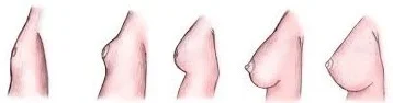
Tanner Stages in Females breasts

Breast Development (Thelarche):

  • Stage 1 (Preadolescent): No glandular tissue; only the papilla elevated.
  • Stage 2: Breast buds appear, along with a small mound of breast and papilla enlargement.
  • Stage 3: Further enlargement, with the breast mound elevating, and areola enlargement.
  • Stage 4: Continued enlargement, areola forms a secondary mound above the breast.
  • Stage 5 (Adult): Mature breast; areola returns to general breast contour.

Tanner Stages in Females pubic

Pubic Hair Development (Pubarche):

  • Stage 1 (Preadolescent): No pubic hair.
  • Stage 2: Sparse, long, downy hair, mostly along the labia.
  • Stage 3: Darker, coarser, curlier hair spreading over the mons pubis.
  • Stage 4: Hair resembles that of an adult, but less in quantity.
  • Stage 5 (Adult): Adult distribution; extends to inner thighs.

Tanner Stages in Males:

Tanner Stages in Males

Genital Development (Gonadarche):

  • Stage 1 (Preadolescent): Testes, scrotum, and penis are at childhood size.
  • Stage 2: Testes and scrotum enlarge; reddening of scrotum.
  • Stage 3: Penis lengthens; continued testicular and scrotal growth.
  • Stage 4: Increased penis size; scrotum darkens.
  • Stage 5 (Adult): Mature genitalia; adult size and shape.

Tanner Stages in Males pubic

Pubic Hair Development (Pubarche):

  • Stage 1 (Preadolescent): No pubic hair.
  • Stage 2: Sparse, long, downy hair at the base of the penis.
  • Stage 3: Darker, coarser, curlier hair spreading over the pubic symphysis.
  • Stage 4: Hair resembles an adult, but less in quantity.
  • Stage 5 (Adult): Adult distribution; extends to inner thighs.
Classification of Precocious Puberty.

Classification of Precocious Puberty.

Precocious puberty can be divided into 2 types

1. Gonadotropin-releasing hormone (GnRH)–dependent (Central Precocious Puberty): GnRH-dependent precocious puberty is more common overall and 5 to 10 times more frequent in girls. In GnRH-dependent precocious puberty, the hypothalamic-pituitary axis is activated, resulting in enlargement and maturation of the gonads, development of secondary sexual characteristics, and oogenesis or spermatogenesis. 

  •  Caused by early maturation of the hypothalamic-pituitary-gonadal axis.
  • Characterized by both breast development and pubic hair sexual maturation in girls, and pubic hair and testicular enlargement in boys.

2. GnRH-independent (peripheral sex hormone effects)Classified as Central or Peripheral Precocious Puberty): GnRH-independent precocious puberty is much less common. Secondary sexual characteristics result from high circulating levels of estrogens or androgens, without activation of the hypothalamic-pituitary axis.

  • Caused by excess secretions of sex hormones from the gonads or adrenal glands.
  • Isosexual precocious puberty: Feminizing signs in girls, masculinization in boys.
  • Heterosexual precocious puberty: Masculine characteristics in girls, feminization in boys.
Conditions Causing Precocious Puberty

Conditions Causing Precocious Puberty:

Central Precocious Puberty: Also known as complete or true precocious puberty, is characterized by the early activation of the hypothalamicpituitarygonadal (HPG) axis, leading to premature sexual development. Several underlying issues in the hypothalamus or pituitary can contribute to the onset of central precocious puberty. Possible causes include:

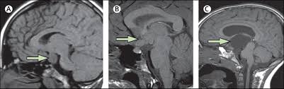
  1. Hypothalamic Haematoma: Formation of a hematoma in the hypothalamus can disrupt the normal inhibitory control of the HPG axis. This leads to the pulsatile release of gonadotropin-releasing hormone (GnRH), initiating premature puberty.
  2. Langerhans Cell Histiocytosis: Langerhans cell histiocytosis, a rare condition involving the proliferation of Langerhans cells, can affect the regulatory mechanisms in the hypothalamus. Dysregulation in the hypothalamus may result in early activation of the HPG axis, triggering precocious puberty.
  3. McCune–Albright Syndrome: Genetic mutations causing McCune–Albright syndrome can lead to abnormal functioning of the hypothalamus. Altered hypothalamic function disrupts the normal timing of puberty onset, causing it to occur prematurely.
  4.  Intracranial Neoplasm: Presence of tumors within the brain can interfere with the normal signaling pathways in the hypothalamus.  Tumor-induced disruptions can lead to the early release of GnRH, initiating the cascade of events leading to central precocious puberty.
  5.  Infection: Infections, especially central nervous system tuberculosis, can cause inflammation and affect the hypothalamic-pituitary axis.  Inflammatory processes may disrupt the normal control mechanisms, triggering premature puberty.
  6. Trauma: Trauma to the brain, such as head injuries, can damage the hypothalamus and pituitary. Structural damage may result in malfunctioning of the regulatory centers, contributing to the early activation of puberty.
  7. Hydrocephalus:  Hydrocephalus, characterized by an accumulation of cerebrospinal fluid in the brain, can exert pressure on the hypothalamus. Pressure-related damage to the hypothalamus may disrupt the normal control of the HPG axis, leading to central precocious puberty.
  8.  Angelman Syndrome: Angelman syndrome, a genetic disorder, can impact neurological functions, including those in the hypothalamus. Altered neural regulation may contribute to the premature activation of the HPG axis, causing central precocious puberty.
  9. Idiopathic or Constitutional:  If no identifiable cause is found. 

Peripheral Precocious Puberty: Peripheral precocious puberty, also referred to as precocious pseudopuberty, involves the premature onset of secondary sexual characteristics due to the influence of sex steroids from abnormal sources.  Causes include:

Isosexual (Feminizing) Conditions in Females:

  • McCune-Albright Syndrome: Genetic mutations leading to overactive endocrine glands, particularly in the ovaries. Excessive estrogen secretion triggers feminizing features prematurely.
  • Ovarian Tumors: Tumors in the ovaries can autonomously produce estrogen. Elevated estrogen levels induce early development of secondary sexual characteristics in females.

Heterosexual (Masculinizing) Conditions in Females:

  • Congenital Adrenal Hyperplasia (CAH): Genetic disorder causing adrenal glands to overproduce androgens. Androgen excess leads to the development of masculinizing features in females.
  • Adrenal Tumors: Tumors in the adrenal glands produce excess androgens. Androgenic influence results in the manifestation of male secondary sexual characteristics.
  • Ovarian Tumors: Ovarian tumors may produce androgens, leading to masculinization. Androgen excess induces the development of male secondary sexual characteristics.

Isosexual (Masculinizing) Conditions in Boys:

  • Congenital Adrenal Hyperplasia (CAH): Genetic disorder causing adrenal glands to overproduce androgens.  Excess androgens result in the development of male secondary sexual characteristics.
  • Leydig Cell Tumors: Tumors in the testes (Leydig cells) produce excess androgens.  Elevated androgen levels lead to the premature appearance of male secondary sexual characteristics.
  • hCG-Secreting Tumors: Tumors producing human chorionic gonadotropin (hCG) stimulate androgen production. Increased androgen levels contribute to the development of male secondary sexual characteristics.

Heterosexual (Feminizing) Conditions in Boys:

  • Feminizing Adrenocortical Tumor: Tumors in the adrenal cortex produce excess estrogen.  Elevated estrogen levels induce feminization in boys.
  • Exogenous Hormones: Introduction of external hormones, often used as a treatment for various conditions. Altered hormonal balance influences the onset of secondary sexual characteristics.

Isosexual and Heterosexual Precocity:

Patients with precocious puberty usually develop phenotypically appropriate secondary sexual characteristics, termed isosexual precocity. In rare cases, the development may be in the opposite direction, known as heterosexual or contrasexual precocity.

  • Example: Aromatase excess syndrome, a rare genetic condition, causes exceptionally high estrogen levels, leading to hyper feminization in both males and females.

Risk Factors:

  • Girls with a high-fat diet, lack of physical activity, or obesity may mature earlier.
  • Exposure to xenoestrogens, such as Bisphenol A in plastics.
  • Pineal tumor secreting chorionic gonadotropin (beta-hCG).
  • Elevated melatonin levels.
  • Familial cases of idiopathic central precocious puberty (ICPP).
  • Mutations in genes like LIN2, LEP, and LEPR, associated with leptin and the leptin receptor.

Diagnosis and Investigations in Precocious Puberty.

Clinical Manifestations: The diagnosis of precocious puberty is based on the premature appearance of secondary sex characteristics, occurring before the known age range.

  • In Boys: Pubic hair or genital enlargement (gonadarche) before 9.5 years.
  • In Girls: Pubic hair (pubarche) before 8 years or breast development (thelarche) before 7 years. Menstruation (menarche) before 10 years.

Blood Tests: Blood tests reveal elevated androgen levels with low cortisol levels.

  • Hormone Levels: Elevated androgen levels, plus low cortisol levels. 

Evaluation – Medical History:

  • Age at onset
  • Sex
  • Pubertal progression
  • Symptoms suggestive of hypothyroidism
  • History of past CNS infection, headache, visual disturbances & seizures.

Physical Examination:

  • Measurements of height, weight, height velocity
  • Pubertal staging according to Tanner’s staging.
  • Evaluate androgen & estrogen effects.
  • Inspection of skin (Café au lait macules in McCune-Albright Syndrome).
  • Examination for signs of hypothyroidism.

Basic Radiology:

  • Bone Age: Determines skeletal maturity, aiding in diagnostic accuracy.
  • Pelvic & Abdominal Sonography: Identifies anomalies or structural abnormalities influencing puberty.

Hormone Evaluation:

  • Intravenous administration of gonadotropin-releasing hormone (GnRH stimulation test) or a GnRH agonist (leuprolide stimulation test) is a helpful diagnostic tool for boys.
  • In girls, the central nature of sexual precocity can be proven by detecting pubertal levels of estradiol (>50 pg/mL), 20-24 hr after stimulation with leuprolide.
Challenges Faced by Precocious Children

Challenges Faced by Precocious Children

The early onset of sexual development poses several challenges:

In Girls:

  • Early bone maturation, potentially reducing adult height.
  • Indication of tumors or serious health issues.
  • Increased risk of becoming an object of adult sexual exploitation.
  • Higher vulnerability to sexual abuse.
  • Elevated risk of teasing or bullying.
  • Mental health disorders.
  • Short stature in adulthood due to advanced bone age.

In Boys:

  • Increased aggressiveness due to hormonal surges.
  • Social pressure to conform to adult norms.
  • Cognitive and social development lagging behind physical appearance.
  • Early maturing boys are more likely to be sexually active and engage in risky behaviors.

Treatment:

  • One possible treatment is with anastrozole. 
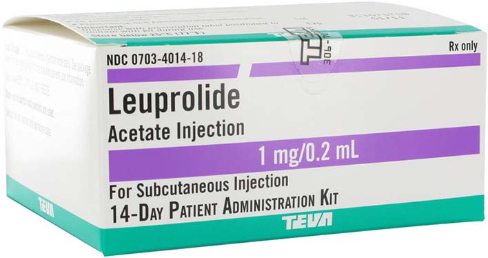
  • GnRH agonists like histrelin acetate (Supprelin LA), triptorelin, or leuprolide may be used. Inj. Leuprolide (0.5-0.3 mg/kg/dose) monthly.
  • Non-continuous use of GnRH agonists stimulates the pituitary gland, releasing follicle-stimulating hormone (FSH) and luteinizing hormone (LH).  Regular use decreases FSH and LH release, but prolonged use carries a risk of osteoporosis. After discontinuation, pubertal changes resume within 3 to 12 months. Regular monitoring is essential during treatment.

Surgery:

  • Tumors of the ovary, testis, and adrenals require surgical removal.
  • Hypothalamic Hamartomas are hazardous and not recommended because they never grow or become malignant.
  • Germ cell, pineal tumors, and hCG-producing suprasellar tumors can be treated by radiotherapy.

Nurses Roles during management of Precocious Puberty.

  1. Assessment and Monitoring: Conduct thorough assessments of patients to gather relevant data. Monitor the progression of secondary sexual characteristics and hormonal levels. Regularly assess the emotional and psychological well-being of the patient.
  2. Patient and Family Education: Provide extensive education about precocious puberty, its causes, and potential treatments. Explain the significance of diagnostic tests and procedures. Offer guidance on the expected course of treatment and potential side effects.
  3. Emotional Support: Offer emotional support to the patient and their family throughout the diagnostic and treatment processes. Address concerns and anxieties related to the condition. Facilitate communication between the healthcare team, patient, and family.
  4. Collaboration with Healthcare Team: Collaborate with endocrinologists, radiologists, and other specialists in the development and implementation of the patient’s care plan. Contribute nursing expertise to the interdisciplinary team.
  5. Administration of Medications: Administer medications as prescribed, such as GnRH agonists, which are commonly used in the management of central precocious puberty. Educate patients and families on medication administration and potential side effects.
  6. Monitoring for Adverse Effects: Monitor patients for any adverse effects of medications or interventions. Report and document any unexpected reactions promptly.
  7. Psychosocial Support: Address psychosocial challenges associated with precocious puberty, such as body image concerns and social interactions. Facilitate support groups or counseling for patients and families.
  8. Advocacy: Advocate for the patient’s needs within the healthcare system. Ensure that the patient’s rights and preferences are respected.
  9. Coordination of Care: Coordinate the various aspects of the patient’s care plan. Ensure smooth transitions between different stages of diagnosis, treatment, and follow-up.
  10. Continuity of Care: Promote continuity of care by maintaining regular follow-up appointments. Facilitate communication between the outpatient and inpatient settings, if necessary.
  11. Patient Safety: Prioritize patient safety during diagnostic procedures and treatment interventions. Educate patients and families on safety measures at home.
  12. Documentation: Maintain accurate and comprehensive documentation of patient assessments, interventions, and outcomes. Ensure that all relevant information is available for the healthcare team.
  13. Patient Advocacy: Advocate for the patient’s holistic well-being, considering physical, emotional, and psychosocial aspects. Address any ethical concerns that may arise during the management process.
  14. Education on Follow-Up Care: Provide detailed instructions for follow-up care, including medications, appointments, and potential lifestyle adjustments.
  15. Promoting Coping Strategies: Facilitate the development of coping strategies for both the patient and their family. Encourage open communication and expression of feelings.

PRECOCIOUS PUBERTY Read More »

CEREBRAL PALSY

CEREBRAL PALSY

CEREBRAL PALSY.

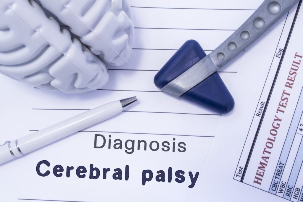
Cerebral palsy is defined as a group of permanent disorders of the development of movement and posture, causing activity limitation, that are attributed to non-progressive disturbances that occurred in the developing fetal or infant brain.”

If your body was a computer, the brain would be the mainframe. It controls mostly everything.

Cerebral Palsy means a brain condition causing paralysis, Therefore its a brain condition that makes the body lose control. It is also considered a neurodevelopmental condition meaning something happens to the brain during its development

Cerebral palsy is caused by abnormal development or damage to the parts of the brain that control movement, balance and posture. Therefore, severity is according to the part of the brain affected.

Cerebral palsy is the most common movement disorder in children. It occurs in about 1 per 323 live births. Cerebral palsy has been documented throughout history, with the first known descriptions occurring in the work of Hippocrates in the 5th century BCE.

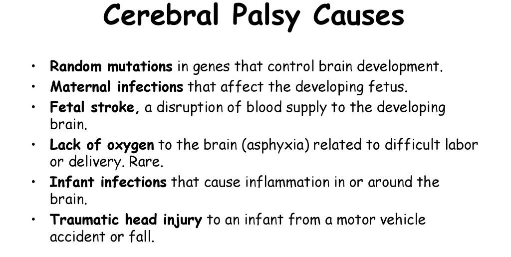
Fetal stroke, a disruption of blood supply to the developing brain. Lack of oxygen to the brain (asphyxia) related to difficult labor or delivery. Rare. Infant infections that cause inflammation in or around the brain. Traumatic head injury to an infant from a motor vehicle accident or fall.

Causes of Cerebral Palsy

Most often, the problems occur during pregnancy; however, they may also occur during childbirth or shortly after birth, however, other causes are unknown.

 Structural problems in the brain are seen in 80% of cases, most commonly within the white matter. More than three-quarters of cases are believed to result from issues that occur during pregnancy. Most children who are born with cerebral palsy have more than one risk factor associated with Cerebral Palsy.

In Africa birth asphyxia, high bilirubin levels, and infections in newborns of the central nervous system are the main cause.

Before Birth (Prenatal Causes)

Intrauterine Development Issues:

  • Exposure to radiation during pregnancy.
  • Infections affecting fetal development.
  • Fetal growth restriction.

Hypoxia of the Brain:

  • Thrombotic events affecting blood flow.
  • Placental conditions leading to reduced oxygen supply.

Genetic Factors:

  • Autosomal recessive inheritance pattern.
  • Inherited cases involving enzymes like glutamate decarboxylase-1.

Prematurity:

  • Births before 28 weeks of gestation carry a higher risk.
  • Between 34 and 37 weeks, the risk is elevated but lower (0.4%).

Multiple-Birth Infants:

  • Increased likelihood, especially with low birth weight.

Genetic Factors in Prematurity:

  • Genetic influences contributing to both prematurity and cerebral palsy.

During Birth (Perinatal Causes)

Birth Trauma:

  • Injuries occurring during labor and delivery.
  • Complications arising just after birth.

Low Birth Weight:

  • Term infants with low birth weight are at risk.

Instrumental Delivery or Emergency C-Section:

  • Use of instruments or emergency Cesarean section.

Placental Issues:

  • Problems with the placenta affecting fetal development.

Meconium Aspiration:

  • Breathing meconium into the lungs during delivery.

Birth Asphyxia:

  • Oxygen deprivation during birth.
  • Seizures occurring just after birth.

After Birth (Postnatal Causes)

  • Toxins: Exposure to toxins in the environment.
  • Severe Jaundice: High bilirubin levels impacting the brain.
  • Lead Poisoning: Environmental exposure to lead.
  • Physical Brain Injury: Trauma causing damage to the brain / Abusive head trauma.
  • Stroke: Disruption of blood flow to the brain.
  • Hypoxia Incidents: Near-drowning incidents impacting oxygen supply to the brain.
  • Infections: Infections during early childhood, such as encephalitis or meningitis.
  • Maternal Infections: Infections in the mother, even if asymptomatic.
  • Chorioamnionitis: Infections of fetal membranes increasing the risk.
  • Identical Twin Death: Hypothesized cases resulting from the death of an identical twin in early pregnancy.
  • Rh Blood Type Incompatibility: Mother’s immune system attacking the baby’s red blood cells.
Risk Factors:
  • Preterm Birth: Major risk factor, occurring in 40-50% of Cerebral Palsy cases.
  • Twin Birth: Being a twin increases the likelihood.
  • Infections During Pregnancy: Toxoplasmosis, rubella, and other infections.
  • Methylmercury Exposure: Environmental exposure during pregnancy.
  • Difficult Delivery and Head Trauma: Traumatic events during the first few years of life.
  • Inherited Genetic Causes: Approximately 2% of cases have a hereditary basis.

A newborn with severe CP may present with:

  1. An irregular posture; their bodies may be either very floppy or very stiff
  2. Birth defects, such as spinal curvature, a small jawbone, or a small head.
  3. Unable to suck, swallow or chew
  4. Lack of control of the head, mouth and trunk
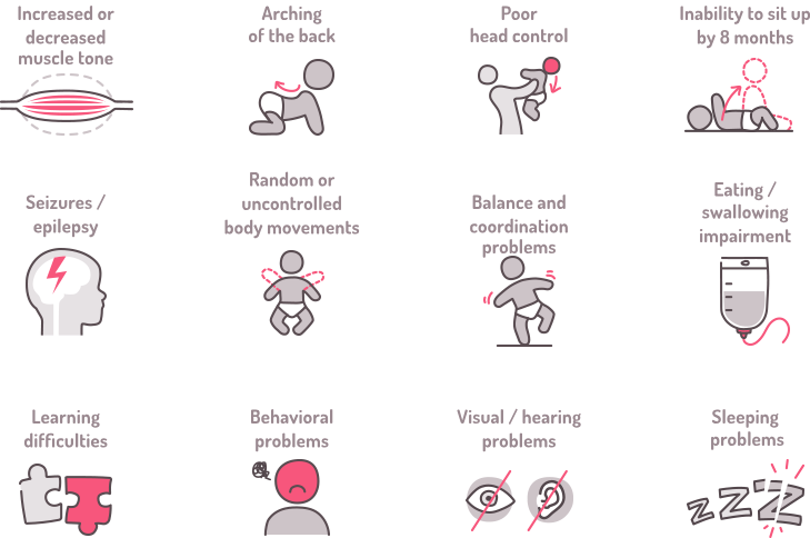
Signs and Symptoms of Cerebral Palsy (CP)

Signs and Symptoms of Cerebral Palsy (CP)

I. Developmental Milestones and Motor Function:

  • Delayed achievement of developmental milestones; Not reaching expected physical and cognitive milestones within typical time frames.
  • Abnormal motor development and coordination; Lack of smooth, coordinated muscle movements during development.
  •  Unusual gait patterns; Unusual walking patterns that may indicate motor control issues.

II. Muscle Tone and Reflexes:

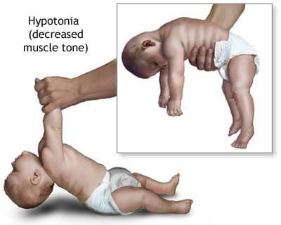
  • Abnormal muscle tone; Muscles may be too tight (spasticity) or too floppy (hypotonia).
  • Spasticity or hypertonia; Increased muscle stiffness, making movement challenging.
  • Hypotonia in some cases; Reduced muscle tone leading to poor muscle control.

III. Joint and Skeletal Abnormalities:

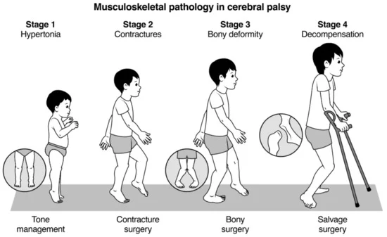
  • Joint contractures; Limited movement in joints due to tight muscles.
  •  Dynamic deformities progressing to static deformities; Irregular bone and joint development due to muscle imbalances.
  • Bone and joint deformities due to unequal growth; Asymmetric bone growth caused by muscle-related stresses.

IV. Coordination and Movement Issues:

  • Difficulty with precise movements; Challenges in performing accurate and controlled movements.
  •  Incoordination and tremors; Lack of coordination and uncontrollable shaking during movement.
  •  Challenges in voluntary muscle control; Difficulty in intentionally controlling muscle actions.

V. Speech and Communication:

  • Speech and language disorders; Difficulty in articulating words or understanding language.
  • Dysarthria (impaired speech due to muscle control); Speech difficulties resulting from poor muscle control.
  • Non-verbal communication in some cases; Reliance on gestures or other non-verbal cues for communication.

VI. Cognitive and Behavioral Aspects:

  • Learning disabilities; Difficulty in acquiring knowledge and skills.
  • Intellectual disabilities in some cases; Below-average intellectual functioning.
  • Behavioral challenges; Emotional or behavioral issues that may impact daily life.

VII. Sensory and Perception Issues:

  •  Visual impairments; Problems with vision or visual processing.
  • Auditory impairments; Hearing difficulties or processing issues.
  • Sensory processing difficulties; Challenges in interpreting and responding to sensory information.

VIII. Seizures:

  • Epilepsy in a significant percentage of cases; Recurrent seizures affecting brain function.

IX. Posture and Balance:

  • Poor posture; Inability to maintain an upright and balanced body position.
  • Balance issues; Difficulty in maintaining stability during movement or at rest.

X. Fine and Gross Motor Skills:

  • Impaired fine motor skills; Difficulty in performing precise tasks with hands and fingers.
  •  Difficulty with gross motor skills; Challenges in performing larger movements like crawling, walking, or jumping.

XI. Feeding and Eating Difficulties:

  • Challenges in chewing and swallowing; Difficulty in the process of biting, chewing, and swallowing food.
  • Gastro-oesophageal reflux; Stomach contents flowing back into the esophagus.
  •  Nutritional concerns; Issues related to inadequate nutrient intake.

XII. Behavioral and Emotional Issues:

  • Emotional challenges due to limitations; Psychological struggles arising from physical constraints.
  • Social difficulties; Challenges in interacting and forming relationships with others.

XIII. Drooling:

  • Excessive drooling due to lack of control; Inability to manage saliva flow.

XIV. Pain and Sleep Issues:

  • Chronic pain; Persistent discomfort or pain.
  • Sleep disturbances; Interruptions in regular sleep patterns.

XV. Orthopedic Complications:

  • Scoliosis; Abnormal sideways curvature of the spine.
  •  Hip dislocation; Displacement of the hip joint.
  • Skeletal deformities; Abnormalities in bone structure and shape.

Pathological effects of Cerebral Palsy on different body functions.

Urinary System: Lower Urinary Tract Symptoms:

  • Excessive storage issues are more prevalent than voiding issues.
  • Pelvic floor overactivity in some cases leads to upper urinary tract dysfunction.

Skeletal System: Bone Development:

  • Increased risk of low bone mineral density.
  • Thin bone shafts (diaphyses) contrasted with enlarged centers (metaphyses).
  • Joint compression from muscular imbalances leads to atrophy of articular cartilage and narrowed joint spaces.
  • Angular joint deformities and hindered bone development due to spasticity and abnormal gait.
  • Height reduction and potential limb length disparities.

Deformities and Conditions:

  • Common deformities include hip dislocation, ankle equinus, planter flexion, and torsional deformities.
  • Scoliosis prevalence increases with higher GMFCS levels.
  • Fracture susceptibility, especially in non-ambulant children, affecting mobility and schooling.

Eating and Nutrition: Feeding Challenges:

  • Sensory and motor impairments result in difficulty preparing food, holding utensils, chewing, and swallowing.
  • Gastro-oesophageal reflux is common; poor sensitivity around the mouth complicates self-feeding.

Nutritional Risks: Feeding difficulties linked to higher GMFCS levels.

  • Dental problems contribute to eating challenges.
  • Risk of undernutrition, particularly in severe cases; drooling and associated complications.

Language and Communication: Speech and Language Disorders:

  • Dysarthria incidence ranges from 31% to 88%; a quarter are non-verbal.
  • Associated with respiratory control, oral-facial muscle movement restrictions, and articulation disorders.
  • Early use of augmentative communication systems may aid language development.
  • Overall language delay is associated with cognitive issues, deafness, and learned helplessness.

Pain and Sleep: Painful Implications:

  • Chronic pain is prevalent, exacerbated by muscle spasms.
  • Pain associated with tight muscles, abnormal posture, and joint stiffness.
  • Hip migration or dislocation as a significant pain source.
  • High rates of sleep disturbance.
  • Chronic sleep disorders due to physical and environmental factors.
  • Babies with Cerebral Palsy may cry more or face challenges in sleep initiation.

Associated disorders.

Cerebral palsy is often accompanied by other disorders of cerebral function. Associated abnormalities may affect cognition, vision, hearing, language, cortical sensation, attention, vigilance, and behavior. Common conditions associated with cerebral palsy include:

Intellectual Disabilities:

  • Around 30-50% of individuals with Cerebral Palsy experience varying degrees of intellectual disability, affecting learning, memory, and problem-solving.
  • The severity can range from mild (requiring some support) to profound (needing extensive assistance).
  • Early intervention and tailored educational programs can significantly improve outcomes.

Seizures:

  • Up to 50% of individuals with Cerebral Palsy have epilepsy, experiencing recurring seizures.
  • Various types of seizures can occur, affecting movement, consciousness, and behavior.
  • Anti-seizure medications and other therapies can help manage seizures and improve quality of life.

Muscle Contractures:

  • These are involuntary muscle shortening and tightening, restricting movement and causing joint pain.
  • They can arise due to muscle imbalances, spasticity, and lack of use.
  • Physiotherapy, stretching, and sometimes surgery can help manage contractures and improve mobility.

Abnormal Gait:

  • Difficulty walking is a common symptom of Cerebral Palsy due to muscle weakness, spasticity, and coordination issues.
  • Different types of abnormal gait patterns exist, impacting balance, stability, and walking efficiency.
  • Assistive devices, orthotics, and gait training can help improve walking patterns and independence.

Osteoporosis:

  • Individuals with Cerebral Palsy have a higher risk of osteoporosis due to reduced bone density, often caused by limited mobility and decreased weight-bearing activity.
  • Bone mineral density checks, dietary adjustments, vitamin D supplementation, and strengthening exercises can help prevent and manage osteoporosis.

Communication Disorders:

  • Speech and language challenges are common in Cerebral Palsy, affecting articulation, fluency, and comprehension.
  • Different types of communication disorders may occur, like dysarthria (motor speech issues), aphasia (language processing difficulties), and apraxia (difficulty planning and executing speech movements).
  • Speech therapy, assistive technology, and alternative communication methods can help individuals communicate effectively.

Malnutrition:

  • Nutritional challenges can arise due to feeding difficulties, gastrointestinal issues, and decreased energy expenditure.
  • This can lead to deficiencies in essential nutrients, impacting growth, development, and overall health.
  • Specialized feeding techniques, nutritional supplements, and dietary adaptations can improve nutritional status.

Sleep Disorders:

  • Individuals with Cerebral Palsy may experience various sleep problems like insomnia, sleep apnea, and restless sleep.
  • These can be caused by medical conditions, muscle tone issues, medications, or environmental factors.
  • Good sleep hygiene practices, addressing underlying medical conditions, and specific sleep therapies can help improve sleep quality.

Mental Health Disorders:

  • Depression and anxiety are more common in individuals with Cerebral Palsy due to chronic health challenges, social limitations, and emotional strain.
  • Early identification, mental health counseling, and support groups can significantly improve mental well-being and quality of life.

Functional Gastrointestinal Abnormalities:

  • Digestive issues like constipation, vomiting, and bowel obstruction can arise due to impaired muscle coordination in the digestive system.
  • Dietary modifications, medications, and bowel management techniques can help manage these issues and improve digestive function.

Classification/Types of Cerebral Palsy

Cerebral Palsy is classified by the types of motor impairment of the limbs or organs, and by restrictions to the activities an affected person may perform. 

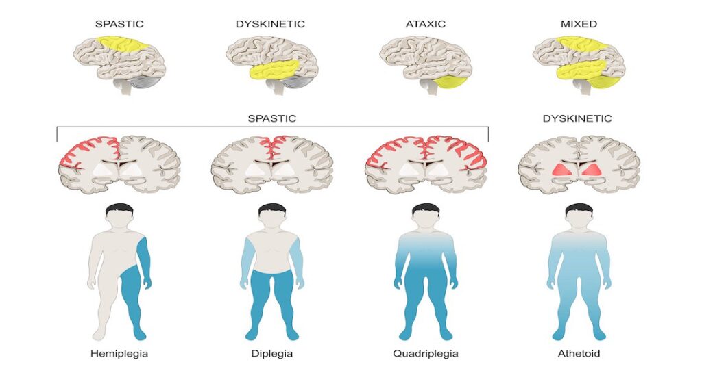
There are three main Cerebral Palsy classifications by motor impairment: 

  1. Spastic
  2. Ataxic
  3. Athetoid/Dyskinetic. 
  • Additionally, there is a Mixed type that shows a combination of features of the other types. 
Spastic Cerebral Palsy:

Spastic Cerebral Palsy where spasticity (muscle tightness) is the exclusive or almost exclusive impairment present and is the most common type, affecting over 70% of cases. It is characterized by hypertonicity (increased muscle tone) and neuromuscular mobility impairment.

This damage impairs the ability of some nerve receptors in the spine to receive gamma-Aminobutyric acid properly, leading to hypertonia in the muscles signaled by those damaged nerves.

  • Pathology: The condition results from damage to the upper motor neurons in the brain, corticospinal tract, or motor cortex, leading to difficulties in the proper reception of gamma-Aminobutyric acid and causing hypertonia in affected muscles.
  • Characteristics:
  1. Clonus: Involuntary muscle contractions.
  2. Muscle Spasms: Resulting from pain or stress due to muscle tightness.
  • Management: Treatment involves orthopedic and neurological interventions throughout life. Physical and occupational therapy, along with medications like antispasmodics, botulinum toxin, or surgical procedures, may be considered.
Ataxic Cerebral Palsy:

Ataxic cerebral palsy is caused by damage to cerebellar structures. Because of the damage to the cerebellum, patients experience problems in coordination, specifically in their arms, legs, and trunk. Ataxic cerebral palsy is known to decrease muscle tone.

  • Prevalence: Accounts for 5-10% of Cerebral Palsy cases.
  • Cause: Ataxic Cerebral Palsy is caused by damage to the cerebellum, impacting coordination, particularly in the arms, legs, and trunk. It results in decreased muscle tone.
  • Symptoms:
  1. The most common manifestation of ataxic cerebral palsy is intention (action) tremor, which is especially apparent when carrying out precise movements, such as tying shoe laces or writing with a pencil.
  • This symptom gets progressively worse as the movement persists, making the hand shake. As the hand gets closer to accomplishing the intended task, the trembling intensifies, which makes it even more difficult to complete.

Athetoid/Dyskinetic Cerebral Palsy:

Athetoid cerebral palsy or dyskinetic cerebral palsy (sometimes abbreviated ADCerebral Palsy) is primarily associated with damage to the basal ganglia in the form of lesions that occur during brain development due to bilirubin encephalopathy and hypoxic-ischemic brain injury.

  • Characteristics:
  • Tonic States: Displays both hypertonia and hypotonia, causing an inability to control muscle tone.
  • Subtypes:
  1. Choreoathetotic: Involuntary movements, primarily in the face and extremities.
  2. Dystonic: Slow, strong contractions, either localized or involving the entire body.
  • Diagnosis: Clinical diagnosis occurs within 18 months, based on motor function assessment and neuroimaging techniques.


Mixed Cerebral Palsy:

Mixed Cerebral Palsy presents a combination of symptoms from different Cerebral Palsy types.

  • Characteristics: Highly heterogeneous and unpredictable, combining features of spastic, ataxic, and athetoid Cerebral Palsy to varying degrees.

Diagnosis and Investigations

1. Clinical Assessment:

History and Physical Examination:

  • Thorough medical history to understand prenatal, perinatal, and postnatal factors.
  • Comprehensive physical examination to assess motor skills, reflexes, muscle tone, coordination, and other developmental milestones.

General Movements Assessment:

  • Especially effective in infants under four months.
  • Observes spontaneous movements to detect abnormalities and assess overall motor function.

2. Neuroimaging:

MRI (Magnetic Resonance Imaging):

  • Preferred over CT due to higher diagnostic yield and safety.
  • Provides detailed images of the brain’s structure, helping identify lesions or abnormalities.
  • Useful when the cause of cerebral palsy is unclear.

CT (Computed Tomography):

  • An alternative when MRI is not feasible.
  • Provides detailed cross-sectional images of the brain.

3. Blood Tests:

Metabolic Screening:

  • Blood tests to rule out metabolic disorders that might present with symptoms similar to cerebral palsy.
  • Includes tests for genetic and biochemical abnormalities.

4. Electroencephalogram (EEG):

  • Detects abnormal brain activity, especially in cases where seizures or epilepsy symptoms are present.
  • Rules out or confirms any underlying electrical abnormalities in the brain.

5. Genetic Testing:

  • Identifies genetic factors that may contribute to cerebral palsy.
  • Useful in cases where there’s a suspicion of a genetic predisposition.

6. Muscle and Nerve Studies:

Electromyography (EMG):

  • Measures electrical activity in muscles.
  • Helps assess the function of the nerves controlling muscles.

Nerve Conduction Studies:

  • Measures the speed at which nerves transmit signals.
  • Assesses the integrity of the nerve pathways.

7. Visual and Auditory Assessments:

Vision and Hearing Tests:

  • Essential to identify and address any associated sensory impairments.
  • Ensures a comprehensive evaluation of the individual’s functional abilities.

8. Developmental and Behavioral Assessments:

  • Assesses cognitive and emotional aspects.
  • Important for understanding the impact of cerebral palsy on overall well-being.

9. Orthopedic Evaluation:

  • X-rays and other imaging techniques to evaluate bone structure and joint health.
  • Helpful in planning orthopedic interventions if deformities are present.

Management of Cerebral Palsy

There is no cure for Cerebral Palsy; however, supportive treatments, medications and surgery may help many individuals.  It needs a team of health workers, which include; a paediatrician, social worker, physiotherapist, speech and language therapist, an occupational therapist, a teacher specializing in helping children with visual impairment, educational psychologist, orthopedic surgeon, a neurologist and a neurosurgeon.

Aims of Management.;

  • To maximize the child’s movements
  • To help the child live well with others
  • To correct disabilities

Much of childhood therapy is aimed at improving gait and walking. Approximately 60% of people with Cerebral Palsy are able to walk independently or with aids at adulthood.

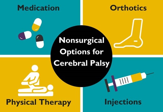
  • Physical therapy: Helps relieve pain and muscle stiffness, as well as improve balance, coordination, and overall mobility. Physical therapists will use specialized equipment to help your child move more freely and live more independently.
  • Occupational therapy: Helps children with cerebral palsy learn how to complete everyday tasks and activities by improving fine motor skills and cognitive abilities.
  • Speech therapy: Helps children to improve their communication and language skills. This type of therapy gives children the confidence to learn and socialize. Speech therapy can also help children who have difficulty eating and swallowing.
  • Communication aids such as computers with attached voice synthesizers.
  • Assistive devices or aids such as  Eye glasses, Hearing aids, Walking aids, Body braces, Wheelchairs, e.t.c.
  • Medication
  • Anticholinergics: Block neurotransmitters, addressing specific symptoms.
  • Anticonvulsants: Suppress neurons to control seizures.
  • Antidepressants: Manage mood-related symptoms.
  • Anti-inflammatories: Reduce pain and inflammation.
  • Baclofen: Muscle relaxer to alleviate stiffness.
  • Benzodiazepines: Treat anxiety, seizures, and insomnia.
  • Botox: Target spasticity.
  • Stool Softeners: Address constipation issues.
  • Surgery: Orthopedic surgery may be used to relieve pain and improve mobility. It may also be needed to release tight muscles or correct bone irregularities caused by spasticity.
  • Selective dorsal rhizotomy (SDR) might be recommended as a last resort to reduce chronic pain or spasticity. It involves cutting nerves near the base of the spinal column. Rhizotomy is a minimally invasive surgical procedure to remove sensation from a painful nerve by killing nerve fibers responsible for sending pain signals to the brain. The nerve fibers can be destroyed by severing them with a surgical instrument or burning them with a chemical or electrical current.    
  • Other surgical measures may include lengthening muscles and cutting overly active nerves.
  • Some affected children can achieve near normal adult lives with appropriate treatment. 

Prognosis

  • Cerebral Palsy is not a progressive disorder (meaning the brain damage does not worsen), but the symptoms can become more severe over time.
  • A person with the disorder may improve somewhat during childhood if he or she receives extensive care
  •  Some individuals with cerebral palsy require personal assistant services for all activities of daily living.
  • Puberty in young adults with cerebral palsy may be delayed due to nutritional deficiencies.
  • Cerebral palsy can significantly reduce a person’s life expectancy, depending on the severity of the condition and the quality of care with which they are provided.
Prevention of Cerebral Palsy.

Prevention of Cerebral Palsy.

Pre-pregnancy and pregnancy:

  • Prenatal care: Early and regular prenatal checkups for monitoring both mother and baby’s health and identifying potential risks like infections, gestational diabetes, or high blood pressure, which can all contribute to Cerebral Palsy.
  • Vaccinations: Ensuring that all recommended vaccinations are received , especially those that protect against infections harmful to the developing fetus, such as rubella, cytomegalovirus, and influenza.
  • Healthy lifestyle: Maintaining a healthy weight, eating a balanced diet rich in vitamins and minerals, exercising regularly, and avoiding smoking, alcohol, and drugs are all essential for a healthy pregnancy and reducing the risk of Cerebral Palsy.
  • Manage chronic conditions: In case of  pre-existing health conditions like diabetes or high blood pressure, actively manage them well to minimize potential complications during pregnancy.

During and after childbirth:

  • Safe delivery practices: Carrying out delivery from a designated hospital or maternity center and managing any associated  birth issued can help ensure a safe and low-risk delivery, minimizing the risk of birth complications that can contribute to Cerebral Palsy.
  • Postpartum care: Regular checkups for both mother and baby after birth for monitoring development and promptly addressing any potential issues.
  • Vaccinations for baby: Ensure the child receives all recommended vaccinations on time, as they protect against potentially Cerebral Palsy-causing infections like meningitis and encephalitis.
  • Preventing head injuries: Implementing safety measures at home and during car rides, using age-appropriate helmets for activities like bike riding, and closely supervising children around water can significantly reduce the risk of head injuries, a potential cause of Cerebral Palsy.

CEREBRAL PALSY Read More »

SEIZURE DISORDERS

SEIZURE DISORDERS

SEIZURE DISORDERS.

Seizure is defined as when there is a non-recurrent abnormal electrical activity in the brain or central nervous system resulting in abnormal motor, sensory or psychomotor experiences.

A seizure is an abnormal, unregulated electrical discharge that occurs within the brain’s cortical gray matter and gradually interrupts normal brain function.

Therefore when a person has recurrent, intermittent tendency to develop a seizure, we say he is having epilepsy.

  • A seizure causes altered awareness, abnormal sensations, focal involuntary movements, or convulsions (widespread violent involuntary contraction of voluntary muscles).
  • About 2% of adults have a seizure at some time during their life. Two thirds of these people never have another one.
  • Seizure disorders are either epileptic or non-epileptic.

Epileptic seizures:

Epilepsy is a neurological disorder in which the brain activity becomes abnormal, causing seizures or periods of unusual behaviour, sensations, and sometimes loss of awareness.

Epilepsy (also called epileptic seizure disorder) is a chronic brain disorder characterized by recurrent seizures that are unprovoked (ie, not related to reversible stressors) and that occur > 24 h apart.

 A single seizure is not considered an epileptic seizure. Epilepsy is often idiopathic, but various brain disorders, such as malformations, strokes, and tumors, can cause symptomatic epilepsy.

Symptomatic epilepsy is epilepsy due to a known cause (eg, brain tumor, stroke). The seizures it causes are called symptomatic epileptic seizures. Such seizures are most common among neonates (see Neonatal Seizure Disorders) and the elderly.


Cryptogenic epilepsy is epilepsy assumed to be due to a specific cause, but whose specific cause is currently unknown.

Non epileptic seizures.

These are provoked by a temporary disorder or stressor (eg, metabolic disorders, CNS infections, cardiovascular disorders, drug toxicity or withdrawal, psychogenic disorders). In children, fever can provoke a seizure (febrile seizures).


Psychogenic nonepileptic seizures (pseudoseizures) are symptoms that simulate seizures in patients with psychiatric disorders but that do not involve an abnormal electrical discharge in the brain.

causes of epilepsy

Etiology of Seizure Disorders:

I. Age-Specific Causes:

A. Before Age 2:

  • Fever: Seizures in young children often result from fevers, a common occurrence in this age group.
  • Birth or Developmental Defects: Structural abnormalities present at birth or developmental issues contribute to seizures.
  • Birth Injuries: Trauma during the birthing process can lead to seizure disorders in infants.
  • Metabolic Disorders: Disorders affecting metabolism may manifest as seizures in early childhood.

B. Ages 2 to 14:

  • Idiopathic Seizure Disorders: Seizures with no identifiable cause, often occurring during childhood, fall under idiopathic seizure disorders.

C. Adults:

  • Cerebral Trauma: Traumatic brain injuries, often resulting from accidents, can trigger seizures in adults.
  • Alcohol Withdrawal: Abrupt cessation of alcohol intake can lead to seizures as the body adjusts.
  • Tumors: The presence of tumors in the brain may cause seizures, especially in adults.
  • Strokes: Disruption of blood flow to the brain, resulting in a stroke, is a significant cause of seizures in adults.
  • Unknown Cause (50%): In many cases, the cause of seizures in adults remains unidentified, highlighting the complexity of diagnosis.

D. The Elderly:

  • Tumors: Tumors in the brain become a more prominent cause of seizures in the elderly.
  • Strokes: Similar to adults, strokes are a common contributor to seizures in the elderly.

E. Reflex Epilepsy

  • Seizures are predictably triggered by external stimuli, such as lights, sounds, or touch, in rare cases known as reflex epilepsy.

F. Cryptogenic and Refractory Epilepsy:

  • Anti-NMDA receptor encephalitis, particularly in young women, is identified as a rare cause, leading to psychiatric symptoms and movement disorders. Ovarian teratoma is associated with this condition.

II. General Causes of Seizures Irrespective of Age:

  • Autoimmune Disorders: Cerebral vasculitis, anti-NMDA receptor encephalitis, and multiple sclerosis may lead to seizures, although rarely.
  • Cerebral Edema: Swelling of the brain tissue, known as cerebral edema, can trigger seizures.
  • Eclampsia, Hypertensive Encephalopathy: Conditions related to high blood pressure during pregnancy can result in seizures.
  • Cerebral Ischemia or Hypoxia: Insufficient blood flow or oxygen to the brain may cause seizures.
  • Cardiac Issues, Carbon Monoxide Toxicity, and Stroke: Conditions affecting the heart, carbon monoxide exposure, drowning, suffocation, and strokes are potential triggers.
  • Head Trauma: Both birth-related injuries and traumatic injuries during life can contribute to seizure disorders.
  • CNS Infections: AIDS, brain abscess, malaria, meningitis, neurocysticercosis, neurosyphilis, rabies, tetanus, toxoplasmosis, and viral encephalitis can lead to seizures.
  • Congenital or Developmental Abnormalities: Structural abnormalities present from birth or those developing during early life contribute to seizures.
  • Drugs and Toxins: Various substances, including drugs and toxins like camphor, ciprofloxacin, cocaine, and others, may induce seizures.
  • Expanding Intracranial Lesions: Hemorrhage, hydrocephalus, and tumors contribute to seizures by causing pressure on the brain.
  • Hyperpyrexia: Extremely high fever, associated with drug toxicity or heatstroke, can lead to seizures.
  • Metabolic Disturbances: Hypocalcemia (linked to hypoparathyroidism), hypoglycemia, and hyponatremia.
  1. Less Common Causes: Aminoacidurias, hepatic or uremic encephalopathy, hyperglycemia, hypomagnesemia, hypernatremia.
  2. Neonatal Cause: Vitamin B6 (pyridoxine) deficiency in neonates.
  • Withdrawal Syndromes: Alcohol, anesthetics, barbiturates, benzodiazepines—withdrawal from these substances can induce seizures.
seizure anticonvulsants

Classification of Seizures

Seizures are classified as generalized or partial.

1. Partial seizures/focal seizures.

In partial seizures, the excess neuronal discharge occurs in one cerebral cortex, and most often results from structural abnormalities.

 Partial seizures may be;

  • Simple : Focal seizures without impairment of consciousness or awareness.
  • Complex : Focal seizures with impairment of consciousness or awareness.

Partial seizures may evolve into a generalized seizure (called secondary generalization), which causes loss of consciousness. Secondary generalization occurs when a partial seizure spreads and activates the entire cerebrum bilaterally. Activation may occur so rapidly that the initial partial seizure is not clinically apparent or is very brief.

Symptoms and Signs of partial seizures.

The manifestation depends on the part of the brain that is affected;

A. Simple Partial Seizures:

  • Aura: Simple partial seizures may begin with auras, such as motor activity, sensory sensations, autonomic changes, or psychic experiences. Auras are simple partial seizures that begin focally. Auras may consist of motor activity or sensory, autonomic, or psychic sensations (eg, paresthesias, a rising epigastric sensation, abnormal smells, a sensation of fear, a déjà vu sensation).
  • Most seizures end spontaneously in 1 to 2 min.
  • Postictal State: Following generalized seizures, a postictal state occurs, characterized by deep sleep, headache, confusion, and muscle soreness; this state lasts from minutes to hours. 
  • Most patients appear neurologically normal between seizures, although high doses of the drugs used to treat seizure disorders, particularly anticonvulsants, can reduce alertness.

B. Jacksonian Seizures:

  • In Jacksonian seizures, focal motor symptoms begin in one hand and then march up the arm (Jacksonian march). Other focal seizures affect the face first, and then spread to an arm and sometimes a leg. Some partial motor seizures begin with an arm raising and the head turning toward the raised arm (called fencing posture).

C. Complex Partial Seizures:

Complex partial seizures are often preceded by an aura. During the seizure, patients may stare. Consciousness is impaired, but patients have some awareness of the environment (eg, they purposefully withdraw from noxious stimuli). The following may also occur:

  • Oral automatisms (involuntary chewing or lip smacking)
  • Limb automatisms (eg, automatic purposeless movements of the hands)
  • Utterance of unintelligible sounds without understanding what they say
  • Resistance to assistance
  • Tonic or dystonic posturing of the extremity contralateral to the seizure focus
  • Head and eye deviation, usually in a direction contralateral to the seizure focus
  • Bicycling or pedaling movements of the legs if the seizure emanates from the medial frontal or orbitofrontal head regions
  • Motor symptoms subside after 1 to 2 min, but confusion and disorientation may continue for another 1 or 2 min. 
  • Postictal amnesia is common.
  • Patients may lash out if restrained during the seizure or while recovering consciousness if the seizure generalizes. However, unprovoked aggressive behavior is unusual.
  • Left temporal lobe seizures may cause verbal memory abnormalities; right temporal lobe seizures may cause visual spatial memory abnormalities.

Generalized seizures

In generalized seizures, abnormal electrical discharge diffusely involves the entire cortex of both hemispheres from the onset, and consciousness is usually lost. Generalized seizures result mostly from metabolic disorders and sometimes from genetic disorders. 

Generalized seizures include the following:

1. Infantile spasms: Characterized by sudden flexion and adduction of the arms and forward flexion of the trunk. Seizures last a few seconds and recur many times a day. They occur only in the first 5 years of life, then are replaced by other types of seizures. Developmental defects are usually present.

2. Typical absence seizures (formerly called petit mal seizures): Consist of a 10- to 30-second loss of consciousness with eyelid fluttering; axial muscle tone may or may not be lost. Patients do not fall or convulse; they abruptly stop activity, then just as abruptly resume it, with no postictal symptoms or knowledge that a seizure has occurred. Absence seizures are genetic and occur predominantly in children. Without treatment, such seizures are likely to occur many times a day. Seizures often occur when patients are sitting quietly, can be precipitated by hyperventilation, and rarely occur during exercise. Neurologic and cognitive examination results are usually normal.

3. Atypical absence seizures: Usually occur as part of the Lennox-Gastaut syndrome, a severe form of epilepsy that begins before age 4 years. They differ from typical absence seizures in the following ways:

  • They last longer.
  • Jerking or automatic movements are more pronounced.
  • Loss of awareness is less complete.
  • Many patients have a history of damage to the nervous system, developmental delay, abnormal neurologic examination results, and other types of seizures. Atypical absence seizures usually continue into adulthood.

4. Atonic seizures: Occur most often in children, usually as part of Lennox-Gastaut syndrome. Atonic seizures are characterized by a brief, complete loss of muscle tone and consciousness. Children fall or pitch to the ground, risking trauma, particularly head injury.

5. Tonic seizures: Occur most often during sleep, usually in children. The cause is usually the Lennox-Gastaut syndrome. Tonic (sustained) contraction of axial muscles may begin abruptly or gradually, then spread to the proximal muscles of the limbs. Tonic seizures usually last 10 to 15 seconds. In longer tonic seizures, a few rapid clonic jerks may occur as the tonic phase ends.

6. Tonic-clonic seizures: Can be primarily generalized or secondarily generalized.

  • Primarily generalized seizures typically begin with an outcry, followed by a loss of consciousness, falling, tonic contraction, and then clonic (rapidly alternating contraction and relaxation) motion of muscles of the extremities, trunk, and head. Urinary and fecal incontinence, tongue biting, and frothing at the mouth sometimes occur. Seizures usually last 1 to 2 minutes, and there is no aura.
  • Secondarily generalized tonic-clonic seizures begin with a simple partial or complex partial seizure and then progress to resemble other generalized seizures.

7. Myoclonic seizures: Brief, lightning-like jerks of a limb, several limbs, or the trunk. They may be repetitive, leading to a tonic-clonic seizure. The jerks may be bilateral or unilateral. Unlike other seizures with bilateral motor movements, consciousness is not lost unless the myoclonic seizure progresses into a generalized tonic-clonic seizure.

8. Juvenile myoclonic epilepsy: An epilepsy syndrome characterized by myoclonic, tonic-clonic, and absence seizures. It typically appears during adolescence. Seizures begin with a few bilateral, synchronous myoclonic jerks, followed in 90% of cases by generalized tonic-clonic seizures. They often occur when patients awaken in the morning, especially after sleep deprivation or alcohol use. Absence seizures may occur in one-third of patients.

GENERAL MANAGEMENT OF SEIZURE DISORDERS.

During the attack; (first aid)

  1. Ensure Safety:
    1. Observe warning signs.

    2. Lay the individual on a flat surface.

    3. Ensure the surrounding environment is safe.

  2. Remove Hazards: Clear the area of dangerous objects like sticks or stones.
  3. Do Not Restrain: Avoid restraining the person during the seizure.
  4. Protect Airways: Do not insert anything into the mouth.
  5. Note Duration: Record the length of the seizure for medical evaluation.
  6. Post-Seizure:
    • Allow the person to rest.

    • Provide refreshments if needed.

Drug Management:

  1. Anticonvulsant Medications:

  • Use medications such as diazepam, phenytoin, sodium valproate, among others.
  • Follow prescribed dosage and administration schedules.

Severe Epileptic Attack (Pediatric/Medical/Psychiatric Emergency):

  1. Anticonvulsant Administration: Administer appropriate anticonvulsants promptly.
  2. Cardio-Respiratory Support: Provide immediate support for cardiac and respiratory functions.
  3. Prevent Falling: Ensure a safe environment to prevent injuries during seizures.
  4. Intravenous Fluids: Administer intravenous fluids to maintain hydration.
  5. Hypoglycemia Prevention: Monitor and maintain blood glucose levels to prevent hypoglycemia.

Long-Term Management:

  1. Individualized Treatment Plans: Develop a personalized treatment plan in collaboration with healthcare professionals.
  2. Regular Medication Adherence: Ensure consistent adherence to prescribed anticonvulsant medications.
  3. Lifestyle Modifications: Encourage a healthy lifestyle with regular sleep patterns, stress management, and a balanced diet.
  4. Trigger Identification: Identify and manage potential triggers, such as stress or lack of sleep.
  5. Seizure Action Plan: Establish a comprehensive seizure action plan in coordination with healthcare providers.
  6. Regular Medical Follow-Up: Schedule routine medical follow-ups to monitor progress and adjust treatment as needed.
  7. Educational Support: Provide educational resources and support for individuals and their families to understand and cope with epilepsy.
  8. Psychosocial Interventions: Integrate psychosocial interventions to address emotional and psychological aspects of living with epilepsy.
  9. Emergency Medication Access: Ensure accessibility to emergency medications in case of prolonged seizures.
  10. Multidisciplinary Approach: Involve a multidisciplinary team, including neurologists, psychologists, and social workers, for holistic care.

Nursing Interventions for Seizure Disorder

Prevent Trauma/Injury:

  • Teach caregivers to recognize warning signs and manage patients during and after seizures.
  • Advise against using breakable thermometers; opt for tympanic thermometers when necessary.
  • Maintain strict bedrest during prodromal signs or auras.
  • Turn the head to the side, suction the airway as needed, and provide support during seizures.
  • Avoid restraint attempts; monitor and document antiepileptic drug (AED) levels, side effects, and seizure frequency.

Promote Airway Clearance:

  • Keep the patient in a lying position on a flat surface.
  • Turn the head to the side during seizure activity.
  • Loosen clothing around the neck, chest, and abdomen.
  • Perform suctioning as needed.
  • Supervise supplemental oxygen or bag ventilation postictally.

Improve Self-Esteem:

  • Assess individual situations contributing to low self-esteem.
  • Avoid over-protectiveness; encourage independence.
  • Support and monitor activities; consider the attitudes and capabilities of significant others.
  • Help individuals understand that their feelings are normal, discouraging guilt and blame.

Enforce Education About the Disease:

  • Review the pathology and prognosis of the condition.
  • Emphasize the lifelong need for treatments.
  • Identify specific trigger factors (flashing lights, hyperventilation, loud noises, video games, TV).
  • Stress the importance of good oral hygiene and regular dental care.
  • Educate on the medication regimen, emphasizing adherence and the significance of not discontinuing therapy without physician supervision.
  • Provide clear instructions for missed doses.

Seizure Documentation:

  • Maintain detailed records of seizure occurrences, duration, and characteristics.
  • Document any changes in the patient’s behavior or aura.

Family Education and Support:

  • Educate family members on seizure first aid and safety measures.
  • Offer emotional support and counseling to both the patient and family members.

Regular Neurological Assessments:

  • Perform routine neurological assessments to monitor changes in seizure patterns or neurological status.

Medication Administration:

  • Administer antiepileptic medications as prescribed, ensuring proper dosage and adherence.

Lifestyle Modifications:

  • Collaborate with the patient to identify and manage lifestyle factors that may trigger seizures.
  • Encourage the establishment of consistent sleep patterns and stress reduction techniques.

Emergency Preparedness:

  • Ensure caregivers are equipped to handle emergencies, providing guidance on when to seek medical attention.

Social Integration:

  • Assist in facilitating social integration for the patient, addressing any potential stigma or discrimination.

FEBRILE CONVULSIONS 

A febrile seizure, also known as a fever fit, is a seizure associated with a high body temperature without any serious underlying health issue.

Primarily occurs in children aged 6 months to 5 years. Usually, seizures last less than five minutes, and the child returns to normal within sixty minutes.

Causes:

  • Familial predisposition to febrile seizures.
  • Linked to fevers exceeding 38 °C (100.4 °F), often triggered by viral illnesses. The risk increases with the height of the temperature.
  • Vaccines, although with a small associated risk, may contribute, including measles/mumps/rubella/varicella, diphtheria/tetanus/acellular pertussis/polio/Haemophilus influenzae type b, and others.

Types:

Simple Febrile Seizures:

  • Short duration (<15 minutes), no focal features.
  • Usually, a single tonic-clonic seizure in a 24-hour period.

Complex Febrile Seizures:

  • Last longer than 15 minutes or occur multiple times within 24 hours.
  • May have focal features.

Febrile Status Epilepticus:

  • Lasts for more than 30 minutes.
  • Occurs in up to 5% of febrile seizure cases.

Diagnosis:

  • Generally clinical, eliminating serious causes such as meningitis and encephalitis.
  • Blood tests, brain imaging, and EEG are typically not required.
  • Verify absence of brain infection, metabolic issues, and prior seizures unrelated to fever.

Management:

First Aid During Seizure:

  • Ensure a safe environment.
  • Remove dangerous objects.
  • Do not restrain the child.
  • Note seizure duration.

Medical Intervention:

  • No routine use of anti-seizure or anti-fever medications.
  • Benzodiazepines (e.g., lorazepam) for seizures lasting over five minutes.

Treatment:

  • Maintain a calm environment.
  • Note seizure start time; call an ambulance if >5 minutes.
  • Place the child on a protected surface.
  • Do not restrain; position on the side to prevent choking.
  • Seek immediate medical attention, especially if the first seizure or concerning symptoms persist.
  • Intravenous lorazepam for prolonged seizures.

Prevention:

  • Proper fever management in children.
  • Avoid exposing babies to excessive heat.

SEIZURE DISORDERS Read More »

INTERSEXUAL DISABILITIES

INTERSEXUAL DISABILITIES

INTERSEXUAL DISABILITIES.

Intersex is an umbrella term used to describe anyone with reproductive or sexual anatomy that doesn’t align with the traditional definitions of “male” or “female.”

Intersex people were previously referred to as hermaphrodites, “congenital eunuchs”, or congenitally “frigid“. Such  terms have  fallen out of  favor; in  particular ,the  term “hermaphrodite” is considered  to  be  misleading and stigmatizing.

The term intersexuality was coined by Richard Goldschmidt in 1917. The first suggestion to replace the term ‘hermaphrodite’ with ‘intersex’ was made by Cawadias in the 1940s.

Normal Sexual Development

Normal Sexual Development

During normal sexual development, humans have two chromosome pairs: XX for females and XY for males

  1. Fertilization: When the sperm fertilizes the egg, it contributes either an X (female) or a Y (male) chromosome, determining the genetic sex of the embryo.
  2. Early Development: In the initial weeks, male and female fetuses are “anatomically indistinguishable.” Primitive gonads start developing around the sixth week of gestation in a bipotential state.
  3. Gonadal Differentiation: Gonads can become testes (male) or ovaries (female) based on subsequent events. By the seventh week, male and female fetuses look identical.
  4. Hormonal Influence: Around the eighth week, XY embryos’ gonads differentiate into functional testes, producing testosterone. In XX embryos, ovarian differentiation occurs around the twelfth week.
  5. Duct System Development: In females, the Mullein duct system becomes the uterus, Fallopian tubes, and inner vagina. In males, Müllerian duct-inhibiting hormone causes regression, while androgens lead to the Wolffian duct system’s development. intersex
  6. Birth: By birth, the fetus is completely “sexed” as male or female. This alignment includes genetic sex (XY or XX), internal and external gonads, and external genital appearance.
Causes of Intersex Conditions

Causes of Intersex Conditions:

Masculinizing:

1. XX Congenital Adrenal Hyperplasia (CAH):

  • Common cause involving abnormal adrenal gland production of virilizing hormones during fetal development. (“Virilizing hormones” refer to hormones that promote the development of male sexual characteristics or traits. The term “virilization” is associated with the development of male secondary sex characteristics, and these hormones are responsible for shaping the male reproductive system and external features.)
  • Detection of CAH genes in the embryo is possible, and early treatment with dexamethasone is controversial but aims to prevent genital masculinization.
  • Some XX-females with CAH may experience partial or complete masculinization, including a large clitoris or even a male appearance.

2. XX Progestin-Induced Virilization:

  • Caused by progestin drug use in the 1950s and 1960s to prevent miscarriage.
  • Individuals have female anatomy but may develop male secondary sex characteristics, including unusually large clitorises.

3. XX Freemartinism:

  • Common in cattle, where female twins born with a male may share blood supply, leading to masculinization of the female.
  • External female appearance, infertility, and behavior resembling a castrated male.

Feminizing:

4. XY Androgen Insensitivity Syndrome (AIS):

  • Individuals with XY chromosomes unable to metabolize androgens, leading to varying degrees of feminization.
  • Complete AIS results in a female appearance, including a vagina, but with undescended or partially descended testes.

5. XY 5-Alpha-Reductase Deficiency (5-ARD):

  • Affects individuals with a Y chromosome, hindering the conversion of testosterone to dihydrotestosterone (DHT).
  • Results in ambiguous genitalia at birth, with effects ranging from male genitalia to female genitalia with mild clitoromegaly.

6. XY Congenital Adrenal Hyperplasia (CAH):

  • In XY individuals with CAH due to 17 alpha-hydroxylase deficiency, virilization is inhibited compared to cases without a Y chromosome.

7. XY Persistent Müllerian Duct Syndrome (PMDS):

  • XY individuals with male chromosomes but an internal uterus and fallopian tubes due to the absence of Müllerian inhibiting factor during fetal development.

8. XY Anorchia:

  • Loss of gonads after 14 weeks of fetal development in XY individuals.
  • Results in the inability to produce hormones for male secondary sex characteristics, leading to feminization.

9. XY Gonadal Dysgenesis:

  • Heterogeneous condition in XY individuals where gonads fail to develop properly.

10. XY Hypospadias:

  • Caused by various factors, including alterations in testosterone metabolism.
  • Urethra opening does not reach the tip of the penis, with severity ranging from mild to severe.

Others:

  • Unusual chromosomal sex variations, including Turner syndrome (XO), Triple X syndrome (XXX), Klinefelter syndrome (XXY) and variants (XXYY, XXXY), XYY syndrome, de la Chapelle syndrome (XX male), Swyer syndrome (XY female).
signs of intersex

Categories/Types of Intersex:

Intersex can either be chromosomal or homonal. Categories/Types can also be causes of intersex.

  1. 46, XX Intersex:
  • Chromosomes and ovaries are female, but external genitals appear male.
  • Often caused by exposure to excess male hormones in utero.
  • Conditions include congenital adrenal hyperplasia (most common), male hormone exposure during pregnancy, tumors in the mother, or aromatase deficiency.
  • Labia fuse, clitoris enlarges, but normal uterus and fallopian tubes are present.
  • Formerly known as female pseudohermaphroditism.

46” refers to the total number of chromosomes in a normal human cell. Humans have 23 pairs of chromosomes, and when you count them all, it adds up to 46 chromosomes. Each parent contributes one chromosome to each pair, resulting in a total of 23 chromosomes from the mother and 23 chromosomes from the father, making a complete set of 46 chromosomes in most cells of the human body.

2. 46, XY Intersex:

  • Chromosomes are male, but external genitals are incompletely formed, ambiguous, or female.
  • Internal testes may be normal, malformed, or absent.
  • Causes include testes problems (e.g., XY pure gonadal dysgenesis), testosterone formation issues, or difficulties in using testosterone (e.g., androgen insensitivity syndrome, 5-alpha-reductase deficiency).
  • 5-alpha-reductase deficiency babies may have varying genitalia that often align with male characteristics during puberty.
  • AIS is the most common cause, where receptors to male hormones don’t function properly.

3. True Gonadal Intersex:

  • Presence of both ovarian and testicular tissue, either in the same gonad (ovotestis) or with 1 ovary and 1 testis.
  • May have XX chromosomes, XY chromosomes, or both.
  • External genitals can be ambiguous, female, or male.
  • Formerly known as true hermaphroditism.
  • Underlying causes are often unknown, but in some animal studies linked to exposure to agricultural pesticides.

4. Complex or Undetermined Intersex Disorders of Sexual Development:

  • Various chromosome configurations beyond 46, XX or 46, XY.
  • Examples include 45, XO (only one X chromosome) and 47, XXY, 47, XXX (extra sex chromosomes, X or Y).
  • Disorders may not lead to a discrepancy between internal and external genitalia but can affect sex hormone levels, overall sexual development, and the number of sex chromosomes.
Intersex types signs and symptoms

SIGNS & SYMPTOMS OF INTERSEX CONDITIONS

The signs and symptoms of intersex conditions can vary based on their underlying causes. 

  1. Ambiguous Genitalia at Birth: The external genitalia may not have the usual male or female appearance, making it challenging to assign a clear gender.
  2. Micropenis: A smaller than usual penis in males, which can be a result of hormonal imbalances or genetic factors.
  3. Clitoromegaly (Enlarged Clitoris): In females, the clitoris is larger than expected, possibly due to hormonal influences during fetal development.
  4. Partial Labial Fusion: In females, there may be a partial fusion of the labia, the folds of skin surrounding the vaginal opening.
  5. Apparently Undescended Testes (May Turn Out to Be Ovaries) in Boys: Testes that appear not to have descended may actually be ovaries, indicating a discrepancy between external appearance and internal structures.
  6. Labial or Inguinal Masses (May Turn Out to Be Testes) in Girls: Masses in the labial or inguinal (groin) area in females may turn out to be testes, highlighting the complexity of internal organ development.
  7. Hypospadias: The opening of the penis is not at the tip, and in females, the urethra opens into the vagina instead of its usual location.
  8. Otherwise Unusual-Appearing Genitalia at Birth: Genitalia may have features that deviate from the typical male or female appearance, leading to uncertainty in gender assignment.
  9. Electrolyte Abnormalities: Imbalances in electrolytes, essential for bodily functions, may be present and require monitoring and management.
  10. Delayed or Absent Puberty: Puberty may be delayed or absent, affecting the development of secondary sexual characteristics such as breast development or facial hair growth.
  11. Unexpected Changes at Puberty: Some individuals may experience unexpected changes during puberty, further complicating the understanding of their sexual development.
  12. Underdeveloped Secondary Sexual Characteristics: Individuals may exhibit underdeveloped or atypical secondary sexual characteristics, such as minimal breast development in females or a lack of facial hair growth in males.
  13. Infertility: Some intersex conditions can lead to infertility due to irregularities in reproductive organ development or hormonal imbalances affecting gamete production.
  14. Atypical Pubic Hair Growth: Unusual patterns or absence of pubic hair growth may be observed, contributing to the complexity of sexual development.
  15. Urinary or Reproductive System Complications:  Intersex conditions may be associated with complications in the urinary or reproductive systems, leading to issues like urinary tract abnormalities or difficulties in conceiving.
  16. Gender Dysphoria: Individuals may experience distress or discomfort with their assigned gender at birth, potentially leading to gender identity concerns and the need for psychological support.
  17. Hormonal Irregularities: Fluctuations in hormone levels can result in various symptoms, including mood swings, fatigue, or irregular menstrual cycles, depending on the specific intersex condition.
  18. Chromosomal Abnormalities: Some intersex variations involve chromosomal abnormalities beyond the typical XX or XY configurations, contributing to the diversity of intersex presentations.
  19. Psychosocial Challenges: Intersex individuals may face unique psychosocial challenges related to societal perceptions, self-identity, and acceptance, emphasizing the importance of supportive environments.
  20. Skeletal and Muscular Variances: Skeletal and muscular development may exhibit differences, affecting overall body structure and physical abilities.
  21. Emotional and Mental Health Factors: The emotional and mental well-being of intersex individuals may be influenced by societal attitudes, disclosure of their intersex status, and coping with unique challenges.
Diagnosis and Investigations

Diagnosis and Investigations

  1. Chromosome Analysis: This test examines the chromosomal composition, determining if there are variations from the normal XX (female) or XY (male) chromosome. It helps identify chromosomal intersex variations.
  2. Hormone Level Testing (e.g Testosterone): Measurement of hormone levels, such as testosterone, provides an overview into the endocrine system’s functioning. Deviations may indicate hormonal imbalances affecting sexual development.
  3. Hormone Stimulation Tests: These tests assess the body’s response to hormonal stimuli, helping evaluate the capacity of endocrine organs to produce and regulate hormones crucial for sexual development.
  4. Electrolyte Tests: Electrolyte tests assess the balance of minerals in the body, helping in the identification of potential abnormalities that may be associated with specific intersex conditions.
  5. Specific Molecular Testing: Molecular testing involves examining genetic material at the molecular level. This can reveal specific genetic variations or mutations associated with intersex conditions.
  6. Endoscopic Exam (Vaginal or Cervical Presence): An endoscopic examination is performed to visually confirm the absence or presence of a vagina or cervix.
  7. Ultrasound or MRI (Evaluation of Internal Organs): Imaging techniques, such as ultrasound or MRI, are used to visualize internal organs like the uterus. This helps determine the presence or absence of internal sex organs.

Challenges Faced by Intersex People:

I. Stigmatization: Intersex individuals face stigma due to misconceptions and lack of understanding about differences in sex development. Stigmatization can have an impact on mental health and well-being.

II. Discrimination from Birth or Discovery: Intersex individuals may experience discrimination immediately upon birth or when their intersex trait is discovered. Discrimination can be seen in many ways, affecting their sense of belonging and acceptance.

III. Infanticide: Unfortunately, some intersex infants face the risk of infanticide, due to cultural beliefs or misinformation surrounding intersex. This creates a severe threat to the lives of newborns with intersex traits.

IV. Abandonment: The discovery of intersex traits in a child may lead to parental distress, and in extreme cases, it can result in abandonment. This abandonment can have profound emotional and psychological effects on the individual.

V. Stigmatization of Families: Families with intersex members may also face stigma from society. The lack of awareness and understanding in communities can lead to judgment and isolation of the intersex.

VI. Unequal Treatment: Intersex individuals may face unequal treatment in many settings, including healthcare, education, and employment. Discrimination based on their intersex status can lead to unequal distribution  opportunities and services.

VII. Mental Health Struggles: Coping with pressures from the community, discrimination, and potential medical interventions can contribute to mental health challenges for intersex individuals. Access to mental health support is very important for their well-being.

VIII. Lack of Inclusive Education: Educational systems may lack inclusivity in addressing intersex variations, leading to misunderstandings among peers and educators. 

X. Inadequate Medical Care: Some intersex individuals may face challenges in accessing competent and sensitive healthcare. Inadequate medical care can result in physical and emotional distress, emphasizing the need for informed healthcare providers.

XI. Limited Awareness and Advocacy: Widespread ignorance about intersex variations contributes to the challenges faced. Increased awareness and advocacy are necessary to promote understanding, tolerance, and equal rights for intersex individuals.


XII. Isolation and Loneliness: The combination of stigma from society and limited understanding can lead to feelings of isolation and loneliness among intersex individuals. 

Medical Management for Intersex Individuals:

1. Diagnosis and Evaluation:

  • Chromosome Analysis: Conduct chromosomal analysis to determine the genetic composition (XX, XY, or other variations).
  • Hormone Levels: Measure hormone levels, including testosterone, estrogen, and other relevant hormones.
  • Imaging Studies: Utilize ultrasound or MRI to assess internal sex organs and identify any anomalies.

2. Hormone Therapy:

  • Feminizing or Masculinizing Hormones: Administer prescribed hormones based on the individual’s gender identity, aiming to align secondary sex characteristics with their affirmed gender.

3. Psychological Support:

  • Mental Health Counseling: Provide psychological support through counseling to address the emotional impact, to promote a positive self-image and coping mechanisms.
  • Support Groups: Connect individuals with support groups to create peer support and shared experiences.

4. Prenatal Counseling:

  • Genetic Counseling: Offer genetic counseling for parents to understand the intersex condition, potential issues, and available options.

Surgical Management for Intersex Individuals

Surgical Management for Intersex Individuals:

5. Genital Reconstruction Surgery:

  • Feminizing Procedures: Include vaginoplasty, clitoroplasty, and labiaplasty for individuals assigned female.
  • Masculinizing Procedures: Involve procedures like phalloplasty and scrotoplasty for individuals assigned male.

6. Gonadectomy:

  • Removal of Gonads: Address concerns related to gonadal cancer risk or hormone imbalances by removing gonads. Determine the appropriate timing for gonadectomy based on individual health considerations.

7. Corrective Surgeries:

  • Hypospadias Repair: For individuals with hypospadias, surgical correction to reposition the urethral opening may be performed.
  • Vaginal or Penile Reconstruction: Tailored surgeries to address specific anatomical variations.

8. Breast Augmentation or Chest Reconstruction:

  • Gender-Affirming Surgeries: Support individuals in achieving a physical appearance aligned with their gender identity.

Nursing Care for Intersex Individuals:

9. Communication and Education:

  • Open Communication: Foster open dialogue, addressing concerns and providing information about medical procedures.
  • Patient Education: Educate individuals and their families about the intersex condition, treatment options, and postoperative care.

10. Preoperative Care:

  • Emotional Support: Offer emotional support before surgery, addressing anxieties and concerns.
  • Physical Preparation: Ensure individuals understand preoperative instructions and are physically prepared for surgery.

11. Postoperative Care:

  • Pain Management: Monitor and manage postoperative pain, ensuring individuals are comfortable.
  • Wound Care: Provide wound care for surgical incisions to prevent infections and promote healing.
  • Emotional Support: Offer psychological support during the recovery period, addressing body image concerns and promoting a positive self-image.

12. Long-Term Follow-Up:

  • Hormone Monitoring: Regularly monitor hormone levels and adjust hormone therapy as needed.
  • Psychosocial Support: Continue providing ongoing psychosocial support, addressing any evolving emotional needs.

13. Advocacy and Dignity:

  • Advocacy: Advocate for the rights and dignity of intersex individuals, promoting inclusive and respectful care.
  • Cultural Sensitivity: Ensure cultural competence and sensitivity in nursing care, respecting diverse identities and backgrounds.

INTERSEXUAL DISABILITIES Read More »

MENINGITIS

MENINGITIS

MENINGITIS

The word meningitis is from Greek μῆνιγξ meninx, “membrane” and the medical suffix –itis, “inflammation“.

Meningitis is an acute inflammation of the protective membranes covering the brain and spinal cord, known collectively as the meninges

Meningitis can be life-threatening because of the inflammation’s proximity to the brain and spinal cord; therefore, the condition is classified as a medical emergency.

WHAT ARE MENINGES? 

The meninges comprise three membranes that, together with the cerebrospinal fluid, enclose and protect the brain and spinal cord (the central nervous system).  There are 3 meninges, namely; the pia mater, the arachnoid mater and the dura mater – this naming is from inwards outwards.

  • The pia mater is a very delicate impermeable membrane that firmly adheres to the surface of the brain, following all the minor contours.
  • The arachnoid mater (so named because of its spider-web-like appearance) is a loosely fitting sac on top of the pia mater. The subarachnoid space separates the arachnoid and pia mater membranes and is filled with cerebrospinal fluid.
  • The outermost membrane, the dura mater, is a thick durable membrane, which is attached to both the arachnoid membrane and the skull. 

The meninges provide a blood brain barrier which prevents infections from blood to spread to the brain, however, some organisms cross this and cause some diseases. They also prevent direct injury to the brain.

Causes of Meningitis and their  Mode of transmission.

Causes of Meningitis and their  Mode of transmission.

1. Bacterial Causes:

  • Streptococcus pneumoniae: Common bacterial cause, transmission through respiratory droplets.
  • Group B Streptococci (subtypes III): Inhabit the vagina, main cause in the first week of life for newborns.
  • Escherichia coli (carrying K1 antigen): Normally found in the digestive tract, affecting newborns during birth.
  • Listeria monocytogenes (serotype IVb): Transmitted by the mother before birth, impacting newborns.
  • Neisseria meningitidis (meningococcus): More common in children around 6 years, transmission through respiratory droplets.
  • Haemophilus influenzae type B: Common in those under 5 years in countries without vaccination, transmission through respiratory droplets.
  • Mycobacterium tuberculosis: More common in people from tuberculosis-endemic countries, transmission through respiratory droplets.
  • Treponema pallidum (syphilis) and Borrelia burgdorferi (Lyme disease): Transmitted through sexual contact (syphilis) and tick bites (Lyme disease).

Note: Aseptic meningitis, where no bacterial infection is demonstrated, is usually caused by viruses.

2. Viral Causes:

  • Enteroviruses: Spread through fecal-oral route.
  • Herpes simplex virus (generally type 2): Transmitted through direct contact with infected lesions (genital sores).
  • Varicella-zoster virus: Causes chickenpox and shingles, transmitted through respiratory droplets.
  • Mumps virus: Spread through respiratory droplets and saliva.
  • HIV: Transmitted through blood, sexual contact, or from mother to child during childbirth or breastfeeding.
  • LCMV (Lymphocytic choriomeningitis virus): Spread through the urine, droppings, saliva, or nesting materials of infected rodents.

3. Fungal Causes:

  • Cryptococcus neoformans: Inhalation of fungal spores from the environment.
  • Coccidioides immitis, Histoplasma capsulatum, Blastomyces spp.: Inhalation of fungal spores from the environment.

4. Parasitic Causes:

  • Eosinophil-predominant CSF indicates parasitic causes.
  • Cerebral malaria: Transmitted through infected mosquitoes.
  • Amoebic meningitis (e.g., Naegleria fowleri): Contracted from freshwater sources.
  • Angiostrongylus cantonensis, Gnathostoma spinigerum, Schistosoma: Various modes of transmission (e.g., contaminated food, water, or snail intermediate hosts).
  • Cysticercosis, Toxocariasis, Baylisascariasis, Paragonimiasis: Different modes of transmission (e.g., ingestion of contaminated food or water).

5. Non infectious Conditions:

  • Neoplastic: Meningitis may result from cancer metastasis to the meninges.
  • Sarcoidosis: An inflammatory condition with an unknown cause.
  • Systemic lupus erythematosus: An autoimmune disorder.
  • Granulomatosis with polyangiitis (Wegener’s): An autoimmune condition affecting blood vessels.
  • Behçet’s disease: An autoimmune condition causing inflammation of blood vessels.
  • Certain drugs may cause meningeal irritation and resemble as meningitis including: Nonsteroidal antiinflammatory drugs (NSAIDs), Intravenous immunoglobulin, Intrathecal agents, Certain antibiotics (eg, trimethoprim-sulfamethoxazole).

Risk Factors for Meningitis:

  1. Immunosuppression: Weakens the immune system.  Use of immunosuppressants (post-organ transplantation), HIV/AIDS, age-related loss of immunity. Associated Pathogens: Staphylococci, Pseudomonas, and other Gram-negative bacteria.
  2. Recent Skull Trauma: Provides an entry point for nasal cavity bacteria into the meningeal space.
  3. Brain and Meninges Devices: Presence of devices like cerebral shunts, extraventricular drains, or Ommaya reservoirs.
  4. Head and Neck Infections: Infections in the head and neck area, such as otitis media or mastoiditis.
  5. Cochlear Implants: Devices for hearing loss.
  6. Persisting Anatomical Defects: Congenital or acquired defects allowing continuity between the external environment and the nervous system.
  7. Extremes of Age: Children, especially below 5 years, and individuals over 50 years old.
  8. Infections (e.g., Endocarditis, Pneumonia): Spread of bacteria clusters through the bloodstream.
  9. Asplenia (Absence of the Spleen):  Lack of a spleen.
Pathophysiology

Pathophysiology of Meningitis:

1. Entry of Organisms:

(a) Routes:

  • Direct Entry: Through open fractures.
  • Blood-Borne: Via the bloodstream.
  • Adjacent Part: From neighboring areas.

2. Bloodstream Invasion: Organisms enter the bloodstream and reach the meninges. Upon reaching the meninges, organisms are identified as foreign.

3. Immune Response Initiation: Recognition triggers a battle between the body’s defense cells and the invading organisms.

Cytokine Release: Astrocytes and microglia release cytokines, recruiting immune cells and stimulating tissues for the immune response.

4. Blood-Brain Barrier Permeability:

  • Vasogenic Edema: Increased permeability leads to cerebral edema, swelling the brain due to fluid leakage from blood vessels.
  • White Blood Cell Influx: Large numbers of white blood cells enter the cerebrospinal fluid (CSF), causing meningeal inflammation and interstitial edema.

5. Cerebral Vasculitis:

  • Inflammation of Blood Vessels: Walls of blood vessels become inflamed, resulting in decreased blood flow.
  • Cytotoxic Edema: Further edema, affecting cells directly.

6. Increased Intracranial Pressure (ICP):

  • Combined Edema Effects: Vasogenic, interstitial, and cytotoxic edema collectively elevate ICP.
  • Impaired Blood Flow: Decreased blood flow makes it challenging for blood to enter the brain.
  • Oxygen Deprivation: Brain cells undergo apoptosis (programmed cell death) due to reduced oxygen supply.

7 .Brain Swelling and Symptoms:

  • CSF Flow Blockade: Brain swelling obstructs cerebrospinal fluid (CSF) flow.
  • Clinical Signs: Severe headache, seizures, and other symptoms manifest.

8. Untreated Progression:

  1. Spread to the Brain: Unchecked inflammation extends to various parts of the brain.
  2. Complications: Encephalitis, increased ICP, brainstem dysfunction, multi-organ dysfunction.
  3. Outcome: Without treatment, progression can lead to death.

CLINICAL FEATURES

  1. Fever: Elevated body temperature. Common and indicative of systemic infection, including meningitis.
  2. Headache: Severe head pain. Present in nearly 90% of bacterial meningitis cases.
  3. Neck Stiffness (Nuchal Rigidity): Increased neck muscle tone and stiffness. Classic symptom, suggests irritation of the meninges. Common in both adults and children with meningitis.
  4. Photophobia: Intolerance to bright light. Reflects heightened sensitivity of the eyes due to meningeal inflammation.
  5. Phonophobia: Intolerance to loud noises. Similar to photophobia, indicative of sensory hypersensitivity.
  6. Irritability (in Small Children): Behavioral changes, increased fussiness.
  7. Unwell Appearance (in Small Children): General discomfort, outward signs of illness. Nonspecific but compliments other symptoms.
  8. Fontanelle Bulging (in Infants): Bulging of the soft spot on a baby’s head. Specific to infants; indicates increased intracranial pressure. Visually noticeable in infants aged up to 6 months.
  9. Leg Pain: Discomfort in the legs. May result from systemic effects of inflammation.
  10. Cold Extremities: Cool hands and feet. Peripheral effects of systemic inflammation. Physical examination reveals cooler-than-normal extremities.
  11. Abnormal Skin Color: Changes in skin tone.  Peripheral circulation disturbances due to inflammation.  Altered skin color noted during examination.
  12. Positive Kernig’s Sign: Pain limits passive extension of the knee. Specific for meningitis; indicates meningeal irritation. Tested with the person lying supine; pain restricts knee movement.
  13. Positive Brudzinski’s Sign: Neck flexion causes involuntary knee and hip flexion. Specific for meningitis; reflects meningeal irritation. Neck flexion triggers involuntary leg movements.
  14. Jolt Accentuation Maneuver: Determines likelihood of meningitis in those with fever and headache. A procedure is done where Rapid horizontal head rotation; worsening headache indicates possible meningitis. Simple bedside test aiding diagnostic decision-making.
  15. Rapidly Spreading Petechial Rash (Meningococcal Meningitis): Small, reddish-purple spots on the skin. Specific to meningococcal meningitis; requires urgent medical attention. May precede other symptoms, aiding early identification.
  16. Confusion or Altered Consciousness: Mental state changes, disorientation. Indicates severe cases with potential neurological involvement. Altered mental status evident during examination.
  17. Vomiting: Forceful expulsion of stomach contents.
  18. Nonspecific Symptoms in Young Children: Irritability, Drowsiness, Poor Feeding:
  • Irritability: Behavioral changes.
  • Drowsiness: Increased sleepiness.
  • Poor Feeding: Reduced appetite or feeding reluctance.

Diagnosis and Investigations

  1. History Taking and Physical Examination:
  • Classic Triad of Diagnostic Signs:
  1. Nuchal Rigidity: Increased neck muscle tone and stiffness.
  2. Sudden High Fever: Elevated body temperature.
  3. Altered Mental Status: Changes in cognitive function.
  • Diagnostic Accuracy: The classic triad is present in only 44–46% of bacterial meningitis cases.
  • Additional Signs: Positive Kernig’s sign or Brudziński sign may be present.

CSF Findings in Different Forms of Meningitis:

  • Parameters Assessed: Glucose levels, Protein levels, White Blood Cell count (predominantly Polymorphonuclear Cells).
  • Diagnostic Differentiation: Discrepancies in CSF composition aid in identifying the type of meningitis.

Blood Tests and Imaging:

  • Inflammatory Markers: C-reactive protein, Complete Blood Count.
  • Blood Cultures: Performed to identify pathogens.
  • Electrolyte Monitoring: Essential for managing complications (e.g., hyponatremia).
  • Imaging (CT or MRI): Recommended before lumbar puncture in 45% of adult cases.
  • Indications: Identify brain masses (tumors or abscesses) or elevated intracranial pressure (ICP).

Lumbar Puncture (Spinal Tap):

  • Procedure: Needle inserted into the dural sac to collect cerebrospinal fluid (CSF).
  • Contraindications: Mass in the brain or elevated ICP.
  • Opening Pressure Measurement: Typically elevated in bacterial meningitis.
  • Appearance of Fluid: Cloudiness may indicate higher levels of protein, white and red blood cells, and/or bacteria.

Specialized Tests for Differentiating Meningitis Types:

-Latex Agglutination Test:

  • Positive Results: Streptococcus pneumoniae, Neisseria meningitidis, Haemophilus influenzae, Escherichia coli, and group B streptococci.
  • Routine Use: Not encouraged unless other tests are inconclusive.

-Limulus Lysate Test:

  • Positive Results: Gram-negative bacteria.

-Polymerase Chain Reaction (PCR):

  • Purpose: Amplify bacterial or viral DNA in CSF.
  • Sensitivity and Specificity: Highly sensitive and specific; detects trace amounts of infecting agent’s DNA.

-Tuberculous Meningitis:

  • Diagnostic Techniques: Ziehl-Neelsen stain (low sensitivity), Tuberculosis culture (time-consuming), increasing use of PCR.

MANAGEMENT OF MENINGITIS.

Meningitis is a potentially life-threatening condition with a high mortality rate if left untreated. Prompt treatment is crucial, and a delay has been linked to a poorer outcome. The initial treatment involves promptly administering antibiotics and sometimes antiviral drugs. Corticosteroids may also be used to prevent complications from excessive inflammation.

Treatment with broad-spectrum antibiotics should not be delayed while confirmatory tests are being conducted. In cases where meningococcal disease is suspected, benzylpenicillin is recommended before transfer to a hospital. Intravenous fluids are administered if there is hypotension or shock, and admission to an intensive care unit may be necessary.

Aims of Management

  • To minimize further complication.
  • To relieve pain.
  • To preserve life.
  • To promote comfort

Immediate Intervention:

  • The patient and relatives are received and admitted to the male medical ward in an isolation room with dim light, on a comfortable bed, and positioned for comfort.
  • Quick assessment of the patient’s condition, including level of consciousness (using the Glasgow Coma Scale), and baseline observations (TPR/BP) are recorded.
  • Relatives and the patient are reassured to alleviate anxiety.
  • The doctor is informed about the patient’s condition.

Meanwhile;

In case of unconsciousness, oxygen administration is instituted.

An Intravenous (IV) line is established for fluid and drug administration, and a blood sample is taken for hematology.

The doctor may request for the following investigations;

  • Cerebral Spinal Fluid analysis for quality, quantity, and nature.
  • Chest x-ray and ultrasound to identify a possible primary site.

Continuous care

  • Catheter insertion for monitoring urine output and fluid balance charting after 24 hours.
  • Nasogastric tube insertion for nutritional support.
  • Tepid sponging is performed to reduce fever and enhance patient comfort.
  • Continuous monitoring of CSF for quality, quantity, and appearance.
  • Collection, disinfection, and safe disposal of all patient discharges to prevent cross-infection.

Following Doctor’s Review and Prescription:

For Streptococcus pneumonia (10-14 day course; up to 21 days in severe cases):

  • Benzyl penicillin 3-4 MU IV or IM every 4 hours
  • Or Ceftriaxone 2 g IV or IM every 12 hours

For Haemophilus influenza (10-day course):

  • Ceftriaxone 2 g IV or IM every 12 hours.

For Neisseria meningitides (up to 14-day course):

  • Benzyl penicillin IV 5-6 MU every 6 hours
  • Or Ceftriaxone 2 g IV or IM every 12 hours
  • Or Chloramphenicol 1 g IV every 6 hours (IM if IV not possible)

For adults above 50 years:

  • Cefotaxime 2g IV every 6 hours
  • Or Ceftriaxone 2 g IV every 12 hours
  • Or Co-trimoxazole 50mg/kg IV daily in 2 divided doses, plus Ampicillin 2g IV every 4 hours
  • Or Co-trimoxazole 50mg/kg IV daily in 2 divided doses.

Meningitis is potentially life-threatening and has a high mortality rate if untreated; delay in treatment has been associated with a poorer outcome.  The first treatment in acute meningitis consists of promptly giving antibiotics and sometimes antiviral drugs. Corticosteroids can also be used to prevent complications from excessive inflammation.

Thus, treatment with wide-spectrum antibiotics should not be delayed while confirmatory tests are being conducted. If meningococcal disease is suspected in primary care, guidelines recommend that benzylpenicillin be administered before transfer to hospital.

Continuous Nursing Care:

  • Reassurance of the patient and relatives.
  • Position change every 2 hours to prevent pressure sores and aspiration.
  • Infusion site cleaning, bed baths, and regular oral care.
  • Proper bed-making and changing of soiled linen.
  • Ensuring a balanced diet.
  • Encouraging patient exercises for healing.
  • Providing a bedpan for bowel opening.
  • Health education about meningitis, its causes, features, and prevention.

Specific Interventions:

Mechanical Ventilation: Required if the level of consciousness is very low or if respiratory failure is evident.

Raised Intracranial Pressure (ICP):

  • Monitoring measures are taken to optimize cerebral perfusion pressure.
  • Various treatments, including medication (e.g., mannitol), are used to decrease intracranial pressure.

Seizures: Treated with anticonvulsants.

  • Hydrocephalus: May require the insertion of temporary or long-term drainage devices, such as a cerebral shunt.

Bacterial Meningitis:

  • Antibiotics Used: Cefotaxime, vancomycin, chloramphenicol, and ampicillin can be used, sometimes in combination.
  • Empirical Therapy: Based on age, history of head injury, recent neurosurgery, and the presence of a cerebral shunt. Ampicillin is recommended for young children, those over 50, and immunocompromised individuals to cover Listeria monocytogenes.
  • Tuberculous Meningitis: Requires prolonged treatment with antibiotics (typically a year or longer).

Steroids:

  • Additional treatment with corticosteroids (usually dexamethasone) shows benefits such as a reduction in hearing loss and better short-term neurological outcomes. Their role differs in children and adults.

Viral Meningitis:

  • Usually requires supportive therapy.
  • Antiviral drugs (e.g., aciclovir) may be used for herpes simplex virus and varicella-zoster virus.
  • Mild cases can be treated at home with conservative measures.

Fungal Meningitis:

  • Treated with long courses of high-dose antifungals (amphotericin B and flucytosine).
  • Frequent lumbar punctures or lumbar drains may be needed to relieve raised intracranial pressure.

Note:

  • Untreated bacterial meningitis is almost always fatal.
  • Viral meningitis tends to resolve spontaneously and is rarely fatal.
  • With treatment, mortality from bacterial meningitis depends on age and the underlying cause. Mortality rates are highest in newborns (20–30%) and adults (19–37%).

 Note; in managing meningitis; (general)

  1. Isolation Precautions: Meningitis, especially of bacterial origin, can be highly contagious. Isolation precautions involve placing the patient in a dedicated room to prevent the spread of the infectious agent to others. Healthcare providers and visitors may need to wear protective gear to minimize exposure.
  2. Initiation of Antimicrobial Therapy: Swift initiation of antimicrobial therapy is paramount. Broad-spectrum antibiotics are administered promptly to target the causative microorganism. This immediate action helps control the infection and improve the chances of a positive outcome.
  3. Maintenance of Optimal Hydration:  Dehydration is a common complication in meningitis due to fever, vomiting, and decreased oral intake. Maintaining optimal hydration involves administering intravenous fluids to prevent dehydration, support overall health, and assist in medication delivery.
  4. Maintenance of Ventilation:  Ensuring adequate ventilation is crucial, especially if the patient exhibits signs of respiratory distress or altered consciousness. Mechanical ventilation may be employed if necessary to assist with breathing and maintain proper oxygen levels.
  5. Reduction of Increased Intracranial Pressure (ICP): Increased intracranial pressure can lead to severe complications. Various measures, such as medications like mannitol, may be employed to reduce intracranial pressure. Monitoring and managing ICP are critical to prevent further damage to the brain.
  6. Management of Bacterial Shock: Bacterial meningitis can lead to septic shock, a life-threatening condition. Managing bacterial shock involves interventions to stabilize blood pressure, improve perfusion, and address systemic inflammatory responses to prevent multiple organ failure.
  7. Control of Seizures: Seizures can occur in meningitis, particularly in the acute phase. Anticonvulsant medications are administered to control and prevent seizures, helping to protect the brain from additional damage.
  8. Control of Temperature: Elevated body temperature is common in meningitis and can worsen outcomes. Temperature control involves antipyretic medications, cooling measures like tepid sponging, and maintaining a comfortable environment to prevent hyperthermia.
  9. Correction of Anemia:  Anemia may develop due to various factors, including inflammation. Correction of anemia involves addressing underlying causes, providing iron supplementation if needed, and ensuring adequate oxygen-carrying capacity in the blood.
  10. Treatment of Complications: Meningitis can lead to various complications, such as neurological deficits, organ dysfunction, and long-term sequelae. Treatment of complications involves targeted interventions to address specific issues, enhance recovery, and improve overall patient outcomes.
  • Intravenous fluids should be administered if hypotension (low blood pressure) or shock is present admit the person to an intensive care unit if deemed necessary.
  • Mechanical ventilation may be needed if the level of consciousness is very low, or if there is evidence of respiratory failure.
  •  If there are signs of raised intracranial pressure, measures to monitor the pressure may be taken; this would allow the optimization of the cerebral perfusion pressure and various treatments to decrease the intracranial pressure with medication (e.g. mannitol).
  •  Seizures are treated with anticonvulsants.
  • Hydrocephalus (obstructed flow of CSF) may require insertion of a temporary or long-term drainage device, such as a cerebral shunt.
prevention of meningitis

Prevention of Meningitis:

Behavioral Measures:

  • Personal Hygiene:  Practicing good personal hygiene, such as regular handwashing, can reduce the risk of bacterial and viral meningitis transmission.
  • Respiratory Etiquette:  Since meningitis can spread through respiratory droplets, avoiding close contact during sneezing, coughing, or kissing helps minimize the risk of transmission.
  • Fecal Contamination Awareness: Viral meningitis, often caused by enteroviruses, can be spread through fecal contamination. Being cautious about hygiene and avoiding behaviors that may lead to contamination helps reduce the risk.

Vaccination:

  • Haemophilus influenzae Type B (Hib) Vaccine: Routine childhood vaccination against Hib has significantly reduced Hib-related meningitis in many countries since the 1980s.
  • Pneumococcal Conjugate Vaccine (PCV):  Vaccination against Streptococcus pneumoniae with PCV reduces the incidence of pneumococcal meningitis, especially in young children.
  • Bacillus Calmette-Guérin (BCG) Vaccine: Childhood vaccination with BCG has been linked to a reduction in the rate of tuberculous meningitis.

Antibiotics:

  • Short-Term Prophylaxis: Administering antibiotics to individuals with significant exposure to specific meningitis-causing agents can serve as short-term prophylaxis. This approach is particularly relevant in risk groups, such as those with basilar skull fractures.

Complications of Meningitis:

  1. Sepsis: Meningitis may trigger sepsis, characterized by a systemic inflammatory response affecting blood pressure, heart rate, temperature, and breathing. It can lead to organ dysfunction due to insufficient blood supply.
  2. Disseminated Intravascular Coagulation (DIC):  Excessive blood clotting in DIC may obstruct blood flow to organs, increasing the risk of bleeding. Gangrene of limbs is a severe complication in meningococcal disease.
  3. Increased Intracranial Pressure (ICP): Swelling of brain tissue may increase pressure inside the skull, leading to herniation. Symptoms include decreased consciousness, loss of pupillary light reflex, and abnormal posturing. Hydrocephalus may result from inflammation obstructing normal cerebrospinal fluid flow.
  4. Seizures:  Seizures, common in the early stages, may persist and lead to epilepsy. They are observed in 30% of cases in children.
  5. Cranial Nerve Abnormalities: Meningitis-induced inflammation may affect cranial nerves, leading to issues with eye movement, facial muscles, and hearing. Visual symptoms and hearing loss may persist post-recovery.
  6. Brain Inflammation and Vascular Issues: Encephalitis and cerebral vasculitis may result in weakness, loss of sensation, or abnormal movement in body parts controlled by the affected brain areas.
  7. Long-Term Consequences: Meningitis can cause long-term complications such as deafness, epilepsy, hydrocephalus, and cognitive deficits, especially if not promptly treated.

MENINGITIS Read More »
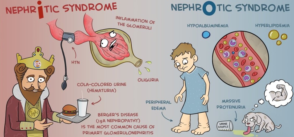
HAEMOPHILUS INFLUENZA INFECTION

HAEMOPHILUS INFLUENZA INFECTION

HAEMOPHILUS INFLUENZA INFECTION.

Haemophilus influenzae is a gram-negative, cocco-bacillary, facultatively anaerobic bacterium that falls within the Coccobacilli group. While it normally resides as a commensal in the nose and throat without causing infections under normal conditions, it can become pathogenic if host defenses are compromised.

The bacterium was initially misattributed as the cause of influenza, later identified correctly as the influenza virus.

Classifications of Haemophilus Influenzae

Classifications of Haemophilus Influenzae

Haemophilus influenzae is classified into two main groups based on the presence or absence of a capsule: Encapsulated and Unencapsulated (non-typeable).

Encapsulated Types:

  • There are six subtypes (a to f) distinguished by alphabetical letters corresponding to their capsular antigens (e.g., Haemophilus influenzae type a, b, c, d, e, and f).
  • Among these, type b (Hib) is the most prevalent and notorious for causing severe diseases.
  • The encapsulated types are susceptible to vaccination, notably the Hib vaccine.

Unencapsulated Types (Non-typeable):

  • Lack capsular serotypes and are generally less invasive but can still cause inflammation.
  • Not affected by the Hib vaccination.

Infections and Diseases:

Haemophilus influenzae infections can lead to various diseases, particularly when the bacterium successfully invades the body. These include:

Invasive Diseases: (For Encapsulated)

Non-invasive Diseases: ( For Non-encapsulated)

Mode of spread of Haemophilus Influenzae:

The primary mode of spread for Haemophilus influenzae is person-to-person transmission through respiratory droplets. The bacterium is spread when an infected person coughs or sneezes, releasing tiny droplets containing the bacteria into the air. These droplets can then be inhaled by individuals in close contact, leading to the colonization of the respiratory tract.

Key points regarding the mode of spread:
  1. Respiratory Droplets: The most common mode of transmission is through respiratory droplets expelled by infected individuals during activities such as coughing, sneezing, or talking.
  2. Close Contact: Transmission is more likely to occur in situations where people are in close contact with an infected person, especially in crowded or confined spaces.
  3. Asymptomatic Carriers: Individuals colonized with Haemophilus influenzae, even if asymptomatic, can still potentially transmit the bacterium to others.
  4. Opportunistic Nature: Haemophilus influenzae is considered an opportunistic pathogen, meaning it takes advantage of weakened immune defenses to cause infections. While it can colonize the respiratory tract without causing disease in healthy individuals, it may lead to infections when the host’s immune system is compromised.
  5. Age Groups: The transmission is particularly significant in settings with a high density of young children, as they are more prone to certain types of invasive Haemophilus influenzae infections, such as Hib (Haemophilus influenzae type b) meningitis.
Risk Factors for Hib Disease:
  1. Household Crowding: Living in a crowded household where people are in close proximity increases the likelihood of person-to-person transmission of the bacterium through respiratory droplets, which can lead to Hib infection.
  2. Large Household Size: Larger households provide more opportunities for the spread of infectious agents. The more people there are in a household, the higher the chances of someone being a carrier of Haemophilus influenzae, increasing the risk of transmission.
  3. Child Care Attendance: Children in daycare settings may have closer contact with each other, facilitating the spread of bacteria. Additionally, young children may not have fully developed immune systems, making them more susceptible to infections like Hib.
  4. Low Socioeconomic Status: Lower socioeconomic status often correlates with limited access to healthcare, crowded living conditions, and potential challenges in maintaining hygiene. These factors collectively contribute to an increased risk of Hib infection.
  5. Low Parental Education Levels: Parents with lower education levels may have less awareness of preventive measures and healthcare practices. This lack of knowledge can impact their ability to protect their children from infectious diseases, including Hib.
  6. School-Age Siblings: Siblings attending school may be exposed to various infectious agents, including Haemophilus influenzae. As carriers, they can potentially transmit the bacterium to younger siblings at home.
  7. Age (Youngest and Oldest): The youngest and oldest individuals are at an elevated risk. Young children often have developing immune systems, and the elderly may have weakened immune defenses, making both age groups more susceptible to severe infections.
  8. Race/Ethnicity (Native Americans): Native Americans may face elevated risks due to a combination of genetic, socioeconomic, and healthcare access factors that can contribute to a higher incidence of Hib disease.
  9. Chronic Diseases (e.g., HIV, Immunodeficiency): Conditions that compromise the immune system, such as HIV, immunodeficiency, or asplenia, reduce the body’s ability to fight infections, increasing the risk and severity of Hib disease.
  10. Prematurity: Premature infants may have underdeveloped immune systems, placing them at a higher risk of infections, including Hib. Their immune systems may not be fully equipped to respond effectively to bacterial threats.
  11. Extremes of Age (Below 5 and Above 65): Both very young children (below 5 years) and the elderly (above 65 years) often have weaker immune responses, making them more vulnerable to severe infections, including those caused by Haemophilus influenzae.
  12. Immunocompromised Individuals: Individuals with compromised immune systems, such as those with HIV/AIDS, cancers, or sickle cell disease, are less capable of mounting an effective immune response against pathogens, increasing the risk of severe Hib infections.
  13. Asplenia: Asplenia (lack of a spleen or non-functional spleen) impairs the immune system’s ability to clear bacteria from the bloodstream, leading to an increased risk of severe Hib infections.
HAEMOPHILUS

Pathophysiology of Haemophilus influenzae Infection:

  1. Entry into the Body: Haemophilus influenzae enters the body through the nasopharynx, commonly the upper respiratory tract.
  2. Colonization in the Nasopharynx: The organisms colonize the nasopharynx, where they may remain shortly or for several months. Some individuals may carry the bacteria without displaying symptoms, becoming asymptomatic carriers.
  3. Multiplication and Immune Recognition: Once inside the body, the organisms start to multiply. The immune system recognizes the presence of the foreign invader, sensitizing immune cells to the threat.
  4. Immune Response Activation: The immune system responds by transporting various defense cells, including cytokines, to the affected area. This mobilization is a defensive reaction against the invading bacteria.
  5. Inflammation Occurs: In response to the interaction between the immune cells and the bacteria, inflammation takes place. This is a protective mechanism designed to eliminate the infectious agent.
  6. Signs and Symptoms of Infection: The inflammatory response leads to signs and symptoms of infection, including fever, weakness, nausea, and other systemic manifestations. These symptoms are indicative of the body’s efforts to combat the infection.

Clinical Features according to Infections and Diseases

Clinical Manifestation

Signs and Symptoms

Pneumonia

  • Fever and chills
  • Cough
  • Shortness of breath
  • Sweating
  • Chest pain
  • Headache
  • Tiredness
  • Fatigue

Bacteraemia

  • Fever and chills
  • Tiredness
  • Anorexia
  • Nausea
  • Vomiting
  • Dyspnea
  • Confusion
  • Irritability

Meningitis

  • Fever
  • Headache
  • Neck stiffness
  • Nausea ± vomiting
  • Photophobia
  • Confusion, decreased mental status.
  • Hearing impairment or neurologic sequelae in survivors
  • Case fatality ratio: 3% to 6%

Epiglottitis

  • Infection and swelling of the epiglottis
  •  Life-threatening airway obstruction

In Children:


Signs and Symptoms

  • Irritability
  • Vomiting feeds
  • Poor feeding and refusal of feeds
  • Lack of interest in everything and inactivity
  • General weakness
  • Drowsiness
  • Decreased reflexes in babies
Diagnosis and Investigations

Diagnosis and Investigations

Clinical Assessment:

  • Medical History: Inquire about the patient’s symptoms, recent illnesses, and exposure to potential sources of infection.
  • Physical Examination: A thorough examination to assess specific signs and symptoms associated with the type of infection, such as lung sounds for pneumonia or neck stiffness for meningitis.

Laboratory Tests:

  • Gram Stain: To visualize the morphology of the bacteria. Gram stain of infected body fluids may reveal small, gram-negative coccobacilli, suggesting H. influenzae infection.
  • Culture: Specimens for culture include CSF, blood, pleural fluid, joint fluid, and middle ear aspirates. Purpose is To isolate and identify H. influenzae. Positive culture for H. influenzae establishes the diagnosis. Detection of antigen or DNA can be used as an adjunct, especially in patients partially treated with antimicrobials.
  • Cerebrospinal Fluid (CSF) Analysis: For suspected cases of meningitis, a lumbar puncture is performed to collect CSF. Analysis of the CSF can reveal the presence of bacteria, white blood cells, and other indicators of infection.
  • Sputum Culture: In cases of pneumonia, a sputum sample may be collected and cultured to identify the causative organism, including Haemophilus influenzae.
  • Polymerase Chain Reaction (PCR): Molecular techniques like PCR can help identify the specific strain of Haemophilus influenzae, providing more detailed information for targeted treatment.

Imaging Studies:

  • Chest X-ray: For suspected pneumonia, a chest X-ray may be conducted to visualize abnormalities in the lungs and confirm the diagnosis.

Management

Aims

  • To minimize further complication.
  • To relieve pain.
  • To preserve life.
  • To promote comfort

Immediate intervention

The patient and relatives are received, admitted to the medical ward. Incase patient has meningitis, they are admitted in an isolation room with dim light on a comfortable bed and positioned in a comfortable position.

Medical Management

  1. Antimicrobial Therapy:
  • Choice of Antibiotics: Effective third-generation cephalosporins such as cefotaxime or ceftriaxone are the first-line treatment and should be initiated immediately. An alternative regimen includes chloramphenicol in combination with ampicillin.
  • Duration of Treatment: A 10-day course of antimicrobial therapy is usually prescribed for severe infections. In cases of penicillin resistance, alternative antibiotics such as ceftriaxone, fluoroquinolones, or macrolides may be considered.

2. Supportive Treatment:

  • Oxygen Therapy: Administered as needed, especially in cases of respiratory distress or pneumonia.
  • Fluid Infusion: Maintaining hydration through intravenous fluid administration is crucial, especially in cases of severe infections where fluid loss may occur.
  • Other Supportive Measures: Depending on the severity and the affected organ system, additional supportive measures may include analgesics for pain relief, antipyretics for fever management, and antiemetics for nausea and vomiting.

4. Monitoring: Regular monitoring of vital signs, blood pressure, and oxygen saturation to assess the patient’s response to treatment.

5.Vaccination: Administration of Haemophilus influenzae type b (Hib) vaccine as a preventive measure, especially in populations at risk, to reduce the incidence of invasive Hib disease.

Nursing care 

1. Admission and Initial Assessment:

  • Vital Signs: Regular monitoring of vital signs, including temperature, heart rate, respiratory rate, and blood pressure.
  • Assessment: Conduct a thorough initial assessment to determine the severity of symptoms and the affected organ systems.

2. Infection Control Measures:

  • Implement standard precautions to prevent the spread of infection.
  • Isolation precautions may be necessary based on the specific type of infection.

3. Hydration and Nutrition:

  • Administer intravenous fluids as prescribed to maintain hydration.
  • Monitor and encourage oral fluid intake if tolerated.
  • Collaborate with the dietitian to provide appropriate nutrition, considering any dietary restrictions or preferences.

4. Medication Administration:

  • Administer prescribed antibiotics promptly and as directed.
  • Monitor for any adverse reactions to medications.

5. Respiratory Support:

  • Administer supplemental oxygen as prescribed for patients with respiratory distress or pneumonia.
  • Monitor respiratory status closely and provide respiratory treatments as needed.

6. Pain Management:

  • Assess and manage pain using appropriate pain management strategies.
  • Administer analgesics as prescribed.

7. Fever Management:

  • Implement measures to manage fever, such as administering antipyretics as prescribed.
  • Employ physical cooling measures (cool compresses, fans) as needed.

8. Neurological Monitoring:

  • For cases involving meningitis, monitor neurological status closely.
  • Assess for signs of increased intracranial pressure.

9. Emotional Support:

  • Provide emotional support to the patient and family, addressing any concerns or fears.
  • Keep the family informed about the patient’s condition and treatment plan.

10. Patient Education:

  • Educate the patient and family about the nature of the infection, treatment plan, and the importance of completing the prescribed antibiotic course.
  • Provide information on preventive measures, such as vaccination.

11. Follow-Up Care:

  • Plan for follow-up care and provide instructions for any necessary post-hospitalization care.
  • Ensure the patient and family understand signs of complications and when to seek medical attention.

12. Collaboration with Other Healthcare Providers:

  • Collaborate with physicians, pharmacists, and other healthcare providers to ensure a coordinated and effective treatment plan.

13. Documentation:

  • Maintain thorough and accurate documentation of assessments, interventions, and patient responses.

Complications of Haemophilus influenzae Infection:

Meningitis Complications:

  • Hearing Impairment: Occurs in 15% to 30% of survivors of meningitis.
  • Neurological Sequelae: Such as cognitive deficits, motor abnormalities, or seizures.

Epiglottitis Complications:

  • Airway Obstruction: Life-threatening complications may arise due to swelling of the epiglottis.
  • Bacteremia Complications:

  • Sepsis: Bacteremia can progress to sepsis, a severe systemic response to infection.
  • Endocarditis: Infection of the heart valves may occur in rare cases.
  • Pneumonia Complications:

  • Respiratory Distress: Severe pneumonia can lead to respiratory failure and the need for mechanical ventilation.
  • Arthritis Complications:

  • Joint Damage: Infective arthritis can result in joint damage and functional impairment.
  • Cellulitis Complications:

  • Abscess Formation: Cellulitis may progress to the formation of abscesses in severe cases.
  • Osteomyelitis Complications:

  • Bone Damage: Invasive infections can lead to osteomyelitis, causing damage to bone tissue.

Prevention of Haemophilus influenzae Infection:

  • Vaccination:
  • Hib Vaccine: Vaccination against Haemophilus influenzae type b (Hib) is highly effective in preventing invasive diseases, including meningitis and bacteremia. It is a routine childhood vaccine.
  • Pneumococcal Vaccine: Protects against pneumonia caused by various bacteria, including some strains of Haemophilus influenzae.

    Routine Immunizations:

  • Ensuring timely administration of routine childhood immunizations as recommended by national vaccination schedules.
  • Good Hygiene Practices:

  • Hand Hygiene: Regular handwashing can help prevent the spread of respiratory infections, including Haemophilus influenzae.
  • Avoiding Crowded Places:

  • Especially during peak seasons of respiratory infections.
  • Prompt Antibiotic Treatment:

  • Early diagnosis and treatment of respiratory infections to prevent complications and the spread of the bacteria.
  • Health Education:

  • Raising awareness about the signs and symptoms of invasive infections and the importance of seeking medical attention promptly.
  • Antibiotic Prophylaxis:

  • In certain cases, antibiotic prophylaxis may be recommended for close contacts of individuals with Haemophilus influenzae infection to prevent secondary cases.
  • Respiratory Etiquette:

  • Encouraging the practice of covering the mouth and nose when coughing or sneezing to prevent the spread of respiratory droplets.
  • Maintaining Healthy Lifestyle:

  • Ensuring good nutrition, regular exercise, and overall well-being to support a healthy immune system.

Quick Quiz

H Influenza Quiz

Paediatrics - mobile-friendly and focused practice.

Privacy: Your details are used only for quiz tracking and certificates.

HAEMOPHILUS INFLUENZA INFECTION Read More »
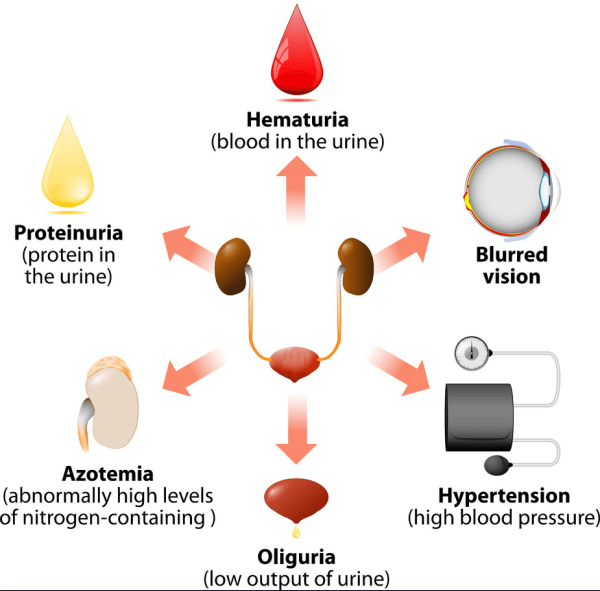
Report Writing

Report Writing

5.0 FINAL RESEARCH REPORT FORMAT

5.1 Outline of a Research Report

According to the UHPAB Guidelines, the Trainee’s research report must not be below 30 pages and consists of the following:

Main Components of a Research Report

  1. Preliminary Pages: Pages before Chapter One numbered in Roman Numerals.
    • a) Title page;
    • b) Preliminary pages which include Declaration, Approval, Dedication, Acknowledgement, Table of contents, List of tables, figures or pictures, Abbreviations/acronyms, Operational definitions, and Abstract;
  2. Main Body:
    • c) Chapter One: Introduction;
    • d) Chapter Two: Literature Review;
    • e) Chapter Three: Methodology;
    • f) Chapter Four: Findings of the Study;
    • g) Chapter Five: Discussions, Recommendations and Conclusions;
  3. References:
    • h) Reference list (Minimum 20 references, APA 7th Edition);
  4. Appendices:
    • Appendix I: Data collection tools
    • Appendix II: Ethical requirements (Consent form, approval letters, introductory letters)
    • Appendix III: Similarity Index Report (Anti-plagiarism max 30%)
    • Appendix IV: Maps showing location of study area
    • Appendix V: Pictures
    • Appendix VI: Information sheets

Each Chapter of the Trainee’s research shall consist of the following:

5.1.1 Chapter One: Introduction

This Chapter should be structured as follows:

  • 1.0 Introduction: Introduce the summary of the chapter in one (1) paragraph;
  • 1.1 Background to the Study: This should be a maximum of two (2) pages. It must be concise and precise;
  • 1.2 Statement of the Problem: This should be half a page;
  • 1.3 Research Objectives:
    • 1.3.1 Purpose of the Study or General Objective;
    • 1.3.2 Specific Objectives: These should be 2-3 SMART objectives;
    • 1.3.3 Research questions;
  • 1.4 Justification of the Study: This should include the rationale for conducting the study;
  • 1.5 Significance of the Study: This should highlight the importance or contribution to academic knowledge or practical use;
  • 1.6 Scope of the Study: Provides the boundary or limits of the research in terms of content, geographical area, and time span.

5.1.2 Chapter Two: Literature Review

This Chapter should be at least five (5) pages and arranged as follows:

  • 2.0 Introduction: This should not be more than a half page;
  • 2.1 Body: Shall be presented with a minimum of five (5) pages.
Note: a) Include a minimum of 20 in-text citations (APA 7th Edition); b) The oldest references should not be beyond 10 years before the trainee’s time of conducting research; c) The sub-headings of the literature review shall be in line with the specific study objectives; d) Include a brief summary of the literature with any gap identified. Use free sources like HINARI, Google Scholar, PubMed.

5.1.3 Chapter Three: Methodology

Chapter three (3) should be structured as follows:

  • 3.0 Introduction;
  • 3.1 Study Design: This should include the rationale;
  • 3.2 Study Setting: This should include the rationale;
  • 3.3 Study Population;
  • 3.4 Sample Size Determination and its Justification: Use simple scientific method;
  • 3.5 Sampling Method/Procedure: This should include the rationale;
  • 3.6 Inclusion and Exclusion Criteria;
  • 3.7 Study Variables;
  • 3.8 Research Instruments/Tools;
  • 3.9 Data Collection Method/Procedure;
  • 3.10 Data Management and Analysis e.g., use of SPSS, Excel;
  • 3.11 Data Presentation: By use of narratives, charts, and tables;
  • 3.12 Quality Control: Measures ensuring validity and reliability;
  • 3.13 Ethical Considerations;
  • 3.14 Limitations to the Study: Constraints affecting sampling/methodology and how to mitigate them;
  • 3.15 Dissemination of Study Findings.

5.1.4 Chapter Four: Findings of the Study

Results should be presented based on the specific objectives and in chronological order:

  • 4.0 Introduction: Description of the sample size and data presentation methods;
  • 4.1 Demographic Characteristics: All socio-demographic characteristics should be presented in one table with frequencies and percentages;
  • 4.2 Research Objective 1: Use tables and figures or summarized narrative statements;
  • 4.3 Research Objective 2: Use tables and figures or summarized narrative statements;
  • 4.4 Research Objective 3: Use tables and figures or summarized narrative statements;
Note: a) Highlight the key findings of the study in relation to the study objectives. b) Use not more than 2 tables or figures for each research objective; c) One can use narrative statements where necessary. After every figure or table, a narrative interpretation should be stated below it.

5.1.5 Chapter Five: Discussions, Recommendations and Conclusions

This Chapter should be structured as follows:

  • 5.0 Introduction:
  • 5.1 Discussion:
    • It is about the interpretation of key results;
    • Comparison with existing literature based on previous studies related to the topic;
    • Follow the order of specific objectives;
    • Comparisons and contrasts should be made in line with the cited literature with correct APA 7th edition in-text citation.
  • 5.2 Recommendations:
    • Recommendations based on key findings derived from the results. It should indicate what is to be done, by whom, how, and when. At least one recommendation for each objective.
  • 5.3 Conclusions:
    • These should be summarized in paragraphs, consistent with study objectives, and cover half a page;
  • 5.4 Implications to Health Profession Practice:
    • Highlight the impact and relevancy of the findings to the Health Profession practice or how the findings will be important in improving Health Profession Practice.

7.0 APPENDICES FOR FINAL RESEARCH REPORT

  • Appendix I: Data collection tool(s) or instrument(s)
  • Appendix II: Ethical requirements (Consent form, approval letters, introductory letters)
  • Appendix III: Similarity Index Report processed by anti-plagiarism checker software (Must not exceed 30%)
  • Appendix IV: Maps showing location of study area
  • Appendix V: Pictures
  • Appendix VI: Information sheets

Key Differences Between a Research Proposal and a Research Report

Feature Research Proposal Research Report
Page Length Minimum 15 pages (excluding preliminary pages). Minimum 30 pages (from Chapter 1 to the last appendix).
Tense Written in the future tense (e.g., "This study will investigate..."). Written in the past tense (e.g., "This study investigated...").
Abstract Not included. Summarizes the completed research (max 300 words, single-spaced).
Declaration Declares originality of the proposed work. Declares the originality of the completed work.
Dedication & Acknowledgement Not applicable. Included to dedicate work and acknowledge actual support received.
List of Tables & Figures Not applicable (tables/figures are not yet finalized). Lists actual tables and figures used in the completed report.
Main Body (Chapters) Chapters 1-3:
  • Chapter 1: Sets the stage for the proposed research.
  • Chapter 2: Summarizes existing research to justify the study.
  • Chapter 3: Details the planned methodology.
Chapters 1-5:
  • Chapter 1-3: Updated to reflect what was actually done.
  • Chapter 4: Presentation of findings (data, statistics).
  • Chapter 5: Discussion, Conclusions, Recommendations, and Implications.
Appendices Includes proposed Budget and Work plan. Omits budget and workplan; adds the Similarity Index Report (anti-plagiarism).

Dissemination of Research Findings

Beyond the completion of a research report, it is crucial to disseminate the findings. Dissemination refers to the strategies employed by the researcher to ensure that individuals concerned with or interested in the research findings become aware of the study and its outcomes.

Strategies for Dissemination of Research Findings

These strategies include:

  • Oral presentations through Continuous Medical Education (CMEs)
  • Poster presentations
  • Seminars
  • Publications in academic journals
  • Conferences
  • Magazines
  • Newspapers etc.

Chapter Four: Findings of the Study

This is the results section of your research report. It primarily involves the presentation of data, often in statistical forms.

Statistical data refers to all numerical descriptions of events, things, or objects. They take the form of counting or measurements, e.g., sex and age distribution of children with diarrheal diseases, clinically diagnosed cases of malaria.

Note that results are presented according to the specific objectives of the study.

Statistical Methods

These are the different means of organizing, analyzing, and interpreting numerical data for a better understanding of a phenomenon, allowing for sound decisions/conclusions.

Statistical methods can be broadly categorized as:

  • Descriptive Statistics
  • Analytical Statistics

Descriptive and Analytical Statistics:

  • Descriptive statistics involves the organization, presentation, and summarization of data.
  • Analytical statistics involves the organization, presentation, summarization, and finding an association between variables.

Statistical Variable

A statistical variable refers to any measurable characteristic that assumes a different value among individuals or subjects, e.g., temperature, blood pressure, age, weight, etc.

Statistical variables can be:

  • Quantitative variables: These can be measured in the form of numbers, as opposed to names or descriptions of events.
  • Qualitative variables: These cannot be measured in the form of numbers but rather names, e.g., degree of pain (like moderate, severe pain); tribe (like Ganda, Nyankole), etc.

Presentation of Data

Data presentation is important in any research study. It helps to summarize all the raw data into information that can be easily read and appreciated by other readers of your work. Data can be presented in the form of tables, figures (i.e., graphs, pie charts, line graphs, histograms, etc.). These form visual aids that help the reader to quickly understand the information.

Tables:

  • Tables help to summarize and give a picture of the size, shape, and distribution of the study findings.
  • These can be presented as:
    • Frequency distribution tables
    • Grouped Frequency distribution tables
  • All socio-demographic characteristics should be presented in one table with frequencies and percentages.

How to Construct a Table:

  • Ensure the table has an appropriate title.
  • The title should be above the table.
  • Every table must be numbered to facilitate easy referencing.
  • Should fit on one page.
  • Column and row headings should be brief and clear.
  • Units of headings should be clearly indicated.

Figures (Graphs, Charts):

  • Figures help to give a valuable supplement to the statistical analysis.
  • They help to show the trends of distribution.
  • When constructing a figure, follow the same guidelines as for a table, but the heading of a figure is usually placed below the figure.

Chapter Five: Discussions, Recommendations, and Conclusions

This section of the research report deals with discussions of the findings, conclusions, and recommendations of the study. In this section, you also highlight the implications of the study findings to Health Profession Practice.

Note that discussions are done according to your specific study objectives.

Key Points in Discussion of Results:

  • Discussion must be based on the major findings of your study.
  • Findings of your study must be related to findings of other previously done studies, i.e., relate your findings to your literature review using correct APA 7th edition in-text citations. Discuss whether the findings are in agreement or disagreement.
  • If your crucial findings do not relate to any literature reviewed, also acknowledge it.

Quick Quiz

Research Report Quiz

Research - mobile-friendly and focused practice.

Privacy: Your details are used only for quiz tracking and certificates.

Report Writing Read More »
References and Appendices

APPENDICES

Research Appendices, Consent, Budget, and Workplan
I. Appendices

Appendices refer to the different supporting documents that contain any additional information needed to enable professionals to follow your research procedures and data analysis.

An appendix is a page containing supplementary material that is not an essential part of the text itself but helps provide a more comprehensive understanding of the research problem. It is also used for information that is too cumbersome or lengthy to be included in the main body of the paper.

Placement and Referencing
  • Appendices appear just after the reference list.
  • In a proposal book, the reference list appears immediately after Chapter Three (Methodology).
  • In a final report, the reference list appears after Chapter Five.
  • Each appendix must be referred to by name (e.g., Appendix A, Appendix B, Appendix C) in the text of the paper.
  • In-text citation example: To refer to the Appendix within your text, write it at the end of the sentence in parentheses. Example: In addition to the limitations of email, Dansons et al. (2012) reviewed studies that focused on international bank employees and college students (see Appendix B for demographic information).
UHPAB Guidelines for Appendices

Every appendix must start on a fresh page and be numbered using Roman numerals (e.g., Appendix I, Appendix II) or standard numerical formats as guided by the institution. Below are the specific requirements for each appendix under the new Uganda Health Professionals Assessment Board (UHPAB) guidelines:

  1. Data Collection Tools (Proposal & Report): Must contain the blank copies of your research instruments, such as questionnaires, interview guides, observational checklists, or scoring sheets used to gather data.
  2. Ethical Requirements & Introductory Letters (Proposal & Report): Includes signed consent forms and participant information sheets. The Introductory Letter issued by the Principal must be signed in triplicate (3 copies): one for the authorities of the study area, one for the IRC file, and one attached here in the final report.
  3. Similarity Index Report (Report Only): Trainees must run their final report through anti-plagiarism software. The printed report showing a similarity index of 30% or less must be attached here to prove academic integrity.
  4. Maps (Proposal & Report): Clear, labeled maps showing the exact location of the study area (district, sub-county, and facility/village).
  5. Pictures (Proposal & Report): Any relevant photographs taken during the study (e.g., of the facility, training, or setup) that support the research, ensuring participant confidentiality is maintained.
  6. Information Sheets (Proposal & Report): Written explanations provided to participants explaining the purpose of the study, their rights, and how their data will be used before they sign the consent form.
  7. Proposed Budget (Proposal Only): A detailed table of estimated financial costs required to complete the study.
  8. Work Plan (Proposal Only): A chronological timeline (usually a Gantt chart) showing the scheduled dates for each research activity from topic formulation to final report submission.
II. The Consent Form

A consent form is the document that proves the informed consent process has taken place.

What is Informed Consent?
Informed consent is the permission granted in the knowledge of the possible consequences, given by the respondent to the researcher for participating in the study (with full knowledge of possible risks and benefits). In research, autonomy is protected by ensuring that the patient consents freely without coercion.
Legal and Ethical Considerations (Ugandan Context)
  • Constitutionally, any person 18 years and above is eligible to give formal consent.
  • For persons below 18 years (principally below 15 years), they will provide assent to agree to participate, while the next of kin or institutional authority will consent as a key witness.
  • The process involves explaining clearly to the prospective participant what the research is about to ensure full understanding.
  • The participant must be allowed to make a free choice whether or not to participate. There must be no coercion of any sort.
Key Features of a Consent Form
  • Statement of Introduction & Researchers Information: Introduce yourself to the participant (names, address, profession, and contact details).
  • Study Title & Purpose of the Study: The official title of the research and a concise explanation of why you are studying that topic.
  • Research Procedures: An overview of the specific procedures involved in the research.
  • Benefits and Potential Risks: Honest description of procedure risks, side effects, or discomforts, along with potential benefits.
  • Confidentiality Protection & Assurance: Description of measures taken to protect participant information and reassurance regarding data security.
  • Voluntary Participation & Participant Rights: A clear statement that participation is voluntary, they have the freedom to withdraw without penalty, and an explanation of their rights during and after the study.
  • Participant Questions & Contact Information: A statement allowing participants to ask questions, including how they can contact the researchers.
  • Statement of Consent (Informed Consent Statement): A section where the participant acknowledges having been explained to, having understood clearly, and accepting to participate.
  • Signature Lines & Date: Designated spaces for the participant's signature, researcher's signature, and the date. (Note: In some sensitive studies, participants sign but do not put their actual names to protect anonymity).
  • Consent Form Copy: Assurance that the participant will receive a copy of the signed and dated consent form.
Sample of Consent Form

Informed Consent Form

Study Title: Factors Associated with Uptake of Malaria Vaccine Among Caretakers of Children Below One Year in Buteebo Village Kampala District – Uganda.

Investigator: Muhindo George (Lyamujungu Health Training Institute).

Purpose of the Study: This study aims to explore the factors influencing the uptake of the malaria vaccine among caretakers of children under one year in Buteebo Village. The findings will help improve health policies and vaccine coverage.

Procedures & Confidentiality: If you agree to participate, you will answer a series of questions related to child immunization. Your participation is completely voluntary. You can withdraw at any time. All information gathered will be treated with absolute confidentiality and used only for academic purposes.

Consent Statement: I have read (or have been read to) the information above. I fully understand the nature, risks, and benefits of participating in this study, and I freely consent to participate.

Participant's Signature/Thumbprint:

...............................................................

Date: ......................................................

Researcher's Signature:

...............................................................

Date: ......................................................

III. Research Budget

A research budget is a line item (tabular) representation of the expenses associated with the proposal project.

  • Budget Justification: Contains more in-depth detail of the costs behind the line items (also called explanatory notes) and explains the use of the funds where it is not immediately evident.
  • Accuracy: Cost estimates need to be as accurate as possible. Reviewers will note both over-estimations and under-estimations.
  • Development: The budget should be developed in consultation with the appropriate project representative. Sponsors customarily specify how budgets should be presented and what costs are allowable.
Sample of Proposed Budget (Proposal Only)

Proposed Budget

The budget below details the estimated financial resources required to execute the research project, from proposal development to final report submission.

No. Item Description Qty Unit Cost (UGX) Total Cost (UGX)
1. Stationery (Printing Paper, Pens, Clipboards) Lumpsum 50,000
2. Internet Data & Airtime (Research and literature review) 4 Months 30,000 120,000
3. Travel / Transport (Data collection visits) 150,000
4. Data Analysis (Consultation and software processing) 1 100,000 100,000
5. Printing & Hardcover Book Binding (Drafts & Final Reports) 3 Copies 25,000 75,000
6. Contingency Fund (Miscellaneous expenses) 40,000
TOTAL ESTIMATED BUDGET (UGX): 535,000

Budget Prepared by: Muhindo George

Signature: ................................................................ Date: ..........................................

IV. Research Timetable / Workplan (Gantt Chart)

In research, a study timetable is referred to as a Work Plan. It is an easy, inexpensive tool that helps you control your study time, giving you perspective on what you need to accomplish and the timeframe available.

The Gantt Chart

A Gantt Chart is a planning tool that shows graphically the order in which various tasks must be implemented (done) and the duration of each activity.

Sample of Work Plan (Proposal Only)

Research Work Plan

The timeline below displays the chronological stages of the research execution, spanning a six-month academic calendar starting from topic selection to final report submission.

Activity / Milestone Month 1 Month 2 Month 3 Month 4 Month 5 Month 6
Topic Formulation and Presentation to IRC X
Literature Review & Proposal Draft Writing X X
Proposal Defense & IRC Approval Form Sign-off X
Data Collection in the Study Area X X
Data Cleaning, Entry, and Statistical Analysis X
Report Writing (Chapters 4 & 5) X
Plagiarism Check, Binding, & Submission to UHPAB X

Work Plan Formulated by: Muhindo George

Signature: ................................................................ Date: ..........................................

Quick Quiz

Appendices Quiz

Research - mobile-friendly and focused practice.

Privacy: Your details are used only for quiz tracking and certificates.

APPENDICES Read More »

References and Appendices

References and Appendices

References and Referencing Styles
I. References and Appendices

References refers to a list of all intext cited works. The researcher is supposed to develop a reference list at the end of your proposal. This list enables the reader or user of this proposal to conveniently retrieve each of the sources of information that the researcher reviewed.

Whenever you use someone else’s words or ideas in your research paper, you must indicate that this information is borrowed by quoting the source of information in the paper itself (in text referencing), and at the end of the paper (reference list). This applies to written sources you have used such as books, articles web pages, e.t.c

Reference is used to tell the reader where ideas from other sources have been used in the research paper. Referencing is a crucial part of successful academic writing, avoiding plagiarism and maintaining academic integrity in your assignments and research.

Purpose/Importance of Referencing: WHY DO WE REFERENCE?
  1. Giving Credit to Others: Referencing is a way of acknowledging and giving credit to the original authors or creators of ideas, theories, and works that you incorporate into your own writing. Example: If you use a quote from a book in your research, proper referencing indicates who wrote that quote originally.
  2. Enhancing Credibility and Authority: By citing reputable sources, referencing lends credibility and authority to your arguments. It shows that your ideas are supported by established knowledge and research. Referencing gives your argument evidence, credibility and authority. Example: Referring to well-known studies or academic papers in your field strengthens the reliability of your statements.
  3. Providing a Trail to the Original Source: References act as signposts, guiding readers to the original works. This allows interested readers to move deeper into the topic by exploring the sources you used. Example: A reader who gets interested in your work can trace it back to the specific research study through your references.
  4. Avoiding Plagiarism: Failure to acknowledge the work of others may lead to plagiarism. Proper referencing is essential to avoid unintentional or intentional plagiarism, demonstrating academic integrity. Example: Copying and pasting a paragraph from a source without proper citation is considered plagiarism.
  5. Distinguishing Your Ideas: Referencing allows you to differentiate your original thoughts from those borrowed from external sources. It is a way of distinguishing your ideas from those of other sources.
  6. Facilitating Fact-Checking: Proper referencing enables others to fact-check your work. Interested readers or researchers can verify the accuracy of your statements by consulting the original sources. Example: Including page numbers in your citations allows readers to locate specific information in the referenced source.
II. Referencing Styles

A referencing style is a set of rules on how to acknowledge the thoughts, ideas and works of others in a particular way.

In your document, referencing is done at two levels; first you need to give a brief reference in the body of text called “in-text citation”, and secondly a detailed reference is provided at the end of the document in the form of a list.

Types of referencing styles

The two commonest styles used are:

  • APA (American psychological Association) style.
  • MLA (Modern Language Association) style.

Other styles are:

  • Vancouver style / author-number system)
  • Chicago style
  • Turabian style
III. APA STYLE OF REFERENCING

APA style uses the author/date method of citation in which the author’s last name and the year of the publication are inserted in the actual text of the paper. It is the style recommended by the American Psychological Association and used in many of the social sciences. It is the Author Prominent Style of Referencing.

General Rules for In-text Citation:
  1. In-text citation utilizes the last name (surname) of the author, followed by a comma and the year of publication. Example:

    (Ghaznavi, 2003).

  2. If a page number follows the publication year, a comma is added. Example:

    (Ghaznavi, 2003, p. 40)

  3. Alternatively, the author’s name can be written outside the bracket. Example:

    Ghaznavi (2003, p. 40) observes…

  4. Punctuation marks come after the citation, not before.
    When you provide an in-text citation in the body of your writing, any punctuation marks, such as commas or periods, should come after the citation.
    For example:
    Correct:

    “This is an important point (Smith, 2021)."


    Incorrect:

    “This is an important point, (Smith, 2021)."


    In the correct example, the period is placed after the citation within the parentheses/brackets. This is to ensure that the citation is clearly associated with the information it is referencing, and punctuation does not interfere with the citation’s structure.

General Rules for APA Reference List
  1. Detailed references are listed on a separate page titled ‘References,’ centered and in bold.
  2. Only sources cited in the work are listed.
  3. Double line spacing is used between each entry.
  4. Each reference has a hanging indent, where the first line is flushed to the left margin, and remaining lines are indented.
Additional Referencing Rules:
  • The list is alphabetically arranged based on the first author’s surname or the first significant word of the title.
  • If sources from the same author have different publication years, references are listed alphabetically by the first author’s name and then chronologically.
  • Titles of larger sources (books, journals) are italicized, while titles of parts within a larger work are enclosed in double quotation marks without italics.
  • Unused but consulted sources can be mentioned under “Bibliography” on a separate page.
Author Variations in In-text Citation and Reference List Entry:
  • For Two Authors: In the in-text citation, only the surnames of the two authors are used, separated by ‘&’. Example:

    (Alvi & Zaidi, 2009).

    In the reference list, both the surname and initials of the two authors are used, separated by ‘&’. Example:

    Alvi, M. H. & Zaidi, R. (2009).

  • For Three to Five Authors: In the in-text citation, only the surnames of the three authors are used, first two separated by a comma (,) and the last two by ‘&’. Example:

    (Alvi, Ghaznavi, Hashmi, Siddiqui & Zaidi, 2009).

    If the same source is cited again in the text, it will appear like this: Example: (Alvi et al., 2009). In the reference list, both the surname and initials of all the authors are used, the last two separated by ‘&’, and the remaining by commas. Example:

    Alvi, M. H., Ghaznavi, K., Hashmi, M., Siddiqui, D. & Zaidi, R. (2009).

  • For 6 to 7 Authors: In the in-text citation, only the surname of the first author is written, followed by ‘et al.’ Example:

    (Alvi et al., 2009).

    In the reference list, both the surname and initials of all the authors are used, the last two separated by ‘&’, and the remaining by commas. Example:

    Alvi, M. H., Ghaznavi, K., Afridi, S., Zaidi, R., Hashmi, M. & Siddiqui, D. (2009).

  • For 8 or More Authors: In the in-text citation, only the surname of the first author is written, followed by ‘et al.’ Example:

    (Alvi et al., 2009).

    In the reference list, write the names of the first six and the last author. The last two names are separated by “……..,” and the remaining by commas. Example:

    Alvi, M. H., Ghaznavi, K., Afridi, S., Zaidi, R., Hashmi, M. & Siddiqui, D.,…., Qureshi, T.R. (2009).

IV. Harvard Style of referencing

Harvard is actually a generic term refers to all the referencing styles that are “author date” based style. This style is most commonly used in U.K and Australia. Developed by Harvard University in the UK and published by the Harvard Law Review Association. The Harvard style and its many variations are used in law, natural sciences, social and behavioural sciences, and medicine.

Harvard Style of referencing-General Rules, citation
  • In in-text citation only the last name (surname) of the author is used, author’s name and year of publication are not separated by a comma (,). For example: (Ghaznavi 2003)
  • A comma (,) is put after the publication year if a page number is mentioned after it. (Ghaznavi 2003, p 40)
  • It is also allowed to write the author’s name out of the bracket. For example: Ghaznavi (2003, p 40) observes ………..
  • Punctuation marks such as comma or full stop are used after the citation and not before them.
General Rules- Harvard Style referencing
  • Detailed references are listed on a separate page at the end of the document.
  • The title ‘References’ is given to the list, placed in center and in bold font.
  • Only those sources are to be listed that has been cited in your work.
  • No reference carries hanging indent.
  • Author’s name and the year are not separated by a comma or a full-stop.
  • Each reference ends up with a full stop (.).
  • If you have used the sources of the same author/s with different years of publication, the references are alphabetically listed first by the first author’s name then chronologically by publication year.
  • In the reference list, the name of an author is written in a way: last name is written first and afterwards initials of the first name/s are written; no full stop is put after the initials. For Example: Khalid Ghaznavi is written as Ghaznavi K. Mohsin Hasan Alvi is written as Alvi MH. The names of the authors are usual presented in capital letters.
V. Revision Questions & Comparisons
Revision Questions
  1. What are the similarities between APA an Harvard styles of referencing?
  2. What are the major differences?
  3. Examine the differences between list of references and bibliography?
Similarities between APA and Harvard styles of referencing:
  • Purpose: Both APA and Harvard styles aim to provide clear and consistent guidelines for citing sources in academic writing. They help to ensure that readers can easily identify and locate the sources used in a research paper or other academic work.
  • Author-date system: Both APA and Harvard styles utilize the author-date system for in-text citations. This means that the author’s last name and the year of publication are included in parentheses within the text to indicate the source of the information.
  • Alphabetical arrangement: Both APA and Harvard styles require that the reference list or bibliography be arranged alphabetically by the author’s last name. This makes it easy for readers to find the full bibliographic information for each source.
Major differences between APA and Harvard styles:
  • In-text citations: APA and Harvard styles differ in the specific format for in-text citations. APA uses the author-date system with parentheses around the author’s last name, the year of publication, and the page number (if applicable). Harvard style omits the parentheses and uses a comma after the author’s last name, followed by a period, the year of publication, and a colon before the page number (if applicable).
  • Reference list vs. bibliography: APA uses a reference list, which includes all sources cited in the paper, whether they are books, articles, websites, or other formats. Harvard uses a bibliography, which includes only sources that are referred to or mentioned in the paper.
  • Formatting: APA and Harvard styles have different formatting requirements for the reference list or bibliography. APA uses specific indentation rules, double spacing, and a hanging indent for each entry. Harvard uses a consistent indentation for all entries, single spacing, and no hanging indent.
5 differences between list of references and bibliography:
  1. Scope: A list of references includes all sources that are cited in the paper, while a bibliography includes only sources that are referred to or mentioned in the paper.
  2. Completeness: A list of references should include complete bibliographic information for each source, while a bibliography may include abbreviated or incomplete information, depending on the style guide.
  3. Purpose: A list of references is primarily used to provide a record of the sources used in the paper, while a bibliography may also serve as a guide for further reading or research.
  4. Ordering: A list of references is normally ordered alphabetically by author’s last name, while a bibliography may be organized differently, such as by topic or chronology.
  5. Labeling: A list of references is labeled as “References,” while a bibliography may be labeled as “Works Cited,” “Bibliography,” or “Literature Cited.”

General Rules for In-text Citation (APA 7th Edition)

In-text citations link the ideas in your writing to the sources listed in your references. APA 7th edition utilizes the last name (surname) of the author, followed by a comma and the year of publication.

  • 1. Basic Citation: Place the surname and year inside parentheses. Example: (Ghaznavi, 2003).
  • 2. Direct Quotes / Page Numbers: If you are directly quoting, add the page number (p. for single page, pp. for multiple pages). Example: (Ghaznavi, 2003, p. 40).
  • 3. Narrative Citation: Alternatively, write the author's surname as part of the sentence, with only the year (and page number if quoting) in parentheses. Example: Ghaznavi (2003, p. 40) observes...
  • 4. Punctuation Placement: All punctuation marks (such as commas or periods) must come after the parenthetical citation, not before.
    • Correct: "This is an important point (Smith, 2021)."
    • Incorrect: "This is an important point, (Smith, 2021)."
Sample Page: In-text Citations in Body Text

Chapter Two: Literature Review

Immunization remains one of the most cost-effective public health interventions globally. Despite its proven benefits, the uptake of new vaccines such as the malaria vaccine remains low in some rural communities (Sebugenyi et al., 2024). According to Nakate (2020), caregiver hesitation is heavily influenced by a lack of sensitization. This issue is further compounded when parents must travel long distances to reach functional health facilities. Indeed, "caregivers residing more than five kilometers from a health facility show a 40% lower likelihood of completing the child's immunization schedule" (Atugonza & Okello, 2020, p. 12).

To solve this, community-based health structures must be strengthened to build trust and increase demand. A similar study conducted in Malawi underscored the importance of training community health workers to address local misconceptions regarding vaccine safety (Simbeye et al., 2024).

In-text Citation & Reference Variations by Number of Authors

Depending on how many authors wrote a source, the citation and reference formats change under APA 7th Edition rules:

  • One Author:
    • In-text: (Nakate, 2020) or Nakate (2020)
    • Reference List: Nakate, T. (2020). Title of the book. Publisher.
  • Two Authors: Use both surnames. Use an ampersand (&) in parenthetical citations, but write the word "and" in narrative citations.
    • In-text Parenthetical: (Atugonza & Okello, 2020)
    • In-text Narrative: Atugonza and Okello (2020)
    • Reference List: Atugonza, M. & Okello, J. (2020). Title of article. Journal Name.
  • Three or More Authors (up to 20): Use the first author's surname followed by "et al." for all in-text citations. In the reference list, write out up to 20 authors.
    • In-text (First & subsequent times): (Sebugenyi et al., 2024) or Sebugenyi et al. (2024)
    • Reference List: List the surnames and initials of all authors up to 20, using an ampersand (&) before the final author.
  • Twenty-one (21) or More Authors:
    • In-text: (Khan et al., 2019)
    • Reference List: List the first 19 authors, then insert an ellipsis (...), and then write the final author's name. Do not use an ampersand. Example: Khan, A., Huynh, T. M., ... Bachert, C. (2019).
General Rules for APA Reference List

At the end of your proposal or report, you must provide a detailed list of all the sources you cited. General rules include:

  • 1. Separate Page: The reference list must start on a separate page titled References, centered and in bold.
  • 2. Match Citations: Only sources cited within the body of your writing are listed in the references.
  • 3. Double Spacing: Use double line spacing throughout the entire reference list.
  • 4. Hanging Indent: Each entry must have a hanging indent, where the first line is flush with the left margin, and all subsequent lines are indented by 0.5 inches (1.27 cm).
  • 5. Alphabetical Order: Arrange entries alphabetically by the first author's surname.
Sample Page: Reference List (APA 7th Edition)

References

Atugonza, M. & Okello, J. (2020). Factors influencing completion rates of childhood immunizations in Uganda. East African Medical Journal, 97(3), 112-120.

Nakate, T. (2020). Community health education and vaccine demand in low-income settings. SAGE Publications Inc.

Sebugenyi, A., Mukasa, R., Wandera, G. & Namaganda, F. (2024). Caregiver attitudes towards the introduction of the RTS,S malaria vaccine. Uganda Journal of Health Sciences, 12(1), 45-53.

Simbeye, A. J., Kumwenda, S., Cohee, L. M., Omondi, D., Masibo, P. K., Wao, H., & Awandu, S. S. (2024). Factors associated with malaria vaccine uptake in Nsanje district, Malawi. Malaria Journal, 23(1), 105. https://doi.org/10.1186/s12936-024-04938-7

Quick Quiz

References Quiz

Research - mobile-friendly and focused practice.

Privacy: Your details are used only for quiz tracking and certificates.

References and Appendices Read More »

Research Methods and Instruments For Data Collection

Research Methods and Instruments For Data Collection

Data Collection Methods and Instruments

Research instruments refers to the tools you are going to use to answer your objectives.

The researcher is supposed to explain the different methods that he/she intends to use during data collection. However, the research must put into consideration the research approach adopted (Qualitative, Quantitative or Mixed Methods), this will help the researcher in choosing the most appropriate research method to adopt.

Furthermore the researcher should as well put into consideration the type of data to be collected whether its primary data, Secondary data or a combination of both primary and secondary data. This will equally guide during the process of selecting the most appropriate data collection methods.

Note that:
The choice of data collection method (s) to be adopted by the researcher depends of the research approach used and the more the data collection methods adopted the more hectic the research but the better the findings.

Some of the data collection methods include; Questionnaire Survey, Interviews, Documentary Review, Focus Group Discussions, Observations and Experiments among others.

Data collection instruments

The primary methods employed for data collection consist of:

Data collection instruments refer to the tools that the researcher will use for collecting the relevant data related to the study objectives.

The researcher is therefore supposed to use this section to discuss the different data collection instruments s/he will adopt during the research, to state the data collection instrument, then briefly explain how it will be structured and the types of questions you expect to ask using the instrument.

For the case of questionnaires and interviews, the researcher must clearly state whether they will be structured or not & whether they will be self-administered or not. Therefore, the researcher is supposed to provide the basic information about the data collection instrument that will be adopted.

Questionnaire or Questionnaire Guide

While as the data collection method was a questionnaire survey, its corresponding data collection instrument is the questionnaire or a questionnaire guide.

The researcher is therefore supposed to briefly explain what a questionnaire is, explain how the questionnaire will be structured, explain the types of questions that you have in the instrument (whether open-ended, closed ended or both), explain whether you will use a Likert scale questionnaire or instrument, explain whether the questionnaire will be mailed, self-administered or web-based and as well explain whether it will be a structured, semi-structured or unstructured instrument.

This section is supposed to enable the reader understand how your questionnaire will be composed.

Interview Guide

In the case that the researcher adopted interviews or interviewing as the data collection method, then the data collection instrument would be the interview guide.

An interview guide is therefore a data collection instrument that supports the researcher through directing an interview process towards the objectives and issues regarding the study.

The researcher is therefore supposed to briefly explain what an interview guide is, explain how the interview guide will be structured, explain the types of questions that will be asked (whether open-ended, closed-ended or mixed), explain whether it will be a structured, unstructured or semi-structured interview, The researcher is also supposed to explain whether it will be a Directly personal interview, Telephone interview, Panel interview, Group interview or a Video conferencing interview.

This section is supposed to enable the reader to understand all the questions of; Who, Why, When, How and Where about your interview guide.

Observation Checklist

While as the data collection method was observation, its corresponding data collection instrument is the observation checklist.

An observation checklist is therefore a list of items that a researcher/ observer intends to observe (look at, hear, taste, smell or touch) during the data collection process.

The researcher is therefore supposed to briefly explain what an observation checklist is, how an observation checklist will be structured, explain the items in the observation checklist and further explain for how long he/she intends to observe. Say everything about your observation checklist.

This section is supposed to explain whether the observation will be; Participatory, Non-participatory or Covert-observation and how it will be executed.

Therefore, this section is supposed to enable the reader understand how your observation checklist will be composed.

Focus Group Discussion Guide

Researchers must note that if the data collection method was Focus Group Discussion, then the data collection instrument is a Focus Group Discussion Guide.

A Focus Group Discussion Guide is a data collection instrument that supports the researcher through streamlining particular processes or advising on how the Focus Group Discussion will be conducted in order to achieve its predetermined objectives.

The researcher is therefore supposed to briefly explain what a Focus Group Discussion Guide is, how the FGDG will be structured, explain the types of questions that will be asked (they are usually open-ended questions), explain the number of members that each FGD will have, explain how long each FGD will last, explain the composition of each FGD, whether the moderator will be the researcher or an appointed research assistant and how many FGDs will be conducted.

Note that;
This section is therefore supposed to enable the reader to understand all the questions of; Who, Why, When, How and Where about your Focus group Discussion Guide.

Experimental Checklist

In case the researcher chose experiment as the data collection method, then the data collection instrument will be the experimental checklist.

An experimental checklist therefore refers to a list of all the relevant scientific procedures that the researcher has to undertake while collecting data about the dependent and independent variables during an experiment.

Quality control methods/ pre-testing

This section can as well be referred to as validity and reliability. It's usually subdivided into two subsection validity and Reliability. The researcher is supposed to use this section to briefly but clearly explain how s/he will test for the validity and reliability of the research instruments.

Validity

Validity refers to the extent to which research instrument correctly measures what the researcher wants to measure. Therefore validity is about credibility or trustworthiness or accuracy or correctness of the research instrument.

The researcher is therefore supposed to provide a brief meaning of validity. Explain how s/he will ensure validity of the instruments. If you are to pre-test ensure that you explain the category of respondents that you will consider for the pilot study and how you will select them. Explain how you will carry-out the pilot study, Explain how you will go about managing the findings of the pre-test, the lessons learnt out of the pre-test and how you will go about redesigning the data collection instrument (s) (just in case the pre-test results deem it necessary).

Reliability

Reliability basically refers to consistency of a research instrument. Therefore reliability of a research instrument can easily be defined as the ability of a research instrument to yield the same results once used repeatedly over a given period of time while measuring the same variable(s).

The researcher is therefore supposed to provide a brief description of reliability. Explain how s/he will ensure reliability of the instrument(s). If you are to pilot test ensure that you explain the category of respondents that you will consider for the pilot study and how you will select them.

Explain how you will carry-out the pilot study, Explain how you will go about managing the findings of the pilot study, the lessons learnt out of the pilot and how you will go about redesigning the data collection instrument (s) (just in case the pilot results deem it necessary).

Note that:
An instrument can easily be reliable without being valid hence the need for testing for both validity and reliability.

A simple example of a reliable but invalid instrument would be offering a test to students and they averagely score 40% in the first sitting, they again score averagely 40% in the 2nd and 3rd sittings but yet the 40% is due to some wrongly set questions, which do note enable these students to excel and score 90's. In this case the instrument is reliable because it consistently provides the same results but invalid because it's not actually measuring what the researcher intended to measure (It's not correct).

Therefore researchers must always pre-test for both validity (correctness) and Reliability (consistency).

Research (Data Collection) Instruments/Tools

The key tools utilized for data collection include:

  • Interview Guides: Structured sets of questions or topics designed to guide an interviewer during face-to-face or key informant interviews.
  • Self-administered Questionnaires: Questionnaires designed for respondents to complete independently, without direct interaction with an interviewer.
  • Key Informant Guides: Structured outlines or questions used when interviewing key informants, individuals with specialized knowledge or experience relevant to the research.
  • Group Discussion Topics: Specific subjects or issues designated for exploration during a focus group discussion to stimulate conversation and elicit diverse perspectives.
  • Observation Checklist/Schedule: A systematic list or plan used by researchers to observe and record specific behaviors, events, or characteristics during the observation method.
  • Library Search: A systematic exploration of existing literature and information sources within a library to gather relevant data for research.
  • Tests: Structured assessments or examinations conducted to measure specific abilities, knowledge, or characteristics of individuals.
  • Use of Diary: The recording of regular, chronological entries detailing events, behaviors, or experiences over time, serving as a method of data collection in research.

The selection of the data collection method is guided by:

  • Accuracy of Information: The degree to which the chosen data collection method ensures precise, reliable, and truthful information from the participants, influencing the method’s appropriateness for the research.
  • Practical Considerations: Factors such as time, available resources, equipment, and personnel, which impact the feasibility and suitability of a particular data collection method for the research.
  • Response Rate of Respondents: The anticipated level of participation and willingness of the target respondents to engage with the chosen data collection method, affecting the method’s effectiveness in gathering sufficient and representative data.
  • Geographical Area Coverage: The extent to which the selected data collection method can efficiently collect information across the intended geographical area, considering the distribution and accessibility of the target population.

Characteristics of a Good Research Instrument

  • The Instrument must be valid and reliable
  • It must be based upon the Conceptual framework.
  • It must gather data suitable for and relevant to the research topic.
  • It must gather data would test the hypotheses or answer the questions under investigation
  • It should be free from all kinds of bias.
  • It must contain clear and definite directions to accomplish it.
  • It must be accompanied by a good cover letter.
  • It must be accompanied, if possible, by a letter of recommendation from a sponsor/school.

Advantages and Disadvantages of Common Research Instruments/Tools

1. Questionnaire/Questionnaire survey

The researcher is supposed to briefly describe this method of data collection, provide a justification for choosing this method of data collection, explain the type of data he/she expects to collect while using this method, the type of instrument that will support this data collection method and as well which category of the population will be subject to this data collection method.

A questionnaire survey is a data collection method mainly aimed towards collecting quantitative data where the researcher designs a set of questions related to the study objectives for purpose of collecting the required data.

Main types of questionnaires include:

  1. Mail questionnaires: This is a form of questionnaire that is prepared and mailed out to the respondent who must be part of the selected sample from a given study population. The main purpose of using a mailed questionnaire is to ensure that the researcher connects with a wider range of respondents. Basic forms of mails include; The E-mail, postal mails and Drop box survey mails among others.
  2. Self-administered questionnaire: This is a form of questionnaire where the respondent is left to read the questions, then fills in the answers by him/herself. This questionnaire is however sometimes filled in the presence of the interviewer(s) who usually stand-by to provide any form of support in case the respondent deems it necessary.
  3. Directly administered questionnaires: This is a form of questionnaire where the interviewer him/herself supports the respondent to fill the questionnaire. In this case the interviewer may keep reading questions for the respondent to answer. The interviewer can therefore support the respondent by rephrasing questions for a better understanding but without compromising the quality of the question. The main disadvantage of such a method is that it can easily lead to interviewer bias.
  4. Web-based questionnaires: This method is sometimes confused with mail questionnaires, however there is a difference between the two as this method is purely through use of web-site links while as mail questionnaire may be postal or email or even hand delivered mails by an agent of the researcher. Web-based questionnaire is becoming so inevitably growing where organizations (Private and Public), Groups and individuals, use internet based questionnaire surveys. This method is so cost effective and quicker but as well less detailed as compared to the other methods. Though its biggest challenge is the fact that it requires the use of internet and computer or high level technology which some potential respondents may not readily have access to, hence a strong limitation.

Types of questions asked include:

  1. Open-ended questions: This refers to a questionnaire which has questions that provide the respondent an opportunity to freely say whatever they deem necessary about the researcher's study or research question. Open-ended questions encourage full and meaningful participation of the respondent through motivating the interviewee to use his/her own knowledge about the subject. Open-ended questions usually help in the collection of qualitative data.
  2. Closed-ended questions: This refers to a questionnaire in which the questions asked have fixed responses that are provided for the respondent to choose from. These questions can be answered by simply ticking Yes or No (Boolean response). They can be answered through use of a Likert scale for example by selecting 5= Strongly Agree, 4 = Agree, 3 = Not Sure, 2 = Disagree and 1 = Strongly Disagree. The respondent is restricted and does not have the ability to exploit his knowledge to the fullest. Closed-ended questions usually help in the collection of quantitative data.
  3. Mixed questions: This is a triangulation of both Closed-ended and Open-ended questions in the same questionnaire. This is the most commonly used type of questionnaire and it helps in the collection of both qualitative and quantitative data. This method is mainly used in social research.
  4. Pictorial questions: This refers to a questionnaire where the researcher decides to use pictures to promote the interest in answering questions. In this case respondents will select their pictures of preference. This method is rarely used but it as well helps in collecting data just like the previous methods. This method is usually used in studies related to the prejudices in children and as well as in other form of social attitudes.

Forms of questionnaire structuring

  1. Structured Questionnaire: This is a form of questionnaire in which a specific set of predetermined questions were prepared by the interviewer in advance. This is the commonly used method by academic researchers.
  2. Unstructured Questionnaire: This is a form of questionnaire in which the survey questions to be asked the respondents are usually not set in advance.

Advantages:

  • Easy administration to respondents across large areas.
  • Respondents can answer at their own convenience.
  • Quick data collection, saving time.
  • Enhances anonymity, allowing respondents to freely address sensitive questions.
  • Eliminates interview bias.
  • Hard to design but easy to use.

Disadvantages:

  • Unsuitable for illiterate respondents.
  • Risk of misinterpretation of questions.
  • Lack of opportunity for researcher probing.
  • Low response rates.
  • No observation of facial expressions.
  • Inflexible tool in terms of respondent approach.

How to Construct a Questionnaire:

  • Keep it brief and attractive.
  • Begin with simple questions, ensuring logical sequencing.
  • Include researcher’s address and a clear title.
  • Provide an introduction, emphasizing the study’s significance, confidentiality, and instructions.
  • Use simple language, avoiding technical terms.
  • Ask specific questions related to research objectives.
  • Avoid leading and double questions.
  • Place sensitive questions at the end.
  • Include a variety of question types.

2. Interview Instrument (Interview Guide/Schedule)

The researcher is supposed to briefly describe this method of data collection, provide a brief justification for choosing this method of data collection, describe which type of data is expected to be collected using this method, which instrument will support this data collection method and the category of the study population to which this method will be applied.

Interviewing is a method of data collection that involves verbal or non-verbal exchanges between an interviewer(s) and an interviewee(s) where the interviewer asks questions related to the study objective(s) and the interviewee provides suitable responses based on his knowledge and experience.

Interviewing is the most commonly used method of data collection in social sciences, it's therefore close to impossible to conduct a social science research without using interviews as a method of data collection. Interviews are usually used by researchers to find out the attitudes and perceptions of the informants about certain study variables.

Types of interviews

While planning to collect data, a researcher who has chosen to use interviewing as one of the data collection methods should as well plan for the specific type of interview he/ she will adopt.

There are 3 basic forms of interviews, these include;

  1. Structured interviews: This is a form of interview where the researcher sets the interview questions in advance, ensures that they are precise and quantified answers/responses are needed. In this form of interview, questions must be asked in a standard way and the researcher ensures that most or all questions are asked. Usually the researcher sets possible answers for the interview, most questions have pre-set responses to be chosen from.
  2. Unstructured / In-depth Interviews: This is a form of interview where the researcher does not set any standard questions for the interview but rather starts up a conversation with an informant on a given topic and then follows the informant to ensure that he/she establishes what is important to discuss or not. In most cases the researcher may start-up the interview with a few open-ended questions but most of the questions asked during the interview will be emerging issues as a result of the interaction between the interviewer and the interviewee. During this kind of interview, as an interviewer; always ensure that you speak minimally and give room to the interviewee to do most of the talking. This interview will help you as a researcher to do in-depth interviewing and subsequently generate sensitive information.
  3. Semi-structural interviews: This is a form of interviewing usually used where the researcher is interested in collecting both qualitative and quantitative data. Therefore the researcher will have both standard questions (where the researcher expects predetermined responses) and open-ended questions (where the respondent is free to say whatever they have to say about a given topic). Therefore semi-structured interviews are simply a triangulation of structured and unstructured interviews.

Administering of interviews

This refers to how the researcher executes the interview. In most cases researchers may use; Telephone interviewing, Face-to-Face interviewing or group interviewing among others. This choice greatly depends on the researchers interests.

Forms of Interview Administration

  1. Directly personal interviews: This is sometimes referred to as face-to-face interviews or one-on-one interviews. This is where the interviewer chooses to personally interact with one informant (interviewee) at ago.
  2. Telephone interview: This is an indirect form of personal interview where the researcher (interviewer) interacts with the interviewee through a telephone conversation. The interviewer will not have the capacity to read the body language of the interviewee. This method can also take the trend of a single interviewer against multiple interviewees, commonly done through conferencing.
    Note that:
    - Telephone interviewing can easily be interrupted by technological breakdown.
    - If well managed this method can support the researcher to collect information in the shortest time possible.
  3. Panel interview: This is a form of interview where a group of interviewers normally ask one interviewee (informant) a set of questions. This form of interview is usually a face -to - face or using modern conferencing technology. A Common example is a job interview where one respondent (interviewee) faces a panel of interviewers.
  4. Group interview: This is a form of interviewing where more than one interviewer interacts with more than one interviewee and all interviewers expect responses from the interviewees.
  5. Video conferencing interviews / internet interviews: This form of interviewing involves the interviewer (researcher) adopting modern technology supported with the use of internet to ensure that he/she directly contacts with his or her respondent (interviewees).
    Note that; This is mainly through; Chart rooms and Video conferencing. It can be a one-to-one, one-to-many, many-to-one or many-to-many (interviewers) to-many (interviewees).

Considerations before an interview

These are issues that a researcher must reflect on before taking any form of interview; they include;

  1. Ensure that you are dressed neutrally (To guard against bias).
  2. Prepare to be friendly to the interviewee(s) but remain professional.
  3. Prepare not to talk academically (Avoid all technical jargons)
  4. Plan well and ensure that you are ready to ask more non-directional questions (non-leading questions or neutral questions).
  5. Prepare to avoid biased language
  6. Prepare probe questions. These will help you to conduct more in-depth interviews.
  7. Prepare a tape recorder (To capture the audio responses or recoding the informants responses).
  8. Prepare a note book.
  9. Prepare pen
  10. Prepare to take notes
  11. Prepare to be a moderate of the session by speaking less and let your interviewee speak more.
  12. Prepare to monitor the interview session i.e., so that it remains a relevant conversation about the study area, within a required time frame and with important issues being raised.
  13. Do a general preparation for the interview such as visiting the washroom before the process.
  14. Prepare the interviewee for the interview by greeting the interviewee, introducing yourself and making a brief suitable talk about the study while assuring the interviewee about confidentiality and motive of the study.
  15. Plan and prepare how you intend to close the interview. Don't forget to thank the informant for the time and resourceful information provided.

Advantages:

  • Higher response rate.
  • Suitable for non-literate respondents.
  • Allows probing.
  • Enables observation of respondent’s non-verbal cues.
  • More control over data collection pace.
  • Identity of respondent is known.
  • Provides an opportunity for follow-up.

Disadvantages:

  • Expensive and time-consuming, especially with a scattered population.
  • Respondents may lack time for interviews.
  • Prone to biases.
  • Limited anonymity.
  • Respondents may give pleasing answers.
  • Embarrassing questions may hinder open responses.
  • Difficulty in tracing respondents.

3. Observation Schedule/Checklist

The researcher is supposed to briefly describe this data collection method, provide a brief justification for choosing this method, describe the type of data he/she expects to collect using this method, the instrument that will support this method of data collection and the category of the study population to which this method will be applied.

Observation as a data collection method therefore refers to the process of the researcher regarding attentively or watching the actions, behaviors and code of conduct for a given population for purposes of making inferences.

The researcher may observe through;

  • Tasting,
  • Touching,
  • Seeing or using his or her senses of sight,
  • Smelling and as well as
  • Hearing.

Forms of observation

There are three types of observation from which researchers may choose the most appropriate method for their study. These include;

  1. Non-participant observation: This is basically a form of observation techniques where the researcher keenly watches the subjects (elements) of the study population with their knowledge and consent but without being part of the situation being observed. Therefore, in this form of observation, the researcher will only concentrate on observing but not a participant. This method allows the researcher to focus fully on players and have little distraction since he/she is not an active participant hence complete and timely data collection. However, the main disadvantage of such a method is that subjects /elements being aware that they are being observed prompts them to behave differently.
  2. Participant observation: This is a form of observation where the researcher gets intensively involved with the study population in their natural environment but with a purpose of collecting data about the behaviors, attitudes and ways of living of a given cohort say religious or occupational groups. To avoid bias, the researchers are supposed to remain objective throughout the research process. Seek permission from relevant authorities but avoid informing a study population that it's under scrutiny since the main objective of the study is to penetrate the subjective worlds of those studied, and to generally see those worlds from the participants point of view. The researcher should avoid imposing his/her own views upon the research elements but rather remain objective enough to collect data that represents the true picture of the research subjects. As compared to non-participant observation, where subjects tend to behave differently because they are aware that they are being studied, in this case subjects are not aware that they are being studied and therefore remain natural; this supports the collection of relevant and valid information.
  3. Covert observation: This form of observation can as well be referred to as hidden observation or undercover observation. This is a form of observation where the researcher or observer is not known to the research subjects, the observer is usually out of sight or not revealed to the observed cohort.

For example;

  • A lecturer may strategically plant a camera in a lecturer room to observe a given group of students.
  • A parent may observe children over a given period of time; this could be behind a one-way glass.
  • A researcher may secretly observe a given community over a given period of time.
Note that;
The main concern in this form of observation is the ethical issue of informed consent. Therefore, researchers must always find ways to go about this for example seeking for permission from relevant authorities, if it's a university you may seek the permission of management but not the students being observed.

While observing, the researcher should do the following;

  1. Ensure that you take notes
  2. Take photos of relevant scenarios
  3. Do video recordings.
  4. Do audio recordings.
  5. Sharpen your memory to capture important information until a point when you can actually record your information
Note that:
While undertaking the above, endeavor to be as private as possible mostly where the study subjects are not aware that they are being observed

Advantages:

  • Oldest research method.
  • Provides reliable, first-hand information.
  • Enables coding and recording real-time behavior.
  • Facilitates clarification of questions.
  • Elicits a high response rate.
  • Allows detailed information gathering using the senses.

Disadvantages:

  • Risk of respondents putting on a show.
  • Time-consuming.
  • Expensive.
  • Inability to observe past events.
  • Influenced by observer weaknesses.

4. Tests

Used for educational research to assess achievement or intelligence quotient.

5. Focus Group Discussion (FGD)

The researcher is supposed to briefly describe the meaning of this method of data collection, provide a brief justification for choosing this method of data collection, describe which type of data is expected to be collected using this method, the instrument to be used and the category of the study population to which this method will be applied.

Focus Group Discussion is therefore a data collection method mainly for collection of qualitative data in which a manageable group of people of a given study population are carefully selected into a discussion guided by a moderator/ facilitator (researcher) to freely articulate their perceptions, attitudes, opinions, beliefs, ideas and experiences about a certain topic of interest.

Qualities of a good Focus Group Discussion;

  1. It must have a moderator who is knowledgeable
  2. The Focus Group (FG) must not be less than 6 (six) members and not more than 12 members.
  3. Members of the FG must share common characteristics which are relevant for the study. For example members may have similar levels of education, professions, Age group & Gender among others parameters.
  4. Members of the FG must be selected from the study population of interest or sampling frame.
  5. The FG must have guidelines to guide procedure.
  6. It must be focused to the study area of interest
  7. Members must be considered equally.
  8. The moderator must have a predetermined list of open-ended questions.
  9. The FG must rely on discussions among all participants rather than dominancy of the moderator and some "I know it all" members.
Remember that:
"Garbage-in- Garbage-Out"
implying that when you review substandard documents, your analysis will yield substandard findings, your inferences will be substandard and as well you will ultimately generate substandard

Functions of the moderator /Facilitator of a FGD;

  1. To introduce the purpose of the discussion to all members
  2. To start the session
  3. To encourage free participation by all members.
  4. To regulate behavior of all members and promote equity and equality
  5. To ensure that the discussion remains in line with the study topic
Note that:
Avoid using one of the members of the FG as a recorder The moderator/Researcher should come along with a rapporteur or secretary who is highly qualified to take minutes, record proceeding, take photos and as well as a video coverage).

Advantages:

  • Gathers a variety of opinions.
  • Reaches a large number in a short time.
  • Encourages mutual checks among group members.
  • Involves directly affected individuals.
  • Provides comfort for those hesitant in larger groups.

Disadvantages:

  • Lacks anonymity.
  • Expensive and time-consuming.
  • Sensitive matters may limit open discussion.
  • Risk of dominance by one participant.
  • Group influence may generate desirable ideas.

6. Telephone Survey

Advantages:

  • Higher response rate than mail surveys.
  • Time-efficient.
  • Eliminates interviewer bias.
  • Covers a broader geographical area.
  • Offers comfort to shy respondents.
  • Cost-effective and convenient.
  • Allows probing during conversation.

Disadvantages:

  • Excludes respondents without telephones.
  • Difficulty in accessing phone numbers.
  • Prone to human weaknesses.

7. Mail Survey

  • Questionnaires mailed to respondents.
  • Applicable for widespread geographical studies.

For additional advantages and disadvantages, refer to those of a questionnaire.

8. Diary Method

  • Records events or occasions in a diary.
  • Provides valuable data on individual work patterns.

9. Experiments

The researcher is supposed to briefly describe the meaning of this method of data collection, describe which type of data is expected to be collected using this method, provide a brief justification for choosing this method of data collection, the instrument of data collection that will be used specifically for this method and the category of the study population to which this method will be applied.

Experiments help the researcher to collect quantitative data. It's therefore, a scientific procedure of data collection where the researcher sets up two equivalent groups/cohorts (Treatment group and Control group) then randomly selects elements into the treatment and control groups and subjects the treatment to an intervention but leaves the control without the intervention and then closely monitors the changes.

Key issues to note;

  1. Subjects are randomly selected into each of the cohorts or there is randomization.
  2. There are two groups, a control where the intervention is not administered and a treatment where the intervention.
  3. There must be no external factors to influence cohorts.
  4. The researcher must closely monitor the two groups.
  5. The treatment and control groups must be equivalent.
  6. The treatment and control groups must not know each other and should not interact.
  7. There must be a specified period of time for the experiment
  8. There must be set guidelines to regulate the experiment.
Note that;
This is the most expensive data collection method but as well very accurate if well regulated. Experiments are usually used in medical or clinical research and usually at 99% to 99.9% level of significance with a margin of error of 0.01 to 0.001, the smaller the margin of error the better and more reliable the findings in a medical or clinical research

Quick Quiz

Methods & Instruments Quiz

Research - mobile-friendly and focused practice.

Privacy: Your details are used only for quiz tracking and certificates.

Research Methods and Instruments For Data Collection Read More »

Want notes in PDF? Join our classes!!

Send us a message on WhatsApp
0726113908

Scroll to Top
Enable Notifications OK No thanks