Nurses Revision

nursesrevision@gmail.com

benign prostatic hyperplasia bph

Benign Prostatic Hyperplasia (BPH)

Benign Prostatic Hyperplasia (BPH) Lecture Notes
I. Introduction and Definition

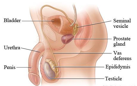
Benign prostatic hyperplasia (BPH) is the benign enlargement, or hypertrophy, of the prostate gland resulting from an increase in the number of epithelial cells and stromal tissue, developing upward into the bladder and obstructing the outflow of urine.

  • It is defined as a noncancerous increase in the size of the prostate gland which involves hyperplasia of prostatic stromal and epithelial cells. This results in the formation of large, fairly discrete nodules in the transitional zone of the prostate, which push on and narrow the urethra, resulting in increased resistance to the flow of urine from the bladder.
  • It is the most common urologic problem in male adults.
Incidence and Epidemiology
  • BPH is common in elderly men over 60 years and above.
  • In many patients older than 50 years, the prostate gland enlarges, extending upward into the bladder and obstructing the outflow of urine by encroaching on the vesicle orifice.
  • About 50% of all men in their lifetime will develop BPH. Of these men, almost half of them will have bothersome lower urinary tract symptoms.
  • 50% of men have evidence of BPH by age 50 years.
  • 75% of men have evidence of BPH by age 80 years.
II. Etiology and Risk Factors

The exact cause of BPH is idiopathic, but several factors are strongly associated with its development:

  • Aging: Prostate gland enlargement rarely causes signs and symptoms in men younger than age 40. About one-third of men experience moderate to severe symptoms by age 60, and about half do so by age 80. Aging occurs along with endocrine factors.
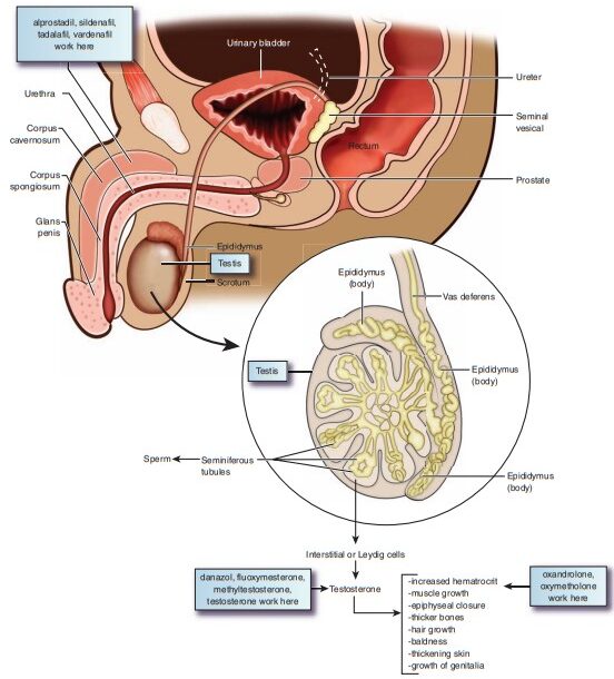
  • Hormonal Factors (Endocrine): Accumulation of Dihydrotestosterone (DHT) in the prostate.
  • Family History: Having a blood relative, such as a father or brother, with prostate problems means a higher likelihood of having problems.
  • Ethnic Background: Prostate enlargement is less common in Asian men than in White and Black men.
  • Diabetes and Heart Disease: Studies show that diabetes, as well as heart disease and the use of beta-blockers, might increase the risk of BPH.
  • Lifestyle and Obesity: Obesity increases the risk of BPH, while exercise can lower the risk.
  • Inflammation: Chronic prostatic inflammation.
  • Moderate Alcohol Consumption: Sometimes implicated, though heavy consumption has complex systemic effects.
III. Pathophysiology of BPH

The outcome of BPH depends on two major factors:

  • Anatomical factors: These involve the physical enlargement of the Prostate gland which produces a physical blockage at the neck of the bladder against urinary flow. This results from increased responsiveness of the prostate gland to androgens and estrogens.
  • Dynamic factors: These result from excessive sympathetic stimulation via alpha-1 receptors in the prostate gland leading to increased tone at the sphincters of the urinary bladder and the prostate.
Step-by-Step Pathological Process:
  1. Due to etiological factors like aging, there is a decrease in systemic testosterone levels.
  2. However, testosterone is actively converted into Dihydrotestosterone (DHT) by the enzyme 5-alpha reductase.
  3. DHT accumulates in the stromal cells of the prostate.
  4. This leads to the enlargement of the prostate (noncancerous increase involving hyperplasia of stromal and epithelial cells).
  5. Large discrete nodules form in the transitional zone, which push on and narrow the urethra.
  6. This results in increased resistance to flow and eventual obstruction of urine flow from the bladder.
Secondary Pathophysiological Effects:
  • Resistance: BPH is a result of complex interactions involving resistance in the prostatic urethra to mechanical and spastic effects.
  • Obstruction: The hypertrophied lobes of the prostate obstruct the bladder neck or urethra, causing incomplete emptying of the bladder and urinary retention.
  • Dilation: Gradual dilation of the ureters (hydroureter) and kidneys (hydronephrosis) can occur due to backpressure.
IV. Clinical Manifestations

The clinical manifestations are broadly divided into obstructive and irritative symptoms (a complex referred to as prostatism), along with generalized systemic symptoms and complications.

A. Obstructive Symptoms (Voiding Symptoms)
  • Hesitancy: Difficulty in starting urination.
  • Decrease in volume and force: A weak urinary stream. Decreased and intermittent force of the stream is a classic sign.
  • Interruption of urinary stream: Intermittency during urination (stopping and starting).
  • Dribbling: Urine dribbles out at the end of urination.
  • Abdominal straining: Using abdominal muscles to force urine out.
B. Irritative Symptoms (Storage Symptoms)
  • Urinary frequency: Frequent trips to the bathroom. Often an early sign.
  • Urinary urgency: The sudden and immediate urge to urinate.
  • Nocturia: Waking up frequently at night to urinate.
  • Sensation of incomplete emptying: Feeling that the bladder has not been completely emptied after voiding.
C. Generalized Symptoms & Advanced Signs
  • Acute urinary retention: Sudden inability to void, leaving more than 60 mL of urine in the bladder after an attempt.
  • Recurrent Urinary Tract Infections (UTIs): Due to stagnant urine.
  • Hematuria: Blood in the urine.
  • Systemic symptoms: Fatigue, anorexia, nausea, vomiting, pelvic discomfort, pain, and epigastric symptoms.
  • Azotemia and Renal Failure: Can result from chronic urinary retention and large residual volumes.
V. Complications of BPH

If left untreated, severe obstruction can lead to:

  • Acute urinary retention
  • Involuntary bladder contractions
  • Bladder diverticula (pouch-like out-pouchings of the bladder wall)
  • Cystolithiasis (bladder stones)
  • Vesicoureteral reflux (urine backing up into ureters)
  • Hydroureter and Hydronephrosis (swelling of ureters and kidneys)
  • Gross hematuria
  • Recurrent UTIs
  • Renal failure
VI. Assessment and Diagnostic Methods
  • History Collection: Obtain a history of voiding symptoms, including onset, frequency of day/night urination, urgency, dysuria, sensation of incomplete emptying, and decreased force of stream. Determine the impact on the patient's quality of life.
  • Physical Examination & Digital Rectal Examination (DRE): Palpate size, shape, and consistency of the prostate. A DRE often reveals a large, rubbery, and nontender prostate gland. Also, perform an abdominal examination to detect a distended bladder.
  • Urinalysis with Culture: To screen for hematuria and UTI.
    • Color/Appearance: May be cloudy, dark brown, or bloody.
    • pH: 7 or greater suggests infection.
    • Microscopy: Bacteria, WBCs, RBCs may be present. WBCs > 11,000/mm³ indicates systemic infection.
    • Urine Culture: May reveal Staphylococcus aureus, Proteus, Klebsiella, Pseudomonas, or E. coli.
  • Urine Cytology: To rule out bladder cancer.
  • Serum Creatinine & BUN: Elevated if renal function is compromised by backpressure.
  • Prostate-Specific Antigen (PSA): A glycoprotein contained in the cytoplasm of prostatic epithelial cells. A normal level is generally < 4.0 ng/mL. PSA is obtained if the patient has at least a 10-year life expectancy and if ruling out prostate cancer changes management. Note: Elevated PSA with a low percentage of free PSA is more likely associated with prostate cancer, whereas BPH elevates total PSA but often maintains a higher percentage of free PSA.
  • Transrectal Prostatic Ultrasound (TRUS): Accurately measures the size of the prostate, amount of residual urine, and locates lesions unrelated to BPH.
  • Cystourethroscopy (Urethrocystoscopy): Endoscopic visualization to view the degree of prostatic enlargement and bladder-wall changes (e.g., trabeculation, bladder diverticulum).
  • Simple Urodynamic Studies:
    • Uroflowmetry: Assesses the degree of bladder obstruction by measuring urinary flow rate.
    • Postvoid Residual (PVR) Measurement: via ultrasound bladder scanner or catheter.
    • Cystometrogram / Cystometry: Measures pressure and volume in the bladder to evaluate detrusor muscle function and tone, identifying bladder dysfunction unrelated to BPH.
  • Intravenous Pyelogram (IVP) with post-voiding film: Shows delayed emptying, upper tract obstruction, and bladder muscle thickening. Voiding cystourethrography may be used instead of IVP to visualize the bladder using local dyes.
  • Complete Blood Studies: Including clotting studies, particularly if surgical intervention is planned.
  • VII. Management of BPH

    The goals of collaborative care and medical management are to:

    1. Restore bladder drainage.
    2. Relieve patient symptoms and improve quality of life.
    3. Prevent and treat complications of BPH.

    The treatment plan depends on the cause, severity of obstruction, and condition of the patient.

    A. Immediate and Conservative Medical Management
    • Immediate Catheterization: If a patient is admitted on an emergency basis because he is unable to void (acute retention), he is immediately catheterized. An urologist may be consulted if an ordinary catheter cannot be inserted (may require a Coudé tip catheter).
    • Suprapubic Cystostomy: An incision into the bladder through the abdomen may be needed to provide urinary drainage if urethral catheterization fails.
    • "Watchful Waiting" (Active Surveillance): For patients with minimal symptoms. Involves monitoring disease progression and modifying diet and lifestyle.
    B. Pharmacological Therapy (Detailed)

    Drug therapy primarily involves three major groups: 5-alpha reductase inhibitors, Alpha-1 selective blockers, and Combination therapy. PDE5 inhibitors and herbal supplements are also utilized.

    Drug Class Mechanism of Action & Clinical Effects Specific Drugs & Dosing Notes Adverse Effects & Nursing Considerations
    5-Alpha Reductase Inhibitors (5-ARIs) Blocks the enzyme 5-alpha reductase, preventing the conversion of testosterone to dihydrotestosterone (DHT) (the principal intraprostatic androgen).

    Effect: Leads to the regression of hyperplastic tissue, actively reducing the mechanical size of the prostate.
    Note: Takes 3 to 6 months of daily use to see effective therapeutic results. Does not provide prompt relief for urinary retention.
    • Finasteride (Proscar): 5mg orally once daily. Competitively inhibits type II 5a-reductase.
    • Dutasteride (Avodart): 0.5mg orally once daily. Inhibits both type I and type II 5a-reductase.
    • Decreased libido, decreased ejaculate volume, and erectile dysfunction.
    • Gynecomastia (rare).
    • Warning: Highly teratogenic to male fetuses. Women of childbearing age should never handle crushed or broken tablets.
    • Lab effect: Artificially lowers serum PSA levels by ~50%. Must adjust PSA interpretation.
    Alpha-1 Adrenergic Receptor Blockers Blocks alpha-1 adrenergic receptors abundant in the prostate, bladder neck, and hyperplastic tissue.

    Effect: Brings about smooth muscle relaxation, resulting in the free flow of urine and rapid relief of dynamic obstruction symptoms.
    Fast-acting compared to 5-ARIs.
    Uroselective (Long-acting):
    • Tamsulosin (Flomax): 0.4 mg once daily, given 30 mins after the same meal daily. Highly selective for Alpha-1A receptors (less BP interference).
    • Silodosin (Rapaflo): 8 mg daily.
    • Alfuzosin (Uroxatral): 10 mg extended-release daily.
    Non-selective (Short/Long-acting):
    • Terazosin (Hytrin): 2-10 mg daily.
    • Doxazosin (Cardura): 1-8 mg daily.
    • Prazosin: 0.5-1 mg given at bedtime initially, titrated to 1 mg BID.
    • Postural (Orthostatic) Hypotension and dizziness (especially with non-selective agents like Terazosin/Doxazosin). Nursing intervention: Administer the first dose at bedtime to avoid "first-dose syncope".
    • Reflex tachycardia, headache, fatigue.
    • Nasal congestion.
    • Retrograde ejaculation (especially Tamsulosin/Silodosin).
    • Intraoperative Floppy Iris Syndrome (IFIS): A severe complication during cataract surgery. Ophthalmic surgeons must be notified if the patient is on Tamsulosin.
    Combination Therapy Combines a 5-ARI with an Alpha-blocker. e.g., Finasteride + Doxazosin, or Dutasteride + Tamsulosin (Jalyn). Provides immediate symptom relief (alpha-blocker) while slowly shrinking the prostate (5-ARI) over months.
    Phosphodiesterase-5 (PDE5) Inhibitors Relaxes smooth muscle in the lower urinary tract. Tadalafil (Cialis): 5 mg daily. Typically prescribed for men who experience both BPH and Erectile Dysfunction concurrently. Not routine for BPH alone.
    Herbal / Phytotherapeutic Therapy Mechanisms vary; generally thought to have anti-inflammatory and mild anti-androgenic effects.
    • Saw Palmetto (Serenoa repens): Commonly used dietary supplement for BPH.
    • African Plum (Pygeum africanum).
    Though commonly used by patients, major clinical guidelines often state there is insufficient evidence to routinely recommend them. Nurses must ask patients about herbal intake as it can interact with prescribed medications.
    C. Dietary and Lifestyle Management
    • Increase intake of fruits and vegetables.
    • Decrease foods high in saturated fat.
    • Avoid excessive intake of caffeine, alcohol, and beer (these have diuretic effects and can irritate the bladder).
    • Drink 50% of your body weight in ounces daily (e.g., if you weigh 150 lbs, drink 75 oz of water daily), but avoid drinking large amounts at once or close to bedtime.
    • Include Saw Palmetto in the diet (as per some holistic guidelines, though clinically debated).
    VIII. Minimally Invasive Therapies

    Minimally invasive therapies are becoming more common as an alternative to watchful waiting and invasive surgical treatment. They generally do not require hospitalization or long-term catheterization and are often performed as outpatient procedures.

    1. Transurethral Microwave Thermotherapy (TUMT):
      • Procedure: An outpatient procedure involving the delivery of microwaves directly to the prostate through a transurethral probe to raise the temperature to about 113°F (45°C). The heat causes the death (necrosis) of tissue, relieving obstruction.
      • Details: Takes about 90 minutes. A rectal temperature probe is utilized to ensure the temperature is kept below 110°F (43.5°C) to prevent rectal tissue damage.
      • Contraindications: Not appropriate for men with rectal problems. Anticoagulant therapy must be stopped 10 days before treatment.
      • Complications & Care: Postoperative urinary retention is common. Patients are sent home with an indwelling catheter for 2 to 7 days to facilitate the passing of small clots and necrotic tissue. Treated with antibiotics, pain medications, and bladder antispasmodics. Occasional bladder spasms, hematuria, and dysuria.
    2. Transurethral Needle Ablation (TUNA):
      • Procedure: Uses low-wave radiofrequency delivered by thin needles placed directly into the prostate gland to produce localized heat/necrosis. Only tissue in direct contact with the needle is affected, allowing high precision.
      • Details: Extent of removal depends on needle length, energy, and duration. Outpatient, local anesthesia with IV/oral sedation, lasts ~30 minutes. Little pain with an early return to normal activities.
      • Complications: Urinary retention (may require a catheter for a short time), UTI, irritative voiding symptoms, and hematuria for up to a week.
    3. Laser Prostatectomy:
      • Procedure: A laser beam is delivered transurethrally through a fiber instrument to cut, coagulate, and vaporize prostatic tissue under visual or ultrasound guidance. Effective alternative to TURP.
      • Types: Visual Laser Ablation of Prostate (VLAP), Contact laser technique.
    4. Photovaporization of the Prostate (PVP):
      • Uses a high-power green laser light to vaporize tissue. Improvements in urine flow are almost immediate. Bleeding is minimal. A catheter is usually inserted for 24-48 hours. Works well for larger glands.
    5. Interstitial Laser Coagulation (ILC):
      • Prostate is viewed through a cystoscope. A laser quickly treats precise areas via the direct placement of interstitial light guides into the tissue.
    6. Intraprostatic Urethral Stents:
      • Used for patients with severe obstruction who are poor surgical candidates. A stent is placed in the urethra to mechanically relieve symptoms.
      • Complications: Chronic pain, infection, and encrustation (stone formation on the stent).
    IX. Invasive Therapy (Surgical Management)

    Invasive treatment of symptomatic BPH involves surgery. The choice depends on the size/location of enlargement and patient factors (age, surgical risk).

    • Intermittent Catheterization: Can temporarily reduce symptoms by bypassing the obstruction, but long-term use should be avoided due to the high risk of infection.
    • Transurethral Resection of the Prostate (TURP):
      • Description: Considered the gold standard for surgical treatment of obstructing BPH. Involves the removal of inner prostate tissue using a resectoscope inserted through the urethra to excise and cauterize the tissue. No external incision is made.
      • Procedure Details: Done under spinal or general anesthesia; requires a 1 to 2-day hospital stay.
      • Post-Operative Irrigation: A large three-way indwelling catheter with a 30-mL balloon is inserted to provide hemostasis (by putting traction on the balloon against the prostatic fossa) and facilitate drainage. The bladder undergoes Continuous Bladder Irrigation (CBI) or intermittent irrigation for the first 24 hours to prevent obstruction from mucus and blood clots.
      • Outcomes & Complications: 80-90% excellent outcome. Complications include bleeding, clot retention, and TURP Syndrome (dilutional hyponatremia caused by the systemic absorption of the irrigation fluid, leading to confusion, nausea, and changes in BP). Urethral strictures or regrowth of the prostate may occur.
    • Transurethral Incision of the Prostate (TUIP):
      • Done under local anesthesia. Indicated for patients with moderate to severe symptoms but small to moderately enlarged prostates. Several small incisions are made into the gland to expand the urethra. Relieves pressure without removing tissue. Similar outcomes to TURP for appropriate candidates.
    • Transurethral Electrovaporization of Prostate (TUVP):
      • Uses electrosurgical desiccation to destroy tissue. Advantages include minimal risk, minimal bleeding, and sloughing. Complications include retrograde ejaculation and hematuria.
    • Open Prostatectomy:
      • Indicated only when the prostate is very large or if there are complicating factors (e.g., large bladder stones).
      • Approaches: Retropubic, Suprapubic, or Perineal approach.
      • Side effects/Complications: Higher risk of erectile dysfunction, heavy bleeding, severe post-operative pain, and a higher risk of infection.
    NURSING MANAGEMENT AND CARE PLAN
    I. Health Promotion & Patient Education
    • Early Detection: Encourage annual DRE and PSA testing for men over 50.
    • Substance Avoidance: Advise reducing the intake of caffeine and alcohol to decrease bladder irritation.
    • Medication Warnings: Strongly advise patients to avoid OTC cold and cough medications containing sympathomimetics (like pseudoephedrine/phenylpropanolamine) or anticholinergics, as these can severely impair voiding and precipitate acute urinary retention.
    • Symptom Reporting: Explain to patients not undergoing treatment the symptoms of complications (retention, cystitis) and encourage prompt reporting.
    • Expectation Management: Advise patients that irritative voiding symptoms do not immediately resolve after the relief of the obstruction; the bladder needs time to heal and adapt.
    II. Preoperative Care
    • Urinary drainage must be restored before surgery (e.g., via catheterization) if the patient is in retention.
    • Antibiotics are administered to prevent intraoperative infection.
    • Psychological Care: Allow the patient to express sexual concerns (fears regarding erectile dysfunction or retrograde ejaculation post-surgery).
    III. Postoperative Care (Especially for TURP)
    • Catheter and Irrigation Management (CBI): Ensure the inflow and outflow of irrigant are strictly monitored to prevent fluid volume overload and bladder rupture. The rate of irrigation is adjusted to keep the output urine light pink to clear.
    • Clot Prevention: Remove clotted blood from the bladder via manual irrigation if the 3-way catheter becomes obstructed.
    • Bleeding Control: Reduce bleeding at the prostate site by applying counterpressure (traction on the catheter balloon).
    • Bladder Spasms: Prevent and treat painful bladder spasms with prescribed antispasmodics (e.g., B&O suppositories, Oxybutynin).
    • Infection Control: Prevent urethral irritation and bladder infection through strict aseptic catheter care.
    • Activity: Straining during bowel movements must be strictly avoided. Provide stool softeners/laxatives. Have the patient practice pelvic floor (Kegel) exercises once the catheter is removed to regain continence.
    IV. Ambulatory and Home Care (Discharge)
    • Care of the urinary catheter (if discharged with one).
    • Managing temporary urinary incontinence (using pads, Kegel exercises).
    • Intake of oral fluids: 2000-3000 mL/day to maintain clear urine and flush out dead tissue/clots.
    • Observing for signs of urinary tract and wound infection.
    • Preventing constipation (dietary fiber, stool softeners).
    • Restrictions: Refrain from heavy lifting, driving, long periods of sitting, and sexual intercourse for 6 to 8 weeks after surgery, until the prostatic fossa is completely healed.
    • Follow-up: Advise strict follow-up visits because urethral strictures may occur, and regrowth of the prostate is possible after TURP.
    V. Nursing Care Plan (Nursing Diagnoses)
    Phase Nursing Diagnosis Nursing Interventions & Rationale
    Preoperative Acute Pain related to bladder distension secondary to enlarged prostate and urinary retention.
    • Insert a urinary catheter as prescribed to rapidly decompress the bladder and provide immediate relief.
    • Assess pain location and intensity to rule out other causes (e.g., pyelonephritis).
    • Administer prescribed analgesics and monitor effectiveness.
    Preoperative Risk for Infection related to an indwelling catheter, urinary stasis, or environmental pathogens.
    • Encourage fluid intake (if not contraindicated) to flush the bladder.
    • Maintain closed urinary drainage system and perform meticulous perineal/meatal care.
    • Monitor for fever, cloudy/foul-smelling urine, and elevated WBCs.
    Postoperative Acute Pain related to bladder irritability, continuous irrigation, distention, presence of the catheter, and surgical trauma (evidenced by moaning, crying, legs drawn to abdomen).
    • Check the catheter for kinks or clots. If obstructed, carefully irrigate manually per protocol. Rationale: Obstruction causes severe painful distention.
    • Administer Belladonna and Opioid (B&O) suppositories or other antispasmodics. Rationale: Directly stops detrusor muscle spasms.
    • Maintain a calm environment and reassure the patient that the urge to void while catheterized is a normal sensation caused by the balloon.
    Postoperative Risk for Imbalanced Fluid Volume (TURP Syndrome) related to systemic absorption of irrigation fluid.
    • Monitor intake and output strictly during CBI. Ensure outflow volume equals or exceeds inflow volume.
    • Monitor for signs of dilutional hyponatremia: confusion, agitation, nausea, vomiting, hypertension, and bradycardia. Notify the physician immediately if noted.
    Postoperative / Home Care Urge Urinary Incontinence related to bladder irritation and poor sphincter control post-catheter removal (evidenced by involuntary leakage).
    • Instruct the patient to perform Kegel exercises 10-20 times per hour while awake. Rationale: Strengthens the pelvic floor and external sphincter.
    • Establish a scheduled voiding routine (e.g., every 2 hours).
    • Provide absorbent pads and meticulous skin care to prevent maceration.
    Postoperative / Home Care Ineffective Therapeutic Regimen Management related to lack of knowledge regarding need for follow-up care and restrictions.
    • Provide written instructions on fluid intake, bowel management, and restricted activities (no heavy lifting/intercourse for 6-8 weeks).
    • Educate on the signs of infection or stricture (weak stream returns) to report to the urologist.
    • Review herbal supplement usage to prevent drug interactions.
    Conclusion

    Thus, BPH is a disease affecting older adults which leads to minor symptoms like urinary frequency to major complications like complete urinary retention and renal failure. Early detection, lifestyle modification, targeted pharmacological treatment, and appropriate surgical management show a very good prognosis.

    REFERENCES
    • Brunner, L. S., & Suddarth, D. S. (2021). Brunner & Suddarth's Textbook of Medical-Surgical Nursing (15th ed.). Wolters Kluwer.
    • Lewis, S. L., Dirksen, S. R., Heitkemper, M. M., & Bucher, L. (2017). Medical-Surgical Nursing: Assessment and Management of Clinical Problems (10th ed.). Elsevier.
    • Katzung, B. G., & Trevor, A. J. (2021). Basic & Clinical Pharmacology (15th ed.). McGraw-Hill Education.
    • American Urological Association (AUA) Guidelines on the Management of Benign Prostatic Hyperplasia.
    • Doenges, M. E., Moorhouse, M. F., & Murr, A. C. (2019). Nursing Care Plans: Guidelines for Individualizing Client Care Across the Life Span (10th ed.). F.A. Davis Company.

    Quick Quiz

    BPH Quiz

    Pharmacology - mobile-friendly and focused practice.

    Privacy: Your details are used only for quiz tracking and certificates.

    Benign Prostatic Hyperplasia (BPH) Read More »

    Erectile dysfunction medications

    Erectile Dysfunction Medications

    Erectile Dysfunction and Pharmacological Management
    I. Overview and Definitions

    Erectile dysfunction (ED) and benign prostatic hyperplasia (BPH) are common urological disorders in males. BPH is a nonmalignant enlargement of the prostate, which occurs naturally as men age. ED, on the other hand, is the inability of the male to attain and maintain an erection sufficient to permit satisfactory sexual intercourse.

    • Erectile Dysfunction (ED): A condition in which the corpus cavernosum does not fill with blood adequately to allow for penile erection. This can result from the aging process and various vascular or neurological conditions.
    • Impotence: A term often used synonymously with ED. It may involve a total inability to achieve an erection, an inconsistent ability to achieve one, or the ability to sustain only brief erections.
    Classification of Erectile Dysfunction
    • Primary Erectile Dysfunction: A condition where a man has never been able to attain and maintain an erection for sexual intercourse throughout his life.
    • Secondary Erectile Dysfunction: Occurs in a man who has a past history of satisfactory sexual performance but currently suffers from impotence.
    II. Physiology of an Erection

    Understanding the physiological mechanism of an erection is crucial for understanding how pharmacological treatments work.

    1. Initiation (Stimulus): Erection begins with sensory stimulus (sight, touch, or thoughts). This stimulates the parasympathetic nervous division.
    2. Neurotransmitter Release: Nerve impulses are transmitted to the erectile tissue of the penis (corpus cavernosum). The nerve endings and endothelial cells release Nitric Oxide (NO).
    3. Smooth Muscle Relaxation (cGMP Pathway): NO diffuses into smooth muscle cells and activates the enzyme guanylyl cyclase. This enzyme catalyzes the conversion of Guanosine Triphosphate (GTP) into cyclic Guanosine Monophosphate (cGMP).
    4. Erection (Tumescence): cGMP causes a reduction in intracellular Calcium (Ca2+) concentration, leading to massive smooth muscle relaxation in the corpus cavernosum. This creates larger intracellular spaces and sinusoids. Blood rapidly flows in, tissue expands, and compresses the emissary veins leaving the penis (venous occlusion). The increased blood volume under pressure results in a rigid erection. (Note: Prostaglandin E1 / PGE1 also continuously maintains erection).
    5. Termination (Detumescence): Brought about by two main events:
      • Activity of the enzyme phosphodiesterase type 5 (PDE-5), which degrades cGMP into an inactive form (5'-GMP), ending smooth muscle relaxation.
      • Stimulation of the sympathetic nervous system, triggering ejaculation and causing contraction of the penile muscles to expel blood.

    Pharmacological Application: Erection relies entirely on adequate penile blood flow and neuro-chemical signaling. Any disease process (vascular blockage) or drug (anticholinergics) that interferes with this pathway will result in ED.

    III. Causes and Risk Factors of Erectile Dysfunction

    ED mainly occurs past middle age and becomes highly common after the age of 65 years. A variety of underlying causes contribute to the disorder:

    • Vascular Diseases: Blood supply to the penis can become blocked or narrowed as a result of atherosclerosis (hardening of the arteries), hypertension, and hyperlipidemia.
    • Neurological Disorders: Conditions that damage the nerves sending impulses to the penis, such as Multiple Sclerosis (MS), stroke, diabetic neuropathy, or spinal cord injuries.
    • Psychological States: Stress, clinical depression, performance anxiety, or a lack of stimulus from the brain.
    • Endocrinal/Hormonal Causes: Low testosterone levels (hypogonadism), thyroid disorders, or hyperprolactinemia.
    • Trauma & Surgery: Pelvic fractures, spinal injuries, or surgical sequelae. Surgeries or radiation targeting the prostate, colon-rectum, or bladder cancer often leave men with ED due to inevitable nerve or vascular damage.
    • Pharmacological Causes (Drugs): Medications used to treat other conditions can negatively impact erections. Examples include anti-hypertensives (beta-blockers, thiazides), antidepressants (SSRIs), H2-receptor antagonists like Cimetidine (Tagamet) and Ranitidine (Zantac), and anti-androgens.
    IV. Classification of Drugs Used to Treat ED

    Drugs for erectile dysfunction are broadly classified into four major groups based on their site and mechanism of action:

    • Peripheral Inhibitors: Act directly on the penile tissue to maintain the environment of erection by inhibiting the breakdown of pro-erectile chemicals (e.g., PDE-5 inhibitors).
    • Peripheral Initiators: Directly stimulate the penile tissue to induce an erection regardless of neural stimulus (e.g., Alprostadil, Papaverine).
    • Central Initiators: Act on the central nervous system (brain) to trigger the neuronal pathways for erection (e.g., Apomorphine).
    • Central Conditioners: Provide a central mood or hormonal environment conducive to an erection (e.g., Androgens/Testosterone, Trazodone).
    V. Peripheral Inhibitors: Phosphodiesterase-5 (PDE-5) Inhibitors

    These are the first-line oral therapies for ED. They act in the penile tissue to maintain the erection environment. Taking one of these tablets will not automatically produce an erection. Sexual stimulation is needed first to cause the release of nitric oxide. PDE-5 inhibitors simply amplify that signal.

    Mechanism of Action (PDE-5 Inhibitors)

    Following sexual arousal, NO is released from synapses in the corpus cavernosum. NO activates guanylyl cyclase, increasing cGMP, which causes smooth muscle relaxation and erection. PDE-5 is the enzyme responsible for the degradation of cGMP. By competitively inhibiting PDE-5, these drugs prevent the breakdown of cGMP, thereby sustaining smooth muscle relaxation, enhancing penile blood flow, and prolonging the erection.

    Drug Name (Brands) Onset & Duration Food Interaction Specific Characteristics / Notes
    Sildenafil
    (Viagra, Kamagra, Penegra, Caverta)
    Onset: ~60 mins
    Duration: 4 hours
    Absorption is significantly delayed by high-fat meals. Also indicated for Pulmonary Hypertension. Weakly inhibits PDE-6 (retina) causing blue/green vision disturbances. Dose: 25-100mg.
    Vardenafil
    (Levitra, Staxyn)
    Onset: ~60 mins
    Duration: 4-5 hours
    Absorption delayed by high-fat meals. More potent than Sildenafil on a mg-per-mg basis. Can rarely cause QT interval prolongation on EKG.
    Tadalafil
    (Cialis, Megalis, Tadarich)
    Onset: 30-120 mins
    Duration: 24-36 hours (The "Weekend Pill")
    Absorption is NOT clinically influenced by food. Long half-life (18 hrs) allows once-daily dosing. Approved for BPH. Inhibits PDE-11 (skeletal muscle) causing back pain/myalgia.
    Avanafil
    (Stendra)
    Onset: 15-30 mins (Quickest)
    Duration: ~6 hours
    Minimal to no food interaction. Highly selective for PDE-5. Less cross-reactivity with PDE-6 or PDE-11, resulting in fewer visual/muscle side effects.
    A. Pharmacokinetics of PDE-5 Inhibitors
    • Metabolism: All PDE-5 inhibitors are predominantly metabolized in the liver by the cytochrome P450 3A4 (CYP3A4) isoenzyme.
    • Administration Timing: Sildenafil and Vardenafil must be timed appropriately (approx. 1 hour prior to anticipated sexual activity) because of food effects and shorter half-lives. Avanafil can be taken 15-30 mins prior.
    B. Adverse Effects
    1. Vasodilatory Effects: Headache, facial flushing, nasal congestion, dizziness, hypotension, and palpitations. (Due to systemic smooth muscle relaxation).
    2. Gastrointestinal: Dyspepsia (heartburn) due to relaxation of the lower esophageal sphincter.
    3. Visual Disturbances (Sildenafil): Loss of blue/green color discrimination. This occurs because Sildenafil also weakly inhibits PDE-6, an enzyme found in the retina that is important in color vision.
    4. Musculoskeletal (Tadalafil): Back pain and myalgias, likely due to the inhibition of PDE-11, an enzyme found in skeletal muscles.
    5. Priapism: A painful, prolonged erection lasting more than 4 hours. All PDE-5 inhibitors have the potential to cause this medical emergency, though it is rare.
    6. Psychological: A feeling of psychological dependency or addiction among users (including recreational use by men without actual ED).
    C. Contraindications & Precautions
    • Nitrates (Absolute Contraindication): Concurrent use with any nitrate medication (Nitroglycerin, Isosorbide) is strictly prohibited. It can precipitate a massive, fatal fall in blood pressure and myocardial infarction. (Nitrates must be avoided for 24 hours after Sildenafil/Vardenafil, and 48 hours after Tadalafil).
    • Coronary Heart Disease: Caution in patients with severe CVD, recent myocardial infarction, or stroke. Sexual activity itself poses a cardiac risk.
    • Alpha-Blockers: Concomitant use (e.g., Tamsulosin, Doxazosin for BPH) may lead to severe orthostatic hypotension.
    • Hepatic/Renal Impairment: Requires dose adjustments.
    • Anatomical Deformities: Caution in patients with Peyronie's disease or conditions predisposing to priapism (Sickle cell anemia, leukemia).
    D. Drug Interactions
    • CYP3A4 Inhibitors: Drugs like Erythromycin, Ketoconazole, Cimetidine, and Grapefruit juice will inhibit the breakdown of PDE-5 inhibitors, dramatically increasing their plasma concentration and toxicity risk.
    • Vitamin K Antagonists: May increase the risk of bleeding.
    VI. Peripheral Initiators: Prostaglandin E1 Analogs

    These drugs bypass the nitric oxide pathway and directly initiate an erection. They are heavily used for patients who are not candidates for oral therapies (e.g., those on nitrates, severe diabetics, post-prostatectomy patients, or non-responders to PDE-5 inhibitors).

    Alprostadil (Brands: Muse, Caverject, Edex)

    Alprostadil is synthetic Prostaglandin E1 (PGE1). It acts locally within the penile tissue, which limits systemic adverse effects.

    • Mechanism of Action: Alprostadil increases the intracellular concentration of cyclic AMP (cAMP) (not cGMP) within the cavernosum tissue. This activates protein kinase, allowing trabecular smooth muscle relaxation, dilation of cavernosal arteries, and compression of venous outflow. Blood is entrapped, and an erection occurs independently of sexual stimulation.
    • Pharmacokinetics: Systemic absorption is minimal and it is quickly metabolized locally.
      • Urethral Suppository (MUSE): Onset is 5-10 minutes.
      • Intracavernosal Injection (Caverject): Onset is 2-25 minutes.
      • Duration: Erection generally lasts for 30 to 60 minutes.
    Routes of Administration
    • Alprostadil Self-Injection (Caverject, Edex): Using a fine needle, the patient injects the drug directly into the base or side of the corpus cavernosum of the penis.
      Combination Therapy: Often, Alprostadil is combined with other vasodilators like Papaverine (direct smooth muscle relaxant) and Phentolamine (alpha-adrenergic blocker). Mixtures are known as Bimix (two drugs) or Trimix (all three drugs).
    • Alprostadil Urethral Suppository (MUSE - Medicated Urethral System for Erection): Involves placing a tiny alprostadil suppository (pellet) inside the penile urethra using a special pre-filled plastic applicator.
    Adverse Effects & Contraindications
    • Local Reactions: Penile pain, urethral burning, and testicular pain (very common).
    • Injection-site Complications: Bleeding, hematoma, ecchymosis, rash, and long-term risk of penile fibrosis (Peyronie's disease-like plaques) from repeated injections.
    • Systemic Reactions: Mild hypotension or headache (rare, due to PGE1-induced vasodilation if it enters systemic circulation).
    • Priapism: A significant risk with injection therapy.
    • Contraindications: Anatomical obstruction, pre-existing penile implants, bleeding disorders, or conditions predisposing to priapism.
    VII. Central Initiators and Conditioners
    A. Central Initiators

    These drugs initiate neuronal pathways for erection directly from the brain.

    • Apomorphine (Uprima): Administered sublingually.
      • Mechanism of Action: It is a dopamine agonist that acts centrally (on the hypothalamus) to stimulate pro-erectile neuronal pathways.
      • Notes: Apomorphine is also famously known for treating Parkinsonism and for being a powerful emetic (induction of vomiting in poisonings). Due to poor efficacy compared to PDE-5 inhibitors and severe side effects, it is rarely used for ED today.
      • Adverse Effects: Severe nausea, vomiting, headache, dizziness. Decreases milk production in lactating mothers (if used for other indications).
    B. Central Conditioners

    These provide a central mood or hormonal environment that facilitates erections.

    • Androgens (Testosterone replacement therapy): Used exclusively if the patient has confirmed hypogonadism (low serum testosterone). Will not correct ED caused by vascular or neurological issues.
    • Trazodone: An atypical CNS anti-depressant. It blocks alpha-adrenergic receptors. It is sometimes used off-label for psychogenic ED, but carries massive adverse effects, particularly a notoriously high risk of causing priapism.
    NURSING CARE PLAN & PERIOPERATIVE MANAGEMENT
    I. Nursing Care Plan for the Patient with Erectile Dysfunction
    No. Nursing Diagnosis Interventions & Rationale
    1 Deficient Knowledge related to proper administration and side effects of PDE-5 inhibitors.
    • Instruct timing and dosing: Teach the patient to take Sildenafil/Vardenafil ~1 hour before sexual activity, not more than once a day. (Rationale: Maximizes efficacy and prevents toxic accumulation).
    • Clarify mechanism: Instruct the patient that sexual stimulation is absolutely required for an erection to occur. The pill is not an aphrodisiac.
    • Dietary advice: Inform them that high-fat meals will delay the onset of Sildenafil/Vardenafil. Avoid grapefruit juice entirely to prevent CYP3A4 toxicity.
    2 Risk for Decreased Cardiac Output related to severe hypotension secondary to drug interactions (Nitrates).
    • Medication Reconciliation: Rigorously review current medications. Advise the patient to NEVER take the drug concurrently with nitrates (Nitroglycerin) or unprescribed alpha-blockers.
    • Emergency instructions: Instruct the patient that if chest pain occurs during or after sex, they must seek emergency help and explicitly tell EMS they took a PDE-5 inhibitor.
    • Monitor hemodynamics: Check baseline blood pressure and pulse before prescribing/administering. Advise against excess alcohol intake which worsens orthostatic hypotension.
    3 Sexual Dysfunction / Situational Low Self-Esteem related to inability to perform sexually and fear of failure.
    • Provide a non-judgmental environment: ED carries a significant stigma. Establish rapport and ensure strict confidentiality.
    • Counseling on expectations: Reassure the patient that pharmacological failure on the first try is common due to anxiety; it often takes a few trials to see optimal results.
    • Include the partner: With the patient's permission, include the partner in education to reduce relationship stress and performance anxiety.
    4 Risk for Injury (Priapism) related to intracavernosal injections (Alprostadil/Trimix) or PDE-5 inhibitors.
    • Educate on Priapism: Teach the patient that an erection lasting longer than 4 hours is a medical emergency that can lead to permanent tissue necrosis.
    • Demonstrate correct injection technique: For Alprostadil, teach the patient to rotate injection sites along the lateral aspects of the penis and compress the site for 5 minutes post-injection to prevent hematomas.
    II. Perioperative Care for Penile Prosthesis / Implant Surgery

    When pharmacological management fails, patients may opt for surgical intervention. A penile prosthesis (inflatable or malleable) is implanted surgically into the corpus cavernosum.

    Pre-Operative Care
    • Infection Control & Scrubbing: A penile implant infection is a devastating complication requiring complete removal of the device. Ensure strict pre-operative antibacterial scrubbing of the lower abdomen, groin, and genitals (often for several days using Chlorhexidine).
    • Prophylactic Antibiotics: Administer broad-spectrum IV antibiotics strictly on time before the initial incision.
    • Diabetes Management: Assess HbA1c and pre-operative blood glucose closely. Poor glycemic control drastically increases the risk of device infection.
    • Patient Education & Counseling: Ensure the patient understands that surgery is irreversible (natural erections will never return once the spongy tissue is destroyed to make room for the cylinders).
    • Urinary Prep: An indwelling Foley catheter will be inserted in the OR; explain this to the patient.
    Post-Operative Care
    • Scrotal Support & Positioning: Provide a scrotal support garment or elevate the scrotum using rolled towels to reduce significant post-operative edema.
    • Cold Compresses: Apply ice packs to the perineal/scrotal area strictly in 20-minute intervals to minimize swelling and pain (do not place ice directly on the skin).
    • Pain Management: Administer prescribed analgesics and antispasmodics (to prevent bladder spasms caused by the catheter).
    • Catheter Care: Maintain the Foley catheter usually for 24 hours post-operatively. Monitor urine output for signs of hematuria or retention upon removal.
    • Wound Care & Drain Management: Monitor the Jackson-Pratt drain (if placed) for excessive sanguineous output. Assess the surgical incision (usually penoscrotal or infrapubic) for erythema or purulent discharge.
    • Device Positioning: The surgeon will typically leave the inflatable device partially inflated to promote hemostasis. Nurses must NEVER attempt to inflate or deflate the device post-operatively without direct orders from the urologist.
    • Discharge Teaching: Advise the patient to avoid sexual intercourse, heavy lifting, or strenuous exercise for 4 to 6 weeks. Teach the signs of implant infection (fever, worsening pain, extreme swelling, redness) that warrant an immediate ER visit.
    References
    • Brunton, L. L., Hilal-Dandan, R., & Knollmann, B. C. (2017). Goodman & Gilman's: The Pharmacological Basis of Therapeutics (13th ed.). McGraw-Hill Education.
    • Katzung, B. G., & Vanderah, T. W. (2021). Basic and Clinical Pharmacology (15th ed.). McGraw-Hill Education.
    • Harding, M. M., Kwong, J., Roberts, D., Hagler, D., & Reinisch, C. (2019). Lewis's Medical-Surgical Nursing: Assessment and Management of Clinical Problems (11th ed.). Elsevier.
    • Burnett, A. L., Nehra, A., Breau, R. H., Culkin, D. J., Faraday, M. M., Hakim, L. S., ... & Shindel, A. W. (2018). Erectile Dysfunction: AUA Guideline. Journal of Urology, 200(3), 633-641.

    Quick Quiz

    Erectile Dysfunction Quiz

    Pharmacology - mobile-friendly and focused practice.

    Privacy: Your details are used only for quiz tracking and certificates.

    Erectile Dysfunction Medications Read More »

    androgens

    Androgens

    Androgens, Anabolic Steroids, and Antiandrogens
    I. Introduction to Androgens

    Androgens are male sex hormones responsible for the development and maintenance of male characteristics. Androgens include Testosterone, which is primarily produced in the testes, and other androgens produced in the adrenal glands.

    • Androgens are chiefly produced in the testes (by Leydig cells) and in small amounts in the adrenal cortex.
    • In females, small amounts of androgens are produced in the ovaries and the adrenal cortex.
    • Testosterone is the most important natural androgen. In an adult male, approximately 8–10 mg is produced daily.
    • Secretion is regulated by gonadotropins (Luteinizing Hormone - LH, and Follicle-Stimulating Hormone - FSH) and Gonadotropin-Releasing Hormone (GnRH).
    • Inadequate production of androgens (hypogonadism) can be due to pituitary malfunction, hypothalamic deficits, testicular atrophy, injury, or removal of testicles.
    Naturally Occurring Androgens
    • Testosterone: The principal androgenic hormone produced by the Leydig cells of the testes.
    • Dehydroepiandrosterone (DHEA): A weaker androgen produced by the adrenal cortex.
    • Androstenedione: Another weak androgen produced by the adrenal glands and gonads, serving as a precursor to testosterone and estrogens.
    II. Common Terminology
    Term Definition
    Anabolic Steroids Synthetic androgens developed with more anabolic (protein-building and muscle-enhancing) effects than androgenic (masculinizing) effects.
    Androgenic Effects Effects associated with the development of male sexual characteristics and secondary characteristics (e.g., deepening of voice, facial/body hair distribution, genital development, acne).
    Androgens Male sex hormones, primarily testosterone, produced in the testes and adrenal glands.
    Hirsutism Abnormal hair distribution associated with male secondary sex characteristics in women (e.g., increased hair on the trunk, arms, legs, and face).
    Hypogonadism Underdevelopment or decreased function of the gonads (testes in males, leading to low testosterone levels).
    Penile Erectile Dysfunction A condition in which the corpus cavernosum does not fill with adequate blood to allow for a penile erection; can be related to aging, vascular, neurological, or hormonal conditions.
    III. Examples of Androgens and Related Drugs
    Drug Name Usual Dosage Usual Indications
    Danazol (Danocrine) 100–600 mg/day PO, depending on use and response Prevention of ovulation for the treatment of endometriosis; prevention of hereditary angioedema; fibrocystic breast disease.
    Fluoxymesterone (Androxy) 5–20 mg/day PO for replacement therapy; 10–40 mg/day PO for certain breast cancers Treatment of delayed puberty in male patients and certain androgen-responsive breast cancers in postmenopausal women.
    Testosterone (Androderm, Depo-Testosterone) 50–400 mg IM every 2–4 weeks (dose varies with preparation). Dermatological patch: 4–6 mg/day. Replacement therapy in hypogonadism, delayed puberty, advanced inoperable breast cancer in women.
    Methyltestosterone (Testred, Virilon) Males: 10–50 mg/day PO. Females: 50–200 mg/day PO. Replacement therapy in hypogonadism; treatment of delayed puberty in males; advanced breast cancers in postmenopausal women.
    PHARMACOLOGY OF TESTOSTERONE
    Classification and Overview
    • Therapeutic Class: Hormone
    • Pharmacological Class: Androgen
    • Pregnancy Category: Category X (Strictly contraindicated in pregnancy due to fetal masculinization).
    • Schedule: Schedule III Controlled Substance (due to high potential for abuse).
    • Dosage: 50–400 mg IM every 2–4 weeks; varies with preparation (e.g., Enanthate, Cypionate). Long-acting depository forms are available. Dermatological patch: 4–6 mg/day, replace patch daily. Topical gels and buccal tablets also exist.
    Effects of Testosterone

    Testosterone has two primary categories of effects on the body:

    1. Anabolic Effects (Growth and Metabolic Functions)
    • Increases protein anabolism and decreases protein catabolism (breakdown).
    • Maintains bone density and counteracts osteoporosis.
    • Regulates fat distribution.
    • Supports muscle growth, strength, and overall body mass.
    • Speeds up recovery from tissue injury.
    • Helps in Red Blood Cell production by stimulating erythropoietin synthesis (erythropoiesis).
    • Increases the retention of nitrogen, sodium, potassium, and phosphorus.
    • Decreases the urinary excretion of calcium.
    2. Androgenic Effects (Sexual Characteristics and Functions)
    • Development and maintenance of male sex organs (penis, scrotum, seminal vesicles, prostate).
    • Spermatogenesis and maturation of spermatozoa.
    • Enhances sex drive (libido) and increases aggression.
    • Stimulates the growth of beard and body hair, while promoting male pattern baldness in genetically susceptible individuals.
    • Stimulates sebaceous glands, often leading to acne.
    • Deepens the voice by enlarging the larynx and thickening the vocal cords.
    • Increased size of the prostate gland.
    Control of Testosterone Secretion (HPG Axis)

    Testosterone production is tightly regulated by the Hypothalamic-Pituitary-Gonadal (HPG) Axis via a negative feedback loop:

    1. The Hypothalamus releases Gonadotropin-Releasing Hormone (GnRH).
    2. GnRH stimulates the Anterior Pituitary Gland (APG) to secrete Follicle-Stimulating Hormone (FSH) and Luteinizing Hormone (LH).
    3. LH stimulates the Leydig cells in the testes to synthesize and secrete testosterone. (FSH stimulates Sertoli cells for spermatogenesis).
    4. Negative Feedback: High levels of serum testosterone exert an inhibitory effect:
      • The APG suppresses the secretion of LH.
      • The Hypothalamus suppresses the release of GnRH.
    Indications of Testosterone
    • Hypogonadism and impotence in males due to testicular, pituitary, or hypothalamic deficiency.
    • Treatment of delayed male puberty.
    • Breast cancer treatment as a palliative, secondary therapy in postmenopausal women with inoperable, hormone-responsive tumors.
    • Prevention of postpartum breast engorgement (historical use).
    • Blockage of FSH and LH release in women to prevent ovulation for the treatment of endometriosis (typically using synthetic derivatives like Danazol).
    • Prevention of hereditary angioedema.
    • Illegally: Sportsmen and bodybuilders often use anabolic steroids and testosterone derivatives to promote musculature and sporting abilities.
    Contraindications and Cautions
    • Allergy: Known hypersensitivity to androgens or vehicle ingredients.
    • Pregnancy and Lactation: Category X. Causes virilization and severe abnormalities in the female fetus. It is not clear whether androgens enter breast milk, but they are contraindicated.
    • Prostate or Breast Cancer in Males: Androgens stimulate the growth of these hormone-dependent carcinomas.
    • Liver Dysfunction and Cardiovascular Disease: Can be exacerbated by fluid retention, altered lipid profiles, and hepatotoxicity.
    • Nephrosis / Nephritis: Due to increased fluid and sodium retention.
    • Black Box Warning (Topical Forms): Risk of virilization (development of male characteristics) in children or women who come into secondary contact with unwashed skin where the drug was applied.
    • Black Box Warning (Danazol): Risk of severe thromboembolic events, fetal abnormalities, peliosis hepatis (blood-filled cysts in the liver), and benign intracranial hypertension.
    • Use with caution in: Patients with Diabetes Mellitus (can alter blood sugar), Benign Prostatic Hyperplasia (BPH - can cause urinary retention), and Sleep Apnea (androgens may stimulate or worsen obstructive sleep apnea).
    Side Effects and Adverse Effects
    In Men:
    • Gynecomastia (breast enlargement due to peripheral conversion of testosterone to estrogen).
    • Testicular atrophy and oligospermia/azoospermia (due to negative feedback shutting down endogenous LH/FSH).
    • Inhibition of normal testicular function and potential impotence post-use.
    • Enlargement of the penis and frequent erections (priapism).
    • Male pattern baldness, acne, and oily skin.
    • Hepatotoxicity (jaundice, drug-induced hepatitis, hepatic carcinoma—especially with oral 17-alpha-alkylated androgens).
    • Psychiatric effects: anxiety, altered mood, depression, increased aggression ("roid rage"), and confusion.
    • Systemic effects: edema, nausea, vomiting, headache, fatigue, abdominal cramps, gingivitis.
    In Women (receiving androgen therapy):
    • Virilization: Acquisition of male sexual characteristics (deepening of the voice, enlargement of the clitoris). These may become irreversible if the drug is not stopped promptly.
    • Amenorrhea and severe menstrual irregularities.
    • Hirsutism (increased facial and body hair distribution) and male-pattern baldness.
    • Acne and oily skin.
    Drug Interactions
    • Anticoagulants (Warfarin): Androgens may increase the action of oral anticoagulants, leading to an increased risk of bleeding. Dosage reductions of warfarin are often required, along with strict PT/INR monitoring.
    • Oral Hypoglycemic Agents and Insulin: Androgens can decrease blood glucose levels, increasing the risk of hypoglycemia. Diabetic drug dosages may need adjustment.
    • Corticosteroids: Concurrent use increases the risk of severe fluid retention and edema formation, which is dangerous for heart failure or renal patients.
    • Hepatotoxic drugs: Increased risk of severe liver damage when combined with oral androgens.
    Nursing Interventions and Involvement
    • Route-Specific Administration:
      • Buccal tablets: Demonstrate placement (between cheek and gum). Warn the patient not to swallow or chew the tablet and allow it to dissolve completely. Instruct the patient not to eat, drink water, or smoke until it is fully dissolved.
      • IM Injections: Administer deep IM into the gluteal muscle (upper outer quadrant) using a Z-track method, as the solutions are oil-based and can be highly irritating.
      • Oral preparations: Give with or before a meal to decrease gastric upset.
      • Transdermal Patches (e.g., Testoderm): Place on clean, dry scrotal skin. Optimal skin contact is achieved by carefully dry-shaving scrotal hair before application. (Note: Androderm patches are applied to non-scrotal skin like the back, abdomen, or thighs).
    • Monitoring Parameters:
      • Monitor strict fluid input and output. Weigh the patient twice a week to detect fluid retention. Assess for peripheral edema and pulmonary congestion.
      • Monitor for changes in secondary sexual characteristics in men and signs of virilization in women (deepening voice, facial hair, clitoromegaly).
      • Monitor Hemoglobin and Hematocrit periodically (risk of polycythemia due to increased erythropoiesis).
      • Monitor urine and serum calcium levels (risk of hypercalcemia, especially in breast cancer patients with bone metastases).
      • Monitor liver function tests (AST, ALT, bilirubin) and lipid profiles (androgens typically lower HDL and raise LDL).
    Patient and Family Teaching
    • Advise male patients to immediately report signs of priapism (prolonged, painful erection), difficulty in urinating (signs of BPH exacerbation), or testicular shrinkage.
    • Instruct all patients to report signs of hypercalcemia (nausea, vomiting, lethargy, muscle weakness), severe edema, unexpected rapid weight gain, swelling of the feet/ankles, unusual bleeding, or signs of hepatitis (jaundice, dark urine, right upper quadrant pain).
    • Explain the strict rationale for prohibiting the illicit use of testosterone and anabolic steroids for increasing athletic performance (severe cardiovascular, hepatic, and psychiatric risks).
    • Females must strictly avoid pregnancy and notify the physician immediately if pregnancy is suspected. Instruct female patients to report signs of virilization immediately, as some changes (voice deepening) can be irreversible.
    • Diabetic patients must monitor their blood sugar closely as hypoglycemia may occur.
    • Emphasize the need for regular follow-up, laboratory tests (lipids, LFTs, CBC, PSA for men), and physical examinations.
    ANABOLIC STEROIDS

    Anabolic steroids are synthetic agents that have been structurally modified to maximize anabolic effects (tissue-building) while minimizing androgenic effects (masculinization). They are derivatives of testosterone and are designed to prevent the conversion to the potent androgen 5-alpha-dihydrotestosterone (DHT).

    Commonly Abused Anabolic Steroids

    All of these drugs are regulated as Schedule III controlled substances, making their use by athletes without a prescription illegal. Notable examples include:

    Drug Name Description and Details
    Nandrolone (Deca-Durabolin) A steroid naturally produced by the body in trace amounts, but synthetically manufactured in high doses. Highly anabolic. Professional athletes like Barry Bonds and Roger Clemens allegedly used nandrolone illegally to enhance performance.
    Stanozolol (Winstrol) A synthetic steroid that is unusual because it can be taken orally and survives first-pass metabolism. Baseball players like Rafael Palmeiro tested positive for its illegal use. Strength athletes use it to quickly build power.
    Oxandrolone (Anavar) An oral synthetic steroid approved for medical use in treating severe weight loss (e.g., post-surgery, chronic infection) and osteoporosis. Bodybuilders abuse it to rapidly create greater muscle mass.
    Methenolone (Primobolan) A mild anabolic steroid with very low androgenic properties, available in both oral and injectable forms.
    Methandrostenolone (Dianabol) One of the most popular and historically abused oral anabolic steroids; highly hepatotoxic and known for rapid muscle and water retention.
    Clinical Uses and Indications of Anabolic Steroids
    • Severe Catabolic States: Acute illness, severe trauma, major surgery, extensive burns, or HIV/AIDS-related muscle wasting. Enhances appetite, improves nitrogen balance, and promotes a feeling of well-being.
    • Osteoporosis: To counteract bone loss and stimulate bone formation, particularly in chronic glucocorticosteroid therapy or refractory postmenopausal osteoporosis.
    • Sub-optimal Growth: Stimulates linear growth in prepubertal boys with growth failure.
    • Renal Diseases: Treatment of severe anemia associated with chronic renal failure (stimulates erythropoietin).
    Contraindications
    • Male patients with known or suspected carcinoma of the breast or prostate.
    • Carcinoma of the breast in females with hypercalcemia (androgenic anabolic steroids may stimulate osteolytic bone resorption).
    • Pregnancy (Category X) due to severe masculinization of the female fetus.
    • Nephrosis or the nephritic phase of nephritis.
    • Severe hepatic dysfunction.
    Side Effects of Anabolic Steroid Abuse
    • Dermatological: Severe cystic acne, oily skin, and hair loss (premature male pattern baldness).
    • Hepatic: Severe liver diseases, including peliosis hepatis (blood-filled cysts in the liver) and hepatic carcinoma.
    • Cardiovascular: Alteration in blood lipids (lowered HDL, elevated LDL), high blood pressure, increased risk of blood clotting, heart attacks, stroke, and left ventricular hypertrophy.
    • Psychiatric: Altered mood, irritability, intense aggression ("roid rage"), severe depression during withdrawal, and suicidal tendencies.
    • Reproductive (Males): Gynecomastia (abnormal mammary gland development due to aromatization to estrogen), shrinking of testicles (testicular atrophy), azoospermia (absence of sperm, leading to infertility), prostate enlargement, and prostate cancer.
    • Reproductive (Females): Menstrual irregularities, infertility, excess facial or body hair (hirsutism), clitoral enlargement, and a deeper, masculine voice.
    • Musculoskeletal: Stunted growth in teens due to premature closure of the epiphyseal growth plates; increased risk of tendon damage/rupture.
    • Infectious: High risk of viral (HIV, Hepatitis B/C) or bacterial infections due to shared or unsterile injection practices.
    • General: Edema (fluid retention) and injury from skin-to-skin transfer of topical testosterone gels.
    Drug Interactions

    Anticoagulants: Anabolic steroids may severely increase sensitivity to oral anticoagulants (e.g., warfarin). The dosage of the anticoagulant often has to be decreased to maintain the prothrombin time (PT/INR) at the desired therapeutic level. Patients receiving oral anticoagulant therapy require very close monitoring, especially when anabolic steroids are started, adjusted, or stopped.

    Patient Information and Teaching

    The physician and nurse should instruct patients to immediately report any of the following effects, which may indicate severe toxicity or virilization:

    • Hoarseness or deepening of the voice (females).
    • Severe acne or oily skin changes.
    • Changes in menstrual periods or completely missed periods.
    • Development of more hair on the face or body (hirsutism).
    • Nausea, vomiting, right upper quadrant abdominal pain, or yellowish skin/eyes (jaundice).
    • Changes in skin color.
    • Ankle swelling or unexpected rapid weight gain (edema).
    ANTIANDROGENS (ANDROGEN ANTAGONISTS)

    Antiandrogens, also known as androgen antagonists or testosterone blockers, are a class of drugs that prevent androgens from mediating their biological effects in the body. They act primarily by blocking the androgen receptor directly, inhibiting the enzymes responsible for androgen synthesis, or suppressing pituitary gonadotropin production.

    Major Classes and Drugs Include:
    • 5-Alpha-Reductase Inhibitors: Finasteride, Dutasteride
    • Androgen Receptor Antagonists (Non-steroidal): Flutamide, Bicalutamide, Nilutamide
    • Androgen Receptor Antagonists (Steroidal): Cyproterone acetate, Spironolactone
    • Gonadotropin Suppressants / Weak Androgens: Danazol
    • Steroidogenesis Inhibitors: Ketoconazole
    Detailed Look at Specific Antiandrogens
    1. Finasteride (and Dutasteride)
    • Available preparations: Tablets (1 mg and 5 mg). Brands: Propecia (1 mg), Proscar (5 mg).
    • Mechanism of Action: It is a specific inhibitor of the enzyme 5-alpha-reductase, which is responsible for converting testosterone to its highly potent active metabolite, 5-alpha-dihydrotestosterone (DHT) in the prostate, liver, and skin. The development of the prostate gland and the miniaturization of hair follicles are dependent on DHT. By lowering serum and tissue levels of DHT, finasteride decreases prostate size and halts hair loss. (Note: Dutasteride inhibits both type 1 and type 2 5-alpha-reductase, making it even more potent and long-acting).
    • Indications & Dose:
      • Benign Prostatic Hyperplasia (BPH): 5 mg once daily. It shrinks the prostate, improving urine flow. Often used in combination with alpha-1 blockers.
      • Androgenetic Alopecia (Male pattern baldness): 1 mg/day for 3 months or more (for men only).
    • Side Effects: Decreased libido, decreased volume of ejaculation, erectile dysfunction/impotence, breast tenderness and enlargement (gynecomastia), and testicular pain.
    • Contraindications: Known hypersensitivity. Women of childbearing potential. Use with caution in hepatic impairment.
    • Nursing Implications:
      • Assess for symptoms of BPH (e.g., feeling of incomplete bladder emptying, weak stream, urgency).
      • Digital rectal examination (DRE) should be done before therapy and periodically during treatment.
      • Monitor baseline and periodic Prostate Specific Antigen (PSA) levels. Note: Finasteride artificially lowers PSA levels by about 50%, which must be accounted for when screening for prostate cancer.
      • The drug can be taken without regard to meals.
    • Patient/Family Teaching:
      • Teratogenic Risk: Finasteride possesses a severe risk to a male fetus (abnormal genitalia development). Pregnant women or women who may become pregnant must absolutely NOT handle crushed or broken tablets.
      • Tell male patients to inform the doctor immediately if their sexual partner is or may become pregnant; additional measures such as discontinuing the drug or strict use of condoms may be necessary, as the drug is secreted in semen.
      • Explain that it may take 3-6 months to see a clinical improvement in BPH or hair loss.
    2. Flutamide, Bicalutamide, and Nilutamide
    • Mechanism: Non-steroidal androgen receptor antagonists. They competitively bind to target cell androgen receptors (e.g., in the prostate), blocking the uptake and binding of testosterone and DHT.
    • Indications: Primarily used in the treatment of metastatic prostate carcinoma (often combined with a GnRH agonist to prevent the initial tumor flare and block adrenal androgens). Flutamide is also used off-label for severe female hirsutism.
    • Dosage Example: Flutamide 250 mg every 8 hours; Bicalutamide 50 mg daily.
    • Side Effects: Severe hot flashes, chills, edema, loose stools/diarrhea, extreme gynecomastia, breast tenderness, and severe hepatotoxicity (especially Flutamide; Bicalutamide is less hepatotoxic and longer-acting).
    3. Spironolactone
    • Mechanism: Primarily a potassium-sparing diuretic (aldosterone antagonist). However, it also acts as a competitive antagonist at the androgen receptor and inhibits ovarian and adrenal steroidogenesis enzymes.
    • Indications (Antiandrogen use): Used off-label heavily in dermatology and gynecology to treat female hirsutism, severe hormonal acne, and Polycystic Ovary Syndrome (PCOS).
    • Side Effects: Hyperkalemia, hypotension, menstrual irregularities, and gynecomastia (in males).
    4. Danazol
    • Mechanism: A synthetic steroid with weak androgenic properties that acts to suppress the pituitary-ovarian axis. It strongly inhibits the release of gonadotropins (LH and FSH), resulting in profound suppression of testicular and ovarian function.
    • Indications: Endometriosis, menorrhagia, fibrocystic breast disease, and prevention of attacks in hereditary angioneurotic edema.
    • Side Effects: Weight gain, acne, hirsutism, deepening of the voice, liver dysfunction, and lipid alterations.
    5. Cyproterone Acetate
    • Mechanism: A steroidal antiandrogen that blocks androgen receptors and possesses strong progestational activity, which suppresses the release of LH and FSH.
    • Indications: Inoperable prostate cancer, severe hypersexuality/sexual deviation in males, and severe hirsutism/acne in females (often formulated with ethinylestradiol as an oral contraceptive, e.g., Diane-35).
    6. Ketoconazole
    • Mechanism: Primarily an imidazole antifungal agent. However, at high systemic doses (e.g., 400 mg every 8 hours), it profoundly inhibits cytochrome P450 enzymes involved in gonadal and adrenal steroid hormone biosynthesis (effectively stopping testosterone production).
    • Indications: Advanced, castration-resistant prostate cancer (off-label) when rapid reduction of testosterone is required.
    • Side Effects: Severe hepatotoxicity, adrenal insufficiency, and significant drug-drug interactions.
    NURSING CARE PLAN FOR ANDROGEN / ANABOLIC STEROID THERAPY
    No. Nursing Diagnosis Interventions & Rationale
    1 Excess Fluid Volume related to sodium and water retention secondary to androgen therapy.
    • Monitor Intake, Output, and Weight: Weigh patient twice weekly under consistent conditions. Rapid weight gain indicates fluid accumulation.
    • Assess for Edema and Congestion: Check for dependent edema (ankles, sacrum), bounding pulses, and auscultate lungs for crackles (signs of heart failure exacerbation).
    • Dietary Modifications: Advise the patient on a low-sodium diet to mitigate fluid retention. Report significant edema to the physician immediately.
    2 Disturbed Body Image related to irreversible masculinizing effects in females (virilization) or feminizing effects in males (gynecomastia).
    • Assess Emotional Status: Monitor the patient's self-concept and provide a non-judgmental, supportive environment to express fears and concerns.
    • Educate on Virilization Signs: Teach female patients to promptly report deepening voice, clitoral enlargement, facial hair growth, and acne. Rationale: Early reporting allows for drug discontinuation before irreversible changes occur.
    • Educate Male Patients: Explain the mechanism of gynecomastia (aromatization of testosterone to estrogen) and discuss potential medical or surgical management if it becomes distressing.
    3 Risk for Impaired Liver Function related to the hepatotoxic nature of 17-alpha-alkylated androgens and anabolic steroids.
    • Monitor Liver Function Tests (LFTs): Routinely check ALT, AST, bilirubin, and alkaline phosphatase as prescribed.
    • Assess for Clinical Signs of Hepatotoxicity: Instruct the patient to report jaundice (yellowing of skin/sclera), dark urine, pale stools, pruritus, or right upper quadrant abdominal pain.
    • Advise Avoidance of Hepatotoxins: Strongly caution the patient against consuming alcohol or using over-the-counter hepatotoxic drugs (like acetaminophen in large doses) during therapy.
    4 Deficient Knowledge / Risk for Abuse related to illicit use for athletic enhancement and complex therapeutic regimens.
    • Provide Comprehensive Education: Teach the patient the prescribed dosage, route, and strict adherence schedules.
    • Explain Severe Risks of Abuse: Clearly outline the life-threatening cardiovascular (MI, stroke), hepatic (peliosis hepatis, cancer), and psychiatric ("roid rage," severe depression) consequences of using mega-doses of anabolic steroids.
    • Medication Handling: For finasteride and topical testosterones, explicitly instruct on the dangers of secondary exposure to pregnant women and children.
    References
    • Vallerand, A. H., & Sanoski, C. A. (2020). Davis's Drug Guide for Nurses (17th ed.). F.A. Davis Company.
    • Burchum, J., & Rosenthal, L. (2021). Lehne's Pharmacology for Nursing Care (11th ed.). Elsevier.
    • Katzung, B. G., & Vanderah, T. W. (2021). Basic & Clinical Pharmacology (15th ed.). McGraw Hill.
    • American Academy of Pediatrics (AAP) and relevant endocrine society guidelines regarding hypogonadism and benign prostatic hyperplasia management.

    Quick Quiz

    Androgens Quiz

    Pharmacology - mobile-friendly and focused practice.

    Privacy: Your details are used only for quiz tracking and certificates.

    Androgens Read More »

    Uterine Relaxants

    Uterine Relaxants

    Uterine Relaxants / Tocolytics

    Uterine relaxants, clinically referred to as tocolytics, are pharmacological agents used to inhibit uterine contractions and delay preterm labor.

    The term "tocolysis" is derived from the Greek words tokos (childbirth) and lysis (loosening/dissolving).

    Clinical Goals of Using Tocolytics

    The objective of tocolytic therapy is rarely to prevent preterm birth indefinitely. Instead, the focus is on short-term prolongation of pregnancy (around 48 hours to 7 days) to allow for critical interventions that improve neonatal outcomes.

    1. The "48-Hour Window" for Corticosteroids: The most significant goal is to delay delivery long enough to administer a full course of antenatal corticosteroids (e.g., Betamethasone or Dexamethasone). These steroids require approximately 48 hours to achieve maximum effect in stimulating fetal surfactant production, which significantly reduces the risk of Respiratory Distress Syndrome (RDS), intraventricular hemorrhage, and necrotizing enterocolitis in the neonate.
    2. In Utero Transport: Delaying delivery provides time to safely transfer the pregnant person to a tertiary care facility with a Neonatal Intensive Care Unit (NICU) equipped to handle highly premature infants.
    3. Magnesium Sulfate for Neuroprotection: For pregnancies less than 32 weeks gestation, tocolysis allows time to administer Magnesium Sulfate for fetal neuroprotection, which reduces the risk and severity of cerebral palsy.
    Key Consideration: Tocolytics are generally indicated between 24 and 34 weeks of gestation when the benefits of delaying delivery outweigh the risks of continuing the pregnancy.

    Classification and Mechanisms of Action

    Tocolytic agents are categorized by their pharmacological class. Each works through a different way to reduce the availability of intracellular calcium or decrease the sensitivity of uterine myofibrils to calcium, thereby inhibiting contractions.

    Class Primary Medication Mechanism of Action
    Beta-Adrenergic Agonists Terbutaline (Brethine) Stimulates β2-receptors in uterine smooth muscle. This increases intracellular cyclic adenosine monophosphate (cAMP), which leads to muscle relaxation.
    Calcium Channel Blockers (CCBs) Nifedipine (Procardia) Inhibits the influx of extracellular calcium ions into the uterine smooth muscle cells (myometrium). Less calcium means the muscles cannot contract effectively.
    Nonsteroidal Anti-inflammatory Drugs (NSAIDs) Indomethacin (Indocin) Blocks the enzyme cyclooxygenase (COX), which inhibits the synthesis of prostaglandins. Prostaglandins are potent stimulators of uterine contractions and cervical ripening.
    Magnesium Sulfate Magnesium Sulfate Acts as a calcium antagonist. It competes with calcium for entry into the cell and displaces calcium from the sarcoplasmic reticulum, effectively "quieting" the electrical activity of the muscle.
    Clinical Note on Preference: Currently, Nifedipine and Indomethacin are often preferred first-line agents due to their oral/rectal ease of administration and generally more favorable maternal side-effect profiles compared to older treatments like Terbutaline or high-dose Magnesium Sulfate.

    Terbutaline (Brethine)

    1. Classification

    • Therapeutic Class: Tocolytic (Uterine Relaxant).
    • Pharmacologic Class: Beta2-Adrenergic Agonist (Sympathomimetic).
    • Legal Classification: Prescription Only (Schedule VI in various jurisdictions).

    2. Available Forms and Precise Dosages

  • Subcutaneous (SC) Injection:
    • Dosage: 0.25 mg administered every 20 minutes to 3 hours.
    • Note: Primarily used for short-term "rescue" tocolysis or to delay delivery for maternal transport/steroid administration.
  • Intravenous (IV) Infusion:
    • Dosage: Start at 2.5–5 mcg/min; increase by 2.5 mcg/min every 20 minutes.
    • Maximum Dose: Generally capped at 25 mcg/min depending on maternal heart rate and uterine response.
  • Oral (PO):
    • Note: No longer recommended for long-term maintenance tocolysis due to FDA Black Box warnings regarding cardiac risks.
  • 3. Indications and Contraindications

    Indications

    • Preterm Labor: Management of preterm labor in pregnancies between 20 and 34 weeks gestation.
    • Uterine Hyperstimulation: Treatment of uterine tachysystole (with or without fetal heart rate changes).

    Contraindications

    • Maternal Cardiac Disease: Pre-existing structural heart disease or arrhythmias.
    • Uncontrolled Hyperthyroidism: Risk of thyroid storm or severe tachycardia.
    • Poorly Controlled Diabetes: Risk of severe hyperglycemia and ketoacidosis.
    • Gestational Age >34 Weeks: Risk-benefit ratio shifts toward delivery.
    • Cervical Dilation >4 cm: Advanced labor where tocolysis is unlikely to be effective.

    4. Side Effects and Adverse Effects

    Maternal Effects

    • Tachycardia: Significant increase in heart rate due to cross-stimulation of Beta1 receptors.
    • Palpitations and Tremors: Common peripheral nervous system side effects.
    • Hypokalemia: Intracellular shifting of potassium; requires monitoring of serum levels.
    • Hyperglycemia: Stimulation of glycogenolysis in the liver.
    • Pulmonary Edema: A severe adverse effect, often associated with fluid overload or concurrent corticosteroid use.
    • Hypotension: Resulting from peripheral vasodilation.

    Fetal/Neonatal Effects

    • Fetal Tachycardia: Direct result of the drug crossing the placenta.
    • Neonatal Hypoglycemia: Reactive hyperinsulinemia in the neonate following maternal hyperglycemia.

    5. Drug Interactions

    • Beta-Blockers: Antagonize the effects of Terbutaline, rendering it ineffective.
    • Corticosteroids: Significantly increases the risk of maternal pulmonary edema.
    • Diuretics: Can exacerbate Terbutaline-induced hypokalemia.

    6. Nursing Care and Administration

    Assessments

    • Vital Signs: Monitor maternal pulse and blood pressure every 15 minutes during IV titration; hold dose if maternal HR >120 bpm.
    • Fetal Heart Rate (FHR): Continuous electronic monitoring to detect tachycardia or distress.
    • Lung Sounds: Auscultate every 4 hours to check for crackles (early sign of pulmonary edema).
    • I/O Monitoring: Strict fluid intake/output tracking; limit fluid intake to 1,500–2,500 mL/24 hours to prevent fluid overload.

    Administration Guidelines

    • IV Setup: Administer via a secondary infusion pump for precise titration.
    • Patient Positioning: Maintain the patient in a lateral recumbent position to maximize placental perfusion and minimize hypotension.
    • Laboratory Review: Monitor blood glucose and potassium levels frequently during prolonged administration.
    • Patient Education: Instruct the patient to report chest pain, shortness of breath, or "racing" heart immediately.

    Salbutamol (Albuterol)

    1. Classification

    • Therapeutic Class: Tocolytic (Uterine Relaxant); Bronchodilator.
    • Pharmacologic Class: Short-Acting Beta2-Adrenergic Agonist (SABA).
    • Legal Classification: Prescription Only (Schedule VI).

    2. Available Forms and Precise Dosages

  • Intravenous (IV) Infusion:
    • Standard Concentration: Often 5 mg diluted in 500 mL of 5% Glucose or 0.9% Normal Saline.
    • Titration: Initial rate of 10 mcg/min (1.2 mL/min of the standard solution).
    • Increments: Increase by 10 mcg/min every 10–20 minutes until uterine contractions cease or side effects become intolerable.
    • Maximum Dose: capped at 45–60 mcg/min.
  • Subcutaneous (SC)/Intramuscular (IM) Injection:
    • Dosage: 100–500 mcg every 4 hours, primarily used for acute stabilization or transport.
  • Oral (PO):
    • Note: Similar to Terbutaline, oral use for maintenance tocolysis is generally avoided due to poor efficacy and high maternal cardiac risk.
  • 3. Indications and Contraindications

    Indications

    • Preterm Labor (Uncomplicated): Short-term (up to 48 hours) management to allow for corticosteroid administration or maternal transfer.
    • External Cephalic Version: To relax the uterus during attempts to turn a breech fetus.
    • Uterine Hypertonus: Emergency management of tetanic contractions during labor.

    Contraindications

    • Cardiac Disease: Pre-existing ischemic heart disease, severe hypertension, or hypertrophic cardiomyopathy.
    • Antepartum Hemorrhage: Conditions such as placenta previa or abruption where delay of delivery may endanger the mother/fetus.
    • Eclampsia/Severe Pre-eclampsia: The cardiovascular strain of Salbutamol can worsen maternal status.
    • Intrauterine Infection: Tocolysis is contraindicated in the presence of chorioamnionitis.
    • Fetal Compromise: Evidence of fetal distress or intrauterine growth restriction (IUGR).

    4. Side Effects and Adverse Effects

    Maternal Effects

    • Reflex Tachycardia: Compulsory monitoring required; hold dose if HR exceeds 130–140 bpm.
    • Hypotension: Due to Beta2-mediated vasodilation of peripheral blood vessels.
    • Tremors and Anxiety: Stimulation of skeletal muscle receptors leads to fine tremors and nervousness.
    • Hypokalemia: Shifts potassium into cells, potentially causing arrhythmias.
    • Metabolic Acidosis/Hyperglycemia: Enhanced glycogenolysis; particularly dangerous in diabetic patients.
    • Chest Pain: May indicate myocardial ischemia in susceptible patients.

    Fetal/Neonatal Effects

    • Fetal Tachycardia: Crosses the placenta directly; fetal HR often mirrors maternal HR increases.
    • Hyperinsulinism: Fetal response to maternal hyperglycemia, leading to neonatal hypoglycemia post-delivery.

    5. Drug Interactions

    • Beta-Adrenergic Blockers: Negate the tocolytic effect (e.g., Propranolol).
    • Corticosteroids (Dexamethasone/Betamethasone): Synergy in causing pulmonary edema and severe hyperglycemia.
    • Non-Potassium Sparing Diuretics: Potentiates the risk of life-threatening hypokalemia.

    6. Nursing Care and Administration Protocols

    Assessments

    • Maternal Heart Rate: Monitor continuously or every 10 minutes during titration; withhold if pulse >140 bpm.
    • Fluid Balance: Maintain strict intake/output records; fluid restriction is often implemented (e.g., <2,000 mL/day).
    • Respiratory Status: Observe for dyspnea, chest tightness, or productive cough (indicators of pulmonary edema).
    • Blood Chemistry: Frequent monitoring of blood glucose and serum potassium.

    Administration Guidelines

    • IV Delivery: Use an infusion pump and a dedicated IV line; never bolus Salbutamol for tocolysis.
    • Dilution: Ensure compatibility with carrier fluids (avoid highly concentrated dextrose if the patient is diabetic).
    • Post-Treatment Monitoring: Continue monitoring for at least 12 hours after the infusion stops for rebound effects or pulmonary complications.
    • Patient Education: Reassure the patient that tremors and palpitations are expected side effects but to report chest pain immediately.

    Nifedipine (Calcium Channel Blocker)

    Legal and Medical Classifications

    • Pharmacological Class: Calcium Channel Blocker (Dihydropyridine derivative). It inhibits the influx of calcium ions through "slow channels" into vascular smooth muscle and myocardium.
    • Therapeutic Class: Tocolytic / Antihypertensive. In obstetrics, it is used off-label to delay preterm labor by relaxing uterine smooth muscle.
    • Pregnancy Category: Category C. Human studies are limited, but it is widely used as a first-line tocolytic due to a more favorable side-effect profile compared to beta-agonists.

    Available Forms and Precise Dosages

    • Oral Immediate-Release (IR) Capsules: Preferred for rapid onset during the initial "loading" phase of tocolysis.
    • Oral Extended-Release (ER/XL) Tablets: Used for maintenance therapy to provide stable plasma concentrations.
    • Loading Dose: 10–20 mg orally every 20 minutes for up to 3 doses. This rapid titration aims to achieve therapeutic levels quickly to stop contractions.
    • Maintenance Dose: 10–20 mg orally every 4–8 hours. The total daily dose should not exceed 120 mg to minimize systemic cardiovascular risks.

    Indications and Contraindications

    • Indications: Management of preterm labor (24–34 weeks gestation). It is used to delay delivery for 48 hours to allow for corticosteroid administration (fetal lung maturation) and GBS prophylaxis.
    • Contraindications (Maternal): Hypersensitivity to nifedipine or other dihydropyridines.
    • Contraindications (Cardiovascular): Severe hypotension (BP < 90/60 mmHg) or preload-dependent cardiac states (e.g., aortic stenosis), as vasodilation may cause critical drops in cardiac output.
    • Contraindications (Obstetric): Intrauterine infection (chorioamnionitis), fetal distress, or premature rupture of membranes (PROM) with signs of infection, where delaying delivery is unsafe.

    Side Effects and Adverse Effects

    • Maternal Hypotension: Peripheral vasodilation reduces systemic vascular resistance, which can lead to lightheadedness or syncope.
    • Reflex Tachycardia: A compensatory mechanism where the heart rate increases in response to a drop in blood pressure caused by vasodilation.
    • Facial Flushing and Headache: Resulting from the dilation of cutaneous and cerebral blood vessels.
    • Peripheral Edema: Increased capillary hydrostatic pressure caused by precapillary vasodilation leads to fluid extravasation into tissues.
    • Fetal Effects: Generally considered safe with minimal impact on fetal heart rate, though severe maternal hypotension can theoretically lead to decreased uteroplacental perfusion and fetal hypoxia.

    Drug Interactions

    • Magnesium Sulfate: Concurrent use is controversial and requires extreme caution. Both are calcium antagonists and can synergistically cause profound hypotension and neuromuscular blockade.
    • Beta-Blockers: May increase the risk of congestive heart failure or severe hypotension by suppressing compensatory tachycardia.
    • Grapefruit Juice: Inhibits CYP3A4 metabolism of nifedipine, significantly increasing plasma concentrations and the risk of toxicity.
    • Antihypertensives: Enhanced hypotensive effect when combined with other vasodilators or diuretics.

    Nursing Care and Administration

    • Blood Pressure Monitoring: Assess BP and pulse immediately before each dose and every 15–30 minutes during the loading phase. Hold medication if BP is < 90/60 or if significant tachycardia is present.
    • Administration Route: Capsules should be swallowed whole. Sublingual administration is strictly contraindicated in obstetrics due to the risk of unpredictable, precipitous drops in blood pressure.
    • Patient Positioning: Maintain the patient in a left lateral recumbent position to maximize uteroplacental perfusion and minimize orthostatic hypotension.
    • Fluid Balance: Monitor Intake and Output (I&O) and assess for signs of pulmonary edema, especially if the patient is receiving concurrent IV fluids or steroids.
    • Fetal Assessment: Continuous electronic fetal monitoring (EFM) is required during the loading phase to ensure fetal well-being remains stable as maternal hemodynamics shift.
    • Patient Education: Instruct the patient to rise slowly from a sitting or lying position to prevent falls from orthostatic hypotension.

    Indomethacin (NSAID)

    Classification

    • Therapeutic Class: Antipyretic, analgesic, nonsteroidal anti-inflammatory drug (NSAID).
    • Pharmacologic Class: Nonselective cyclooxygenase (COX) inhibitor.
    • Pregnancy Category: Category B (first and second trimester); Category D (third trimester/after 30 weeks gestation).

    Available Forms and Dosage

    • Oral (Capsules): 25 mg and 50 mg.
    • Rectal (Suppositories): 50 mg.
    • Loading Dose: 50 mg to 100 mg administered orally or rectally.
    • Maintenance Dose: 25 mg to 50 mg every 6 hours for a maximum of 48 hours.
    • Dosage Limitation: Treatment is strictly limited to 48 hours to prevent premature closure of the fetal ductus arteriosus.
    • Gestational Age Limit: Typically not administered after 32 weeks gestation due to increased fetal risks.

    Indications

    • Preterm Labor Suppression: Used as a first-line or second-line tocolytic, particularly for early preterm labor (less than 30–32 weeks).
    • Polyhydramnios Management: Reduces fetal urine production to lower amniotic fluid volume.

    Contraindications

    • Maternal Asthma: Risk of bronchospasm (aspirin-sensitive asthma triad).
    • Active Peptic Ulcer Disease: Risk of gastrointestinal hemorrhage or perforation.
    • Coagulation Disorders: Interference with platelet aggregation increases bleeding risk.
    • Renal Impairment: Reduces renal blood flow and glomerular filtration rate.
    • Gestational Age >32 Weeks: High risk for fetal ductus arteriosus constriction.
    • Suspected Fetal Heart Defect: Especially ductal-dependent lesions.

    Side Effects and Adverse Effects

    Maternal

    • Gastrointestinal Distress: Nausea, vomiting, and dyspepsia due to inhibition of protective prostaglandins in the gastric mucosa.
    • Platelet Dysfunction: Increased bleeding time because COX-1 inhibition prevents thromboxane A2 production.
    • Peripheral Edema: Prostaglandin inhibition in the kidneys leads to sodium and water retention.

    Fetal

    • Premature Closure of Ductus Arteriosus: Inhibition of PGE2 (which keeps the ductus patent) causes constriction, potentially leading to pulmonary hypertension.
    • Oligohydramnios: Reduced fetal renal blood flow leads to decreased urine output, lowering amniotic fluid volume.
    • Necrotizing Enterocolitis (NEC): Possible reduction in fetal mesenteric blood flow.
    • Intraventricular Hemorrhage (IVH): Related to alterations in fetal cerebral blood flow and platelet function.

    Drug Interactions

    • Anticoagulants/Antiplatelets: Potentiates bleeding risks.
    • ACE Inhibitors/Diuretics: Increases risk of renal failure due to combined effects on renal hemodynamics.
    • Lithium: May increase lithium levels to toxic ranges by decreasing renal clearance.

    Nursing Care and Assessments

    Administration

    • Administer with Food/Milk: Reduces gastric irritation and risk of ulceration.
    • Rectal Administration: Consider if the patient is experiencing significant nausea or vomiting.
    • Adherence to 48-Hour Limit: Meticulous tracking of start time to prevent prolonged fetal exposure.

    Maternal Assessments

    • Gastrointestinal Screening: Monitor for epigastric pain or occult blood in stool.
    • Respiratory Assessment: Check for wheezing or shortness of breath, especially in patients with a history of allergies.
    • Renal Function Monitoring: Track Intake and Output (I&O) and serum creatinine if therapy is repeated.

    Fetal Assessments

    • Ultrasound Evaluation: Assessment of amniotic fluid index (AFI) before and during therapy to detect oligohydramnios.
    • Fetal Echocardiogram: Indicated if therapy exceeds 48 hours to monitor for ductal constriction (evidenced by increased flow velocity).
    • Continuous Fetal Monitoring: Assessing for non-reassuring heart rate patterns related to decreased placental or fetal perfusion.

    Magnesium Sulfate (MgSO4)

    Legal and Medical Classifications

    • Legal Classification: Class B Controlled Drug (Prescription Only Medication).
    • Medical Classification: Anticonvulsant, Tocolytic, Electrolyte Replenisher, and Osmotic Laxative.

    Available Forms and Strengths

    • Form: Sterile aqueous solution for intravenous (IV) or intramuscular (IM) injection.
    • Common Strength: 50% solution (5 g/10 ml), 20% solution (2 g/10 ml), or 10% solution (1 g/10 ml).

    Indications

    • Preeclampsia and Eclampsia: Used primarily to prevent and control seizures. It acts as a CNS depressant and blocks neuromuscular transmission.
    • Fetal Neuroprotection: Administered in anticipated preterm birth (usually <32 weeks) to reduce the risk of cerebral palsy.
    • Tocolysis: Utilized as a secondary agent to inhibit uterine contractions by competing with calcium at the cellular level, thereby decreasing myometrial contractility.
    • Hypomagnesemia: Correction of magnesium deficiency.
    • Severe Asthma: Bronchodilation via smooth muscle relaxation.

    Dosage and Administration Protocols

    Loading Dose (Total 14 g)

    • Intravenous (IV) Component: 4 g of MgSO4 administered slowly over 15–20 minutes.
      • Preparation: Draw 8 ml of 50% MgSO4 (4 g) into a 20 ml syringe. Add 12 ml of water for injection to create a 20% solution (4 g in 20 ml).
    • Intramuscular (IM) Component: 10 g administered immediately following the IV dose.
      • Preparation: Draw 10 ml of 50% MgSO4 (undiluted 5 g) into each of two 20 ml syringes. Add 1 ml of 2% Lignocaine to each to minimize injection site pain.
      • Administration: Deep IM injection into each buttock (Z-track method recommended).

    Maintenance Dose

    • Dosage: 5 g of 50% MgSO4 every 4 hours.
    • Preparation: 10 ml of 50% MgSO4 plus 1 ml of 2% Lignocaine.
    • Duration: Continued for 24 hours after the last seizure or 24 hours post-delivery.
    • Breakthrough Seizures: If a seizure occurs before the next scheduled dose, an additional 2 g may be given slowly IV.

    Contraindications

    • Myasthenia Gravis: Magnesium inhibits acetylcholine release at the neuromuscular junction, which can precipitate a fatal myasthenic crisis.
    • Renal Impairment: Magnesium is excreted solely by the kidneys; impaired clearance leads to rapid toxic accumulation.
    • Myocardial Damage/Heart Block: Magnesium affects cardiac conduction and can exacerbate existing heart blocks.
    • Hypocalcemia: High magnesium levels further suppress calcium, risking tetany or cardiac arrest.

    Side Effects and Adverse Effects

    Maternal Effects

    • Flushing and Diaphoresis: Result of peripheral vasodilation.
    • Nausea and Vomiting: Gastric irritation and CNS effects.
    • Muscle Weakness: Inhibition of neuromuscular transmission.
    • Hypotension: Due to smooth muscle relaxation in the vascular walls.

    Fetal/Neonatal Effects

    • Neonatal Hypotonia: "Floppy baby" syndrome due to neuromuscular blockade crossing the placenta.
    • Respiratory Depression: Reduced drive in the neonate if maternal levels are high.
    • Reduced Fetal Heart Rate (FHR) Variability: CNS depressant effect on the fetus.

    Toxicity (Hypermagnesemia)

    • Loss of Deep Tendon Reflexes (DTRs): Occurs at 7–10 mEq/L; magnesium interferes with the release of acetylcholine at the motor endplate.
    • Respiratory Depression: Occurs at >10–12 mEq/L; paralysis of respiratory muscles.
    • Cardiac Arrest: Occurs at >25 mEq/L; profound electrical conduction interference.

    Drug Interactions

    • Calcium Channel Blockers (e.g., Nifedipine): Synergistic effect can lead to severe hypotension and neuromuscular blockade.
    • Neuromuscular Blockers: Potentiates the effect of agents like vecuronium.
    • Digitalis: Magnesium toxicity can be masked or cardiac arrhythmias exacerbated.

    Nursing Care and Assessments

    Pre-Administration Assessment

    • Verify presence of Patellar Reflex (Knee Jerk): If absent, the drug must be withheld as this is the first sign of toxicity.
    • Respiratory Rate (RR): Must be >12–16 breaths/min.
    • Urinary Output: Must be >30 ml/hr (or 100 ml/4 hours) to ensure adequate drug excretion.

    Monitoring during Therapy

    • Continuous Fetal Monitoring: Assessing for loss of variability or bradycardia.
    • Strict Intake/Output: Use of a Foley catheter to monitor renal clearance accurately.
    • Bed Rest/Safety: Side rails up and seizure precautions due to sedation and muscle weakness.

    Management of Toxicity

    • Antagonist: Calcium Gluconate (10% solution).
    • Dose: 1 g (10 ml) given slowly IV over 3 minutes.
    • Mechanism: Calcium directly antagonizes the neuromuscular and cardiac effects of magnesium.

    Uterine Relaxants Read More »

    Drugs used in labor

    Drugs used in Labor

    Drugs Used in Pregnancy, Labour, and Puerperium
    I. Introduction to Obstetric Pharmacology

    Drugs used in obstetrics have a profound impact on the outcome of both the mother and the baby. The physiological changes of pregnancy alter the pharmacokinetics of drugs, and the placental barrier allows many medications to cross into the fetal circulation.

    • First Trimester (Organogenesis): Drugs used during the first trimester can produce severe congenital malformations. The period of greatest risk is from the 3rd to the 11th week of pregnancy.
    • Second and Third Trimesters: During this period, drugs can affect the growth and functional development of the fetus, or they can have direct toxic effects on fetal tissues.
    PART 1: DRUGS USED DURING PREGNANCY

    The primary drugs used routinely or for specific conditions during pregnancy include nutritional supplements, antihypertensives, and diuretics.

    A. Nutritional Supplements
    1. Folic Acid
    • Preparation: Injection - 10ml vial (5mg/ml with 1.5% benzyl alcohol); Tablets - 0.4mg, 0.8mg, 1mg.
    • Action: Stimulates normal erythropoiesis and nucleoprotein synthesis. Essential for fetal cellular division and DNA synthesis.
    • Indications:
      • Prevention and treatment of megaloblastic or macrocytic anemia during pregnancy to prevent fetal damage.
      • Prevention of fetal neural tube defects (e.g., spina bifida, anencephaly).
    • Contraindications: Untreated Vitamin B12 deficiency (can mask pernicious anemia).
    • Adverse Effects: Abdominal cramps, diarrhea, rash, irritability, nausea, or bloating.
    • Dosage & Route: 0.4mg (400mcg) OD orally. For parenteral use: 0.4 - 0.8mg IM or subcutaneously daily.
    • Nursing Considerations: Patients with a history of fetal neural tube defects in a previous pregnancy should increase folic acid intake 1 month before and 3 months after conception (often up to 4-5mg daily). Patients with intestinal malabsorption may require parenteral administration.
    2. Iron (Ferrous Fumarate / Iron Sucrose)
    • Preparation: Tablets (90mg, 200mg, 300mg, 325mg, 350mg). Each 100mg provides 33mg of elemental iron. Injection (Iron Sucrose): 20mg elemental iron/ml in 5ml and 10ml single-dose vials.
    • Action: Provides elemental iron, an essential component in the formation of hemoglobin, preventing maternal and fetal hypoxia.
    • Indications: Iron deficiency anemia; routine supplement during pregnancy.
    • Contraindications: Primary hemolytic anemia, peptic ulcer disease, ulcerative colitis, repeated blood transfusions.
    • Adverse Effects: Metallic taste, temporarily stained teeth (liquid forms), nausea or vomiting, GI irritation, black stools (harmless but should be explained to the patient).
    • Dosage & Route: Oral: 30mg OD. IV/IM: Dose is 15mg/kg body weight (max 1000mg in a single injection), given deep IM or diluted in 100ml of NS for slow IV infusion.
    • Nursing Considerations:
      • Advise patient to avoid taking the tablet with milk or antacids (calcium decreases absorption). Vitamin C (citrus juice) enhances absorption.
      • Caution the patient not to crush the tablet.
      • Caution not to blindly substitute one iron salt for another, as the amount of elemental iron varies significantly.
      • Advise the patient to report severe constipation or changes in stool color.
    3. Calcium (Calcium Citrate)
    • Preparation: Tablets (250mg, 500mg). Each tablet typically contains 211mg (10.6 mEq) of elemental calcium.
    • Action: Replaces calcium stores depleted by fetal bone development and maintains maternal calcium levels.
    • Indications: Calcium supplement during pregnancy to prevent maternal osteopenia and fetal skeletal issues.
    • Contraindications: Cancer patients with bone metastasis, hypercalcemia, hypophosphatemia, renal calculi.
    • Adverse Effects: Headache, irritability, hypercalcemia, chalky taste, nausea, or vomiting.
    • Dosage & Route: 500mg OD orally.
    • Nursing Considerations: Advise patient to take oral calcium 1 to 1.5 hours after meals if GI upset occurs. Monitor calcium levels closely if the patient has mild renal impairment. Report abdominal pain, nausea, or vomiting.
    B. Antihypertensive Drugs in Pregnancy

    Hypertensive disorders of pregnancy (e.g., pre-eclampsia) require careful pharmacological management to prevent stroke or eclampsia without harming the fetus.

    Safe Choices for Pregnancy:
    1. Labetalol Hydrochloride (Alpha and Beta Blocker):
      • Preparation: Tablets (100mg, 200mg, 300mg); Injection (5mg/ml in 20ml vial).
      • Action: Reduces peripheral vascular resistance as a result of combined alpha and beta blockade.
      • Indications: Chronic hypertension, Hypertensive emergencies (severe pre-eclampsia).
      • Contraindications: Hypersensitivity, bronchial asthma, hepatic/heart failure, prolonged hypotension, severe bradycardia.
      • Adverse Effects: Dizziness, fatigue, nausea, vomiting, headache, vertigo.
      • Dosage: Oral: 50mg or 100mg OD/BD. IV: 20mg IV bolus over 2 mins; wait 10 min, if no response give 40mg slow bolus (up to 300mg total).
      • Nursing: Keep patient supine for 3 hours after IV infusion. Monitor BP frequently. In diabetic patients, monitor glucose closely (beta-blockers can mask hypoglycemia). Advise slow position changes to minimize dizziness.
    2. Nifedipine (Calcium Channel Blocker):
      • Preparation: Capsules (10mg, 20mg); Tablets (20mg, 30mg, 60mg, 90mg).
      • Action: Inhibits calcium ion influx across cardiac and smooth muscle cells, decreasing contractility and oxygen demand. Dilates coronary arteries and arterioles.
      • Indications: Hypertension, classic chronic stable angina pectoris (also used off-label as a tocolytic).
      • Contraindications: Heart failure, hypotension, severe GI narrowing.
      • Adverse Effects: Dizziness, syncope, heart failure, muscle cramps, peripheral edema.
      • Dosage: 5-20mg OD orally.
      • Nursing: Monitor BP and HR regularly. Advise the patient strictly to avoid grapefruit juice (inhibits metabolism, causing toxicity). Watch for signs of heart failure.
    3. Methyldopa (Centrally Acting Alpha Blocker):
      • Preparation: Tablets (250mg, 500mg); Injection (50mg/ml).
      • Action: Inhibits the central vasomotor center, decreasing sympathetic outflow to the heart, kidneys, and peripheral vasculature.
      • Indications: Chronic hypertension in pregnancy, hypertensive crisis.
      • Contraindications: Hepatic disease or liver cirrhosis, lactating mothers (can cause severe depression).
      • Adverse Effects: Decreased mental acuity, sedation, headache, depression, bradycardia, hepatic necrosis, hepatitis.
      • Dosage: 250mg BD or TDS (max 2g daily).
      • Nursing: Monitor BP regularly. Monitor patient's Coombs test result (can cause positive direct Coombs and hemolytic anemia). Report involuntary movements. Weigh daily and notify provider if weight gain exceeds 2 lbs/week.
    4. Hydralazine Hydrochloride (Vasodilator):
      • Preparation: Tablets (10mg, 25mg, 50mg, 100mg); Injection (20mg/ml).
      • Action: Direct-acting peripheral vasodilator that relaxes arteriolar smooth muscle.
      • Indications: Severe essential hypertension, hypertensive emergencies in pre-eclampsia.
      • Contraindications: Coronary artery disease, rheumatic heart disease, stroke, severe renal impairment.
      • Adverse Effects: Neutropenia, leukopenia, thrombocytopenia, orthostatic hypotension.
      • Dosage: 25mg BD (increase to 50mg BD if needed). IV: 5mg diluted in 10ml of NS given as a slow IV push.
      • Nursing: Monitor BP, pulse, and weight frequently. Monitor for muscle/joint pain, fever, or sore throat (drug-induced lupus syndrome). Take with food to increase absorption.
    Antihypertensives CONTRAINDICATED in Pregnancy
    These drugs must be avoided because they can cause poor fetal renal function, congenital malformations, or intrauterine growth restriction (IUGR).
    1. ACE Inhibitors (e.g., Captopril, Enalapril, Lisinopril) Causes fetal renal agenesis, oligohydramnios, and neonatal anuria.
    2. Minoxidil Causes hypertrichosis and severe fetal abnormalities.
    3. Sodium Nitroprusside Risk of fetal cyanide toxicity.
    4. Diltiazem Teratogenic effects seen in animal models.
    5. Atenolol & 6. Propranolol Associated with severe IUGR, fetal bradycardia, and hypoglycemia.
    C. Diuretics

    Diuretics are rarely used routinely in pregnancy due to the risk of decreasing placental perfusion, but they are indicated in specific, life-threatening fluid overload states.

    Furosemide (Lasix)
    • Indications during pregnancy: Pregnancy-induced hypertension (PIH) with massive edema, Eclampsia with pulmonary edema, severe anemia in pregnancy with heart failure, prior to blood transfusion in severe anemia, or as an adjunct to certain antihypertensive drugs.
    • Preparation: Tablets (20mg, 40mg, 80mg, 500mg); Injection (10mg/ml).
    • Action: Inhibits sodium and chloride reabsorption at the proximal/distal tubules and the ascending Loop of Henle, causing profound diuresis.
    • Contraindications: Anuria, hepatic cirrhosis, allergy to sulfonamides.
    • Adverse Effects: Maternal: Weakness, fatigue, muscle cramps, severe hypokalemia. Fetal: May cause decreased placental perfusion leading to fetal compromise; fetal hyponatremia.
    • Dosage: 40mg tablet daily following breakfast. In acute conditions (pulmonary edema), administered parenterally 40-120mg daily.
    • Nursing Considerations: Monitor weight, BP, and pulse routinely. Strict strict I/O charting. Watch for signs of hypokalemia (muscle weakness, ECG changes). Administer in the morning to prevent nocturia. Monitor uric acid if the patient has gout. Protect medication from direct sunlight to prevent photosensitivity reactions.
    PART 2: TOCOLYTIC AGENTS (UTERINE RELAXANTS)

    These drugs inhibit uterine contractions and are used to prolong pregnancy. In women who develop premature uterine contractions, tocolytic drugs are administered (along with absolute bed rest and sedation) in an attempt to arrest preterm labor and allow time for fetal lung maturation (usually via maternal corticosteroid administration).

    1. Isoxsuprine Hydrochloride (Duvadilan)
    • Preparation: Tablets (10mg); Injection (10mg/ml).
    • Action: Acts directly on vascular smooth muscle, causes cardiac stimulation, and profound uterine relaxation (beta-adrenergic agonist properties). Dilates veins and arteries, increasing blood flow to certain parts of the body.
    • Indications: Prevention of preterm labor, inhibition of uterine contractions.
    • Contraindications: Hypersensitivity, postpartum state, active maternal bleeding.
    • Adverse Effects: Maternal hypotension, tachycardia, nausea/vomiting, pulmonary edema, cardiac arrhythmias, hyperglycemia, or hypokalemia.
    • Dosage: Initial IV drip: 100mg in 5% Dextrose at a rate of 0.2mcg/minute. Continue at least 2 hours after contractions cease. Maintenance: IM 10mg 6-hourly for 24 hours, or orally 10mg 6-8 hourly.
    • Nursing Considerations: Assess BP and pulse constantly during treatment. Take BP lying and standing (orthostatic hypotension is highly common). Monitor intensity and length of uterine contractions and continuous Fetal Heart Sound (FHS) monitoring. Advise slow position changes to prevent fainting.
    2. Ritodrine Hydrochloride (Yutopar)
    • Preparation: Injection (5ml ampoule = 10mg/ml); Tablets (10mg).
    • Action: A selective Beta-2 adrenergic agonist that acts directly on vascular smooth muscle to cause uterine relaxation.
    • Indications: Prevention and arrest of preterm labor.
    • Contraindications: Hypersensitivity, Eclampsia/severe pre-eclampsia, maternal hypertension, dysrhythmias, maternal hyperthyroidism.
    • Adverse Effects: Hyperglycemia, headache, restlessness/sweating, chills/drowsiness, nausea/vomiting, altered maternal & fetal heart tones, and palpitations.
    • Dosage: Initial IV drip: 100mg in 5% Dextrose at 0.1mg/min, gradually increased by 0.05mg/min. Continue for at least 2 hours after contractions cease. Maintenance: Tablet 10mg 6-8 hourly (PO 10mg given 30 mins before stopping IV, then 10mg q2h for 24h, then 10-20mg q4h, not to exceed 120mg/day).
    • Nursing Considerations:
      • Assess maternal and fetal heart tones continuously.
      • Monitor fluid intake strictly to prevent fluid overload and pulmonary edema; discontinue if dyspnea or crackles occur.
      • Administer only clear solutions via an infusion pump for precise titration.
      • Position the patient in the left lateral recumbent position to decrease hypotension and increase renal/placental blood flow.
    PART 3: DRUGS USED IN LABOUR

    Drugs used in labor can be grouped according to the effect they have on the uterus and maternal nervous system: Uterine Stimulants (Oxytocics/Abortifacients), Analgesics, Anticonvulsants, and Anticoagulants.

    A. Uterine Stimulants / Motility Drugs (Oxytocics)

    Oxytocics are drugs that have the power to excite contractions of the uterine muscles. They are used to assist/augment labor, induce abortion (abortifacients), or prevent/treat postpartum hemorrhage.

    1. Oxytocin (Pitocin, Syntocinon)
    • Classification: Class B controlled drug; Medical class: Oxytocic. It is an octapeptide synthesized in the hypothalamus and stored in the posterior pituitary.
    • Preparation: Sterile solution for injection. Syntocinon (5 units/ml), Pitocin (10 units/ml). Oxytocin nasal solution (40 units/ml).
    • Action: Acts directly on myofibrils producing powerful, rhythmic uterine contractions. Also stimulates milk ejection (let-down reflex) by contracting myoepithelial cells in the breasts.
    • Pharmacokinetics: Absorption is immediate following IV injection. Distributed throughout extracellular fluid; small amounts cross the placenta. Metabolized rapidly in liver/kidneys and excreted in urine.
    • Indications:
      • Pregnancy/Labor: Induction of labor, augmentation of labor (hypotonic uterine contractions), oxytocin challenge test.
      • Abortions: To induce abortion, expedite expulsion of hydatidiform mole, or stop bleeding following evacuation. Cases of Intra-Uterine Fetal Death (IUFD).
      • Postpartum: Active management of 3rd stage of labor, prevent and treat postpartum hemorrhage (PPH), initiate milk let-down in breast engorgement.
    • Contraindications: Hypertonic uterine action, fetal or maternal distress, grand multipara (risk of rupture), multiple pregnancy, contracted pelvis / cephalo-pelvic disproportion (CPD), malpresentation (breech, brow), history of LSCS or previous uterine scar, obstructed labor.
    • Adverse Effects: Uterine rupture (most severe), fetal bradycardia, hypoxia and anoxia leading to birth asphyxia, maternal hypotension, neonatal jaundice. High doses given in electrolyte-free fluids can cause water retention and severe water intoxication (due to ADH-like effect).
    • Dosage & Route:
      • Induction/Augmentation: Controlled IV infusion (e.g., 5-10 IU in 500ml/1L of RL or 5% Dextrose), initially at a very slow rate (e.g., 2-5 drops/min) titrated to contraction pattern.
      • PPH Prevention: 5-10 IU slow IV or deep IM after delivery of the anterior shoulder or placenta.
    • Nursing Considerations: Assess strict Intake/Output ratio (water intoxication risk). Monitor continuous fetal heart rate and maternal BP. Evaluate the length and duration of contractions; immediately stop infusion and notify physician if contractions last >60-90 seconds (hyperstimulation) or if fetal deceleration occurs. Ensure a crash cart is available.
    2. Ergot Derivatives (Ergometrine & Methergine)
    • Preparation: Natural or semi-synthetic. Ergometrine (0.25mg/0.5mg ampoules, 0.5mg tablets); Methergine (0.2mg ampoules, 0.5mg tablets).
    • Action: Acts directly on the myometrium to produce tetanic (sustained) uterine contractions, which rapidly compresses torn blood vessels at the placental site, decreasing bleeding.
    • Indications: Therapeutic: To stop atonic uterine bleeding following delivery, abortion, or mole expulsion. Prophylactic: Against excessive hemorrhage, administered after delivery of the anterior shoulder or following delivery.
    • Contraindications: Suspected plural (multiple) pregnancy (do not give until the last twin is out), organic cardiac disease, severe pre-eclampsia, eclampsia, or hypertension.
    • Adverse Effects: Sudden rise in BP due to potent vasoconstriction. Nausea, vomiting, headache. Prolonged use in puerperium may interfere with lactation (decreases prolactin concentration).
    • Dosage & Route: 0.2mg IM for active management of 3rd stage. For severe PPH, 1 ampoule given slowly IV over 60 seconds. For excessive lochia/subinvolution: 1 tablet (0.125mg) TDS for 3 days.
    • Nursing Considerations: NOTE: As the drug produces tetanic contractions, it should NEVER be used for induction of labor or abortion. Assess BP before administration. Have emergency hypertensive drugs available.
    3. Syntometrine
    • Preparation: A combination ampoule containing Ergometrine 0.5mg + Oxytocin (Syntocinon) 5 IU in 1 ml.
    • Action: Combines the rapid onset of oxytocin (2-3 mins) with the sustained tetanic action of ergometrine (hours).
    • Indications: Active management of the third stage of labor; severe PPH.
    • Contraindications: Same as Ergometrine (hypertension, severe pre-eclampsia, cardiac disease, undiagnosed 2nd twin).
    • Adverse Effects: Retained placenta (if given too early), retained 2nd twin (fatal IUFD if trapped), severe hypertension, dyspnea.
    • Nursing Considerations: Syntometrine should always be administered IM, never as an IV bolus due to the severe hypertensive spike risk.
    4. Prostaglandins (Abortifacients & Cervical Ripening)

    Prostaglandins are synthesized from arachidonic acid. They have powerful oxytocic effects and uniquely sensitize the myometrium to oxytocin while actively softening (ripening) the cervix.

    • Preparations:
      • PGE1 (Misoprostol): Tablets (100mcg, 200mcg, 600mcg).
      • PGE2 (Dinoprostone / Prostin E2 / Cervidil): Tablets (0.5mg); Vaginal Gel (0.5mg in 2.5ml syringe); Vaginal insert/pessary.
      • PGF2-alpha (Dinoprost / Carboprost): Injection (125mcg, 250mcg).
    • Action: PGF2-alpha acts predominantly on the myometrium (intense contractions). PGE2 and PGE1 act mainly on the cervix (breakdown of collagen, increasing elasticity) and also stimulate contractions.
    • Indications:
      • Cervical ripening prior to induction of labor.
      • Induction of abortion during 2nd trimester & expulsion of hydatidiform mole / IUFD.
      • To stop severe refractory atonic PPH (Misoprostol 600-800mcg PR/SL, or Carboprost IM).
    • Contraindications: Hypersensitivity, placenta previa (grade 3/4), multiparous mothers (high risk of rupture), previous Caesarean section (for Misoprostol labor induction), cephalopelvic disproportion, uterine fibroids, pelvic inflammatory disease (PID).
    • Adverse Effects: Headache, dizziness, fever, severe shivering/chills, explosive diarrhea, nausea, vomiting, hypotension, joint swelling. Major risk: Uterine hyperstimulation and rupture.
    • Dosage & Route (Misoprostol): For induction: 25-50mcg vaginally or orally every 4 hours. For PPH: 600mcg-800mcg rectally or sublingually. (Note: The user text mentions 100mcg q12h for induction, but standard WHO/ACOG guidelines mandate 25mcg for live viable term fetuses to prevent hyperstimulation).
    • Nursing Considerations:
      • Assess vital signs, respiratory rhythm, vaginal discharge.
      • Administer antiemetic/antidiarrheal preparations prior to giving Carboprost.
      • For Dinoprostone gel/suppository: Warm the package under warm water. Advise patient to remain supine for 10-15 minutes after vaginal insertion.
      • Evaluate length and duration of contractions; monitor for fetal distress and maternal fever/chills.
    B. Analgesics and Spasmolytics in Labor
    1. Valethamate Bromide (Epidosin)
    • Classification: Cervical spasmolytic / Anticholinergic.
    • Preparation: Injection (1 ampoule = 8mg/ml).
    • Action: It is both a central and peripheral antimuscarinic agent. It acts as a competitive inhibitor of acetylcholine at the muscarinic receptor, inducing smooth muscle relaxation and facilitating cervical dilation.
    • Indications: To accelerate cervical dilatation in the active first stage of labor. Symptomatic relief of GI tract and ureteric colic.
    • Contraindications: Paralytic ileus, Myasthenia Gravis, severe hypertension, closed-angle glaucoma, severe cardiovascular disorders.
    • Adverse Effects: Dryness of mouth, intense thirst, dilatation of pupils (blurred vision), palpitations, tachycardia, giddiness.
    • Dosage: 8mg deep IM or slow IV. May be repeated after 4 hours if necessary.
    • Nursing Considerations: Advise the patient to report any blurred vision or severe dry mouth. Instruct the patient to get up from the bed carefully and slowly due to giddiness and orthostatic risks. Monitor maternal and fetal heart rate (can cause maternal tachycardia).
    2. Tramadol Hydrochloride
    • Preparation: Injection (50mg/ml); Tablets (50mg, 100mg, 200mg).
    • Action: Binds to mu-opioid receptors and weakly inhibits the reuptake of norepinephrine and serotonin. Provides centrally acting analgesia.
    • Indications: Moderate to severe pain relief. Safe to be given during labor as it does not cause severe depression to the fetal respiratory center (unlike pethidine or morphine), hence safer for the baby.
    • Contraindications: Breastfeeding mothers (long-term), hypersensitivity, hepatic impairment, increased Intracranial Pressure (ICP).
    • Adverse Effects: Dizziness, headache, malaise, hypertonia, nausea, vomiting.
    • Dosage: 50 to 100mg IM 6-hourly or as required.
    • Nursing Considerations: Monitor patient cardiovascular and respiratory status. Monitor patients at risk for seizures (tramadol lowers seizure threshold). Monitor bowel and bladder function (urinary retention).
    C. Anticonvulsants in Obstetrics
    Magnesium Sulphate (MgSO4)
    • Preparation: Injection: 1 ampoule = 2ml containing 1gm MgSO4 (typically available as 20% or 50% solutions).
    • Action: Decreases acetylcholine release at motor nerve terminals, acting as a profound anticonvulsant and reducing neuromuscular irritability. It decreases intracranial edema and induces mild diuresis. Its peripheral vasodilatation effect improves uterine blood supply. It also acts as a CNS and uterine muscle depressant (tocolytic).
    • Indications:
      • Drug of choice for lowering seizure threshold in severe pre-eclampsia and eclampsia.
      • Used in preterm labor to decrease uterine activity (Neuroprotection for preterm fetus).
    • Contraindications: Heart block, severe myocardial damage, impaired renal function (excreted solely by kidneys), pregnant women actively progressing in normal labor (will arrest contractions).
    • Adverse Effects:
      • Maternal: Severe CNS depression, flushing, sweating, muscular paresis, respiratory paralysis, circulatory collapse.
      • Fetal: Tachycardia, hypoglycemia, hypotonia, respiratory depression at birth.
    • Dosage & Route:
      • Seizure Control (Pritchard Regimen): Loading dose: 4g IV slowly (20% solution over 5-10 mins), followed immediately by 10g IM (5g in each buttock of 50% solution). Maintenance: 5g IM 4-hourly into alternate buttocks.
      • Tocolytic effect: 4g IV slowly over 10 mins, followed by an IV drip of 1-2g/hr.
      • Therapeutic Serum Level: 4 - 7 mEq/L.
    • Nursing Considerations (Toxicity Monitoring is Critical!):
      • Assess vital signs 15 mins after IV dose. Use an infusion pump.
      • Monitor the 4 strict criteria before administering any repeat dose:
        1. Reflexes: Patellar (knee-jerk) reflex MUST be present.
        2. Respirations: Must be > 12 breaths/minute.
        3. Urine Output: Must be > 30 ml/hr (or 100ml in 4 hours).
        4. Antidote: 10% Calcium Gluconate (10ml IV) MUST be readily available at the bedside to treat magnesium toxicity.
      • Provide seizure precautions (padded side rails, decreased stimuli).
      • Discontinue infusion immediately in the event of severe hypotonia, mental confusion, lethargy, or fetal distress.
    PART 4: DRUGS GIVEN DURING PUERPERIUM

    During the postpartum period (puerperium), maternal care focuses on recovery, pain relief, and initiating or suppressing lactation. Standard supplements like Iron, Folic Acid, and Calcium are continued to replenish depleted maternal stores.

    1. Acetaminophen (Paracetamol)
    • Preparation: Tablets (500mg); Suppository; Oral Solution; IV infusion.
    • Action: Produces central analgesia by inhibiting prostaglandin synthesis in the CNS and blocking pain impulse generation.
    • Indications: Mild to moderate postpartum pain (episiotomy, afterpains), fever.
    • Contraindications: Severe liver disease, hypersensitivity.
    • Adverse Effects: Generally safe, but overdose causes severe hepatotoxicity. Rare: neutropenia, hemolytic anemia, hypoglycemia, urticaria.
    • Dosage: 500mg to 1000mg orally thrice or four times a day (max 4g/day).
    • Nursing Considerations: Advise the patient not to exceed the prescribed dose. Warn about taking concurrent OTC cold medications that also contain acetaminophen. Take after meals to prevent GI upset.
    2. Lactation Suppressants (Bromocriptine Mesylate)

    NURSING ALERT: While historically used to dry up breast milk, the FDA and global health authorities have withdrawn the approval of Bromocriptine for routine lactation suppression due to severe, life-threatening complications (maternal stroke, seizures, myocardial infarction, and death). It is now restricted strictly to medical conditions like prolactinomas or severe psychiatric illness where lactation is absolutely contraindicated, and safer alternatives are unavailable. Conservative management (firm bra, ice packs, analgesics) or Cabergoline are preferred.

    • Preparation: Tablets (0.8mg, 2.5mg).
    • Action: A potent dopamine receptor agonist that directly blocks the release of prolactin from the anterior pituitary gland.
    • Indications: Medical suppression of lactation (e.g., stillbirth, neonatal death, severe breast abscess, HIV mothers where formula is safe), Pregnancy with prolactinoma, Infertility, Amenorrhea.
    • Adverse Effects: Severe dizziness or lightheadedness (orthostatic hypotension), confusion, hallucinations, severe paradoxical hypertension, seizures, myocardial infarction.
    • Dosage: 2.5mg tablet orally once a day (if strictly indicated).
    • Nursing Considerations: Monitor patient for severe adverse cardiovascular reactions. Assess orthostatic vital signs before initiation. Instruct patient to take the drug with a meal. Warn the patient: The drug rapidly restores fertility; it may lead to early postpartum conception. Advise barrier contraception and test for pregnancy if menses do not resume normally.
    3. Coagulant for the Newborn: Vitamin K (Phytonadione)
    • Rationale: At birth, the newborn's sterile colon lacks the normal bacterial flora necessary for synthesizing fat-soluble Vitamin K. Therefore, newborns have a transient, dangerous decrease in Prothrombin and clotting factors (II, VII, IX, X) during the first 5 to 8 days of life, predisposing them to Hemorrhagic Disease of the Newborn.
    • Preparation: Injection (2ml vial = 2mg/ml or 1mg/0.5ml pediatric ampoules).
    • Action: Promotes the hepatic formation of clotting factors II, VII, IX, and X.
    • Indications: Routine prophylaxis to prevent neonatal bleeding problems.
    • Adverse Effects: Pain and edema at the injection site. Allergic reactions (urticaria, rash). Rare: Hyperbilirubinemia (especially in premature infants given high doses).
    • Dosage & Route: 0.5mg to 1mg IM administered into the vastus lateralis muscle within 1 hour of birth.
    • Nursing Considerations: Meticulously document the administration to prevent accidental double-dosing. Observe the injection site for local inflammation. Observe the infant for bleeding (usually peaks on the 2nd or 3rd day if unprotected) and monitor for physiological jaundice.
    PART 5: EFFECTS OF MATERNAL MEDICATIONS ON FETUS & BREASTFEEDING
    A. Teratogenic Effects During Pregnancy
    1. Early Embryogenesis (Days 1-14): Drugs taken by the mother reach the conceptus through tubal/uterine secretions by diffusion. The "All or Nothing" rule applies: the harmful effect on the blastocyst is usually death/miscarriage. If it survives, there is a lower chance of specific organ anomalies but high risk of overall disruption.
    2. Organogenesis (2nd - 12th week): The period of maximum vulnerability. Drugs crossing the placenta can cause serious, irreversible gross congenital malformations (Teratogenesis).
    3. Second & Third Trimester: Transfer of drugs takes place rapidly through the utero-placental circulation.
      • Maternal physiological hemodilution lowers serum albumin concentration. Because the albumin binding capacity is decreased, more free (unbound, active) drug is available for placental transfer.
      • Increased utero-placental blood flow, increased placental surface area, and decreased thickness of the placental membrane late in pregnancy dramatically increase drug transfer.

    Rule of Thumb: Fetotoxic or teratogenic drugs are prescribed ONLY when the maternal benefits clearly outweigh the potential fetal risks. Minimum therapeutic dosage is used for the shortest possible duration after mandatory counseling.

    Established Teratogenic Maternal Medications Known Fetal Effects
    1. Cytotoxic Drugs / Antimetabolites (e.g., Methotrexate) Multiple severe fetal malformations, skeletal anomalies, and spontaneous abortion.
    2. Androgenic Steroids & Hydroxyprogesterone Virilization / Masculinization of the external genitalia of a female offspring.
    3. Lithium Increased congenital heart malformations (Ebstein's anomaly) when used in 1st trimester. Neonatal goiter, hypotonia, and cyanosis ("floppy baby syndrome").
    4. Diethylstilbestrol (DES) Vaginal adenosis, clear cell adenocarcinoma, vaginal stenosis, cervical hoods, and severe uterine hypoplasia in female offspring.
    5. Antithyroid drugs & Radioactive Iodine Congenital hypothyroidism, irreversible neurodevelopmental delay, and neonatal goiter.
    B. Maternal Drug Intake & Breastfeeding

    Maternal drug intake can have adverse effects on lactation itself (suppressing supply) and on the baby via transmission in breast milk. The transfer of drugs into breast milk depends on several pharmacologic factors:

    • Chemical properties & Molecular weight: Small molecules pass easily.
    • Degree of protein binding: Highly protein-bound drugs stay in maternal plasma; low protein binding means higher milk concentration.
    • Lipid solubility: Highly lipid-soluble drugs easily cross into the fat-rich breast milk.
    • Ionic dissociation & Tissue pH: Breast milk is slightly more acidic than plasma (pH 7.2 vs 7.4). Weak basic drugs become ionized and trapped in milk ("ion trapping").
    • Drug concentration gradient & Exposure time.
    Specific Drugs and their Effects on Lactation / Neonate:
    • Bromides: Rash, severe drowsiness, poor feeding in the neonate.
    • Iodides: Risk of neonatal hypothyroidism.
    • Chloramphenicol: Bone marrow toxicity and "Gray Baby Syndrome".
    • Metronidazole: Anorexia, blood dyscrasias, weakness, neurotoxic disorders (pump and dump recommended for 12-24 hours after a stat dose).
    • Anticoagulants (Warfarin/Heparin): Depending on the type, can pose a risk of hemorrhagic tendency in the fragile newborn (Heparin is generally safe as it does not cross into milk; Warfarin is highly protein bound but must be monitored).
    • Isoniazid (INH): Anti-DNA activity and risk of hepatotoxicity.
    • Diazepam, Opiates, Phenobarbitone: Profound sedation effect, causing poor sucking reflex, lethargy, and failure to thrive in the infant.
    • Oral Contraceptive Pills (Estrogen-containing): Severe suppression of lactation and milk volume.
    • Bromocriptine / Ergot derivatives: Complete suppression of lactation.
    CONCLUSION & BIBLIOGRAPHY
    Conclusion
    • No drug should be administered to a woman during pregnancy, labor, and birth unless the woman is fully informed of the known risks and the relevant areas of uncertainty regarding the effects of the drug on the physiologic and neurologic development of her baby.
    • The drugs that are used daily in obstetrics can have a huge impact on the outcome of both mother and child.
    • Therefore, obstetric providers and nurses need to have a very clear, evidence-based understanding of the mechanisms of action, doses, maternal-fetal side effects, and antidotes of the most commonly used drugs.
    Bibliography / References
    • Jacob, Annamma. A Comprehensive Textbook of Midwifery & Gynecological Nursing (3rd Edition). Jaypee Brothers Medical Publishers (P) Ltd, pp. 604-619.
    • Dutta, D.C. Textbook of Obstetrics (7th Edition). New Central Book Agency (P) Ltd, p. 666.
    • Debdas, A.K. Drug Handbook in Obstetrics (3rd Edition). Jaypee Brothers Medical Publishers, New Delhi.
    • Wolters Kluwer. Nursing Drug Handbook (32nd Edition). Lippincott Williams & Wilkins, London.
    • World Health Organization (WHO) / ACOG Guidelines on the Management of Postpartum Haemorrhage and Induction of Labour.
    • FDA Drug Safety Communications regarding Bromocriptine for Lactation Suppression.

    Quick Quiz

    During Pregnancy Drugs

    Pharmacology - mobile-friendly and focused practice.

    Privacy: Your details are used only for quiz tracking and certificates.

    Drugs used in Labor Read More »

    Fertility Drugs/ Gonadotropin Drugs drugs

    Infertility Drugs/Gonadotropin Drugs

    Pharmacotherapy of Infertility

    Infertility drugs are pharmacological agents designed to stimulate the reproductive system, primarily to induce ovulation in females, promote spermatogenesis in males, or support early pregnancy. These therapies are indicated for individuals who cannot conceive after a minimum of one year of regular, unprotected intercourse.

    I. Introduction to Infertility
    • Humans are actually one of the least fertile species on earth. The most fertile couple only has about a 20% chance of conception taking place in any given month.
    • Fertility status begins to decline progressively with age.
    • Approximately 10% of the population suffers from fertility problems, and this number is increasing every year due to delayed childbearing, lifestyle factors, and environmental exposures.
    Key Definitions:
    • Infertility: Defined as the inability to conceive after regularly having unprotected sex for at least one year.
    • Sterility: Defined as the absolute, irreversible failure to conceive.
    II. Female Infertility
    A. Aetiology of Female Infertility

    Female infertility can stem from multiple physiological, anatomical, and immunological disruptions:

    1. Hormonal Causes:
      • Hypothalamic-pituitary hormone deficiency: Lack of GnRH, FSH, or LH.
      • Polycystic Ovarian Disease (PCOD/PCOS): Characterized by hyperandrogenism, chronic anovulation, and polycystic ovaries.
      • Corpus Luteal Phase Defect: Either the duration of the luteal phase is shortened, or there is decreased production of progesterone, which interferes with proper endometrial receptivity and embryo implantation.
      • Decreased Progesterone.
      • Hyperprolactinemia: Increased prolactin suppresses GnRH pulsatility.
      • Hypothyroidism / Hyperthyroidism.
    2. Infection: Pelvic Inflammatory Disease (PID) due to Tuberculosis (T.B.), Gonorrhea, or Chlamydia can cause severe tubal scarring.
    3. Immunological: Presence of cervical or serum anti-sperm antibodies that immobilize or destroy sperm.
    4. Anatomical/Structural: Endometriosis, Retroversion of the uterus, or Uterine Fibroids (leiomyomas).
    5. Congenital: Hypoplasia or malformation of the genital tract, congenital tubal blockage.
    6. Others: Poor or lack of cervical mucus (hostile cervical mucus).
    7. Risk Factors: Advanced maternal age, smoking, alcohol consumption, being overweight/obese, excessive strenuous exercise, and high caffeine intake.
    B. Management Modalities
    • Pharmacological Drugs (Ovulation inducers, hormones).
    • Surgical treatment (Corrective surgery, laparoscopic ovarian drilling, tubal reconstruction).
    • Assisted Reproduction Techniques (ART) like IUI (Intrauterine Insemination) and IVF (In Vitro Fertilization).
    • Counseling and Lifestyle Modifications.
    • Adoption.
    III. Detailed Pharmacology of Female Infertility Drugs

    Women without primary ovarian failure (meaning they have functioning ovaries) who cannot get pregnant are candidates for fertility drugs. These drugs work either directly to stimulate follicles or stimulate the hypothalamus to increase FSH and LH levels. Given in sequence with hCG to maintain the follicle, they treat infertility in women whose partners are fertile, or are used to stimulate multiple follicles for harvesting ova for IVF.

    1. Clomiphene Citrate (Clomid)

    First-line therapy for anovulatory infertility, especially in PCOD.

    • Mechanism of Action: It is a selective estrogen receptor modulator (SERM) that acts as a pure estrogen antagonist at estrogen receptors (ERα & ERβ) in the hypothalamus. By blocking estrogen's negative feedback, it induces the release of GnRH, leading to increased FSH and LH secretion. This stimulates the ovaries to enlarge and triggers follicular growth and ovulation.
    • Pharmacokinetics: Orally active. Half-life (T1/2) is about 6 days (it deposits in adipose tissues). Metabolism and excretion are primarily through the bile.
    • Indications: Anovulation, PCOD, and oligospermia in males.
    • Dosage: Starting dose is 50 mg/day orally for 5 days. It is typically started within 5 days of the onset of menstruation (preferably day 2 to day 6, or day 5 to day 9). If ovulation does not occur, the dose can be doubled (100 mg/day) in subsequent cycles.
    • Success Rate: Ovulation rate is around 80%, but the pregnancy rate is lower (approx. 40%). Most evidence points toward a lesser pregnancy rate if the dose exceeds 100 mg.
    • Clinical Guidelines: Cumulative pregnancy rate rises until 10 cycles. The RCOG recommends that up to 12 cycles of treatment should be considered, though prolonged use increases risks. Ovulation induction should be monitored via ovarian ultrasound.
    • Adverse Effects:
      • Anti-estrogenic effects: Regression of estrogen-induced proliferative endometrium and hostile effect on cervical mucus (making it thick and impenetrable to sperm).
      • Systemic: Hot flushes, headache, fluid retention, nausea, bloating, gastric upset, vertigo, allergic dermatitis.
      • Ovarian: Polycystic ovaries, multiple pregnancies (6-10% risk of twins), ovarian enlargement.
      • Long-term risks: Osteoporosis (if treated >8 months), and potential association with epithelial ovarian cancer with prolonged use.
    2. Aromatase Inhibitors (Letrozole, Anastrozole)
    • Mechanism of Action: They reversibly inhibit the aromatase enzyme, which blocks the peripheral conversion of androgens to estrogens. The resulting drop in estrogen levels removes the negative feedback on the hypothalamus and pituitary, increasing FSH output and driving follicular development. Unlike Clomiphene, they do not deplete estrogen receptors centrally or peripherally, leaving cervical mucus and the endometrial lining intact.
    • Indications: Increasingly preferred as first-line therapy for PCOD/PCOS, especially in Clomiphene-resistant patients.
    • Dosage: Letrozole is typically given at 2.5 mg to 5 mg orally once daily for 5 days, or as a single 20 mg dose on day 3 of the menstrual cycle.
    3. Gonadotropins (Menotropins, FSH, LH, hCG)

    Gonadotropins are glycoproteins with a variable half-life. They are highly effective but very expensive, have high complication rates (like OHSS), and require close ultrasound and hormonal monitoring. Pregnancy wastage (miscarriage) is similar to clomiphene, but the multiple pregnancy rate is much higher (approx. 20%).

    Drug Type Description / Brand Names Mechanism & Indications
    FSH (Follicle-Stimulating Hormone) Urofollitropin (purified from urine), Follitropin alpha & beta (rFSH - recombinants). Brand: Folgest 75 IU Causes follicular growth and specifically affects granulosa cells. Preferred in PCOD patients and heavily used in IVF to stimulate multiple follicles.
    LH (Luteinizing Hormone) Lutropin (rLH), Luveris (lutropin alfa). Causes ovulation and the development of the corpus luteum.
    hMG (Human Menopausal Gonadotropins / Menotropins) Contains equal amounts of FSH and LH (75 IU FSH + 75 IU LH per ampoule). Brands: Pergonal, Humegon, Pregnorm Indicated for deficient pituitary gonadotropin production or Clomiphene-failed PCOD. Dose: 1 injection IM/day for 10 days, followed by hCG.
    hCG (Human Chorionic Gonadotropin) Secreted from syncytiotrophoblasts of the fetus (LH analogue). Brands: Chorex, Profasi, Pregnyl, Ovidrel (Choriogonadotropin alfa - rHCG) Maintains corpus luteum (secretes progesterone) until the placenta takes over. Used to trigger final follicular maturation and induce ovulation exactly 24-36 hours after administration (Dose: 5000-10000 IU).
    Standard hMG + hCG Protocol for Ovulation Induction:
    1. Perform baseline oestradiol assay and ultrasound scanning.
    2. Administer hMG, 2 ampoules (75 IU each) per day for 3 days.
    3. Repeat oestradiol. If doubled, monitor hMG dosage; if not, increase hMG dosage by 50% for 3 days. Repeat until oestradiol doubles.
    4. Perform ultrasound scan every 2-3 days till the size of the leading follicle becomes 14-20 mm.
    5. 24 hours later, administer hCG 5000 IU. Recommend IUI or natural timed intercourse.
    6. Administer a second injection of hCG 3000 IU 7 days later for luteal support.
    7. Await onset of menses or perform urine pregnancy test.

    Note: Recombinant versions (rFSH, rLH, rHCG) are more purified, more expensive, and have largely replaced urine preparations in developed countries. They are administered IM or SC and do not elicit antibodies like older urine-derived preparations could.

    4. GnRH Agonists and Antagonists

    Natural GnRH is a decapeptide synthesized in the hypothalamus. Its half-life is 2-8 minutes and it cannot be given orally. It is an alternative to hMG and an adjuvant in IVF, with less risk of hyperstimulation than hMG, but it is very expensive.

    A. GnRH Agonists:
    • Mechanism: Initial administration causes a "flare" (initial stimulation of release of FSH and LH). Continuous, prolonged administration leads to desensitization and down-regulation of pituitary gonadotropes, ultimately causing a complete loss of FSH and LH release (medical menopause).
    • Examples & Dosing:
      • Leuprolide acetate: 1 mg SC daily.
      • Buserelin acetate: 40 mcg/kg/d, in divided doses or 1 mg SC daily.
      • Nafarelin: 200 - 400 mcg intranasally, twice daily.
      • Triptorelin microspheres: 4 mg IM monthly.
      • Goserelin acetate: 3.6 mg SC monthly implant in the anterior abdominal wall.
    • Uses in Infertility:
      • Pulsatile Fashion (using a pump): Promotes synthesis and release of FSH/LH to mimic natural physiology (10-20 mcg IV over 1 min every 90 min). Used for Hypothalamic Hypogonadism (Kallmann syndrome).
      • Continuous Fashion (IVF Regimens): Used in Controlled Ovarian Hyperstimulation. Continuous pretreatment suppresses endogenous FSH/LH, preventing an untimely, premature release of LH from the pituitary before follicles are fully mature.
    • Adverse Effects: Hyperstimulation, multiple pregnancy. Prolonged use (>6 months) causes severe estrogen deprivation: hot flushes, loss of libido, vaginal dryness, osteoporosis, emotional lability. (May require Add Back Therapy with low-dose estrogen/progesterone).
    B. GnRH Antagonists:
    • Mechanism: Act by competitive inhibition of binding of endogenous GnRH to its receptors. They suppress release of LH and FSH immediately from the onset of administration, without the initial stimulation (flare) seen with agonists.
    • Examples: Ganirelix, Cetrorelix (Cetrotide), Abarelix, Degarelix.
    • Advantages: Quick suppression of gonadotropins, less ovarian hyperstimulation, and a shorter duration of injections. Very expensive and can cause the release of histamine at the injection site (Ganirelix releases less histamine).
    • Use: Given in IVF regimens to rapidly prevent the LH surge during ovarian stimulation.
    5. Dopamine (D2) Agonists

    Used to treat infertility caused by hyperprolactinemia (prolactin suppresses GnRH). They inhibit prolactin release from the anterior pituitary and restore the normal cyclic release of gonadotropins.

    • Bromocriptine (Parlodel): A selective D2 agonist. Initiated at 1.25 mg at bed time every day for 7 days. Dose is gradually increased by 1.25 mg/week to a usual dose of 2.5 mg three times a day with food, until hyperprolactinemia is corrected (max 30mg daily). Side effects include severe nausea, vomiting, orthostatic hypotension, headache, and constipation.
    • Cabergoline (Dostinex): A long-acting oral tablet. Dose is 0.25 - 1 mg twice weekly. Highly preferred due to better efficacy and dramatically better tolerability than bromocriptine.
    • Pergolide: Available as vaginal tab and injectables. Causes acute reduction in tumor size and prolactin levels.
    • Quinagolide: 25-150 μg daily followed by a maintenance dose.
    6. Other Specific Agents & Adjuvants
    • Tamoxifen: A SERM and anticancer drug evaluated for infertility. Improves folliculogenesis via direct action on the ovary. Has a beneficial effect on cervical mucus and endometrium compared to Clomiphene. Dose in CC-resistant PCOD: 20-40 mg OD for 5 days.
    • Metformin: An insulin sensitizer used in insulin-resistant patients (common in PCOD) to lower androgens and restore spontaneous ovulation.
    • N-acetyl cysteine (NAC): 1.2 gm daily. PCOD patients often have raised levels of homocysteine. NAC acts as a mucolytic and insulin sensitizer.
    • Prednisolone: A corticosteroid used in anovulation with high androstenedione. Dose: 5 mg at night and 2.5 mg in the morning until spontaneous ovulation occurs.
    • Progesterone: Used to treat Corpus Luteal Phase Defect to support the endometrium and maintain implantation. Dose: 100 mg IM or 300-600 mg micronized vaginal tablets daily.
    • Danazol: The first FDA-approved drug for Endometriosis. It is a synthetic steroid with progesterone-like, hypoestrogenic, and hyperandrogenic effects causing atrophy of the endometrium. It suppresses the LH surge, preventing ovulation. Side effect: masculinizing/virilizing effects (acne, voice deepening, hirsutism).
    IV. Male Infertility
    A. Aetiology of Male Infertility
    Impaired Production / Function Impaired Delivery of Sperm General Health and Lifestyle
    • Impaired shape (morphology) or movement (motility/asthenozoospermia)
    • Low sperm concentration (oligospermia)
    • Varicocele
    • Undescended testis (Cryptorchidism)
    • Testosterone deficiency (male hypogonadism)
    • Genetic defects / Infections
    • Sexual issues (Erectile/Ejaculatory dysfunction)
    • Retrograde ejaculation
    • Blockage of epididymis or ejaculatory ducts
    • No semen (ejaculate)
    • Hypospadia
    • Anti-sperm antibodies
    • Cystic fibrosis (congenital absence of vas deferens)
    • Stress / Malnutrition
    • Cancer and its treatments
    • Alcohol and drugs
    • Advanced Age
    • Diabetes Mellitus (DM)
    • Pesticides
    • Overheating of testicles
    B. Pharmacotherapy of Male Infertility
    1. Hypogonadotropic Hypogonadism
    • Clomiphene Citrate: 25 mg/day for 24 days & gap of 6 days. Duration: 6 months. It blocks estrogen receptors centrally, causing an increase in FSH/LH, which stimulates the testes to produce testosterone and sperm.
    • hCG (Human Chorionic Gonadotropin): 1000-2500 IU twice/thrice a week IM or SC. Mimics LH to stimulate Leydig cells to produce testosterone. Yields 90% spermatogenesis. Can add hMG 75-150 u thrice a week.
    • FSH: Has higher specific activity for stimulating Sertoli cells for spermatogenesis. Can be given with hCG.
    • GnRH (Pulsatile): Alternative to hCG/hMG for Kallmann syndrome. Administered 50ug/kg 2 hrly SC. Not effective in defective spermatogenesis if FSH is already raised.
    2. Idiopathic Male Infertility
    • Antiestrogens (Clomiphene and Tamoxifen): Stimulate the secretion of FSH & LH by blocking estrogen and testosterone receptors in the hypothalamus. Tamoxifen acts directly on spermatogenesis by interfering with the testicular estrogen receptor.
    • Combination: Tamoxifen + Testosterone Undecanoate (40 mg TDS) helps in spermatid differentiation, increases sperm number, and improves sperm function.
    • Anastrozole (Aromatase Inhibitor): Blocks the conversion of testosterone to estrogen. Used in males with severe infertility and a low testosterone/estrogen ratio to increase sperm count and motility and correct hormonal abnormalities.
    • FSH therapy: For severe male infertility to improve sperm structure and functions (75-150 IU for 2 months).
    3. Androgen Replacement
    • Androgens: Used to correct testosterone deficiency in patients with primary hypogonadism.
      • Testosterone enanthate: 250mg every 3-4 weeks IM.
      • Testosterone undecanoate: 120-160 mg/day orally.
      • Cutaneous applications (scrotal patches or gels) are also available.
    4. Specific Adjuvants for Male Infertility
    • Mast Cell Stabilizers (Ketotifen): Inhibits the release of histamine and vasoactive substances from mast cells. Prevents progressive intratesticular fibrosis and blocks harmful effects of mast cell products (like IL-6 or tryptase) on spermatozoa. Improves sperm count and motility.
    • Pentoxifylline: A methylxanthine derivative. It inhibits phosphodiesterase (PDE), which increases sperm cAMP. This directly increases sperm count and motility. Dose: 400-600 mg TDS Orally for 3-6 months.
    • Methylprednisolone: An immunosuppressant used for the treatment of antisperm antibodies. Dose: 40-60 mg for 4-6 weeks in decreasing doses (for low-grade auto-immune orchitis).
    • Antioxidants (Tocopherol / Vitamin E): A fat-soluble vitamin. It has a protective action on lipid peroxidation in sperm membranes by scavenging free oxygen radicals. Useful in asthenozoospermia and abnormal acrosome reactions. Dose: 300-600 mg daily.
    • Treatment for Emission & Ejaculatory Failure (Retrograde Ejaculation): Often secondary to retroperitoneal lymphadenectomy or Diabetes Mellitus. Treated with Alpha-sympathomimetics and Anticholinergics which tighten the bladder neck during ejaculation:
      • Midodrine: 5-15 mg IV.
      • Imipramine: 25-75 mg oral.
      • Brompheniramine: 8 mg TDS.
    V. General Contraindications and Adverse Effects
    Contraindications of Fertility Drugs
    • Allergy to fertility drug: Prevent hypersensitivity.
    • Primary ovarian failure: These drugs only work to stimulate functioning ovaries. If there are no viable eggs, stimulation is ineffective.
    • Thyroid or adrenal dysfunction: Drugs have profound effects on the hypothalamic-pituitary axis which can exacerbate endocrine dysfunction.
    • Ovarian cysts: Can be hyper-stimulated by the drugs and can rupture or become dangerously larger.
    • Pregnancy: Strictly contraindicated due to the potential for serious fetal effects and teratogenicity.
    • Idiopathic uterine bleeding: Can represent an underlying problem (like endometrial cancer) that could be exacerbated by the stimulatory effects of estrogens and fertility drugs.
    • Lactation: Risk of adverse effects on the nursing infant.
    • Thromboembolic disease: Increased risk of thrombus formation due to high circulating estrogen levels during ovarian stimulation.
    • Women with respiratory diseases: Alterations in fluid volume, pleural effusions (seen in OHSS), and blood flow can severely overtax the respiratory system.
    Adverse Effects of Fertility Drugs
    • Ovarian Hyperstimulation Syndrome (OHSS): A severe, life-threatening complication characterized by massive ovarian enlargement, abdominal pain, distention, ascites, pleural effusion, shock, and hemoconcentration.
    • Greatly increased risk of multiple births: Twins, triplets, or more. This inherently carries a higher risk of premature birth and birth defects.
    • Systemic Effects: Headache, massive fluid retention, nausea, bloating.
    • Gynecological Effects: Uterine bleeding, ovarian enlargement, gynecomastia in males.
    • Febrile reactions: Possibly due to the stimulation of massive progesterone release.
    NURSING CARE PLAN & MANAGEMENT
    No. Nursing Diagnosis Interventions & Rationale
    1 Acute Pain related to headache, rapid fluid retention, GI upset, or severe ovarian enlargement (OHSS).
    • Assess nature and severity of pain: Helps differentiate between normal ovarian twinges and life-threatening Ovarian Hyperstimulation Syndrome (OHSS).
    • Monitor daily weight and abdominal girth: Rapid fluid shifting, ascites, and extreme bloating are hallmark signs of severe OHSS requiring immediate medical intervention.
    • Administer prescribed analgesics cautiously: Provide comfort while avoiding NSAIDs if bleeding risks are elevated.
    2 Deficient Knowledge regarding complex drug therapy regimens, timing of intercourse/IUI, and potential risks.
    • Educate on strict medication timing: Explain that drugs like hCG must be given at specific hours to trigger ovulation correctly prior to intercourse or IUI/IVF procedures.
    • Instruct on signs of OHSS and ectopic pregnancy: Teach the patient to report sudden weight gain, severe abdominal pain, shortness of breath, or decreased urine output immediately.
    • Provide written calendars: Infertility protocols are highly complex; written schedules improve adherence and reduce anxiety.
    3 Situational Low Self-Esteem & Disturbed Body Image related to the diagnosis of infertility, necessity of fertility drugs, and physical changes (bloating, weight gain).
    • Provide emotional support and active listening: Validate feelings of frustration, inadequacy, or grief over the inability to conceive naturally.
    • Refer to support groups or counseling: Connecting with others facing infertility can reduce feelings of isolation.
    • Reassure regarding physical changes: Explain that bloating and weight gain are temporary side effects of hormone therapy and fluid retention.
    4 Sexual Dysfunction related to alterations in normal hormone control, scheduled intercourse, and loss of spontaneity.
    • Acknowledge the stress of "timed intercourse": Discuss how medicalizing sex can cause performance anxiety or marital strain.
    • Encourage open communication between partners: Facilitate discussions to maintain intimacy outside of the procreative window.
    5 Risk for Impaired Tissue Perfusion (Cardiopulmonary, Peripheral) related to increased risk for thrombus formation secondary to hyperestrogenism.
    • Monitor for signs of deep vein thrombosis (DVT) or pulmonary embolism (PE): Assess for unilateral leg swelling, calf pain, sudden shortness of breath, or chest pain.
    • Encourage adequate hydration and mobility: Prevents hemoconcentration and stasis of blood, which are precursors to clot formation.
    References
    • Maheshwari, N. (2017). Pharmacotherapy of INFERTILITY [PDF Presentation].
    • Shaw's Textbook of Gynaecology (Reference cited within the PDF material).
    • American Academy of Pediatrics (AAP) Guidelines on Pediatric Choking and First Aid (Note: Erroneously included in source material but acknowledged for completeness).
    • Standard Pharmacological Guidelines for Assisted Reproductive Technology (ART) and Ovulation Induction.

    Quick Quiz

    Infertility Quiz

    Pharmacology - mobile-friendly and focused practice.

    Privacy: Your details are used only for quiz tracking and certificates.

    Infertility Drugs/Gonadotropin Drugs Read More »

    Estrogen Receptor Modulators

    Estrogen Receptor Modulators

    Estrogen Receptor Modulators

    Estrogen Receptor Modulators are agents that either stimulate or block specific estrogen receptor sites.

    They are used to stimulate specific estrogen receptors to achieve therapeutic effects of increased bone mass without stimulating the endometrium and causing other less desirable effects i.e. these drugs stimulate the estrogen receptors in the body so as to produce estrogen as needed by the body.

    Examples of Estrogen Receptor Modulators.

    Two available estrogen receptor modulators are raloxifene (Evista) and toremifene (Fareston).

    Raloxifene

    Dose : 60 mg/day Orally.

    Indications : Used therapeutically to stimulate specific estrogen receptor sites, which results in an increase in bone mineral density without stimulating the endometrium in women; reduces risk of invasive breast cancer in  postmenopausal women with osteoporosis who are at
    high risk for invasive breast cancer

    Toremifene

    Dose : 60 mg/day orally until disease progression occurs.

    Indications: Used as an antineoplastic agent because of its effects on estrogen receptor sites for treatment of advanced breast cancer in postmenopausal women with estrogen receptor–positive and estrogen
    receptor–unknown tumors

    Contraindications of Estrogen Receptor Modulators
    • Allergy to estrogen receptor modulators.
    • Contraindicated in pregnancy and lactation because of potential effects on the fetus or neonate. 
    •  History of venous thrombosis or smoking. Increased risk of blood clot formation if smoking and estrogen are combined.
    Adverse Effects of Estrogen Receptor Modulators
    • Raloxifene has been associated with GI upset, nausea, and vomiting.
    • Changes in fluid balance may cause headache, dizziness, visual changes, and mental changes.
    • Specific estrogen receptor stimulation may cause hot flashes, skin rash, edema, and vaginal bleeding.
    Clinically Important Drug–Drug Interactions
    •  Cholestyramine: reduced raloxifene absorption
    • Highly protein-bound drugs (e.g. diazepam, ibuprofen, indomethacin, naproxen): interference on binding sites
    • Warfarin: decreased prothrombin time if taken with raloxifene

    Nursing Considerations

    1.  Assess for the mentioned cautions and contraindications (e.g. drug allergies, cardiovascular diseases, metabolic bone disease, history of thromboembolism, etc.) to prevent any complications.
    2. Perform a thorough physical assessment (e.g. bowel sounds, skin assessment, vital signs, mental status, etc.) to establish baseline data before drug therapy begins, to determine effectiveness of
      therapy, and to evaluate for occurrence of any adverse effects associated with drug therapy.
    3. Assist with pelvic and breast examinations. Ensure specimen collection for Pap smear and obtain a history of patient’s menstrual cycle to provide baseline data and to monitor for any adverse
      effects that could occur.
    4. Arrange for ophthalmic examination especially for patients who are wearing contact lenses because hormonal changes can alter the fluid in the eye and curvature of the cornea, which can
      change the fit of contact lenses and alter visual acuity.
    5. Monitor laboratory test results (e.g. urinalysis, renal and hepatic function tests, etc.) to determine possible need for a reduction in dose and evaluate for toxicity.

    Nursing Diagnoses
    •  Ineffective tissue perfusion related to changes in the blood vessels brought about by drug therapy and risk of thromboemboli
    • Excess fluid volume related to fluid retention
    • Acute pain related to systemic side effects of gastrointestinal (GI) pain and headache


    Implementation with Rationale
    These are vital nursing interventions done in patients who are taking female sex hormones and estrogen receptor modulators:

    •  Administer drug with food to prevent GI upset.
    • Provide analgesic for relief of headache as appropriate.
    • Provide small, frequent meals to assist with nausea and vomiting.
    • Monitor for swelling and changes in vision or fit of contact lenses to monitor for fluid retention and fluid changes.
    • Provide comfort measures to help patient tolerate drug effects.
    • Provide safety measures (e.g. adequate lighting, raised side rails, etc.) to prevent injuries.
    • Educate client on drug therapy to promote understanding and compliance.


    Evaluation

    Here are aspects of care that should be evaluated to determine effectiveness of drug therapy:

    •  Monitor patient response to therapy (palliation of signs and symptoms of menopause, prevention of pregnancy, decreased risk factors for coronary artery disease, and palliation of certain cancers).
    • Monitor for adverse effects (e.g. GI upset, edema, changes in secondary sex characteristics, headaches, thromboembolic episodes, and breakthrough bleeding).
    • Evaluate patient understanding on drug therapy by asking patient to name the drug, its indication, and adverse effects to watch for.
    • Monitor patient compliance to drug therapy

    Estrogen Receptor Modulators Read More »

    Fertility Drugs/ Gonadotropin Drugs drugs

    Gonadotropin drugs

    GONADOTROPINS

    Gonadotropins are fertility medications given by injection that contain follicle-stimulating hormone (FSH) alone or combined with luteinizing hormone (LH).

    Gonadotropins are hormones that stimulate the gonads, which are the sex organs in the body. 

    Gonadotropins are produced by the pituitary gland, which is a small gland located at the base of the brain. The release of gonadotropins is regulated by the hypothalamus.


    In females, the gonads are the ovaries, and in males, they are the testes.

    Gonadotropins are a class of medications used to treat infertility and disorders associated with reproductive functions.

    Types of Gonadotropins

    There are two main types of gonadotropins:

    1. Follicle-stimulating hormone (FSH): This hormone stimulates the growth and development of follicles in the ovaries of females and sperm production in the testes of males.

    Females

    Males

    – Normal Ovarian Function: FSH is useful for the development and maturation of follicles in the ovaries, which contain the eggs. This ensures regular ovulation and fertility.

    – Estrogen Production: FSH stimulates the production of estrogen by the growing follicles. Estrogen is for the development of female secondary sexual characteristics, menstrual cycle regulation, and overall reproductive health.

    – Improved Egg Quality: FSH contributes to the development of healthy eggs, increasing the chances of successful fertilization and pregnancy.

    – Fertility Treatment: FSH is a key component of fertility treatments like in vitro fertilization (IVF) to stimulate multiple egg production.

    – Sperm Production: FSH is essential for the production of sperm in the testes. It stimulates the Sertoli cells, which are responsible for nourishing and supporting sperm development.

    – Improved Sperm Quality: FSH contributes to the production of healthy, motile sperm, increasing the chances of fertilization.

    2. Luteinizing hormone (LH): This hormone triggers ovulation in females and testosterone production in males.

    Females

    Males

    – Ovulation: LH triggers the release of the mature egg from the follicle (ovulation), which is essential for fertilization.

    – Corpus Luteum Formation: After ovulation, LH stimulates the formation of the corpus luteum, which produces progesterone. Progesterone is for maintaining the uterine lining for potential pregnancy.

    – Hormonal Balance: LH plays a role in regulating the production of estrogen and progesterone, contributing to hormonal balance in the female body.

    – Fertility Treatment: LH is used in fertility treatments to trigger ovulation and support the development of the corpus luteum.

    – Testosterone Production: LH stimulates the Leydig cells in the testes to produce testosterone. Testosterone is essential for male sexual development, sperm production, and overall health.

    – Secondary Sexual Characteristics: LH-driven testosterone production is responsible for the development of male secondary sexual characteristics like facial hair, muscle mass, and deepening of the voice.

    – Libido and Sexual Function: Testosterone, produced under the influence of LH, plays a crucial role in libido and sexual function.

    GONADOTROPIN DRUGS (Fertility Drugs)

    Gonadotropin Drugs/Fertility drugs are agents that stimulate the female reproductive system.

    Fertility drugs are medications used to help women who are having trouble getting pregnant. They work by stimulating the ovaries to produce more eggs, increasing the chances of conception.

    Indications for Fertility Drugs:

    1. Treatment of infertility in women with functioning ovaries whose partners are fertile: This is a broad category encompassing various causes of infertility, including:

    • Anovulation: When a woman doesn’t ovulate regularly, fertility drugs can stimulate ovulation and increase the chances of pregnancy.
    • Polycystic Ovarian Syndrome (PCOS): PCOS often causes irregular ovulation. Fertility drugs can help regulate ovulation and improve fertility.
    • Endometriosis: This condition can affect ovulation and egg quality. Fertility drugs can help stimulate ovulation and improve chances of conception.
    • Premature Ovarian Failure: In some cases, women experience premature ovarian failure, leading to low egg reserves. Fertility drugs can help stimulate limited egg production.
    • Unexplained Infertility: When the cause of infertility is unknown, fertility drugs can be used to stimulate ovulation and see if it improves chances of pregnancy.

    2. Used to stimulate multiple follicle development for harvesting of ova for in vitro fertilization (IVF): This is a crucial aspect of IVF, where multiple eggs are needed for fertilization and embryo transfer.

    3. Menotropins are used to stimulate spermatogenesis in men with low sperm counts and otherwise normally functioning testes: While not directly related to female fertility, this highlights the broader application of fertility drugs in both men and women.

    Contraindications for Fertility Drugs:
    1. Allergy to fertility drug: Prevent hypersensitivity reactions.
    2. Primary ovarian failure: These drugs only work to stimulate functioning ovaries.
    3. Ovarian cysts: Can be stimulated by the drugs and can become larger.
    4. Pregnancy: Due to the potential for serious fetal effects.
    5. Idiopathic uterine bleeding: Can represent an underlying problem that could be exacerbated by the stimulatory effects of these drugs.
    6. Lactation: Risk of adverse effects on the baby.
    7. Thromboembolic disease: Increased risk of thrombus formation.
    8. Women with respiratory diseases: Alterations in fluid volume and blood flow can overtax the respiratory system.
    Adverse Effects:
    • Greatly increased risk of multiple births and birth defects.
    • Ovarian overstimulation: abdominal pain, distention, ascites, pleural effusion.
    • Others: headache, fluid retention, nausea, bloating, uterine bleeding, ovarian enlargement, gynecomastia, and febrile reactions possibly due to stimulation of progesterone release.
    • Fluid retention is a common side effect of fertility medications, because;

      Hormonal Changes: Fertility drugs increase estrogen levels, which can lead to fluid retention. Estrogen promotes sodium retention in the body, and sodium attracts water, causing fluid buildup.

      Increased Blood Flow: Fertility drugs increase blood flow to the ovaries and uterus, which can lead to fluid buildup in the pelvic area.

    Drugs used in treatment of infertility

    Name

    Clinical uses and dosage

    Contraindications

    Clomifene


    • Available in tablet form of 50mg

    • Brand name Clomid

    Infertility due to failure to ovulate.

    Given 50 mg daily × 5/7

    Starting from the 5th day of the cycle ,

    Increase to 100mg ×5/7

    From day 5-10 if no response.

    Pregnancy.

    Bromocriptine


    • Available in tablet form of 2.5mg

    Female infertility associated with hyperprolactinemia

    Dosage 1.25 – 2.5mg

    Bid × 3-7 days with food.

    Inhibition of lactation 2.5mg bid with meals × 14 days.


    Severe ischemic heart disease

    Uncontrolled hypertension

    Pregnancy

    Breast feeding.

    Subfertility, Ovulation Induction, and Fertility Drugs
    I. Major Causes of Subfertility in Couples

    Subfertility (or infertility) is generally defined as the inability to conceive after 12 months of regular, unprotected intercourse. A clinical approach to subfertility requires an understanding of its major causes:

    • Unexplained: 28%
    • Male factors: 24%
    • Ovarian dysfunction (Anovulation): 21%
    • Tubal factors: 14%
    • Other causes: 13%

    Reference: Jose-Miller AB, et al. Am Fam Physician 2007;75:849-56, 857-8.

    II. Pre-Conception Counseling

    Preconception care is a broad term that refers to the process of identifying social, behavioral, environmental, and biomedical risks to a woman's fertility and pregnancy outcome, and then reducing these risks through education, counseling, and appropriate intervention.

    Key Advice and Interventions Given During Pre-Conception:
    • BMI (Body Mass Index): Counseling on weight optimization (weight loss for obese patients is a primary strategy to restore ovulatory cycles).
    • BP (Blood Pressure): Monitoring and optimization of cardiovascular health.
    • Lifestyle Counseling: Smoking cessation, reducing alcohol intake, managing stress, and maintaining a healthy diet.
    • Garbh Sanskar / Spiritual: Mental and spiritual preparation for pregnancy to reduce maternal stress.
    • Folic Acid Supplementation: To prevent neural tube defects.
    • Anemia Correction: Screening and treating iron-deficiency or other forms of anemia.
    • Sugar / Thyroid: Strict glycemic control for diabetics and optimization of TSH levels.
    • Correction of Endocrine Factors: Addressing underlying hormonal imbalances (e.g., hyperprolactinemia).
    • Thalassemia Screen: Genetic screening wherever indicated.
    Pre-conceptional Counseling - Vaccination
    • FOGSI recommends vaccination counseling as a part of pre-pregnancy counseling (for unvaccinated women).
    • History of occurrence of vaccine-preventable diseases, previous vaccinations administered, and allergic reactions to vaccinations must be recorded.
    • Rubella, Hepatitis B, and Varicella vaccination should be given, preferably during the postmenstrual period.
    • Pregnancy should be deferred for 3 months in the case of the Rubella vaccine.
    III. Clinical Approach to Ovulation Induction

    Ovulation induction is the method for treating anovulatory infertility. The clinical approach requires an understanding of the causes of anovulation. Goals of ovulation induction include inducing mono-follicular rather than multi-follicular development, leading to mono-ovulation, a singleton pregnancy, and the birth of a healthy newborn.

    A. WHO Categories of Ovulation Disorders

    Historically, The World Health Organization (WHO) categorized ovulation disorders into three groups. Patients eligible for ovulation induction belong either to WHO group I or to WHO group II.

    Group Gonadotropin Levels Estrogen Secretion Cause / Characteristics
    WHO Class I Low Low Hypothalamic-pituitary failure (Hypogonadotropic hypogonadal anovulation).
    WHO Class II Normal Normal Hypothalamic-pituitary-ovarian axis failure (Normogonadotropic normoestrogenic anovulation).
    WHO Class III High Low Ovarian failure (Hypergonadotropic hypoestrogenic anovulation).
    B. Modern Classification: The Four Most Common Ovulatory Disorders

    Most experts have moved away from the strict WHO terminology. The modern approach focuses on the specific pathology:

    1. Polycystic Ovary Syndrome (PCOS)
      • Corresponds to WHO Class 2. This is the most common cause of anovulation.
      • Almost all women in this category have PCOS when using the Rotterdam criteria for diagnosis.
      • Treatment Approach:
        • First-line: Letrozole (superior to clomiphene, especially in restoring ovulation and improving live-birth rates regardless of BMI) or Clomiphene citrate. Weight loss and lifestyle changes (Grade 2B recommendation).
        • Second-line: Gonadotropin injections (FSH) or Laparoscopic Ovarian Diathermy (ovarian drilling) using electrocautery or laser.
        • Third-line: In Vitro Fertilization (IVF).
    2. Hypogonadotropic Hypogonadism (HA)
      • Corresponds to WHO Class 1.
      • Hypothalamic causes include functional hypothalamic amenorrhea (FHA) and isolated gonadotropin-releasing hormone (GnRH) deficiency.
      • Multiple factors contribute to FHA: eating disorders (anorexia nervosa), excessive exercise, and stress.
      • Rarely, it presents as primary amenorrhea due to complete congenital GnRH deficiency (Idiopathic hypogonadotropic hypogonadism, or Kallmann syndrome if associated with anosmia).
      • Infiltrative diseases and tumors of the hypothalamus/pituitary can also cause it due to diminished GnRH or gonadotropin release.
    3. Primary Ovarian Insufficiency (POI)
      • Corresponds to WHO Class 3. Formerly referred to as premature ovarian failure (POF).
      • Defined as menopause before age 40 years. Occurs in only 1% of all women but accounts for 5-10% of cases of anovulation.
      • In most cases, the follicle pool is exhausted due to accelerated follicle loss of unknown origin.
      • The only effective fertility option is IVF with donor oocytes. No ovulation induction strategy has been shown to be effective.
      • Women with POI have other health issues related to estrogen deficiency, including an increased risk of osteoporosis and cardiovascular disease if estrogen is not replaced.
    4. Hyperprolactinemia
      • Did not have a separate WHO category. Accounts for 5 to 10% of women with anovulation.
      • Hyperprolactinemia inhibits gonadotropin secretion, presumably by inhibiting GnRH.
      • Most patients have oligomenorrhea or amenorrhea. Serum gonadotropin concentrations are usually normal or decreased.
      • Treatment: Ovulation induction with dopamine agonists (Bromocriptine or Cabergoline) (Grade 2C).
    IV. Gonadotropins and Ovarian Stimulation

    Gonadotropins are a class of fertility medications given by injection that contain follicle-stimulating hormone (FSH) alone or combined with luteinizing hormone (LH). They stimulate the gonads (ovaries in females, testes in males) and are regulated by the hypothalamus via GnRH.

    A. Mechanism of Action in Females and Males
    Hormone Role in Females (Ovaries) Role in Males (Testes)
    Follicle-Stimulating Hormone (FSH) - Stimulates growth, development, and maturation of ovarian follicles.
    - Stimulates estrogen production by the growing follicles.
    - Improves egg quality and is a key component of IVF.
    - Essential for spermatogenesis.
    - Stimulates Sertoli cells to nourish and support sperm development.
    - Improves sperm quality and motility.
    Luteinizing Hormone (LH) - Triggers the release of the mature egg (ovulation).
    - Stimulates the formation of the corpus luteum, which produces progesterone to maintain the uterine lining.
    - Regulates estrogen/progesterone balance.
    - Stimulates Leydig cells to produce testosterone.
    - Drives male secondary sexual characteristics.
    - Crucial for libido, sexual function, and sperm maturation.
    B. Types of Gonadotropin Preparations

    Since their introduction in 1961, various formulations have been used. Evidence indicates no significant difference in effectiveness (clinical pregnancy or live-birth rates) or OHSS rates between different preparations (Cochrane Database 2015).

    • hMG (Human Menopausal Gonadotropins): Extracted from the urine of postmenopausal women. The ratio of LH to FSH bioactivity is 1:1 (e.g., Pergonal, Humegon).
    • Highly Purified hMG (HP-hMG)
    • Urinary FSH (uFSH): Refinement of the crude preparation to isolate FSH. Purified uFSH has some LH activity.
    • Recombinant human FSH (rhFSH): Available since 1996, >99% purity. Does not contain LH activity. Appealing due to ease of subcutaneous administration and batch-to-batch consistency. *Note: Long-acting rhFSH is registered for IVF but not advised for basic ovulation induction.*

    Clinical Note: Women with hypogonadotropic hypogonadism who have very low serum LH (<0.5 IU/L) need exogenous hCG or recombinant LH alongside rhFSH to maintain adequate estradiol biosynthesis and follicle development.

    C. Candidates for Gonadotropin Therapy
    • Women with PCOS who have not ovulated or conceived with weight loss, clomiphene, or letrozole therapy (second-line).
    • Hypogonadotropic anovulatory women with hypopituitarism or hypothalamic amenorrhea (HA) who do not have access to pulsatile GnRH therapy.
    • Used to stimulate multiple follicle development for harvesting ova in IVF.
    • Used to stimulate spermatogenesis in men with low sperm counts and normal functioning testes.
    D. Ovarian Stimulation Protocols (Non-IVF Cycles)

    Because ovarian sensitivity to FSH varies, specific protocols are needed to achieve the formation of a single dominant follicle and avoid multiple pregnancies/OHSS. The starting dose depends on Age, Antral Follicle Count, AMH, and previous response.

    1. Conventional Gonadotropin Protocol

    Starting dose of FSH is 150 IU/day. However, this regimen is associated with an unacceptably high multiple pregnancy rate (up to 36%) and Ovarian Hyperstimulation Syndrome (OHSS) (up to 14%). It has largely been abandoned for PCOS patients.

    2. Low-Dose, Step-Up Protocol (First Choice for PCOS)

    Designed to allow the FSH threshold to be reached gradually, minimizing excessive stimulation.

    • Initial SC or IM dose of FSH is 37.5 to 75 IU/day.
    • The dose is increased only if, after 14 days, no response is documented on ultrasonography and serum estradiol monitoring.
    • Increments of 37.5 IU are given at weekly intervals up to a maximum of 225 IU/day.
    • Detection of an ovarian response is an indication to continue the current dose until hCG can be given to stimulate ovulation.
    3. Low-Dose, Step-Down Protocol

    Mimics normal physiological cycles more closely.

    • Therapy with 150 IU FSH/day is started shortly after spontaneous or progesterone-induced bleeding.
    • Continued until a dominant follicle (>10 mm) is seen on TVS.
    • The dose is then decreased to 112.5 IU/day, followed by a further decrease to 75 IU/day three days later.
    • Continued until hCG is administered. (Robustness in everyday practice remains evaluated; step-up is preferred first).
    4. Combination Protocols (CC / Letrozole + Gonadotropins)
    Day of Cycle Drug and Dose
    Days 2, 3, 4 Clomiphene Citrate (CC) 50 mg OR Letrozole 2.5 mg
    Days 5, 6 CC 50 mg / Letrozole 2.5 mg + Gonadotropin
    Days 7, 8 Gonadotropins alone
    Day 9 onwards Ultrasound monitoring and dose adjustments

    Advantages of CC + Gonadotropins: Higher pregnancy rate than CC alone, more cost-effective, lesser multiple pregnancy rate than gonadotropins alone, lower incidence of OHSS compared to conventional regimes.
    Disadvantages: Anti-estrogenic effect of CC on the endometrium.
    Letrozole + Gonadotropins: Used to reduce the requirement of gonadotropins and side effects. However, some studies found slightly lower pregnancy rates compared to FSH alone.

    E. Monitoring Ovarian Stimulation
    • Transvaginal Ultrasound (TVS): The primary tool to monitor follicular diameter (number & size) and the pattern/thickness of the endometrium. Baseline D2 scan is performed. Scans in the late follicular phase are done every 2-3 days.
    • Serum Estradiol: Measurements are useful; preovulatory concentrations above normal ranges may predict OHSS.
    • Serum Progesterone: Sometimes useful before hCG to determine if a premature LH surge has occurred (routine measurement not suggested).
    • Dose Adjustments (IUI Cycle):
      • Lead follicle >10mm, 2-3 follicles: Keep same dose.
      • 4-6 follicles: Keep same dose or taper.
      • >6 follicles: Taper and look for OHSS.
      • Lead follicle <10mm: Increase the dose.
    F. The Ovulatory Trigger (hCG)

    hCG acts as a surrogate for the natural LH surge to induce final oocyte maturation and ovulation.

    • When to give: Administered on the day at least one follicle appears mature.
      • HCG at 18-20 mm (CC + Gonadotropin cycles).
      • HCG at 20-22 mm (CC alone cycles).
    • Dose: 5000 - 10,000 IU of urinary hCG, OR 250 mcg of recombinant hCG (Ovidrel).
    • Withholding Trigger: Strict attention to follicle number is essential. To minimize the risk of multiple gestation and OHSS, if three or more follicles >15 mm are present, or serum estradiol is extremely high, stimulation should be stopped, hCG withheld, and barrier contraception advised.
    G. GnRH Analogs in Ovulation Induction
    • GnRH Agonists: Used to avoid interference from endogenous gonadotropin secretion. However, use of GnRH analogues in standard IUI cycles is not recommended as they do not significantly improve pregnancy rates.
    • GnRH Antagonists (Cetrorelix, Ganirelix): Indicated for conversion of IUI to IVF cycles (flexibility) and programming to avoid weekends. However, Cochrane reviews concluded antagonists were not helpful in improving pregnancy rates for basic IUI. Protocols:
      • Lubeck Protocol: Antagonist started when follicle reaches 14mm, or from day 6 of stimulation (0.25mg/day until hCG injection).
      • French Protocol: Single 3mg dose of antagonist given when E2 is 150-200 pg/ml and follicle is 14mm.
    V. Complications and Outcomes of Gonadotropin Therapy
    1. Multiple Gestation

    The risk of multiple gestation is increased with clomiphene, but to a much greater extent with gonadotropin therapy. Adherence to low-dose protocols and strict trigger-withholding criteria is required.

    2. Ovarian Hyperstimulation Syndrome (OHSS)

    OHSS is a potentially life-threatening complication of ovulation induction caused by excessive ovarian stimulation. It leads to massive fluid shifts from the intravascular space to the third space due to increased vascular permeability.

    • Mild/Moderate: Abdominal pain, bloating, distention, nausea, diarrhea, ovarian enlargement. Managed with OPD care.
    • Severe: Ascites, pleural effusion, severe hemoconcentration, oliguria, thromboembolism. Requires In-patient care or ICU admission.
    3. Cancer Risks (Ovarian, Breast, and Others)
    • Ovarian Cancer: Early studies suggested an association with borderline ovarian tumors. However, best evidence (systematic review of 11 case-control and 14 cohort studies of 182,972 women) shows no convincing evidence of an excess risk of invasive ovarian tumors with fertility drug therapy. Infertility itself is an independent risk factor. Note: Due to one study suggesting increased risk, women should generally not receive more than 12 cycles of clomiphene citrate.
    • Breast Cancer: There does not appear to be an increased risk of breast cancer in women treated with fertility drugs. In a prospective cohort, women who used fertility drugs and conceived had the same risk as non-users.
    • Other Cancers: No association found with melanoma, thyroid, cervical, or colon cancers.
    4. Risks in Offspring

    A large population-based study found that childhood tumor risk was not increased. Congenital malformation risk does not appear to be increased with oral ovulation induction agents, though ongoing monitoring is warranted.

    5. Pregnancy Outcomes

    Gonadotropin therapy following CC failure is the classical approach. Cumulative singleton live-birth rates of 71% over 24 months have been reported, confirming it as an effective means before proceeding to IVF. Success rates are negatively influenced by age, obesity, and insulin resistance.

    VI. Luteal Phase Support

    Luteal phase support is utilized to ensure the endometrium is receptive for implantation.

    • Required: Definitely required when using Gonadotropins AND GnRH analogs (Agonist or Antagonist). Supported by meta-analysis showing distinct clinical benefit.
    • Empirical: Often given empirically in most Letrozole and CC cycles, although meta-analyses show no significant benefit for CC alone (benefit is seen in CC + Gonadotropin cycles).
    Progesterone and Dydrogesterone
    • Progesterone: Administered via different routes: oral, intramuscular, or vaginal (suppositories/gels).
    • Dydrogesterone: Structurally and pharmacologically similar to natural progesterone. Has good oral bioavailability, a good safety and tolerability profile, and crucially, no androgenic effects on the fetus.
    VII. When Should the Gynecologist Refer a Patient to an IVF Specialist?

    Early referral benefits include a reasonable chance of achieving pregnancy, an increase in live birth rate, and a reduction in recurrent pregnancy loss.

    • Unexplained Infertility (Idiopathic): IVF is the method of choice if the duration of infertility is 3 years or longer. If the woman is older than 36 years, IVF may be considered earlier. (IUI merits consideration before IVF).
    • Hormonal Disturbances: IVF is the method of choice in case of anovulatory cycle abnormalities if 12 cycles of treatment with ovulation induction have been unsuccessful.
    COMPREHENSIVE FERTILITY DRUGS & FEMALE REPRODUCTIVE SYSTEM PHARMACOLOGY

    Drugs that affect the female reproductive system typically include Female Sex Hormones, Estrogen Receptor Modulators, Fertility Drugs (Gonadotropins), Uterotonics, and Abortifacients.

    A. Female Sex Hormones (Estrogens and Progestins)

    Used for hormone replacement therapy (HRT), oral contraceptives, treating hypogonadism, endometriosis, dysmenorrhea, and palliative cancer care.

    1. Estrogens (e.g., Estradiol, Premarin, Estropipate)
    • Functions: Stimulates development of female sex characteristics, thickens endometrial lining, maintains bone mineral density, prevents atherosclerosis (increases HDL, lowers LDL), and maintains vaginal lubrication/libido.
    • Dosages:
      • Estradiol: 1–2 mg/day orally, or 1–5 mg IM every 3–4 weeks, or 2–4 g intravaginal cream daily.
      • Conjugated Estrogens (Premarin): 0.3–1.25 mg/day orally.
    • Contraindications: Idiopathic vaginal bleeding, estrogen-dependent cancers (Breast Cancer), History of CVA/Thromboembolism (increases clotting factors), Hepatic dysfunction, Pregnancy, Lactation.
    • Drug Interactions:
      • Barbiturates, rifampin, tetracyclines, phenytoin: Decrease serum estrogen levels.
      • Grapefruit juice & St. John’s wort: Inhibit metabolism, altering effectiveness.
      • Nicotine: Drastically increases risk of thrombi and emboli.
    2. Progestins / Progesterone (e.g., Medroxyprogesterone, Etonogestrel)
    • Functions: The "pregnancy hormone". Transforms proliferative endometrium into secretory endometrium. Prevents uterine contractions. Inhibits GnRH, FSH, and LH in contraceptives.
    • Dosages:
      • Etonogestrel (Implanon): 68 mg implanted sub-dermally for up to 3 years.
      • Medroxyprogesterone (Provera): 5–10 mg/day PO for 5–10 days (amenorrhea) or 150 mg deep IM every 3 months for contraception.
    • Contraindications: PID, STD, Endometriosis, Hepatic/Renal disorders, Epilepsy, Asthma, Migraines (can exacerbate fluid retention).

    General Adverse Effects of Female Sex Hormones: Peripheral edema, chloasma, nausea, vomiting, bloating, withdrawal bleeding, hepatic adenoma, photosensitivity, corneal changes.

    B. Specific Fertility Drugs Expanded
    Drug Name & Class Indications & Mechanism Standard Dosages & Administration
    Clomiphene Citrate (Clomid)
    Selective Estrogen Receptor Modulator (SERM)
    Indication: First-line for anovulatory infertility (WHO Group II).
    Mechanism: Binds to estrogen receptors in the hypothalamus, blocking negative feedback. This increases GnRH, driving up FSH/LH release to stimulate ovulation.
    50 mg daily for 5 days. Starting within 5 days of onset of menstruation (usually Day 2 or Day 5). If no response, increase to 100 mg daily for 5 days in subsequent cycles.
    Letrozole (Femara)
    Aromatase Inhibitor
    Indication: Superior first-line for PCOS anovulation.
    Mechanism: Blocks the conversion of androgens to estrogens, dropping systemic estrogen, triggering a release of FSH from the pituitary without depleting peripheral estrogen receptors like CC.
    2.5 mg to 7.5 mg daily for 5 days (typically Days 3-7 of cycle).
    Bromocriptine & Cabergoline
    Dopamine Agonists
    Indication: Hyperprolactinemic infertility, galactorrhea, benign breast disease.
    Mechanism: Mimics dopamine to inhibit pituitary secretion of prolactin.
    Bromocriptine: Initially 1.25 mg at bedtime, increased gradually to 2.5 mg BID/TID with meals (max 30mg/day).
    Cabergoline: Given once or twice weekly due to longer half-life.
    Menotropins (hMG: Pergonal, Humegon)
    Gonadotropins
    Indication: CC-resistant anovulation, IVF stimulation, male hypogonadotropic hypogonadism.
    Mechanism: Direct stimulation of ovaries (contains 1:1 FSH and LH).
    Given via daily SC/IM injections. Dosages vary widely based on step-up or step-down protocols (e.g., 37.5 to 150 IU daily) guided by ultrasound.
    Chorionic Gonadotropin (hCG: Pregnyl, Ovidrel)
    Ovulatory Trigger
    Indication: Triggers final oocyte maturation and ovulation.
    Mechanism: Structurally similar to LH; mimics the natural mid-cycle LH surge.
    5,000 to 10,000 IU (urinary) or 250 mcg (recombinant) given when lead follicle is mature (18-22mm).
    Cetrorelix (Cetrotide) / Ganirelix
    GnRH Antagonists
    Indication: Prevents premature LH surges in controlled ovarian stimulation (IVF). 0.25 mg daily starting on day 5 or 6 of FSH stimulation, continued until hCG trigger day.
    Contraindications & Adverse Effects of Fertility Drugs
    • Contraindications: Primary ovarian failure (drugs need functioning ovaries to work), Undiagnosed uterine bleeding, Ovarian cysts (drugs will exacerbate them), Pregnancy, Thromboembolic disease, Uncontrolled thyroid/adrenal dysfunction.
    • Adverse Effects:
      • Ovarian Hyperstimulation Syndrome (OHSS): Abdominal pain, ascites, pleural effusion.
      • Multiple Gestations: Greatly increased risk.
      • Fluid Retention: Common due to high estrogen levels promoting sodium retention, and increased pelvic blood flow.
      • Others: Headache, nausea, bloating, visual disturbances (especially with Clomiphene - indicates immediate cessation), gynecomastia (in men), and febrile reactions.
    NURSING CARE PLAN & MANAGEMENT FOR FERTILITY DRUG THERAPY
    No. Nursing Diagnosis Interventions & Rationale
    1 Deficient Knowledge regarding drug therapy, administration techniques, and monitoring schedules.
    • Educate on Injection Technique: Teach patients how to self-administer SC injections (e.g., rhFSH, hCG) safely, rotate sites, and dispose of sharps.
    • Explain Monitoring Requirements: Emphasize the absolute necessity of attending scheduled transvaginal ultrasounds and blood draws to monitor follicular growth and prevent OHSS.
    2 Risk for Impaired Tissue Perfusion related to increased risk of thrombus formation secondary to high estrogen states.
    • Counsel against Smoking: Strongly advise women taking fertility drugs or estrogens to stop smoking due to the compounded risk of thrombotic events.
    • Monitor for VTE Signs: Instruct patients to report sudden leg pain, swelling, chest pain, or shortness of breath immediately.
    3 Acute Pain / Fluid Volume Excess related to fluid retention, GI upset, and ovarian enlargement (Risk for OHSS).
    • Monitor for OHSS: Educate the patient on warning signs of severe OHSS: rapid weight gain, severe abdominal pain, bloating, decreased urination, and shortness of breath. Require daily weight monitoring.
    • Provide Comfort Measures: Recommend loose clothing and mild analgesics (approved by the physician) for minor bloating and headaches.
    4 Situational Low Self-Esteem / Anxiety related to infertility diagnosis, invasive treatments, and hormonal mood swings.
    • Provide Psychological Support: Women receiving these drugs require extensive comfort measures. Hormonal fluctuations exacerbate anxiety. Provide a non-judgmental space to express fears.
    • Discuss Outcomes: Clearly explain the risk of multiple births and set realistic expectations for success rates per cycle to manage disappointment.
    5 Sexual Dysfunction related to scheduled intercourse, pelvic discomfort, and altered normal hormone control.
    • Open Communication: Normalize the stress that "timed intercourse" places on a relationship. Encourage open communication between partners.
    Key Nursing Reminders
    • Women receiving reproductive hormones should receive an annual medical examination, including breast examination and Pap smear.
    • Drugs used in the treatment of specific cancers in males (like estrogens for prostate cancer) require education about the possibility of estrogenic effects (gynecomastia).
    • Not indicated during pregnancy or lactation due to fetal risks. Always rule out pregnancy before starting a new cycle of ovulation induction.
    References
    • Jose-Miller AB, et al. Major Causes of Subfertility. Am Fam Physician 2007;75:849-56, 857-8.
    • Messinis IE. Ovulation induction - When. Hum Reprod 2005;20(10):2688-2697.
    • Lunenfeld B. Historical perspectives in gonadotrophin therapy. Hum Reprod Update 2004;10:453.
    • Weiss NS, Nahuis M, Bayram N, et al. Gonadotrophins for ovulation induction in women with polycystic ovarian syndrome. Cochrane Database Syst Rev 2015.
    • Balen AH. Practical considerations while using gonadotropins. Mol Cell Endocrinol. 2013 Jul 5;373(1-2):77-82.
    • Fauser BC, Van Heusden AM. Manipulation of human ovarian function: physiological concepts and clinical consequences. Endocr Rev 1997; 18:71.
    • White DM, Polson DW, Kiddy D, et al. Induction of ovulation with low-dose gonadotropins in PCOS. J Clin Endocrinol Metab 1996; 81:3821.
    • van Santbrink EJ, et al. Gonadotrophin induction of ovulation using a step-down dose regimen. Hum Reprod 1995; 10:1048.
    • Cohlen et al. GnRH analogue in combination with gonadotropins. Cochrane Database Syst Rev 2007 Apr 18;(2):CD005356.
    • Rizzuto I, Behrens RF, Smith LA. Risk of ovarian cancer in women treated with ovarian stimulating drugs for infertility. Cochrane Database Syst Rev 2013; CD008215.
    • Brinton LA, et al. Breast cancer risk associated with ovulation-stimulating drugs. Hum Reprod 2004; 19:2005.
    • Hill MJ, et al. Progesterone luteal support after ovulation induction and intrauterine insemination. Fertility and Sterility Vol. 100. No. 5. Nov 2013.
    • Ben-Ami I, et al. When Should the Gynecologist Refer a Patient to an IVF Specialist? Textbook of Assisted Reproductive Techniques. 2012; 18–30.

    Quick Quiz

    Gonadotropins Quiz

    Pharmacology - mobile-friendly and focused practice.

    Privacy: Your details are used only for quiz tracking and certificates.

    Gonadotropin drugs Read More »

    pneumonia in children

    Pneumonia in Children

    Pediatric Pneumonia Lecture Notes
    Pediatric Pneumonia

    Pneumonia remains a leading cause of morbidity and mortality in children worldwide, especially in developing countries. Its epidemiology and etiology differ significantly from adults, largely due to variations in immune system maturity, exposure patterns, and anatomical differences.

    Pneumonia is an acute inflammatory condition of the lung parenchyma caused by an infection.

    • lung parenchyma is the the functional tissue of the lungs, specifically the alveoli and bronchioles.

    This inflammation leads to the filling of the alveolar spaces with exudate, cells, and fluid, a process known as consolidation. This consolidation impairs gas exchange, leading to symptoms such as cough, fever, chills, and difficulty breathing.

    In simpler terms, pneumonia is an infection that inflames the air sacs in one or both lungs. The air sacs may fill with fluid or pus (purulent material), causing cough with phlegm or pus, fever, chills, and trouble breathing.

    Classifications of Pneumonia

    Pneumonia can be classified in various ways, each providing a different lens through which to understand its cause, presentation, and management.

    A. By Etiology (Cause of Infection):

    This classification focuses on the specific microorganism responsible for the infection.

    1. Bacterial Pneumonia: The most common type, often more severe than viral pneumonia.
      • Common Pathogens:
        • Streptococcus pneumoniae (Pneumococcus): The most frequent cause of community-acquired bacterial pneumonia.
        • Haemophilus influenzae.
        • Staphylococcus aureus (including MRSA).
        • Klebsiella pneumoniae.
        • Mycoplasma pneumoniae (often called "walking pneumonia" due to milder symptoms).
        • Chlamydophila pneumoniae.
        • Legionella pneumophila (Legionnaires' disease).
    2. Viral Pneumonia: Often milder than bacterial pneumonia but can be severe, especially in infants, elderly, and immunocompromised individuals.
      • Common Pathogens:
        • Influenza viruses (Types A and B).
        • Respiratory Syncytial Virus (RSV).
        • Adenoviruses.
        • Parainfluenza viruses.
        • Human Metapneumovirus.
        • Coronaviruses (e.g., SARS-CoV, MERS-CoV, SARS-CoV-2).
    3. Fungal Pneumonia: Less common, usually affecting individuals with weakened immune systems or those exposed to large amounts of fungi in the environment.
      • Common Pathogens:
        • Pneumocystis jirovecii (PCP pneumonia, common in HIV/AIDS patients).
        • Histoplasma capsulatum (Histoplasmosis).
        • Coccidioides immitis (Coccidioidomycosis or Valley Fever).
        • Blastomyces dermatitidis (Blastomycosis).
        • Aspergillus species.
    4. Parasitic Pneumonia: Rare, caused by parasites, usually seen in immunocompromised individuals or those who have traveled to endemic areas.
      • Common Pathogens:
        • Toxoplasma gondii.
        • Strongyloides stercoralis.
    5. Aspiration Pneumonia: Occurs when foreign material (e.g., food, liquid, vomit, stomach contents) is inhaled into the lungs, leading to inflammation and often secondary bacterial infection.
      • Causes: Impaired swallowing mechanisms, altered consciousness, gastroesophageal reflux.
    6. Chemical Pneumonia (Pneumonitis): Lung inflammation caused by inhaling irritating chemicals or toxic gases, rather than an infectious agent. This is not an infection but can predispose to one.
      • Causes: Inhalation of smoke, noxious fumes, or gastric acid.
    B. By Anatomical Location (Area of Lung Affected):

    This classification describes the pattern of lung involvement as seen on chest imaging.

    1. Lobar Pneumonia: Affects a large, continuous area of an entire lobe of a lung. Often caused by Streptococcus pneumoniae.
      • Appearance: Typically seen as a dense, homogeneous consolidation on chest X-ray.
    2. Bronchopneumonia (or Lobular Pneumonia): Characterized by patchy consolidation centered around the bronchi and bronchioles, often affecting multiple lobes. More common in infants, young children, and the elderly.
      • Appearance: Patchy infiltrates on chest X-ray, often bilateral and basal.
    3. Interstitial Pneumonia: Involves the interstitial spaces of the lung (the tissue between the alveoli and capillaries), rather than primarily the air sacs. More commonly associated with viral or atypical bacterial infections.
      • Appearance: Reticular or reticulonodular patterns on chest X-ray.
    4. Miliary Pneumonia: A form of pneumonia characterized by the wide dissemination of an infectious agent (Mycobacterium tuberculosis) throughout the lung tissue in small, discrete lesions resembling millet seeds.
      • Appearance: Fine, diffuse nodular infiltrates throughout both lungs on chest X-ray.
    C. By Duration:

    This classification refers to the time course of the illness.

    1. Acute Pneumonia: Rapid onset and progression of symptoms, typically resolving within days to a few weeks with appropriate treatment. Most common form.
    2. Chronic Pneumonia: Persistent symptoms and radiological findings lasting for weeks to months, or even longer. Often associated with specific pathogens (e.g., Mycobacterium tuberculosis, fungi) or underlying conditions.
    D. By Clinical Grounds / Acquisition Setting:

    This is one of the most clinically relevant classifications, as it guides initial empiric treatment decisions.

    1. Community-Acquired Pneumonia (CAP): Pneumonia acquired outside of hospitals or long-term care facilities.
      • Common Pathogens: Streptococcus pneumoniae, Mycoplasma pneumoniae, Chlamydophila pneumoniae, Haemophilus influenzae, influenza virus.
    2. Hospital-Acquired Pneumonia (HAP) / Nosocomial Pneumonia: Pneumonia that develops 48 hours or more after hospital admission and was not incubating at the time of admission.
      • Common Pathogens: Often more virulent and antibiotic-resistant bacteria, such as Pseudomonas aeruginosa, Staphylococcus aureus (MRSA), Klebsiella species, Escherichia coli.
    3. Ventilator-Associated Pneumonia (VAP): A subtype of HAP that develops in patients who have been mechanically ventilated for more than 48 hours.
      • Common Pathogens: Similar to HAP, often highly resistant organisms.
    Etiology of Pneumonia

    The etiology refers to the specific agents or organisms responsible for causing pneumonia. As discussed in Objective 1, these can be broadly categorized.

    A. Common Bacterial Pathogens:

    These are the most frequent causes of pneumonia, especially bacterial pneumonia.

    1. Streptococcus pneumoniae (Pneumococcus):
      • Description: The leading cause of community-acquired bacterial pneumonia (CAP) in all age groups, particularly in adults.
      • Characteristics: Gram-positive coccus, typically arranged in pairs (diplococci). Has a polysaccharide capsule that protects it from phagocytosis.
      • Risk Factors: Old age, chronic lung disease, recent viral infection, immunocompromised status.
    2. Haemophilus influenzae:
      • Description: A common cause of both CAP and HAP, especially in individuals with chronic obstructive pulmonary disease (COPD) or other underlying lung conditions.
      • Characteristics: Gram-negative coccobacillus.
      • Risk Factors: COPD, cystic fibrosis, alcoholism.
    3. Staphylococcus aureus:
      • Description: Can cause severe pneumonia, often seen as HAP or as a complication of viral infections (e.g., influenza). Methicillin-resistant S. aureus (MRSA) is a significant concern, especially in VAP and HCAP.
      • Characteristics: Gram-positive coccus, often arranged in clusters. Produces various toxins.
      • Risk Factors: Recent influenza, injection drug use, skin/soft tissue infection, hospitalization, surgical procedures.
    4. Klebsiella pneumoniae:
      • Description: A common cause of HAP and, less frequently, severe CAP, particularly in individuals with alcoholism or diabetes. Known for causing "currant jelly" sputum.
      • Characteristics: Gram-negative rod, often encapsulated.
      • Risk Factors: Alcoholism, diabetes, chronic lung disease, hospitalization.
    5. Pseudomonas aeruginosa:
      • Description: A significant cause of HAP and VAP, particularly in immunocompromised patients, those with cystic fibrosis, or prolonged hospital stays. Difficult to treat due to antibiotic resistance.
      • Characteristics: Gram-negative rod.
      • Risk Factors: Cystic fibrosis, bronchiectasis, mechanical ventilation, broad-spectrum antibiotic use, immunocompromised state.
    6. Mycoplasma pneumoniae:
      • Description: A common cause of "atypical pneumonia" or "walking pneumonia" in young adults and school-aged children. Causes milder, but prolonged, symptoms.
      • Characteristics: Lacks a cell wall, making it resistant to many common antibiotics (e.g., penicillin).
    7. Chlamydophila pneumoniae:
      • Description: Another cause of atypical pneumonia, often with milder symptoms.
      • Characteristics: Obligate intracellular bacterium.
    8. Legionella pneumophila:
      • Description: Causes Legionnaires' disease, a severe form of pneumonia often associated with contaminated water sources (e.g., air conditioning systems, hot tubs).
      • Characteristics: Gram-negative rod, fastidious growth requirements.
    B. Common Viral Pathogens:

    Viruses are a very common cause of pneumonia, especially in children. They can also predispose to secondary bacterial infections.

    1. Influenza Viruses (A and B): Seasonal epidemics cause widespread respiratory illness, including primary viral pneumonia and often secondary bacterial pneumonia.
    2. Respiratory Syncytial Virus (RSV): The most common cause of lower respiratory tract infections in infants and young children, often leading to bronchiolitis and pneumonia.
    3. Adenoviruses: Can cause a range of respiratory illnesses, including pneumonia, particularly in children and immunocompromised individuals.
    4. Parainfluenza Viruses: Common cause of croup, but can also cause bronchiolitis and pneumonia, especially in children.
    5. Coronaviruses (e.g., SARS-CoV-2): Various coronaviruses can cause respiratory infections, with SARS-CoV-2 (COVID-19) being a notable cause of severe viral pneumonia and acute respiratory distress syndrome (ARDS).
    C. Common Fungal Pathogens:

    More prevalent in immunocompromised individuals or specific geographic regions.

    1. Pneumocystis jirovecii: Causes Pneumocystis pneumonia (PCP), a common and severe opportunistic infection in individuals with HIV/AIDS.
    2. Endemic Fungi (e.g., Histoplasma capsulatum, Coccidioides immitis, Blastomyces dermatitidis): Found in specific geographic areas. Exposure to spores can lead to pneumonia, especially in immunocompromised individuals.
    3. Aspergillus species: Can cause invasive aspergillosis, a severe pneumonia, primarily in severely immunocompromised patients (e.g., transplant recipients, leukemia patients).
    D. Aspiration of Gastric Contents/Foreign Material:

    Not an infectious agent itself, but the aspiration of acidic gastric contents or other foreign material can cause a severe chemical pneumonitis, which then often becomes secondarily infected by oral flora (anaerobic bacteria).

    Pathogenesis of Pneumonia

    Pathogenesis describes the sequence of events that leads to the development of pneumonia, from initial exposure to clinical symptoms.

    A. Normal Host Defenses:

    The respiratory tract has several protective mechanisms to prevent infection:

    1. Upper Airway Filtration: Nasal hairs, turbinates, and mucous membranes filter out large particles.
    2. Epiglottis and Cough Reflex: Protect the lower airways from aspiration.
    3. Mucociliary Escalator: Ciliated epithelial cells line the trachea and bronchi, moving mucus (which traps pathogens) upwards for expectoration or swallowing.
    4. Alveolar Macrophages: Phagocytic cells in the alveoli that engulf and destroy pathogens and debris.
    5. Humoral and Cellular Immunity: Antibodies (IgA, IgG) and T lymphocytes provide specific immunity.
    Mechanisms of Pathogen Entry:

    Pneumonia develops when pathogens overcome or bypass these host defenses.

    1. Aspiration (Most Common): Microaspiration of oropharyngeal secretions containing pathogens is the most frequent route. This happens constantly in small amounts, but typically the host defenses clear them. Impaired consciousness, dysphagia, or presence of a nasogastric tube increases the risk of significant aspiration.
    2. Inhalation: Airborne pathogens (e.g., viruses, Mycoplasma, Legionella, fungi) can be inhaled directly into the lower respiratory tract.
    3. Hematogenous Spread: Pathogens from a distant site of infection (e.g., endocarditis, IV drug use, abdominal sepsis) can travel through the bloodstream to the lungs.
    4. Direct Spread: Less common, but can occur from contiguous infected sites (e.g., empyema spreading to lung, trauma).
    Pathophysiology:

    Once pathogens reach the lower respiratory tract and evade local defenses, a series of events leads to inflammation and consolidation:

    1. Colonization and Multiplication: Pathogens colonize the alveoli and/or terminal bronchioles and begin to multiply.
    2. Immune Response and Inflammation:
      • Alveolar Macrophages: Are typically the first line of defense. If overwhelmed, they release cytokines (e.g., TNF-alpha, IL-1, IL-6, IL-8).
      • Neutrophil Recruitment: These cytokines attract neutrophils from the bloodstream into the alveolar spaces.
      • Increased Vascular Permeability: The inflammatory response causes vasodilation and increased permeability of the alveolar-capillary membrane.
    3. Fluid Exudation and Consolidation:
      • Plasma fluid, red blood cells, and fibrin leak into the alveolar spaces.
      • Neutrophils and bacteria fill the alveoli.
      • This mixture of fluid, cells, and debris leads to the characteristic consolidation seen in pneumonia, where the lung tissue becomes dense and airless.
    4. Impaired Gas Exchange:
      • The consolidated alveoli can no longer participate in gas exchange.
      • This leads to ventilation-perfusion mismatch (areas are perfused but not ventilated), resulting in hypoxemia (low blood oxygen).
      • The increased work of breathing due to decreased lung compliance and airway obstruction can also lead to hypercapnia (high blood carbon dioxide) in severe cases.
    5. Tissue Damage: The inflammatory process and release of bacterial toxins can cause damage to the alveolar and bronchial epithelial cells, impairing mucociliary function and further propagating inflammation.
    6. Resolution: With effective immune response and/or antibiotic treatment, the inflammation subsides, macrophages clear cellular debris, and the exudate is reabsorbed, allowing the lung to return to normal function.
    Etiology of Pediatric Pneumonia (Causative Agents)

    The pathogens responsible for pneumonia vary significantly by age group.

    A. Neonates (Birth to 1 Month):
  • Pneumonia in neonates is often acquired perinatally (from the mother during birth) or nosocomially (in the hospital).
  • Bacterial:
    • Group B Streptococcus (GBS): Common cause of early-onset neonatal sepsis and pneumonia.
    • Gram-negative enteric bacilli: Escherichia coli, Klebsiella pneumoniae.
    • Listeria monocytogenes.
  • Viral: Less common primary cause, but can be involved (e.g., Herpes Simplex Virus - HSV).
  • B. Infants (1 Month to 6 Months):
  • Transition period, with a mix of perinatal pathogens and increasing community-acquired pathogens.
  • Bacterial:
    • Streptococcus pneumoniae (pneumococcus): Increasingly common.
    • Haemophilus influenzae (non-typeable or type b if unvaccinated).
    • Staphylococcus aureus: Can cause severe disease.
  • Atypical Bacteria:
    • Chlamydia trachomatis: Can cause afebrile pneumonia, often associated with conjunctivitis, transmitted from mother during birth. Presents at 2-12 weeks of age.
    • Bordetella pertussis (whooping cough): Can cause severe pneumonia, especially in unvaccinated infants.
  • Viral (Most Common Overall):
    • Respiratory Syncytial Virus (RSV): The leading cause of bronchiolitis and pneumonia in infants.
    • Parainfluenza viruses: (Types 1, 2, 3).
    • Adenovirus: Can cause severe and prolonged disease.
    • Influenza viruses: (A and B).
    • Human Metapneumovirus.
  • C. Preschool Children (6 Months to 5 Years):
  • Viral (Still Most Common):
    • RSV, Influenza, Parainfluenza, Adenovirus, Human Metapneumovirus, Rhinovirus.
  • Bacterial:
    • Streptococcus pneumoniae (Pneumococcus): Remains the most frequent bacterial cause.
    • Haemophilus influenzae (non-typeable).
    • Staphylococcus aureus (including MRSA).
    • Streptococcus pyogenes (Group A Strep): Less common but can cause severe pneumonia.
  • Atypical Bacteria:
    • Mycoplasma pneumoniae: Becomes more common in this age group, though classically associated with school-aged children.
  • D. School-Aged Children and Adolescents (> 5 Years):
  • The spectrum of pathogens begins to resemble that of adults.
  • Atypical Bacteria (Increasingly Common):
    • Mycoplasma pneumoniae: The most common cause of "atypical pneumonia" or "walking pneumonia."
    • Chlamydophila pneumoniae.
  • Bacterial:
    • Streptococcus pneumoniae.
    • Staphylococcus aureus (including MRSA).
    • Haemophilus influenzae.
    • Streptococcus pyogenes.
  • Viral:
    • Influenza A and B.
    • Adenovirus.
  • E. Less Common but Important Causes (Across Age Groups):
  • Tuberculosis (Mycobacterium tuberculosis): Consider in endemic areas or with risk factors.
  • Fungal Pneumonia: (e.g., Pneumocystis jirovecii pneumonia - PCP) primarily in immunocompromised children.
  • Aspiration Pneumonia: In children with feeding difficulties, GERD, or neurological impairment.
  • Clinical Presentation of Pneumonia in Children

    Recognize age-specific manifestations and indicators of severity to ensure timely intervention.

    I. General Signs and Symptoms of Pneumonia in Children
  • Cough: May be dry, moist, or productive (though young children rarely expectorate sputum). Can sometimes be the only prominent symptom.
  • Tachypnea (Increased Respiratory Rate): Often the most sensitive and specific sign of pneumonia in children, especially in infants. Defined as:
    • < 2 months: ≥ 60 breaths/min
    • 2-11 months: ≥ 50 breaths/min
    • 1-5 years: ≥ 40 breaths/min
    • 5 years: ≥ 20 breaths/min
  • Fever: Present in many cases, but can be absent, especially in neonates, young infants, or immunocompromised children.
  • Dyspnea (Difficulty Breathing): Manifested as increased work of breathing.
  • Lethargy / Irritability: Non-specific signs of illness in children.
  • Poor Feeding / Decreased Oral Intake: Common in infants and young children.
  • Chest Pain: More common in older children, often pleuritic (sharp, worse with breathing).
  • Abdominal Pain: Can be referred pain from diaphragmatic irritation, especially in lower lobe pneumonia.
  • II. Age-Specific Clinical Manifestations
    A. Neonates (Birth to 1 Month):
  • Pneumonia in neonates is often subtle and non-specific, making diagnosis challenging.
  • Non-specific Signs:
    • Respiratory Distress: Tachypnea (often the earliest sign), grunting, nasal flaring, retractions (subcostal, intercostal, suprasternal).
    • Apnea: Pauses in breathing, especially in premature infants.
    • Cyanosis (bluish discoloration) or pallor.
    • Lethargy, irritability, hypotonia.
    • Poor feeding, vomiting.
    • Temperature instability (hypothermia is common, fever less so).
    • Jaundice.
  • Physical Exam: May reveal decreased breath sounds, crackles (rales), or wheezing.
  • B. Infants (1 Month to 1 Year):
  • More overt signs of respiratory illness are typically present.
  • Key Signs:
    • Tachypnea: Always a critical sign.
    • Retractions: Subcostal, intercostal, suprasternal, supraclavicular.
    • Nasal Flaring.
    • Grunting: Short, low-pitched sounds during expiration, attempting to increase end-expiratory pressure.
    • Cough: Can be prominent, may be paroxysmal, especially with Pertussis or viral causes like RSV.
    • Fever.
    • Poor feeding, decreased activity.
    • Wheezing (more common with viral pneumonia/bronchiolitis).
  • Physical Exam: Crackles, decreased breath sounds, dullness to percussion (if consolidation is significant).
  • C. Toddlers and Preschoolers (1 Year to 5 Years):
  • Similar to infants, but with more verbal communication of symptoms.
  • Key Signs:
    • Tachypnea.
    • Cough: Often harsh and persistent.
    • Fever.
    • Dyspnea, increased work of breathing.
    • Lethargy, irritability, decreased playfulness.
    • Decreased appetite.
    • Abdominal pain: Can be a presenting complaint, particularly with lower lobe pneumonia irritating the diaphragm.
  • Physical Exam: Crackles, rhonchi, decreased breath sounds, dullness to percussion.
  • D. School-Aged Children and Adolescents (> 5 Years):
  • Clinical presentation begins to resemble adult pneumonia.
  • Key Signs:
    • Cough: Can be productive with sputum, especially in bacterial pneumonia.
    • Fever and Chills.
    • Dyspnea / Shortness of Breath.
    • Pleuritic Chest Pain: Sharp pain worsened by breathing or coughing.
    • Headache, malaise, myalgia.
    • Abdominal pain.
    • "Atypical" Pneumonia (e.g., Mycoplasma pneumoniae): Often presents with more insidious onset, low-grade fever, persistent dry cough, headache, and malaise, sometimes called "walking pneumonia."
  • Physical Exam: Crackles, egophony, decreased breath sounds, dullness to percussion.
  • III. Indicators of Severe Pneumonia / Respiratory Distress in Children

    Rapid recognition of these signs is critical for determining the need for hospitalization and intensive care.

  • Inability to Feed/Drink: Especially in infants and young children.
  • Severe Respiratory Distress:
    • Severe Tachypnea (respiratory rate significantly above age-appropriate limits).
    • Severe Retractions (all types, especially supraclavicular, tracheal tug).
    • Grunting.
    • Nasal Flaring.
    • Central Cyanosis: Bluish discoloration of the tongue, lips, and nail beds, indicating hypoxemia.
    • Head Bobbing: Especially in infants.
  • Altered Mental Status: Lethargy, extreme irritability, difficult to arouse, confusion.
  • Hypoxemia: SpO2 < 90% (or lower, depending on altitude and clinical context) on room air.
  • Signs of Dehydration.
  • Signs of Shock: Tachycardia, poor perfusion, hypotension (a late sign in children).
  • Diagnostic Approaches for Pneumonia in Children
    Clinical Assessment (The Most Important Step):
  • History:
    • Onset and duration of symptoms (fever, cough, respiratory distress, feeding difficulties).
    • Exposure history (sick contacts, daycare, travel).
    • Vaccination status.
    • Risk factors (prematurity, underlying medical conditions).
    • Medication history.
  • Physical Examination:
    • General Appearance: Alertness, activity level, signs of distress.
    • Vital Signs: Respiratory rate (most sensitive sign of pneumonia), heart rate, temperature, blood pressure.
    • Respiratory Examination:
      • Inspection: Work of breathing (retractions, nasal flaring, grunting), cyanosis, symmetry of chest movement.
      • Palpation: Tactile fremitus (may be increased over consolidation, but difficult in young children).
      • Percussion: Dullness over consolidated areas or pleural effusion.
      • Auscultation:
        • Crackles (rales): Suggestive of alveolar inflammation/fluid.
        • Bronchial breath sounds: Over consolidated lung tissue.
        • Wheezing: More common in viral causes or with underlying reactive airway disease.
        • Decreased or absent breath sounds: May indicate consolidation or pleural effusion.
    • Other Systems: Assess for dehydration, cardiac involvement, neurological status.
  • Pulse Oximetry:
    • Essential non-invasive test in all children suspected of having pneumonia.
    • Measures oxygen saturation (SpO2). Hypoxemia (SpO2 < 90-92% on room air) is a strong indicator of severity and often guides hospitalization and oxygen therapy.
    Chest Radiography (CXR):
  • Indications:
    • Typically not recommended for routine diagnosis of uncomplicated community-acquired pneumonia in children who can be managed as outpatients and whose diagnosis is clear clinically.
    • Recommended for:
      • Children with severe pneumonia.
      • Uncertain diagnosis, or if differential diagnoses like foreign body aspiration are considered.
      • Failure to respond to initial empiric therapy.
      • Suspicion of complications (e.g., pleural effusion, empyema, abscess).
      • Recurrent pneumonia.
  • Findings:
    • Lobar Consolidation: Suggests bacterial pneumonia.
    • Interstitial Infiltrates: More characteristic of viral or atypical pneumonia.
    • Bronchial Wall Thickening/Peribronchial Cuffing: Common in viral infections.
    • Pleural Effusion, Empyema, Pneumothorax: Indicate complications.
    • Hyperinflation: Common in viral bronchiolitis.
  • Limitations:
    • Cannot reliably distinguish between bacterial and viral pneumonia.
    • Poor correlation between radiological findings and clinical severity.
    • Radiation exposure.
  • Laboratory Tests:
  • Blood Cultures:
    • Generally NOT recommended for routine CAP in outpatient settings.
    • Consider for: Hospitalized children with severe pneumonia, immunocompromised children, suspicion of bacteremia. Low yield (typically < 1-2%).
  • Complete Blood Count (CBC) with Differential:
    • Not routinely recommended for uncomplicated CAP.
    • May show leukocytosis with neutrophilia in bacterial infection, or lymphocytosis in viral infection, but findings can overlap and are not definitive.
  • Inflammatory Markers (e.g., C-reactive protein (CRP), Procalcitonin):
    • May be elevated in bacterial infections, but also in severe viral infections.
    • Not routinely used for initial diagnosis but can sometimes aid in differentiating bacterial from viral, or monitoring response to treatment.
  • Viral Diagnostics (e.g., Nasopharyngeal Swabs for PCR):
    • Recommended for: All hospitalized infants and young children with suspected viral pneumonia/bronchiolitis (e.g., RSV, influenza, adenovirus, parainfluenza).
    • Important for infection control, cohorting patients, and avoiding unnecessary antibiotic use.
    • Does not rule out bacterial co-infection.
  • Sputum Culture:
    • Difficult to obtain in young children, often contaminated by upper airway flora. Not routinely recommended.
  • Pleural Fluid Analysis:
    • If pleural effusion is present, diagnostic thoracentesis may be performed to identify the pathogen and guide treatment for empyema.
  • Tuberculin Skin Test (TST) / Interferon-Gamma Release Assay (IGRA):
    • Consider in children with persistent or recurrent pneumonia, or risk factors for tuberculosis.
  • Serology for Atypical Pathogens (e.g., Mycoplasma pneumoniae, Chlamydia pneumoniae):
    • Can be useful for retrospective diagnosis, but acute and convalescent titers are needed, so not helpful for acute management.
  • Differential Diagnoses

    Many conditions can mimic pneumonia in children due to similar respiratory symptoms.

    1. Upper Respiratory Tract Infection (URI) / Common Cold: Often presents with cough, rhinorrhea, low-grade fever. Absence of tachypnea and significant work of breathing usually differentiates it from pneumonia.
    2. Bronchiolitis: Common in infants < 2 years, primarily caused by RSV. Presents with cough, rhinorrhea, tachypnea, prominent wheezing, and crackles. Often difficult to distinguish clinically from viral pneumonia, and they can coexist.
    3. Asthma Exacerbation / Reactive Airway Disease: Wheezing, cough, dyspnea. History of recurrent episodes or triggers may point to asthma.
    4. Foreign Body Aspiration: Sudden onset of choking, coughing, dyspnea, particularly in toddlers. Can lead to unilateral wheezing or recurrent localized pneumonia. A high index of suspicion is needed. CXR may show unilateral hyperinflation or atelectasis.
    5. Croup (Laryngotracheobronchitis): "Barking" cough, inspiratory stridor, hoarseness, typically worse at night. Primarily affects the upper airway.
    6. Pertussis (Whooping Cough): Prolonged paroxysmal cough, often followed by a "whooping" sound and post-tussive emesis. Can cause severe pneumonia in infants.
    7. Heart Failure: Tachypnea, cough, poor feeding, hepatomegaly, often in infants with congenital heart disease. CXR may show cardiomegaly and pulmonary edema.
    8. Pulmonary Edema: Can result from fluid overload, acute kidney injury, or cardiac dysfunction.
    9. Pleural Effusion (without underlying pneumonia): Can cause dyspnea and decreased breath sounds, but usually related to other causes (e.g., malignancy, autoimmune disease).
    10. Tuberculosis: Consider in endemic areas or with risk factors, especially for persistent cough, failure to thrive, or abnormal CXR.
    Medical Management for Pediatric Pneumonia

    Aims: The medical management of pediatric pneumonia aims to eradicate the causative pathogen, alleviate symptoms, prevent complications, and provide supportive care tailored to the child's age and severity of illness.

    I. General Principles of Management
    1. Assessment of Severity: The initial step is to assess the severity of pneumonia to determine the appropriate level of care (outpatient vs. inpatient, general ward vs. ICU). Key indicators include respiratory distress (tachypnea, retractions, grunting, nasal flaring), hypoxemia (SpO2 < 90-92%), inability to feed, lethargy, and signs of dehydration.
    2. Empiric Antibiotic Therapy:
      • Rationale: While viral etiologies are common, bacterial pneumonia can be severe and life-threatening. Clinical signs often overlap, and rapid viral testing may not be immediately available. Therefore, empiric antibiotic treatment is crucial, especially in moderate to severe cases, to cover likely bacterial pathogens.
      • De-escalation: Once a pathogen is identified (e.g., strong evidence of viral infection) or if the child rapidly improves, antibiotics may be discontinued or narrowed.
    3. Supportive Care: This is the cornerstone of management for all types of pneumonia (viral and bacterial) and focuses on maintaining oxygenation, hydration, nutrition, and comfort.
    II. Specific Management Strategies
    A. Antimicrobial Therapy (Based on your provided text and general guidelines):

    The choice of antibiotic depends on the child's age, severity of illness, local resistance patterns, and immunization status.

    1. For Infants under 2 months with Severe Pneumonia (Hospitalized):
      • First-line combination: Ampicillin (150-200 mg/kg/day in divided doses IV) plus Gentamycin (5-6 mg/kg/day IV).
        • Rationale: Covers common neonatal pathogens like Group B Streptococcus and Gram-negative enteric bacilli.
      • Alternative if Penicillin Not Available/Suitable: Cefotaxime (IV).
      • If Condition Does Not Improve/Suspicion of S. aureus: Add Cloxacillin (IV) to cover Staphylococcus aureus.
      • Duration: Typically 10 days, but can be individualized based on clinical response and pathogen.
    2. For Older Children 2 months to 5 years (Hospitalized with Severe Pneumonia):
      • First-line: Ceftriaxone (IV, 50-100 mg/kg/day once daily) or Ampicillin plus Gentamycin.
        • Rationale: Ceftriaxone provides broad-spectrum coverage against Streptococcus pneumoniae and Haemophilus influenzae. Ampicillin + Gentamycin is an alternative.
      • Consideration for Atypical Pathogens (e.g., Mycoplasma): If atypical pneumonia is suspected (e.g., persistent cough, gradual onset, older child), a macrolide (e.g., Azithromycin) may be added or used alone depending on clinical suspicion and local guidelines.
      • Duration: Typically 7-10 days.
    3. For Children with Non-Serious Pneumonia (Outpatient Management):
      • First-line: Amoxicillin (oral, 80-90 mg/kg/day divided twice daily).
        • Rationale: Effective against Streptococcus pneumoniae, the most common bacterial cause in this age group, and has a good safety profile.
      • Alternative if Amoxicillin Allergy or Suspected Atypical Pathogen: Macrolide (e.g., Azithromycin, Erythromycin) may be considered.
      • Duration: Typically 5-7 days for uncomplicated cases.
    B. Symptomatic Management and Supportive Care:
    1. Fever Management:
      • Paracetamol (Acetaminophen): Administer for fever (and pain) as per weight-based dosing.
      • Tepid Sponging: Can be used as an adjunctive measure if the child is uncomfortable or has very high fever, but should not be the sole method of fever reduction and can cause discomfort.
      • Goal: Improve comfort and reduce metabolic demands, not necessarily to normalize temperature.
    2. Respiratory Support:
      • Positioning: Nurse patient in a semi-sitting up position or with the head elevated to aid breathing and improve lung expansion.
      • Airway Clearance:
        • Nasal Irrigation: With 0.9% sodium chloride to clear nasal passages, especially important in neonates and infants who are obligate nasal breathers.
        • Assisted Coughing/Suctioning: If the child is unable to clear secretions effectively. Suctioning should be performed gently and only when necessary to avoid trauma or laryngospasm.
        • Chest Physiotherapy (CPT) / Chest Exercises: Can be helpful, especially in cases with significant secretions or atelectasis, but evidence for routine use in uncomplicated pneumonia is mixed.
      • Monitoring for Increased Respiratory Distress: Continuous assessment of respiratory rate, work of breathing, and oxygen saturation is paramount.
      • Bronchodilators: Administer bronchodilators (e.g., inhaled salbutamol) if there is evidence of bronchospasm or significant wheezing, especially in children with a history of asthma or bronchiolitis.
      • Oxygen Therapy:
        • Indication: Administer oxygen where hypoxemia (SpO2 < 90-92% on room air) or cyanosis has occurred.
        • Delivery Methods: Nasal cannula, oxygen mask, high-flow nasal cannula (HFNC) for more severe cases.
        • Goal: Maintain SpO2 > 90-92% (or higher, depending on clinical scenario).
    3. Fluid and Nutritional Support:
      • Hydration: Promote adequate rehydration.
        • Oral Fluids: Encourage frequent sips of oral fluids (water, breast milk, rehydration solutions) as tolerated.
        • Intravenous (IV) Fluids: In children with severe respiratory difficulty, vomiting, or inability to take oral fluids, place an IV line and give fluids cautiously. Typically, start with 70-80% of normal maintenance fluids to avoid fluid overload, which can worsen pulmonary edema. Resume oral fluids as soon as possible.
      • Nutrition:
        • Breastfeeding on Demand: For infants, if they are able to suck effectively and without severe respiratory distress. Breast milk provides vital antibodies and nutrients.
        • Well-Balanced Nutrition: For older children. If oral intake is poor due to dyspnea or fatigue, nasogastric tube (NGT) feeding may be necessary to ensure adequate caloric and fluid intake.
    C. General Care and Monitoring:
    1. Observations: Regular and frequent monitoring of respiratory rate, temperature, heart rate, and oxygen saturation is essential to assess response to treatment and detect deterioration.
    2. Hygiene: Maintain good personal and environmental hygiene to prevent further infections and transmission.
    3. Keep Patient Warm and Dry: Ensure comfortable body temperature and clean, dry clothing/bedding.
    4. Change Position: Regularly change the patient's position to prevent skin breakdown, promote lung expansion, and facilitate secretion drainage.
    5. Rest: Provide adequate rest periods to conserve the child's energy.
    6. Pain Management: Treat any associated pain (e.g., pleuritic chest pain) with analgesics like paracetamol or ibuprofen.
    Nursing Diagnoses for Pediatric Pneumonia

    These diagnoses guide the nurse in identifying patient needs and planning individualized care.

    1. Ineffective Airway Clearance related to increased tracheobronchial secretions, ineffective cough (especially in young children), and inflammation, as evidenced by adventitious breath sounds (crackles, rhonchi), ineffective or absent cough, nasal flaring, tachypnea, dyspnea, pallor/cyanosis, poor feeding.
    2. Impaired Gas Exchange related to alveolar-capillary membrane changes (inflammation, exudate), ventilation-perfusion mismatch, as evidenced by tachypnea, dyspnea, hypoxemia (SpO2 < 90-92%), cyanosis, restlessness/irritability/lethargy, abnormal blood gases.
    3. Ineffective Breathing Pattern related to inflammation, pain (pleuritic), and fatigue, as evidenced by tachypnea, dyspnea, use of accessory muscles, shallow respirations, retractions, grunting.
    4. Risk for inadequate Fluid Volume related to fever, increased insensible fluid loss (tachypnea), decreased oral intake, and vomiting, as evidenced by dry mucous membranes, decreased urine output, poor skin turgor, sunken fontanelles (infants), absent tears.
    5. Inadequate protein energy intake related to anorexia, dyspnea, fatigue, increased metabolic needs, and difficult feeding, as evidenced by reported inadequate intake, weight loss/poor weight gain, refusal to eat/drink, fatigue during feeding.
    6. Hyperthermia related to infectious process and increased metabolic rate, as evidenced by elevated body temperature, flushed skin, tachycardia, tachypnea, irritability.
    7. Acute Pain related to inflammation of lung parenchyma/pleura or generalized body aches, as evidenced by verbal reports of pain (older child), grimacing, guarding, restlessness, crying, irritability, withdrawal.
    8. Activity Intolerance related to imbalance between oxygen supply and demand, generalized weakness, and fatigue, as evidenced by verbal reports of fatigue (older child), decreased play/activity, exertional dyspnea, abnormal heart rate/blood pressure response to activity.
    9. Excessive Anxiety (Child/Parent) related to dyspnea, threat to health status, hospitalization, unfamiliar environment, and fear of unknown outcomes, as evidenced by restlessness, crying, apprehension, irritability, verbalization of concerns.
    10. Inadequate Health Knowledge (Parents) related to disease process, treatment regimen, home care, and prevention, as evidenced by questions, inaccurate follow-through of instructions, verbalization of concerns.
    Specific Nursing Interventions for Pediatric Pneumonia

    These interventions are tailored to the child's age and developmental stage, focusing on gentle, non-threatening approaches.

    A. For Ineffective Airway Clearance / Impaired Gas Exchange / Ineffective Breathing Pattern:
    Intervention Detail/Rationale
    1. Continuous Respiratory Assessment Monitor respiratory rate, depth, rhythm, effort (retractions, nasal flaring, grunting), breath sounds, SpO2 (continuous pulse oximetry is often used), skin color (for cyanosis) every 1-4 hours or more frequently as needed.
    2. Positioning Place the child in a semi-Fowler's position (head of bed elevated 30-45 degrees) or position of comfort to promote lung expansion. Avoid positions that might impede breathing.
    3. Airway Management
    • Nasal Care: Perform nasal saline irrigation and gentle suctioning, especially before feeds and sleep, for infants and young children to clear nasal passages.
    • Encourage Coughing: For older children, encourage deep breathing and effective coughing. For younger children, provide chest physiotherapy (percussion, vibration) as prescribed, followed by suctioning or assisted coughing if appropriate, to mobilize secretions.
    • Suctioning: Perform gentle nasopharyngeal or oropharyngeal suctioning only when necessary to remove visible secretions that the child cannot clear. Use appropriate catheter size and technique to avoid trauma.
    4. Oxygen Therapy
    • Administer warmed, humidified oxygen via nasal cannula, mask, hood, or tent as prescribed, to maintain SpO2 > 90-92%.
    • Monitor oxygen flow rate and ensure patency of delivery device.
    • Minimize crying and agitation to conserve oxygen.
    5. Administer Medications Give bronchodilators, antibiotics, and other prescribed respiratory medications (e.g., corticosteroids) on time and monitor for effectiveness and side effects.
    6. Maintain Hydration Ensure adequate hydration to thin secretions. (See Fluid and Nutrition section).
    B. For Risk for Inadequate Fluid Volume / Inadequate protein energy intake:
    Intervention Detail/Rationale
    1. Monitor Fluid Balance Strictly monitor intake (oral, IV, NGT) and output (urine, stools, emesis). Assess for signs of dehydration (e.g., dry mucous membranes, sunken fontanelles, poor skin turgor, decreased urine output).
    2. Promote Hydration
    • Oral: Offer small, frequent amounts of preferred clear fluids (e.g., Pedialyte, water, diluted juice). For infants, encourage frequent, shorter breastfeeds or formula feeds if tolerated.
    • IV Fluids: Administer IV fluids as prescribed, monitoring for signs of overhydration (e.g., crackles, edema).
    3. Optimize Nutrition
    • Small, Frequent Meals: Offer small, frequent, nutrient-dense meals or snacks.
    • Rest Before Feeds: Allow rest periods before feeding to conserve energy.
    • NGT/OGT Feeding: If the child has significant respiratory distress, is unable to feed orally, or is losing weight, administer feeds via nasogastric or orogastric tube as prescribed.
    • Consult Dietitian: For specialized nutritional assessment and planning.
    C. For Hyperthermia / Acute Pain:
    Intervention Detail/Rationale
    1. Monitor Temperature Assess temperature regularly.
    2. Fever Management
    • Administer antipyretics (e.g., Paracetamol, Ibuprofen) as prescribed, ensuring correct dose based on weight.
    • Remove excessive clothing, use lightweight blankets.
    • Tepid sponging may be used if the child is uncomfortable, but avoid chilling.
    3. Pain Assessment Use age-appropriate pain scales (e.g., FLACC scale for non-verbal children, Faces Pain Scale for older children).
    4. Pain Management
    • Administer analgesics (e.g., Paracetamol, Ibuprofen) as prescribed.
    • Utilize non-pharmacological methods: comfort positioning, distraction (toys, stories, music), parental presence, gentle touch.
    D. For Activity Intolerance:
    Intervention Detail/Rationale
    1. Balance Rest and Activity Organize care to allow for uninterrupted rest periods.
    2. Encourage Age-Appropriate Activity Gradually increase activity as tolerated, monitoring for signs of fatigue or respiratory distress.
    3. Assist with ADLs Provide assistance with activities of daily living as needed to conserve energy.
    E. For Excessive Anxiety (Child/Parent):
    Intervention Detail/Rationale
    1. Child
    • Provide a calm, reassuring presence.
    • Use age-appropriate language to explain procedures.
    • Allow comfort items (e.g., blanket, toy) and parental presence.
    • Use distraction techniques during procedures.
    2. Parents
    • Provide clear, consistent information about the child's condition, treatment plan, and prognosis.
    • Answer questions honestly and empathetically.
    • Encourage participation in care, as appropriate.
    • Address their fears and concerns, and provide emotional support.
    • Refer to social work or spiritual care if needed.
    F. For Inadequate Health Knowledge (Parents):
    Intervention Detail/Rationale
    1. Assess Learning Needs Determine what parents already know and what information they need.
    2. Educate on
    • Disease Process: What pneumonia is, what to expect during recovery.
    • Medications: Name, purpose, dose, frequency, side effects, importance of completing full antibiotic course.
    • Home Care: How to manage fever, cough, recognize worsening symptoms, return precautions (when to seek medical attention).
    • Nutrition and Hydration: Importance of maintaining intake, encouraging small, frequent feeds.
    • Prevention: Hand hygiene, avoiding sick contacts, importance of immunizations (influenza, PCV, Hib).
    • Follow-up: Importance of follow-up appointments.
    3. Teach-Back Method Have parents demonstrate or verbalize understanding of key information.
    4. Provide Written Materials For reinforcement.
    Prevention Strategies for Pediatric Pneumonia

    Effective prevention can significantly reduce the global burden of this disease.

    I. Vaccination

    Vaccines are one of the most effective tools in preventing severe pneumonia and its complications in children.

    1. Pneumococcal Conjugate Vaccine (PCV):
      • Targets: Streptococcus pneumoniae, the leading bacterial cause of pneumonia, meningitis, and sepsis in children.
      • Impact: Dramatically reduced the incidence of invasive pneumococcal disease and pneumonia in vaccinated children and, through herd immunity, in unvaccinated individuals.
      • Recommendation: Universal vaccination for infants, typically administered in a series of doses (e.g., PCV13, PCV15, PCV20 depending on national guidelines).
    2. Haemophilus influenzae type b (Hib) Vaccine:
      • Targets: Haemophilus influenzae type b, another significant bacterial cause of pneumonia, meningitis, and epiglottitis.
      • Impact: Led to a near elimination of invasive Hib disease in vaccinated populations.
      • Recommendation: Universal vaccination for infants, typically administered in a series of doses.
    3. Influenza (Flu) Vaccine:
      • Targets: Seasonal influenza viruses (Type A and B), which can directly cause viral pneumonia or predispose to secondary bacterial pneumonia.
      • Impact: Reduces the risk of influenza illness, hospitalizations, and deaths.
      • Recommendation: Annual vaccination for all children 6 months of age and older, especially those with underlying chronic conditions.
    4. Measles, Mumps, Rubella (MMR) Vaccine:
      • Targets: Measles virus, which can cause severe pneumonia directly and also predispose to secondary bacterial pneumonia due to its immunosuppressive effects.
      • Impact: Significantly reduced measles-associated pneumonia and mortality.
      • Recommendation: Universal vaccination for children.
    5. Pertussis (Whooping Cough) Vaccine (DTaP/Tdap):
      • Targets: Bordetella pertussis, which can cause severe pneumonia, especially in unvaccinated infants.
      • Impact: Reduces the incidence and severity of pertussis.
      • Recommendation: Universal vaccination for infants and booster doses for older children/adolescents. Tdap is also recommended for pregnant women to provide passive immunity to newborns.
    6. Respiratory Syncytial Virus (RSV) Immunization (Passive):
      • Targets: RSV, the leading cause of bronchiolitis and pneumonia in infants.
      • Palivizumab (Synagis): A monoclonal antibody given monthly during RSV season to high-risk infants (e.g., premature infants, those with chronic lung disease, significant congenital heart disease).
      • Newer options: Maternal RSV vaccine and longer-acting monoclonal antibodies are emerging.
      • Impact: Reduces the severity and hospitalization rates due to RSV in vulnerable infants.
    II. Improved Nutrition

    Malnutrition significantly impairs the immune system, making children more susceptible to infections, including pneumonia, and increasing the severity of illness.

    1. Exclusive Breastfeeding: For the first 6 months of life, breast milk provides essential antibodies and immune factors that protect infants from respiratory infections.
    2. Appropriate Complementary Feeding: After 6 months, introduce nutritious, age-appropriate complementary foods alongside continued breastfeeding up to 2 years and beyond.
    3. Adequate Overall Nutrition: Ensure children receive a balanced diet rich in vitamins and minerals to support a robust immune system. Addressing micronutrient deficiencies (e.g., Vitamin A, Zinc) can also be important.
    III. Environmental and Hygiene Measures

    Reducing exposure to pathogens and irritants is critical for preventing pneumonia.

    1. Improved Indoor Air Quality:
      • Reduce Exposure to Indoor Air Pollution: Promote the use of clean cooking fuels and improved cooking stoves to reduce exposure to biomass fuel smoke.
      • Avoid Tobacco Smoke Exposure: Strict avoidance of passive (secondhand) smoke exposure from parents/caregivers, as it irritates airways, impairs ciliary function, and increases susceptibility to respiratory infections.
    2. Good Hand Hygiene:
      • Frequent Handwashing: Educate children and caregivers on the importance of frequent and thorough handwashing with soap and water, especially after coughing/sneezing, before eating, and after using the toilet.
    3. Reduce Crowding: Minimizing overcrowding, especially in daycare settings or households, can reduce the transmission of respiratory pathogens.
    4. Clean Water and Sanitation: Access to clean water and adequate sanitation can indirectly prevent infections that weaken the immune system.
    IV. Health Promotion and Access to Care
    1. Early Recognition and Treatment of Illnesses: Promptly seek medical attention for respiratory symptoms to prevent progression to severe pneumonia.
    2. Management of Underlying Conditions: Effectively manage chronic conditions like asthma, cystic fibrosis, and congenital heart disease, which predispose children to pneumonia.
    3. HIV Prevention and Treatment: In regions with high HIV prevalence, preventing mother-to-child transmission and ensuring access to antiretroviral therapy for children with HIV are crucial, as HIV-positive children are at much higher risk of severe and recurrent pneumonia.
    4. Community Health Programs: Implement and support community-based health programs that promote child health, provide education, and improve access to primary healthcare services, especially in underserved areas.
    5. Antibiotic Stewardship: While a treatment strategy, responsible antibiotic use also plays a role in prevention by limiting the development of antibiotic-resistant bacteria, which could make future pneumonia harder to treat.

    Quick Quiz

    Pneumonia Quiz

    Paediatrics - mobile-friendly and focused practice.

    Privacy: Your details are used only for quiz tracking and certificates.

    Pneumonia in Children Read More »

    Asthma in children

    Asthma in Children

    Paediatric Asthma Lecture Notes
    Paediatric Asthma

    Asthma is a chronic reversible inflammatory disease of the airways characterized by an obstruction of airflow.

    Asthma can be defined as:

    • A chronic inflammatory disorder of the airways.
    • Characterized by airway hyperresponsiveness (AHR), leading to recurrent episodes of wheezing, breathlessness, chest tightness, and coughing.
    • These episodes are associated with widespread, but variable, airflow obstruction within the lung that is often reversible spontaneously or with treatment.

    In simpler terms, a child with asthma has airways that are always a bit "twitchy" or sensitive (inflammatory), making them overreact to various triggers. When they react, the airways narrow, causing the typical asthma symptoms. This narrowing is usually temporary and can be relieved.

    • Inflammation causes recurrent typical characteristics of recurrent episodes of wheezing(occurs during expiration), breathlessness, chest tightness, and coughing, which respond to treatment with bronchodilators.
    • Many inflammatory mediators play a role; mast cells, eosinophils, T-lymphocytes, macrophages, neutrophils, and epithelial cells.
    • No precise cause but genetic and triggers are associations
    Pathophysiology in Children

    The pathophysiology of asthma involves a complex interplay of genetic predisposition, environmental exposures, and immunological responses that lead to characteristic changes in the airways.

  • Airway Inflammation: This is the central and most important feature of asthma. The airways of children with asthma are chronically inflamed, even when they are asymptomatic.
    • Immune Cells Involved:
      • Eosinophils: Key inflammatory cells, recruited to the airways, releasing mediators that damage epithelial cells and contribute to bronchoconstriction.
      • Mast Cells: Reside in the airway mucosa; when activated by allergens or other stimuli, they release potent bronchoconstrictive and inflammatory mediators (e.g., histamine, leukotrienes, prostaglandins).
      • T-lymphocytes (Th2 cells): Predominantly involved in allergic asthma, producing cytokines (e.g., IL-4, IL-5, IL-13) that promote B-cell production of IgE, eosinophil differentiation and survival, and mucus production.
      • Macrophages & Neutrophils: Also contribute to the inflammatory process, especially in severe asthma or in asthma triggered by viral infections.
    • Structural Changes: Chronic inflammation can lead to remodeling of the airway wall over time, including:
      • Epithelial damage/shedding: Increases airway sensitivity.
      • Subepithelial fibrosis: Thickening of the basement membrane.
      • Smooth muscle hypertrophy and hyperplasia: Increase in the size and number of smooth muscle cells, contributing to greater airway narrowing.
      • Mucus gland hyperplasia and hypersecretion: Leads to excessive, tenacious mucus production that can plug airways.
      • Angiogenesis: Formation of new blood vessels, contributing to airway edema.
  • Airway Hyperresponsiveness (AHR):
    • This refers to the exaggerated bronchoconstrictor response of the airways to various stimuli that would cause little or no effect in healthy individuals.
    • It's a consequence of the underlying inflammation and structural changes. The smooth muscle cells contract more easily and forcefully.
    • Common stimuli include allergens, irritants (smoke, fumes), cold air, exercise, viral infections, and certain chemicals.
  • Reversible Airflow Obstruction: During an asthma exacerbation, several factors lead to narrowing of the airways:
    • Bronchoconstriction: Contraction of the airway smooth muscle, rapidly reducing the airway lumen.
    • Airway Edema: Swelling of the airway walls due to inflammation and increased vascular permeability.
    • Increased Mucus Production and Plugging: Thick, tenacious mucus can further block smaller airways.
    • This obstruction causes characteristic symptoms like wheezing (due to air trying to pass through narrowed airways), shortness of breath, and cough.
    • The reversibility (either spontaneously or with bronchodilator medication) is a hallmark feature distinguishing asthma from other obstructive lung diseases.
  • Summary

    The pathophysiology in asthma is reversible and airway inflammation leads to airway narrowing.

    • Trigger Factor. When a person is exposed to a trigger, it causes airway inflammation and mast cells are activated.
    • Activation. When the mast cells are activated, it releases several chemicals called mediators. These chemicals perpetuate the inflammatory response, causing increased blood flow, vasoconstriction, hypersecretion of mucus, the attraction of white blood cells to the area, airway muscle constriction and bronchoconstriction.
    • Narrow Breathing Passages. Acute bronchoconstriction due to allergens results from a release of mediators from mast cells that directly contract the airway.
    • Asthma features: As asthma becomes more persistent, the inflammation progresses and other factors may be involved in the airflow limitation, Signs include wheezing, cough, dyspnea, chest tightness. etc.
    Asthma Phenotypes in Children

    It's important to recognize that asthma isn't a single disease but rather a syndrome with different presentations, especially in children:

    1. Early-Onset (Viral-Induced) Wheezing/Asthma:
      • Often triggered by viral respiratory infections (e.g., RSV, rhinovirus) in infancy and early childhood.
      • May not involve significant allergic sensitization.
      • Many children with viral-induced wheezing "grow out of it" by school age, but a subset will go on to develop persistent asthma.
      • This phenotype is often characterized by neutrophilic inflammation.
    2. Allergic (Atopic) Asthma:
      • The most common phenotype in older children and adults.
      • Strong association with atopy (a genetic predisposition to develop allergic reactions), often coexisting with eczema and allergic rhinitis.
      • Triggered by exposure to common allergens (e.g., dust mites, pollen, pet dander).
      • Characterized by eosinophilic inflammation and IgE-mediated responses.
      • Often persists into adulthood.
    3. Other Phenotypes: Less common but include exercise-induced bronchoconstriction, occupational asthma, and severe asthma that is difficult to control.
    Asthma Severity Classification (Levels of Asthma)

    The Global Initiative for Asthma (GINA) guidelines, widely used internationally, classify asthma into categories based on symptom frequency, nocturnal awakenings, reliever use, and interference with normal activity. Lung function measurements (FEV1 and FEV1/FVC ratio) are also considered for older children capable of performing spirometry.

    1. Intermittent Asthma: Asthma is considered intermittent if without treatment any of the following are true:
      • Daytime symptoms: ≤ 2 days per week.
      • Nighttime awakenings: ≤ 2 times per month.
      • Reliever (SABA) use: ≤ 2 days per week.
      • Interference with normal activity: None.
      • Exacerbations: Infrequent, usually mild.
      • Lung Function (for children > 5 years capable of spirometry):
        • FEV1 > 80% predicted.
        • FEV1/FVC: Normal.
      • Recommendation: No daily controller medication is typically needed, but a short-acting beta-agonist (SABA) is used for quick relief of symptoms.
    2. Mild Persistent Asthma: Asthma is considered mild persistent if without treatment any of the following are true:
      • Daytime symptoms: > 2 days per week but not daily.
      • Nighttime awakenings: 3-4 times per month.
      • Reliever (SABA) use: > 2 days per week but not daily.
      • Interference with normal activity: Minor limitation.
      • Exacerbations: May affect activity.
      • Lung Function (for children > 5 years):
        • FEV1 > 80% predicted.
        • FEV1/FVC: Normal.
      • Recommendation: Requires daily low-dose inhaled corticosteroid (ICS) or a leukotriene receptor antagonist (LTRA) as a controller medication, in addition to SABA for quick relief.
    3. Moderate Persistent Asthma: Asthma is considered moderate persistent if without treatment any of the following are true:
      • Daytime symptoms: Daily.
      • Nighttime awakenings: > 1 time per week but not nightly.
      • Reliever (SABA) use: Daily.
      • Interference with normal activity: Some limitation.
      • Exacerbations: May require oral corticosteroids.
      • Lung Function (for children > 5 years):
        • FEV1 60-80% predicted.
        • FEV1/FVC: Reduced by 5%.
      • Recommendation: Requires daily low-to-medium dose ICS plus a long-acting beta-agonist (LABA), or medium-dose ICS, in addition to SABA for quick relief.
    4. Severe Persistent Asthma: Asthma is considered severe persistent if without treatment any of the following are true:
      • Daytime symptoms: Continual.
      • Nighttime awakenings: Often nightly.
      • Reliever (SABA) use: Several times per day.
      • Interference with normal activity: Extreme limitation.
      • Exacerbations: Frequent, may require oral corticosteroids, hospitalizations.
      • Lung Function (for children > 5 years):
        • FEV1 < 60% predicted.
        • FEV1/FVC: Reduced by > 5%.
      • Recommendation: Requires daily high-dose ICS plus LABA and, potentially, oral corticosteroids, or other advanced therapies (e.g., biologics), in addition to SABA for quick relief.
    Risk Factors for Developing Asthma

    These are factors that increase a child's susceptibility to developing asthma. They often represent a combination of genetic predisposition and early-life environmental exposures.

    1. Genetic Predisposition/Family History:
      • Atopy: The strongest identifiable risk factor. Atopy is a genetic tendency to develop allergic diseases (asthma, allergic rhinitis, eczema). Children with a personal history of atopic dermatitis (eczema) or allergic rhinitis are at significantly higher risk for asthma.
      • Parental Asthma: Children with one asthmatic parent have a 2-3 fold increased risk of developing asthma; if both parents have asthma, the risk is even higher (up to 6-fold). This highlights the strong hereditary component.
    2. Environmental Exposures in Early Life:
      • Exposure to Tobacco Smoke:
        • Maternal Smoking during Pregnancy: Increases the risk of wheezing and asthma in offspring, potentially due to altered lung development.
        • Secondhand Smoke Exposure (Passive Smoking): A well-established risk factor for developing asthma and a major trigger for exacerbations. It irritates airways, impairs lung growth, and increases susceptibility to respiratory infections.
      • Early Life Viral Respiratory Infections:
        • Respiratory Syncytial Virus (RSV) and Rhinovirus: Severe infections, especially in infancy, are strongly associated with recurrent wheezing and an increased risk of developing persistent asthma, particularly in genetically susceptible individuals.
        • The link is complex; these infections might unmask underlying airway hyperresponsiveness or contribute to airway remodeling.
      • Allergen Exposure:
        • Early sensitization to perennial indoor allergens: (e.g., house dust mites, pet dander from cats/dogs, cockroaches) can contribute to the development of allergic asthma, especially in genetically predisposed children.
        • The "hygiene hypothesis" suggests that reduced exposure to certain microbes in early life might shift the immune system towards an allergic (Th2) response.
      • Air Pollution: Exposure to outdoor air pollutants (e.g., particulate matter, ozone, nitrogen dioxide from traffic) can increase the risk of asthma development and exacerbations.
    3. Other Factors:
      • Low Birth Weight/Prematurity: Premature infants, especially those with bronchopulmonary dysplasia (BPD), have a higher risk of developing recurrent wheezing and asthma-like symptoms.
      • Obesity: Growing evidence suggests a link between childhood obesity and an increased risk of developing asthma, particularly non-allergic phenotypes.
      • Gastroesophageal Reflux Disease (GERD): While GERD can be a trigger for existing asthma, severe or chronic GERD in infancy may also be a risk factor for developing respiratory symptoms.
      • Sex: Before puberty, boys are more likely to have asthma than girls. This trend often reverses after puberty.
    Triggers for Asthma Exacerbations

    Triggers are specific stimuli that can cause airways to narrow and provoke asthma symptoms in a child who already has asthma. Identifying and avoiding these triggers is a cornerstone of asthma management.

  • Allergens:
    • Indoor Allergens:
      • House Dust Mites: Found in bedding, carpets, upholstered furniture.
      • Pet Dander: From cats, dogs, birds, rodents.
      • Cockroach Allergens: Found in droppings and body parts, especially in urban environments.
      • Molds: Indoors (damp areas like bathrooms) and outdoors.
    • Outdoor Allergens:
      • Pollen: From trees, grasses, weeds (seasonal).
  • Irritants:
    • Tobacco Smoke: Both secondhand and thirdhand smoke (residue on surfaces).
    • Air Pollution: Outdoor pollutants (ozone, particulate matter, sulfur dioxide, nitrogen dioxide).
    • Strong Odors/Fumes: Perfumes, cleaning products, paint fumes, deodorizers, cooking odors.
    • Chemical Sprays: Hair spray, aerosols.
    • Wood Smoke/Fireplace Smoke.
    • Dust: General household dust (distinct from dust mite allergen).
  • Respiratory Infections:
    • Viral Infections: The most common trigger for asthma exacerbations in children, especially in infants and preschoolers. Viruses like rhinovirus (common cold), RSV, influenza, and parainfluenza can cause significant airway inflammation and trigger wheezing episodes.
    • Bacterial Infections: Less common as direct triggers, but can sometimes lead to exacerbations.
  • Exercise:
    • Exercise-Induced Bronchoconstriction (EIB): Occurs when airways narrow during or after physical activity, often exacerbated by cold, dry air. It is a common manifestation of asthma, not a separate condition, but can also occur in non-asthmatic individuals.
  • Weather Changes / Meteorological Factors:
    • Cold Air: Can directly irritate and narrow airways.
    • Changes in Temperature or Humidity.
    • Thunderstorms: Can worsen asthma, possibly by increasing airborne allergen levels (e.g., pollen fragments).
  • Emotional Factors / Stress:
    • Strong Emotions: Crying, laughing, anger, anxiety, stress can sometimes trigger or worsen asthma symptoms, likely through vagal nerve stimulation or changes in breathing patterns.
  • Gastroesophageal Reflux Disease (GERD):
    • Acid reflux into the esophagus can indirectly trigger bronchoconstriction through vagal reflexes or microaspiration into the airways.
  • Certain Medications:
    • Nonsteroidal Anti-Inflammatory Drugs (NSAIDs): (e.g., ibuprofen, aspirin) can trigger asthma in a small subset of sensitive individuals (aspirin-exacerbated respiratory disease, AERD).
    • Beta-blockers: (even eye drops) can worsen asthma by causing bronchoconstriction.
  • Clinical Presentation of Asthma in Children

    The clinical presentation of asthma in children is highly variable, influenced by the child's age, the severity of the asthma, and the specific triggers involved. It's often referred to as "the great masquerader" because its symptoms can overlap with other common childhood respiratory illnesses.

    I. Cardinal Symptoms of Asthma

    Regardless of age, asthma is primarily characterized by a constellation of recurrent respiratory symptoms, often worse at night or in the early morning, or in response to exercise or other triggers.

    1. Wheezing:
      • A high-pitched, whistling sound produced by air passing through narrowed airways, usually heard on exhalation but can be heard on inhalation in severe cases.
      • It's the most recognized symptom, but its absence does not rule out asthma, especially in young children or during a severe attack (where airflow might be too limited to produce a sound – "silent chest").
    2. Cough:
      • Can be dry, persistent, hacking, or can produce sputum (though less common in young children).
      • Often worse at night, with exercise, or after exposure to triggers.
      • Sometimes, cough is the only symptom, leading to a diagnosis of "cough-variant asthma."
    3. Shortness of Breath (Dyspnea):
      • Difficulty breathing, often described by older children as feeling "winded" or "out of breath."
      • In younger children, this may manifest as rapid breathing (tachypnea) or increased work of breathing.
    4. Chest Tightness:
      • A constricting sensation in the chest, often described by older children as feeling like "an elephant sitting on my chest" or "a band squeezing my chest."
      • Younger children may rub their chest or be irritable.
    Age-Specific Presentations

    The way these cardinal symptoms manifest and are described can differ significantly between infants/toddlers and older children/adolescents.

    A. Infants and Young Children (typically < 5-6 years old):

    Diagnosing asthma in this age group is challenging because:

    • Their airways are smaller and more prone to obstruction.
    • They often have frequent viral infections that cause wheezing, and many "outgrow" this viral-induced wheezing.
    • They cannot verbally describe symptoms.
    • Objective lung function tests are difficult to perform.

    Common Manifestations:

    • Recurrent episodes of wheezing and coughing, often following a viral infection (e.g., "always getting colds that go to their chest").
    • Persistent cough, especially at night or with activity.
    • Increased work of breathing:
      • Tachypnea (rapid breathing).
      • Nasal flaring.
      • Retractions: Sucking in of skin between ribs (intercostal), below ribs (subcostal), or above clavicles (supraclavicular/substernal).
      • Grunting: A short, low sound heard at the end of exhalation, indicating partial closure of the glottis to maintain lung volume.
      • Head bobbing (in severe cases).
    • Feeding difficulties: Interruptions in feeding due to breathlessness.
    • Irritability and restlessness: Due to hypoxemia and respiratory distress.
    • Fatigue or lethargy: In severe cases.
    • Prolonged expiratory phase.
    B. Older Children and Adolescents (typically > 5-6 years old):

    In this age group, symptoms become more similar to adult asthma and they are better able to communicate their symptoms.

    • Classic Symptoms: Recurrent wheezing, coughing, shortness of breath, chest tightness.
    • Exercise-Induced Symptoms: Cough, wheezing, or shortness of breath that starts during or shortly after physical activity. This is a very common presentation in this age group.
    • Nocturnal Symptoms: Symptoms that wake them from sleep (cough, wheezing, dyspnea).
    • Seasonal Patterns: Symptoms worsening during specific seasons (e.g., pollen season).
    • Symptoms after exposure to specific triggers: (e.g., pets, dust, smoke).
    • Decreased activity or avoidance of sports due to breathlessness.
    • Poor performance in school (due to nocturnal symptoms or exacerbations).
    Asthma Exacerbations (Asthma Attacks)

    An asthma exacerbation is an acute or subacute episode of progressively worsening shortness of breath, cough, wheezing, or chest tightness, or a combination of these symptoms.

  • Signs of a Mild-to-Moderate Exacerbation:
    • Increased respiratory rate.
    • Use of accessory muscles (mild).
    • Audible wheezing.
    • Cough.
    • Children may be anxious.
    • Able to speak in full sentences.
    • Oxygen saturation (SpO2) often > 92-94%.
    • Peak Expiratory Flow (PEF) or FEV1: 50-80% of personal best or predicted.
  • Signs of a Severe Exacerbation (Requires urgent medical attention):
    • Severe dyspnea, child struggles to breathe.
    • Speech limited to single words or phrases.
    • Use of accessory muscles (prominent retractions, sternocleidomastoid use).
    • Loud wheezing, or absent wheezing ("silent chest" - very ominous sign indicating severe airflow obstruction).
    • Cyanosis (bluish discoloration of lips, nail beds) - a late sign of hypoxemia.
    • Confusion, drowsiness, altered consciousness (ominous signs).
    • Tachycardia and possibly bradycardia (in very severe cases).
    • SpO2 < 92%.
    • PEF or FEV1: < 50% of personal best or predicted.
  • Status Asthmaticus: A severe, life-threatening asthma exacerbation that is refractory to standard bronchodilator and corticosteroid therapy. This is a medical emergency requiring aggressive management.
  • Diagnostic Approaches

    The diagnosis of asthma is largely clinical, based on a recurring pattern of respiratory symptoms and response to asthma medications.

    A. Clinical History (The most important component):

    A detailed history should be obtained from the child (if old enough) and caregivers, focusing on:

    1. Symptom Characteristics:
      • Recurrent episodes of wheezing, coughing, shortness of breath, chest tightness.
      • Timing: Worse at night, in the early morning, or seasonally.
      • Triggers: What provokes symptoms (e.g., exercise, cold air, allergens, viral infections, strong odors, emotional stress).
      • Response to Medications: Improvement with bronchodilators (e.g., albuterol/salbutamol).
    2. Family History:
      • Parental history of asthma, allergies, eczema.
      • Siblings with asthma.
    3. Personal History:
      • History of atopic dermatitis (eczema), allergic rhinitis (hay fever).
      • History of viral-induced wheezing in infancy.
      • Recurrent pneumonia or bronchitis.
      • Hospitalizations or emergency department visits for respiratory symptoms.
      • Environmental exposures (tobacco smoke, pets, mold).
    4. Impact on Daily Life:
      • School absences.
      • Limitations on physical activity or sports.
      • Sleep disturbances.
    B. Physical Examination:

    Often normal between exacerbations, but during an exacerbation, findings may include:

    1. Audible Wheezing: On auscultation (inspiration, expiration, or both). Absence of wheezing (silent chest) can be an ominous sign of severe obstruction.
    2. Increased Work of Breathing: Tachypnea, retractions (intercostal, subcostal, supraclavicular), nasal flaring, prolonged expiratory phase.
    3. Cyanosis: Bluish discoloration of lips/nail beds (a late sign of severe hypoxemia).
    4. Tachycardia: Increased heart rate.
    5. Hyperinflation: Barrel chest, especially in chronic, poorly controlled asthma.
    6. Allergic Stigmata: Nasal crease, allergic shiners (dark circles under eyes), pale/boggy nasal mucosa (suggesting allergic rhinitis).
    C. Objective Tests (When feasible):
    1. Spirometry with Bronchodilator Reversibility (for children typically ≥ 5-6 years old):
      • Gold standard for diagnosis and monitoring in cooperative children.
      • Procedure: Measures forced expiratory volume in 1 second (FEV1) and forced vital capacity (FVC).
      • Asthma Findings: Obstructive pattern (reduced FEV1, reduced FEV1/FVC ratio).
      • Reversibility: A significant improvement in FEV1 (usually ≥ 12% increase) after administration of a short-acting bronchodilator (e.g., albuterol) confirms reversible airflow obstruction, a hallmark of asthma.
    2. Peak Expiratory Flow (PEF) Monitoring:
      • Measures the maximum speed of exhalation.
      • Can be used at home for daily monitoring of lung function in older children (>5-6 years) to detect worsening asthma and guide management.
      • Less sensitive than spirometry and effort-dependent, but useful for identifying personal best and variability.
    3. Bronchial Provocation Tests (e.g., Methacholine Challenge):
      • Used when asthma is suspected but spirometry is normal and reversibility is absent.
      • Patient inhales increasing doses of a bronchoconstricting agent (e.g., methacholine). A significant drop in FEV1 indicates airway hyperresponsiveness.
      • Usually performed in specialized centers.
    4. Allergy Testing (Skin Prick Test or Specific IgE Blood Test):
      • Identifies specific allergens that trigger symptoms, helping with avoidance strategies.
      • Positive tests support a diagnosis of allergic asthma but do not, by themselves, diagnose asthma.
    5. Fractional Exhaled Nitric Oxide (FeNO):
      • Measures the level of nitric oxide in exhaled breath, which is often elevated in eosinophilic airway inflammation (a type of asthma inflammation).
      • Can be useful as an adjunctive tool in diagnosis and for monitoring response to inhaled corticosteroids.
    6. Therapeutic Trial:
      • In young children (< 5 years) where objective tests are difficult, a diagnosis can sometimes be made based on a significant improvement in symptoms (e.g., reduction in wheezing episodes, cough, improved activity) with a trial of asthma controller medication (e.g., low-dose inhaled corticosteroid).
    Challenges in Diagnosing Asthma in Young Children (<5 years)
    • Non-specific Symptoms: Cough and wheezing are common with viral infections.
    • Difficulty with Objective Tests: Cannot perform spirometry or PEF.
    • "Transient Early Wheezers": Many infants wheeze with viral infections but do not develop chronic asthma.
    • Predictive Indices: The Asthma Predictive Index (API) uses a combination of major (parental asthma, eczema, allergic sensitization) and minor (other allergic conditions, wheezing unrelated to colds) criteria to predict which wheezing infants are more likely to develop persistent asthma.
    Differential Diagnoses for Pediatric Asthma

    It's crucial to rule out other conditions that can cause similar respiratory symptoms.

    1. Infections:
      • Bronchiolitis: (Especially in infants, usually RSV-related).
      • Viral Tracheobronchitis (Croup): Inspiratory stridor, barking cough.
      • Pneumonia: Fever, localized crackles/rhonchi, infiltrates on chest X-ray.
      • Pertussis (Whooping Cough): Paroxysms of coughing followed by inspiratory "whoop."
    2. Upper Airway Obstruction:
      • Foreign Body Aspiration: Sudden onset of coughing, choking, unilateral wheezing. Always consider in any child with new onset or unexplained unilateral wheezing.
      • Laryngomalacia/Tracheomalacia: Stridor, often worse when crying or feeding.
      • Vocal Cord Dysfunction: Paradoxical vocal cord movement leading to inspiratory obstruction.
      • Enlarged Adenoids/Tonsils: Can cause noisy breathing and obstructive sleep apnea.
    3. Congenital/Structural Abnormalities:
      • Cystic Fibrosis (CF): Chronic cough, recurrent infections, failure to thrive, steatorrhea.
      • Congenital Heart Disease: Symptoms of heart failure (tachypnea, poor feeding, sweating with feeds).
      • Tracheoesophageal Fistula/H-type fistula: Recurrent aspiration, coughing with feeds.
      • Bronchopulmonary Dysplasia (BPD): History of prematurity and chronic lung disease.
      • Airway Malformations: Tracheal stenosis, vascular rings.
    4. Gastrointestinal Issues:
      • Gastroesophageal Reflux Disease (GERD): Reflux leading to chronic cough or aspiration.
    5. Immunodeficiency:
      • Recurrent infections, failure to thrive.
    6. Other:
      • Alpha-1 Antitrypsin Deficiency: Rare, but can cause early-onset emphysema.
      • Primary Ciliary Dyskinesia: Chronic sinusitis, bronchiectasis, situs inversus.
    Medical Management Strategies for Pediatric Asthma

    The goal of asthma management in children is to achieve and maintain good asthma control, which means:

    • Minimizing chronic symptoms: Day and night.
    • Preventing severe exacerbations: Reducing emergency room visits and hospitalizations.
    • Maintaining normal (or near-normal) lung function.
    • Maintaining normal activity levels: Including participation in sports and play.
    • Avoiding adverse effects from asthma medications.

    Asthma management is guided by a stepwise approach, where treatment is "stepped up" if control is not achieved and "stepped down" when control is maintained for a period. This approach is personalized and outlined in the child's Asthma Action Plan.

    Key Components of Asthma Management
    1. Patient and Family Education: This is paramount.
      • Understanding asthma (what it is, triggers, goals of treatment).
      • Proper use of inhalers and devices (spacers are critical for children).
      • Recognizing worsening symptoms and knowing when to seek help.
      • Adherence to medication regimens.
      • Development of a personalized Asthma Action Plan.
    2. Environmental Control and Trigger Avoidance:
      • Identifying and reducing exposure to known allergens (dust mites, pet dander, mold, pollen).
      • Eliminating exposure to tobacco smoke (e.g., parental smoking cessation).
      • Avoiding irritants (strong odors, air pollution).
      • Managing co-morbid conditions (e.g., allergic rhinitis, GERD).
    3. Pharmacological Therapy: Medications are generally divided into two main categories:
      • Controller Medications (Preventive): Taken daily, long-term, to reduce airway inflammation and prevent symptoms.
      • Reliever Medications (Quick-Relief): Taken as needed to rapidly open airways and relieve acute symptoms during an exacerbation.
    Pharmacological Therapy: Controller Medications

    These medications are the cornerstone of long-term asthma control, addressing the underlying inflammation.

    1. Inhaled Corticosteroids (ICS):
      • Mechanism: Anti-inflammatory agents that reduce airway inflammation, mucus production, and airway hyperresponsiveness. They are the most effective long-term controller medication for persistent asthma.
      • Examples: Fluticasone, Budesonide, Mometasone, Beclomethasone, Ciclesonide.
      • Delivery: Via metered-dose inhaler (MDI) with a spacer/valved holding chamber (VHC) or nebulizer.
      • Dosing: Taken daily. Doses are categorized as low, medium, or high, based on age and specific product.
      • Side Effects: Generally well-tolerated. Local side effects (oral candidiasis/thrush, dysphonia) can be minimized by using a spacer and rinsing the mouth after use. Systemic effects (e.g., growth suppression) are minimal at recommended doses and outweighed by the benefits of asthma control.
    2. Long-Acting Beta2-Agonists (LABA):
      • Mechanism: Bronchodilators that provide long-lasting (up to 12 hours) relaxation of airway smooth muscle.
      • Examples: Salmeterol, Formoterol.
      • Important Note: LABAs should NEVER be used alone in asthma. They must always be used in combination with an ICS, typically in a single inhaler device (e.g., Fluticasone/Salmeterol, Budesonide/Formoterol). This is because while they relax muscles, they do not treat the underlying inflammation, and monotherapy can lead to worsened outcomes.
      • Role: Added to ICS therapy when asthma is not well-controlled on ICS alone (e.g., moderate persistent asthma).
    3. Leukotriene Receptor Antagonists (LTRAs):
      • Mechanism: Block the action of leukotrienes, inflammatory mediators that contribute to bronchoconstriction, mucus secretion, and airway inflammation.
      • Example: Montelukast (oral tablet/granules).
      • Role: Can be used as an alternative or add-on therapy for mild persistent asthma, especially if there's an allergic component or exercise-induced bronchoconstriction. Also helpful for co-morbid allergic rhinitis. Generally less potent than ICS.
    4. Other Controller Medications (for severe/uncontrolled asthma, used by specialists):
      • Systemic Corticosteroids: Oral prednisone/prednisolone are used for short bursts during severe exacerbations but are not for long-term daily control due to significant systemic side effects. Long-term oral corticosteroids are reserved for the most severe, refractory cases.
      • Immunomodulators/Biologics: (e.g., Omalizumab, Mepolizumab, Reslizumab, Benralizumab) are monoclonal antibodies targeting specific inflammatory pathways (e.g., IgE, IL-5) for children with severe, persistent allergic or eosinophilic asthma not controlled by standard therapy.
      • Cromolyn Sodium/Nedocromil: Mast cell stabilizers, rarely used now due to less efficacy compared to ICS.
    Pharmacological Therapy: Reliever Medications (Quick-Relief)

    These medications provide rapid relief of acute symptoms and are used on an as-needed basis.

    1. Short-Acting Beta2-Agonists (SABAs):
      • Mechanism: Rapidly relax airway smooth muscle, leading to bronchodilation within minutes.
      • Examples: Albuterol (Salbutamol outside the US), Levalbuterol.
      • Delivery: Via MDI with a spacer/VHC or nebulizer.
      • Role: Used for acute symptom relief (wheezing, cough, shortness of breath) during an asthma attack or before exercise (for EIB).
      • Important Note: Frequent SABA use (>2 days/week, not including pre-exercise use) indicates poorly controlled asthma and signals a need to step up controller therapy.
    2. Systemic Corticosteroids (Oral/IV):
      • Mechanism: Powerful anti-inflammatory agents.
      • Role: Used for short courses (e.g., 3-5 days) during moderate to severe asthma exacerbations to reduce airway inflammation and prevent progression to severe lung damage. They are not quick-relief in the same way as SABAs but are critical for resolving inflammation during attacks.
    Stepwise Approach to Management (Simplified)

    This is a general guide, with specific dosages and choices tailored to the individual child.

    • Step 1: Intermittent Asthma: SABA as needed.
    • Step 2: Mild Persistent Asthma: Low-dose ICS daily OR LTRA daily. SABA as needed.
    • Step 3: Moderate Persistent Asthma: Medium-dose ICS daily OR Low-dose ICS + LABA daily. SABA as needed.
    • Step 4: Moderate-Severe Persistent Asthma: Medium-dose ICS + LABA daily OR High-dose ICS daily. SABA as needed.
    • Step 5-6: Severe Persistent Asthma: High-dose ICS + LABA daily, possibly with additional therapies (e.g., LTRA, biologics, oral corticosteroids). SABA as needed.
    Practically,

    General Principles: Stepwise approach based on symptom control. Inhaled route preferred. Use spacers for children/poor technique.

    Reliever Therapy (For symptom relief): Short-Acting Beta2-Agonists (SABA) - e.g., Salbutamol inhaler 100-200mcg (1-2 puffs) PRN.

    Controller Therapy (Regular prevention - based on severity step):

    • Step 1 (Intermittent): SABA PRN only.
    • Step 2 (Mild Persistent): Low-dose Inhaled Corticosteroid (ICS) - e.g., Beclomethasone 100-200mcg BID. Plus SABA PRN.
    • Step 3 (Moderate Persistent): Low-dose ICS + Long-Acting Beta2-Agonist (LABA) - e.g., Salmeterol/Fluticasone or Budesonide/Formoterol combination inhaler OR Medium/High-dose ICS. Plus SABA PRN. (UCG suggests high-dose ICS first). Consider adding Aminophylline 200mg BID (adults - less preferred now).
    • Step 4 (Severe Persistent): High-dose ICS + LABA +/- other controllers (e.g., LTRA, Theophylline, Tiotropium). Consider regular low-dose oral Prednisolone (specialist). Plus SABA PRN.
    Acute Asthma Attack Management:
    • Mild/Moderate (Outpatient/HC3): Salbutamol inhaler (via spacer) 2-10 puffs OR Nebulized Salbutamol 2.5-5mg. Repeat Q20-30min PRN for 1 hour. Oral Prednisolone 1mg/kg (max 50mg) daily for 3-5 days.
    • Severe (Referral/HC4/Hospital): Oxygen (aim SpO2 >94%). High-dose Salbutamol (nebulized or MDI+spacer, repeated frequently). Add Ipratropium Bromide nebulized (250-500mcg) Q20-30min initially. Systemic Corticosteroids (Oral Prednisolone or IV Hydrocortisone 100mg Q6H). Consider IV Aminophylline (loading + infusion - use with caution, specialist input).
    • Life-Threatening (Hospital/ICU): As for Severe, plus consider IV Magnesium Sulphate, potential need for intubation/ventilation.

    Rescue Course Oral Steroids: Short course (3-5 days) of Prednisolone can be used at any step for exacerbations.

    Nursing Diagnoses for Pediatric Asthma

    Nursing diagnoses provide a framework for individualized care based on the child's response to their health condition. Here are some key nursing diagnoses relevant to pediatric asthma:

    1. Ineffective Airway Clearance related to bronchoconstriction, increased mucus production, and airway inflammation, as evidenced by wheezing, cough, dyspnea, abnormal breath sounds, and use of accessory muscles.
      Rationale: Directly addresses the primary physiological impairment in asthma.
    2. Impaired Gas Exchange related to altered oxygen supply (bronchoconstriction, mucous plugging) and alveolar-capillary membrane changes (inflammation) as evidenced by hypoxemia, tachypnea, restlessness, and abnormal blood gas values.
      Rationale: Focuses on the consequence of compromised airway clearance on oxygenation and ventilation.
    3. Ineffective Breathing Pattern related to bronchoconstriction, anxiety, and fear, as evidenced by tachypnea, dyspnea, nasal flaring, retractions, and prolonged expiratory phase.
      Rationale: Addresses the altered mechanics of breathing often seen during an exacerbation.
    4. Activity Intolerance related to imbalance between oxygen supply and demand, and fatigue secondary to increased work of breathing, as evidenced by verbal reports of fatigue, shortness of breath on exertion, and reluctance to participate in age-appropriate activities.
      Rationale: Highlights the impact of asthma on the child's ability to engage in normal life.
    5. Excessive Anxiety (Child and/or Parent) related to acute illness, fear of suffocation, potential for serious complications, and insufficient knowledge of disease process/management, as evidenced by restlessness, irritability, crying, verbalization of concerns, and difficulty sleeping.
      Rationale: Recognizes the emotional toll of a chronic illness and acute exacerbations.
    6. Inadequate Health Knowledge (Child and/or Parent) regarding disease process, triggers, medication regimen, and emergency management, as evidenced by verbalized questions, inaccurate follow-through of instructions, and recurrent exacerbations.
      Rationale: Addresses the critical need for education in managing a chronic condition effectively.
    7. Risk for Ineffective Therapeutic Regimen Management related to complexity of medication schedule, lack of resources, cultural beliefs, or insufficient support systems.
      Rationale: Proactive diagnosis to identify potential barriers to adherence.
    8. Risk for Infection related to compromised respiratory system and altered immune response (especially if on oral steroids).
      Rationale: Children with asthma are often more susceptible to respiratory infections, which are also common triggers.
    Specific Nursing Interventions for Pediatric Asthma
    A. During an Acute Exacerbation:
    Intervention Detail/Rationale
    1. Assess Respiratory Status Frequently
    • Monitor respiratory rate, effort, depth, and rhythm.
    • Auscultate lung sounds for wheezing, diminished breath sounds.
    • Assess for use of accessory muscles, nasal flaring, retractions.
    • Monitor oxygen saturation (SpO2) via pulse oximetry.
    • Assess level of consciousness, restlessness, and anxiety.
    • Evaluate skin color and capillary refill.
    2. Administer Medications as Ordered
    • Bronchodilators (SABAs): Administer via MDI with spacer/VHC or nebulizer. Ensure proper technique and assess response (decreased wheezing, improved SpO2, reduced work of breathing). Monitor for side effects (tachycardia, tremors).
    • Corticosteroids (Oral/IV): Administer as prescribed to reduce inflammation.
    3. Maintain Patent Airway and Optimize Breathing
    • Position child upright or in a position of comfort (e.g., tripod position) to facilitate breathing.
    • Provide supplemental oxygen as ordered to maintain SpO2 > 92-95%.
    • Encourage slow, deep breathing (if age-appropriate).
    • Encourage effective coughing to clear secretions.
    4. Reduce Anxiety (Child and Parents)
    • Maintain a calm environment.
    • Stay with the child, providing reassurance.
    • Explain procedures and what to expect in simple, age-appropriate language.
    • Involve parents in care as much as possible, providing clear updates.
    5. Monitor for Worsening Status
    • Be vigilant for signs of respiratory failure (decreased level of consciousness, cyanosis, bradycardia, absent breath sounds/wheezing, exhaustion).
    • Prepare for potential intubation and mechanical ventilation in severe cases.
    B. For Long-Term Management and Education (Critical Role):
    Intervention Detail/Rationale
    1. Educate on Asthma Pathophysiology and Triggers
    • Explain what asthma is in simple terms (inflammation, bronchoconstriction, mucus).
    • Help identify specific triggers for the child and discuss avoidance strategies (e.g., dust mite control, pet dander reduction, smoking cessation for parents).
    • Emphasize the importance of flu and pneumonia vaccines.
    2. Medication Education
    • Purpose: Differentiate between controller (preventive, daily) and reliever (rescue, as needed) medications.
    • Administration Technique: Demonstrate and have child/parent return-demonstrate proper use of MDIs with spacers, nebulizers, and dry powder inhalers. Emphasize rinsing mouth after ICS.
    • Adherence: Discuss the importance of daily controller medication use even when feeling well.
    • Side Effects: Explain potential side effects and how to manage them.
    3. Asthma Action Plan (AAP) Teaching
    • Review the individualized AAP with child and parents.
    • Ensure understanding of "Green," "Yellow," and "Red" zones, and the corresponding actions.
    • Teach how to recognize early warning signs of an exacerbation.
    • Instruct on when to use reliever medications and when to seek emergency care.
    • If applicable, teach how to use a peak flow meter and interpret readings.
    4. Promote Self-Management Skills
    • Encourage older children to participate in their own care and decision-making.
    • Develop problem-solving skills for managing symptoms at school, during activities, etc.
    5. Support and Resources
    • Provide emotional support and validate fears/concerns.
    • Refer to support groups, asthma camps, and community resources.
    • Advocate for the child's needs at school (e.g., medication administration, reduced physical activity during exacerbations).
    6. Nutritional Support and Hydration
    • Encourage adequate fluid intake to thin secretions (during exacerbations and generally).
    • Address any concerns related to appetite or feeding difficulties.
    7. Monitor Growth and Development
    • Regularly assess growth parameters, especially in children on long-term ICS, although significant growth suppression is rare at therapeutic doses.
    • Monitor for psychosocial impacts of chronic illness.

    Quick Quiz

    Asthma Quiz

    Paediatrics - mobile-friendly and focused practice.

    Privacy: Your details are used only for quiz tracking and certificates.

    Asthma in Children Read More »

    Want notes in PDF? Join our classes!!

    Send us a message on WhatsApp
    0726113908

    Scroll to Top
    Enable Notifications OK No thanks