
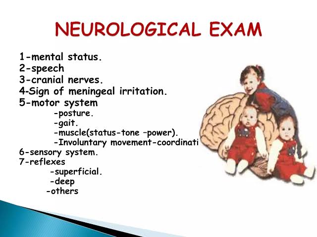
The Central Nervous System (CNS) assessment is one of the most critical, complex, and vital components of a full body physical examination. Unlike other systems where pathology is often localized, neurological issues can manifest anywhere in the body, requiring a highly structured, logical approach.
By the end of this comprehensive session, the nursing student should be able to:
- Prepare the patient and equipment for a safe, respectful neurological examination.
- Perform a structured CNS assessment: mental status, cranial nerves, motor system, reflexes, sensation, coordination, and gait.
- Recognise common abnormal findings and relate them to likely neurological anatomical localisation.
- Identify red flags requiring urgent medical escalation.
- Document neurological findings clearly and communicate them using a concise clinical summary.
A neurological assessment is not just a collection of tests; it is a vital tool to map the integrity of the brain, spinal cord, and peripheral nerves.
| What the Examination Tells You | What it Changes in Clinical Practice |
|---|---|
|
|
DETECT (Find the abnormality) ➔ LOCALISE (Determine where in the nervous system the problem originates) ➔ ESCALATE (Get help if it's life-threatening) ➔ DOCUMENT (Record the baseline accurately).
A complete neurological exam follows a predictable, systematic sequence to ensure no pathways are missed. The sequence can be shortened for a rapid screen, then expanded according to specific findings.
- Mental Status: Level of consciousness, orientation, language, and memory.
- Cranial Nerves (I-XII): Testing the brainstem and specialized sensory/motor functions of the head and neck.
- Motor System: Muscle bulk, tone, and power.
- Reflexes: Deep tendon reflexes and plantar responses.
- Sensation: Spinothalamic (pain/temperature) and Dorsal Column (vibration/proprioception) pathways.
- Coordination: Cerebellar function.
- Gait & Balance: Integration of motor, sensory, and cerebellar systems.
- Introduce & Consent: Introduce yourself, confirm patient identity, explain the examination (some neuro tests seem strange to patients), and obtain consent.
- Assess Immediate Stability First: Ensure Airway, Breathing, Circulation, and blood glucose are stable, especially when altered consciousness is present (hypoglycemia closely mimics a stroke).
- Environment: Provide privacy, expose only what is necessary, and always compare right with left symmetrically.
- Modify as Needed: Check whether severe pain, recent injury, severe weakness, dizziness, or cervical-spine (neck) injury risk will force you to modify the examination.
- Penlight / torch (for pupillary reflexes).
- Reflex hammer (for deep tendon reflexes).
- 128-Hz tuning fork (specifically for vibration sense).
- Cotton wool / tissue (for light touch sensation).
- Blunt neurotip or disposable pin (for pain/pinprick sensation).
- Snellen or near-vision chart (for Cranial Nerve II).
- Tongue depressor (for Cranial Nerves IX, X).
- Familiar objects (coin, key) for testing cognition/stereognosis.
The patient's history heavily guides the physical examination and helps localise the lesion before you even touch them.
- Headache, seizures, loss of consciousness (syncope).
- Weakness, numbness, tingling (paraesthesia), neuropathic pain.
- Visual changes (blurring, double vision/diplopia), speech difficulty (slurring, inability to find words), swallowing difficulty (dysphagia), or hearing disturbances.
- Tremor, imbalance, frequent falls, gait changes.
- Bladder or bowel dysfunction (incontinence or retention - major red flags for spinal cord compression).
| Element | Clinical Implication / Localisation |
|---|---|
| Onset & Time Course |
|
| Distribution |
|
| Associated Features |
|
Clinical Pearl: Always establish the Previous Baseline. Knowing if a neurological finding (like a slight facial droop) is genuinely new today, or if they have had it for 5 years since a previous stroke, dictates your entire emergency response.
Useful at first contact or during routine vital sign observation.
- LOOK: Alertness, speech clarity, facial symmetry, spontaneous posture.
- ASK: Name, place, time, current symptoms (tests orientation and speech fluency).
- ARMS: Assess for pronator drift (patient holds arms out, palms up, eyes closed; if one arm drifts down and pronates, it indicates mild upper motor neuron weakness), test gross power.
- LEGS: Test gross power, ask them to stand if safe.
- EYES: Check pupils, gaze, gross visual fields.
- WALK: Observe gait and balance if safe.
| Domain | How to Assess & Abnormal Findings |
|---|---|
| Alertness | Observe if awake spontaneously, responds to voice, or requires pain to rouse. Abnormal: Drowsy, difficult to arouse, responds only to pain. |
| Interaction & Affect | Have a normal conversation. Assess eye contact, appropriate responses, and emotional state. Abnormal: Flat affect, emotional lability (crying/laughing inappropriately), perseveration. |
| Orientation | Ask: "What is your name? Where are we? What is the date/year? Why are you here?" |
| Attention | Ask the patient to say the months of the year backward, or spell "WORLD" backwards. |
| Memory | Immediate: Repeat 3 words immediately. Recent: Recall those 3 words after 5 minutes. Remote: Verifiable past personal facts/historical events. |
| Language | Ask them to name common objects (pen, watch), repeat a phrase ("No ifs, ands, or buts"), follow a 3-step command, and write a sentence. |
| Higher Function | Assess abstraction (proverb interpretation, e.g., "Don't cry over spilled milk"), calculations, and praxis (ability to perform learned motor tasks, like pretending to brush teeth). |
Documentation Tip: Document exactly what you observed—not merely "oriented" or "confused." Write: "Patient alert, unable to recall the year or their location, short-term memory 0/3 words at 5 mins."
A standardized tool for assessing the depth and duration of impaired consciousness and coma.
- Eye Opening (E) - Max 4: Spontaneous (4), To sound/voice (3), To pressure/pain (2), None (1).
- Verbal Response (V) - Max 5: Oriented (5), Confused conversation (4), Inappropriate words (3), Incomprehensible sounds (2), None (1).
- Motor Response (M) - Max 6: Obeys commands (6), Localises to pain (5), Normal flexion/withdrawal to pain (4), Abnormal flexion/decorticate posturing (3), Extension/decerebrate posturing (2), None (1).
Practical Rules: Always use the best observed response. Trend the components over time (e.g., E3V4M5 = 12), not just the total score. A falling GCS (e.g., dropping by 2 or more points) is a massive clinical deterioration signal—reassess ABCs and escalate promptly.
Organize the 12 Cranial Nerves (CN) by function to make assessment logical and fluid.
- Visual Acuity: Test one eye at a time using a Snellen chart. Have them wear their corrective lenses.
- Visual Fields (Confrontation): Sit at patient's eye level, 1 meter away. Cover opposite eyes. Bring a wiggling finger in from the periphery in all 4 quadrants. This detects hemianopia (blindness over half the field of vision, common in strokes).
- Fundoscopy: Use an ophthalmoscope to inspect the optic disc, vessels, and retina (looking for papilledema/swelling).
- Pupils & Light Reflex (CN II in, CN III out): Assess size, symmetry, and reactivity in ambient light. Shine light from the side to observe direct (same eye) and consensual (opposite eye) constriction.
- Red Flag: Anisocoria (unequal pupils) + ptosis (drooping eyelid) + ophthalmoplegia (paralysis of eye muscles) = CN III palsy. This can indicate an expanding intracranial aneurysm or brain herniation—a profound medical emergency!
- Eye Movements (H-Pattern): Keep the patient's head still. Ask them to follow your finger as you draw a large "H" in the air, moving through the six cardinal positions of gaze. Look for nystagmus (jerky eye movements), limitations, or reports of diplopia (double vision).
- CN VI Palsy: The affected eye cannot abduct (look outward laterally).
- CN IV Palsy: The patient cannot look downward and inward; they often tilt their head to compensate for double vision.
- Sensory: Test facial sensation using light touch and pinprick in all three divisions: V1 (Ophthalmic/forehead), V2 (Maxillary/cheeks), V3 (Mandibular/jaw).
- Motor: Ask the patient to clench their teeth tightly; palpate the masseter and temporalis muscles for equal strength.
- Corneal Reflex: Afferent limb is CN V, efferent is CN VII. (Assessed only when clinically indicated).
- Motor: Inspect resting symmetry. Ask the patient to: Raise eyebrows, close eyes tightly against resistance, smile broadly, show teeth, and puff out cheeks.
- Clinical Pearl: Differentiating a stroke (Upper Motor Neuron) from Bell's Palsy (Lower Motor Neuron). An UMN lesion (stroke) typically spares the upper forehead because the forehead has bilateral cortical representation. A LMN lesion (Bell's Palsy) paralyses the entire half of the face, including the forehead.
- Hearing: Whisper test or rubbing fingers together near the ear. Use Rinne and Weber tuning fork tests if hearing loss is detected to differentiate conductive vs. sensorineural hearing loss.
- Assessment: Listen to the voice for hoarseness or nasal quality. Ask patient to say "Ahhh"—observe the uvula and soft palate. The palate should elevate symmetrically, and the uvula should remain midline. If there is a lesion, the uvula deviates away from the side of the lesion.
- Note: Do not provoke the gag reflex routinely in every patient; assess it only when clinically necessary (e.g., assessing aspiration risk).
- Motor: Ask the patient to shrug their shoulders against your hands' resistance (Trapezius muscle), and turn their head side-to-side against resistance (Sternocleidomastoid muscle).
- Assessment: Ask the patient to stick their tongue straight out. Look for wasting (atrophy), fasciculations (twitching), and deviation. A lower motor neuron lesion causes the tongue to deviate toward the side of the lesion (the weak side gets pushed over by the strong side).
Assess the motor system in a strictly logical sequence: Inspect ➔ Tone ➔ Power.
- Inspection: Look at posture, involuntary movements (tremors, chorea), muscle bulk (atrophy/wasting), asymmetry, and fasciculations (muscle twitches indicative of LMN disease).
- Tone: Ask the patient to relax completely ("go floppy"). Move their joints (wrists, elbows, knees, ankles) passively through a range of motion.
- Hypertonia/Spasticity: Increased resistance, common in Upper Motor Neuron (UMN) lesions like stroke.
- Hypotonia/Flaccidity: Decreased resistance, common in Lower Motor Neuron (LMN) lesions.
- Power: Test key muscle groups systematically against your resistance. Always stabilise the proximal joint and compare side to side.
0: No muscle contraction visible.
1: Flicker or trace of contraction.
2: Active movement, but cannot overcome gravity (moves sideways on bed).
3: Active movement against gravity, but not against any resistance.
4: Active movement against gravity and some resistance.
5: Normal power against full resistance.
Note: Pain-limited effort is NOT the same as true neurological weakness. Document if pain prevented full testing.
| Region | Action Tested | Primary Nerve Root |
|---|---|---|
| Upper Limb | Shoulder abduction (Deltoid) Elbow flexion (Biceps) Elbow extension (Triceps) Wrist extension (Extensors) Finger abduction (Interossei) Grip strength | C5 C5–C6 C7 C6–C7 T1 C8–T1 |
| Lower Limb | Hip flexion (Iliopsoas) Knee extension (Quadriceps) Knee flexion (Hamstrings) Ankle dorsiflexion (Tibialis Anterior) Plantar flexion (Gastrocnemius) Great-toe extension (EHL) | L1–L3 L3–L4 L5–S1 L4–L5 S1–S2 L5 |
DTRs are stretch reflexes that assess the integrity of the sensory nerve, the specific spinal cord segment, the motor nerve, the neuromuscular junction, and the muscle itself.
- Technique: Ensure the patient is relaxed. Expose the limb. Use a tendon hammer with a quick, brisk, swinging tap directly on the tendon. Compare both sides.
- Key Reflexes & Roots: Biceps (C5-C6), Supinator/Brachioradialis (C5-C6), Triceps (C7-C8), Knee/Patellar (L3-L4), Ankle/Achilles (S1-S2).
- 0: Absent
- 1+: Reduced / sluggish
- 2+: Normal
- 3+: Brisk
- 4+: Very brisk with clonus (rhythmic, oscillating movements)
Clinical Pearl: Reflexes are pivotal for localization.
Upper Motor Neuron (UMN) lesions (brain/spinal cord, e.g., stroke, MS) ➔ result in Hyperreflexia (3+, 4+) + increased tone + weakness.
Lower Motor Neuron (LMN) lesions (peripheral nerves, e.g., neuropathy, radiculopathy) ➔ result in Hyporeflexia/Areflexia (0, 1+) + decreased tone + wasting + fasciculations.
This is a superficial reflex used to assess the corticospinal (upper motor neuron) pathway.
- Technique: Use a blunt object (like the back of a reflex hammer). Stroke the lateral sole of the foot from the heel toward the forefoot, then curve medially across the ball of the foot. Observe the great toe.
- Interpretation:
- Normal Adult Response: Flexor (downgoing) great toe and curling of other toes.
- Positive Babinski Sign: Extensor (upgoing) great toe with fanning of the other toes. This is highly indicative of an Upper Motor Neuron lesion in adults.
- Infants: An extensor plantar response is completely normal in babies up to 1-2 years old because their corticospinal tracts are not yet fully myelinated.
Test with the patient’s eyes closed to ensure they are feeling the stimulus, not just seeing you touch them.
| Sensation & Pathway | Technique | Normal / Abnormal Response |
|---|---|---|
| Light Touch (Peripheral nerves & Spinothalamic) | Use cotton wool. Lightly touch dermatomes. Ask patient: "Tell me when you feel the touch." | Normal: Identifies stimulus. Abnormal: Hypoesthesia (reduced), Hyperesthesia (increased/painful), Anesthesia (absent). |
| Pain / Pinprick (Spinothalamic Tract) | Apply gentle sharp stimuli with a neurotip. Ask "Is it dull or sharp?" | Correctly differentiates sharp from dull. |
| Temperature (Spinothalamic Tract) | Use a warm or cold object (often a cold tuning fork). | Correctly identifies hot vs. cold. |
| Vibration Sense (Dorsal Column Pathway) | Strike a 128 Hz tuning fork. Place on a bony prominence (distal interphalangeal joint of finger or big toe). Ask: "Can you feel the buzzing? Tell me when it stops." | Patient feels vibration and correctly identifies the exact moment it stops. |
| Joint Position Sense (Proprioception) (Dorsal Column Pathway) | Hold the sides of the patient's distal finger or toe. Move it slightly up or down. Ask: "Is it moving up or down?" | Correctly identifies direction of movement with eyes closed. |
These test the sensory cortex's ability to interpret complex sensory data.
- Stereognosis: Place a familiar object (coin, key) in the patient's hand with their eyes closed. Ask them to identify it by feeling it.
- Graphesthesia: Draw a number or letter on the patient's palm with a blunt object. Ask them to identify it.
- Two-Point Discrimination: Apply two blunt points simultaneously on the skin. Find the minimum distance at which they can distinguish two points rather than one.
The cerebellum is located at the back of the brain and coordinates balance, posture, muscle tone, and smooth, accurate voluntary movements. A lesion here does not cause weakness; it causes clumsiness and lack of coordination.
Dysdiadochokinesia (difficulty with rapid alternating movements)
Ataxia (uncoordinated, wide-based, drunken-like gait)
Nystagmus (jerky eye movements)
Intention tremor (tremor that gets worse as the finger approaches the target)
Slurred, scanning speech (staccato, irregular rhythm)
Hypotonia (decreased muscle tone)
- Finger-Nose Test: Assesses upper limb coordination and dysmetria (past-pointing). Ask patient: "Touch your nose, then touch my finger, and repeat." Move your finger to different positions. The movement should be smooth and accurate without overshooting or shaking (intention tremor).
- Rapid Alternating Movements (Dysdiadochokinesia test): Ask the patient to rapidly pronate and supinate their hands on their thighs, or rapidly tap their thumb against their fingers. Movements should be fast and rhythmic.
- Heel-Shin Test: Assesses lower limb coordination. Ask the patient (lying down) to "Place your heel on the opposite knee and slide it smoothly down your shin."
- Gait Assessment: Ask the patient to walk normally, turn, and walk heel-to-toe (tandem gait). Cerebellar lesions produce a wide-based, unsteady, veering gait.
- Romberg Test: Technically a test of proprioception (dorsal column), but helps assess balance. Ask the patient to stand with feet together, eyes open. Then ask them to close their eyes.
Positive Romberg: The patient maintains balance with eyes open, but sways heavily or falls when eyes close (indicating sensory ataxia/dorsal column disease). If they fall with eyes open and closed, it indicates a cerebellar issue.
Consider meningitis or subarachnoid hemorrhage when a patient presents with headache, fever, photophobia, altered mental status, or meningism.
- Neck Stiffness (Nuchal Rigidity): After ruling out cervical spine trauma, place your hands behind the patient's head and gently, passively flex the neck forward. Resistance and pain indicate meningeal irritation.
- Kernig's Sign: Flex the patient's hip and knee to 90 degrees, then attempt to straighten the leg at the knee. Severe pain in the lower back and resistance to extension is a positive sign.
- Brudzinski's Sign: As you passively flex the patient's neck forward, their hips and knees involuntarily flex to relieve the tension on the spinal cord.
Neurological diagnosis relies on combining signs rather than a single finding.
| Pattern / Lesion Location | Typical Clinical Clues |
|---|---|
| Upper Motor Neurone (UMN) (Brain/Spinal Cord) | Weakness + increased tone (spasticity) + brisk reflexes (hyperreflexia) + extensor plantar response (Babinski). |
| Lower Motor Neurone (LMN) (Peripheral Nerve) | Weakness + wasting (atrophy) / fasciculations + reduced tone (flaccidity) + reduced/absent reflexes. |
| Cerebellar | Ataxia, dysmetria, intention tremor, nystagmus, dysarthria (speech). No marked weakness. |
| Peripheral Neuropathy | Distal sensory loss ("stocking-glove") ± distal weakness and reduced reflexes at ankles. |
| Spinal Cord | Bilateral long-tract signs (paraplegia/quadriplegia) ± distinct sensory level (loss of sensation below a specific dermatome) ± bowel/bladder sphincter dysfunction. |
If you see any of the following, use local emergency pathways (e.g., Code Stroke) and document the exact time symptoms were first noted or the "last known well" time.
- Sudden focal weakness, speech, or language disturbance (Classic stroke symptoms).
- New seizure with persistent reduced consciousness (Status epilepticus).
- Rapidly progressive weakness (e.g., ascending weakness in Guillain-Barré syndrome causing respiratory failure).
- Acute severe headache with neurological deficit or altered consciousness ("Worst headache of my life" = Subarachnoid hemorrhage).
- New pupil abnormality with ptosis/ophthalmoplegia (CN III compression).
- Spinal cord warning signs: bilateral weakness + distinct sensory level + bowel/bladder retention or incontinence (Spinal Cord Compression or Cauda Equina Syndrome).
Example of Excellent Documentation:
"Alert and conversant; oriented to person, place and time. Speech fluent. Pupils 3 mm, equal and reactive to light; visual fields full to confrontation; extraocular movements full, no nystagmus. Face symmetric; tongue midline. Normal muscle bulk and tone. Power 5/5 in all major upper and lower muscle groups. Reflexes 2+ and symmetric; plantar responses flexor bilaterally. Light touch, pinprick, vibration and joint position intact. Finger–nose and heel–shin smooth and accurate. Gait steady with normal base and arm swing."
Rule of thumb: Document abnormal findings precisely (side, distribution, grade, severity) and explicitly list relevant negatives (e.g., "No facial droop").
Scenario: A 58-year-old develops sudden right-arm and right-leg weakness with difficulty speaking 45 minutes ago. He is alert. Pupils are equal and reactive. Face shows right lower facial weakness. Right limbs are power 3/5 with brisk reflexes.
- What is the likely neurological pattern? Upper Motor Neuron (UMN) lesion, specifically involving the Left Cerebral Hemisphere (causing right-sided contralateral weakness, right lower facial weakness, and aphasia).
- Which additional focused findings would you check immediately? ABCs, blood glucose (to rule out hypoglycemia mimicking stroke), and perform a rapid stroke scale (like the NIHSS or FAST).
- What information must be documented precisely? The exact time of onset ("Last known well time" - crucial for clot-busting tPA administration), vital signs, and the specific deficits.
- What is your next action? Immediate escalation! This is a sudden focal deficit. Activate the local acute stroke pathway and prepare for urgent transport to CT scan.
- Use the same sequence every time. Compare right with left.
- Describe what you see before trying to name a complex syndrome.
- Interpret findings as patterns that localise a lesion (e.g., UMN vs LMN).
- Safety and escalation come before completing the entire checklist.
- Serial examinations (doing the exam repeatedly over time) are often much more informative than a single snapshot.
- Dover AR, Innes JA, Fairhurst K, eds. Macleod’s Clinical Examination. 15th ed. Elsevier; 2023.
- National Institute for Health and Care Excellence (NICE). Suspected neurological conditions: recognition and referral (NG127). Updated October 2023.
- Glasgow Coma Scale. Official educational resources. GlasgowComaScale.org.
- Bickley LS. Bates’ Guide to Physical Examination and History Taking. 13th ed. Wolters Kluwer; 2021.
- OpenStax. Anatomy & Physiology: The Cranial Nerves. CC BY 4.0.
Quick Quiz
CNS Examination Quiz
Health Assessment
Privacy: Your details are used only for quiz tracking and certificates.
CNS Examination Quiz
Health Assessment
Preparing questions...
Choose your answer and keep your streak alive.
Great effort.
Here is your quick performance summary.