
Glaucoma is damage to the optic nerve caused by increased pressure inside the eye (intraocular pressure/IOP). In babies, this pressure makes the eye enlarge because the eye wall (sclera and cornea) is still soft, immature, and stretchy. The name "buphthalmos" means "ox-eye" because the eye looks like a cow's eye—big, bulging, and cloudy.
Physiological Expansion (Aqueous Humor Pathway): The eye constantly produces fluid (aqueous humor) in the ciliary body. This fluid flows through the pupil and drains out of the eye through the trabecular meshwork (located in the angle where the iris meets the cornea). If this drain is blocked, fluid builds up, pressure spikes, and it physically crushes the delicate optic nerve fibers at the back of the eye.
READ MORE ABOUT GLAUCOMA BY CLICKING HERE- Developmental defect: The drainage system inside the eye (trabecular meshwork) does not form properly before birth (trabeculodysgenesis).
- Genetic: Often autosomal recessive (both parents carry the gene).
- Associated with other systemic conditions: Aniridia (missing iris), Neurofibromatosis, Sturge-Weber syndrome (port-wine stain on face).
| Sign | Description & Mechanism |
|---|---|
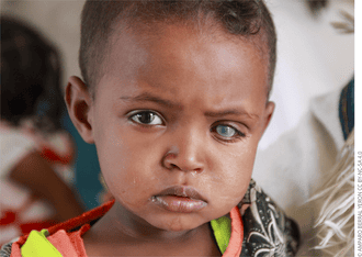
| Buphthalmos | Enlarged, protruding eye—usually one eye first, then both. The high pressure physically stretches the elastic infant sclera. |
| Corneal clouding (oedema) | The clear front window of the eye looks milky/blue-white. High pressure forces fluid into the corneal tissue, causing it to swell and lose transparency. |
| Photophobia | Baby squeezes eyes shut in light, turns away from windows. The swollen cornea scatters light painfully. |
| Epiphora (tearing) | Constant watering of eyes—not just when crying. Often mistaken for a blocked tear duct! |
| Blepharospasm | Forceful blinking or squeezing eyelids together due to severe pain and light sensitivity. |
| Irritability | Baby cries excessively and refuses to feed due to agonizing headache-like pain from high eye pressure. |
🧠 MNEMONIC: "B-E-P-C" for Buphthalmos Signs
- Big eye (Buphthalmos & Blepharospasm)
- Epiphora (Constant tearing)
- Photophobia (Hates light)
- Corneal clouding (Looks blue/white)
These drugs only BUY TIME for surgery—they do NOT cure congenital glaucoma. The anatomy of the drain is physically blocked and must be surgically opened.
- Topical beta-blockers: Timolol eye drops. Mechanism: Reduce aqueous humour production by the ciliary body. (Watch for bradycardia/bronchospasm in babies!).
- Carbonic anhydrase inhibitors: Acetazolamide tablets. Mechanism: Inhibit the enzyme needed to secrete fluid into the eye.
- Hyperosmotic agents: Mannitol IV. Mechanism: Emergency use only—acts as a massive osmotic sponge in the blood, rapidly drawing fluid out of the eye to drop pressure before surgery.
| Surgery | When Used & Description | Nursing Notes |
|---|---|---|
| Goniotomy | First-line; a tiny blade is used to cut open the blocked drainage channels from the inside. | Requires a clear cornea and operating microscope; 80-90% success rate in babies. |
| Trabeculotomy | Used if the cornea is too cloudy for goniotomy; opens the drainage pathway from the outside. | Post-op: Monitor closely for hyphema (blood pooling in the front of the eye). |
| Trabeculectomy | Usually for older children; creates a completely new, artificial drainage bypass pathway (a "bleb"). | High risk of infection; lifelong follow-up needed. |
| Tube shunts (Ahmed/Baerveldt) | Used when other surgeries fail; places a physical silicone drainage tube into the eye. | Parents must learn to massage the tube area if blockage occurs. |
- Monitor IOP: Daily using a tonopen or gentle finger palpation (a rock-hard, firm eye = dangerously high pressure).
- Check for hyphema: Blood layering in the anterior chamber. Keep the child positioned upright/elevated to let gravity settle the blood away from the visual axis.
- Cycloplegic drops (Atropine): Prevents painful spasms and scarring; dilates the pupil. Warn parents about extreme light sensitivity.
- Steroid drops: Reduce surgical inflammation. Must watch closely for "steroid-induced glaucoma" (a paradoxical spike in pressure).
- Patching: If only one eye was operated on, patch the good eye to prevent amblyopia.
- Glaucoma is a lifelong disease—even after successful surgery, the child needs check-ups every 3-6 months for life.
- Medication compliance is CRITICAL—missing drops can cause irreversible blindness in a matter of hours.
- Teach siblings to be gentle—absolutely no rough play around the eyes (the enlarged eyes are stretched thin and can easily rupture).
- If the child complains of severe headache, nausea/vomiting, or sees colored halos around lights = ACUTE GLAUCOMA ATTACK → MEDICAL EMERGENCY.
Retinoblastoma is a malignant (cancerous) tumour of the retina. It is the most common primary intraocular malignancy in children. It can affect one eye (unilateral) or both eyes (bilateral). If detected early, the survival rate is >95%. If left untreated, it spreads down the optic nerve into the brain and kills the child.
Physiological Expansion (Tumour Growth): The tumour arises from immature retinal cells (retinoblasts). It can grow inward toward the vitreous jelly (endophytic), causing white "seeds" to float in the eye, or outward toward the choroid (exophytic), causing retinal detachment.
- Genetic (Germline/Hereditary): RB1 tumour suppressor gene mutation.
Mechanism (Knudson's Two-Hit Hypothesis): The child inherits one broken RB1 gene in every cell of their body. When the second copy breaks by random chance, cancer forms.
Because every cell is affected, it is hereditary, almost always affects BOTH eyes (bilateral), and drastically increases the risk of other cancers later in life (like osteosarcoma bone cancer or melanoma). - Non-hereditary (Somatic): Spontaneous mutation of BOTH copies of the RB1 gene in just one single retinal cell. More common, affects only one eye, and is not passed to children.
- Age profile: 90% diagnosed before age 5; median age of diagnosis is 18 months.
| Sign | What You See & Why |
|---|---|
| Leukocoria (White Pupil) | Most common first sign (60%). You are literally looking through the pupil and seeing the white, calcified tumour sitting on the retina. |
| Strabismus (Crossed eyes) | Second most common sign (20%). The tumour destroys central vision, so the brain lets the blind eye drift. |
| Red, painful eye | Inflammation mimicking infection (often mistakenly treated as conjunctivitis, causing fatal delays). |
| Proptosis | The eye bulging forward out of the socket. Indicates advanced disease where the tumour has grown massive. |
| Hyphema | Blood in the front of the eye (tumour vessels are fragile and bleed easily). |
| Orbital cellulitis-like picture | Swollen, red eyelids resulting from tumour necrosis and massive inflammation. |
| Pseudohypopyon | A white mass settling in the bottom of the front of the eye. These are actual tumour seeds floating in the fluid! |
🧠 MNEMONIC: "WHITE + CROSS" for Retinoblastoma
- WHITE:
- White pupil (leukocoria)
- Hereditary risk (always ask family history)
- Inflammation (red eye)
- Tumour seeds (pseudohypopyon)
- Eye bulging (proptosis)
- + CROSS:
- CROSSed eyes (strabismus)
- Red reflex absent
- Orbital swelling
- Second eye involved (bilateral)
- Siblings need screening
- Group A-E: Based on tumour size, location, and presence of seeding. Group A is tiny; Group E means the eye is destroyed and must be removed.
- Extraocular extension: The absolute worst-case scenario. The tumour has spread outside the eye (down the optic nerve to the brain). Prognosis drops drastically.
| Treatment | Indication | Nursing Care & Notes |
|---|---|---|
| Enucleation (removing the eye) | Large tumour filling >50% of eye; no useful vision left; glaucoma present. | Fit a prosthetic (glass) eye 4-6 weeks post-op. Deep psychosocial care—counsel parents about body image; the child will grieve the loss of the eye. |
| Chemoreduction | Bilateral disease; used to shrink tumours before local therapy to try and save the eyes. | Administer systemic chemotherapy via port. Monitor for neutropenia, vomiting, hair loss; strictly protect from infection. |
| Focal therapies (laser, cryotherapy) | Small tumours, or after chemoreduction has shrunk them. | Multiple sessions required under anaesthesia; nurse must dilate the pupil before each session. |
| Plaque radiotherapy | Residual tumour near the optic disc. | A radioactive plaque is sewn to the outside of the eye over the tumour. Nurse must teach radiation safety (limited contact with pregnant women/young children). |
| External beam radiotherapy (EBRT) | Extensive bilateral disease; used as a last resort. | High risk of causing second cancers (like bone cancer) in hereditary RB patients. Causes cataracts and severe dry eye. |
| Intra-arterial chemotherapy (IAC) | Advanced unilateral disease. | Directs chemo right into the ophthalmic artery. Requires interventional radiology. Nurse must monitor for stroke and bleeding at the groin access site. |
- Genetic counselling:
- Hereditary RB means a 50% chance each future child will inherit the gene.
- ALL siblings of the patient need eye exams under anaesthesia every 3-6 months until age 7.
- Parents should be screened for retinoma (a benign precursor tumour).
- Psychosocial support:
- Parents often carry massive guilt and blame themselves—reassure them it is a random genetic mutation and not their fault.
- Connect them to support groups for families who chose enucleation.
- Long-term surveillance:
- Hereditary RB survivors need an MRI every 6 months to check for a pineal gland tumour in the brain (called trilateral RB, which is highly fatal).
- Avoid CT scans: The radiation from CT scans vastly increases the risk of triggering second cancers in these genetically vulnerable children. Use MRI instead.
ANY white pupil (Leukocoria) or new-onset squint in a child under 5 = CANCER until proven otherwise.
Do NOT assume it is just a cataract or an infection. Do NOT prescribe antibiotic drops and send them home. You must make an urgent, fast-tracked referral to the Uganda Cancer Institute or Mulago Eye Department. Early detection prevents enucleation and literally saves the child's life.
❓ Applied Clinical Question
Case: A 2-year-old boy presents with a white pupil in his left eye and is diagnosed with unilateral Retinoblastoma. The ophthalmologist recommends immediate enucleation of the left eye. The parents are devastated and refuse, asking if you can just give him chemotherapy instead so he keeps his eye. How do you counsel them?
Answer: With immense empathy, explain that when a tumour is large enough to cause a white pupil, it has likely destroyed the vision in that eye entirely. Chemotherapy might not penetrate a massive tumour well enough. Explain that the eye is a direct pathway to the brain (via the optic nerve). Enucleation guarantees the cancer is removed from the body before it reaches the brain and becomes fatal. Reassure them that with a modern prosthetic eye, the child will look completely normal and live a long, healthy life.
Quick Quiz
Glaucoma Quiz
Surgical Nursing - mobile-friendly and focused practice.
Privacy: Your details are used only for quiz tracking and certificates.
Glaucoma Quiz
Surgical Nursing
Preparing questions...
Choose your answer and keep your streak alive.
Great effort.
Here is your quick performance summary.