
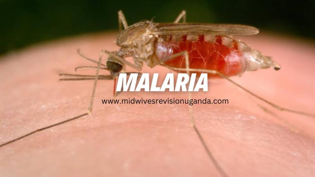
Malaria is an acute febrile, potentially life-threatening infectious disease caused by protozoan parasites of the genus Plasmodium and transmitted to humans through the nocturnal bite of an infected female Anopheles mosquito.
The disease manifests characteristically with paroxysms of severe shaking chills, high-spiking fevers, generalized body aches, and profuse drenching sweats, corresponding directly to the synchronous rupture of infected red blood cells.
There are five identified species of Plasmodium known to cause disease in human beings:
- Plasmodium falciparum: Responsible for practically all severe, complicated, and fatal malaria worldwide. It is the overwhelmingly dominant parasite in Uganda and sub-Saharan Africa. It has a high propensity for high parasite densities and vascular sequestration.
- Plasmodium vivax: Capable of causing classic, debilitating relapsing malaria due to dormant liver stages (hypnozoites). Prevalent in Asia and the Americas, though less common in West/Central Africa due to the protective Duffy antigen-negative phenotype.
- Plasmodium ovale: Also forms dormant hypnozoites in hepatocytes, leading to late relapses months to years later. Endemic predominantly in tropical Africa.
- Plasmodium malariae: Causes quartan malaria (fever spikes every 72 hours). It can persist asymptomatically in the human bloodstream at low levels for decades and is associated with chronic immune-complex nephrotic syndrome.
- Plasmodium knowlesi: A zoonotic parasite naturally infecting long-tailed and pig-tailed macaques in Southeast Asia; can cause rapid, life-threatening hyperparasitaemia in humans.
In Uganda, malaria is transmitted primarily by two highly efficient mosquito vectors:
- Anopheles gambiae complex (the most efficient vector globally)
- Anopheles funestus
Both species breed primarily in clean, stagnant fresh water collections. They are strictly nocturnal, anthropophilic (prefer feeding on human blood), and endophagic/endophilic (they prefer to enter homes, bite indoors at night, and rest on internal walls after feeding).
- Malaria remains the single leading cause of illness and death in Uganda.
- It accounts for approximately 25% to 40% of all outpatient health facility attendances, 20% of inpatient hospital admissions, and 9% to 14% of all inpatient hospital deaths.
- The disease extracts a devastating physical and economic toll, causing chronic severe anaemia, low birth weight, impaired child physical and cognitive development, and catastrophic household expenditures.
- The populations at highest risk of severe disease, complications, and death are:
- Children under 5 years of age (due to lack of acquired semi-immunity).
- Pregnant women (due to immunological changes and parasite sequestration in the placenta).
- Non-immune individuals (such as international travelers and residents of non-endemic highland areas).
- Individuals living with HIV/AIDS or severe malnutrition.
For sustainable malaria transmission to occur within an ecosystem, four interrelated biological and environmental criteria must be met:
- Human Reservoir: The presence of human hosts carrying viable sexual forms of the parasite (gametocytes) in their peripheral blood. Notably, asymptomatic individuals often serve as silent reservoirs.
- Breeding Sites: Presence of stagnant, unpolluted water bodies (pits, road puddles, agricultural ditches, brick-making excavations, ponds, marshes, animal footprints).
- Climatic Conditions: Warm, humid conditions with relative humidity greater than 60%. Parasite sporogony within the mosquito requires a mean daily ambient temperature consistently above 18°C for P. falciparum.
- Vector Density & Human Contact: Sufficient densities of long-lived female Anopheles mosquitoes biting susceptible individuals.
In many rural communities, there is a widespread belief that eating unwashed mangoes, consuming freshly harvested maize or millet, walking in heavy rain, or drinking dirty water directly causes malaria. Health workers must clearly explain that these beliefs arise because the rainy harvest seasons coincide directly with the explosive multiplication of mosquito breeding sites, resulting in a spike in infectious mosquito bites, not nutritional or waterborne transmission.
The female mosquito undergoes complete metamorphosis (Egg ➔ Larva ➔ Pupa ➔ Adult) alongside a continuous gonadotrophic cycle:
- Night 1 (Feeding): The female mosquito seeks a blood meal to obtain protein necessary for egg development. If unsuccessful, she rests and tries again the following night.
- Day 2 (Resting & Digestion): She rests indoors in dark, cool, humid corners. Digestion of the blood meal occurs, and her ovaries develop eggs.
- Night 2 (Half-Gravid): Continues to rest as egg maturation proceeds; may relocate to other resting surfaces.
- Day 3 (Fully Gravid): Egg maturation is fully complete.
- Day 3 at Dusk (Oviposition / Egg Laying): The gravid female flies to a suitable water body and deposits 50 to 150 eggs on the water surface. She then immediately seeks another blood meal, repeating the cycle.
- Larval Stage (2–3 Days): Eggs hatch into aquatic larvae that breathe via surface spiracles and filter-feed on microscopic algae. Over 3 days, they undergo 3 successive moults.
- Pupal Stage (2–3 Days): After the 3rd moult, larvae become non-feeding, comma-shaped pupae that breathe through two cephalothoracic air trumpets. Internal transformation into the adult form takes place.
- Emergence: The pupal skin splits along the dorsal surface, and the soft adult climbs out, rests on the surface to dry and harden its cuticle, and flies away.
- Mating: Males form swarms at twilight. Mating takes place in flight, and spermatozoa are deposited into the female's spermatheca to fertilize eggs throughout her lifespan.
The life cycle requires two hosts: a definitive host (the female Anopheles mosquito, where sexual reproduction occurs) and an intermediate host (humans, where asexual reproduction occurs).
- Inoculation: During a blood meal, an infected mosquito injects saliva containing motile, thread-like sporozoites into the human dermis and capillary bed.
- Exo-Erythrocytic (Hepatic) Stage: Within 30 to 45 minutes, sporozoites migrate through the bloodstream and invade hepatocytes (liver parenchymal cells).
- Inside liver cells, each sporozoite multiplies asexually by schizogony over 7 to 14 days, transforming into a massive liver schizont containing up to 30,000 to 40,000 daughter merozoites. This phase is clinically completely silent (asymptomatic).
- Hypnozoite Formation: In P. vivax and P. ovale infections, a proportion of sporozoites do not divide immediately but enter a dormant "sleeping" state called hypnozoites. These remain latent in liver tissue for weeks, months, or years, periodically reactivating to cause clinical relapses.
- Erythrocytic Stage (Asexual Cycle): The liver schizont ruptures, discharging waves of merozoites into the bloodstream. These merozoites rapidly invade red blood cells (erythrocytes).
- Inside the red cell, the parasite consumes host hemoglobin and develops into a ring-form, which matures into an active, amoeboid trophozoite.
- The trophozoite undergoes nuclear division to form an erythrocytic schizont containing 8 to 24 merozoites.
- The erythrocyte eventually bursts, releasing new merozoites, metabolic toxins (hemozoin), and pyrogenic cellular debris into systemic circulation. The released merozoites immediately invade fresh erythrocytes, repeating the cycle every 48 hours (in P. falciparum, P. vivax, P. ovale) or 72 hours (in P. malariae).
- Gametocytogenesis (Sexual Differentiation): After several erythrocytic cycles, a small fraction of intra-erythrocytic merozoites differentiate into male and female sexual forms known as microgametocytes and macrogametocytes, respectively. These forms do not cause symptoms and circulate freely, waiting to be ingested by a biting mosquito.
- Ingestion: When a female Anopheles feeds on an infected human, she ingests erythrocytes containing mature gametocytes.
- Gametogenesis: Within the mosquito midgut, erythrocytes lyse. The male microgametocyte undergoes exflagellation, producing eight whiplike, highly motile microgametes.
- Fertilization: A microgamete fertilizes a female macrogamete, resulting in a diploid zygote.
- Ookinete Formation: Within 18 to 24 hours, the zygote elongates into a motile, worm-like ookinete. The ookinete penetrates the peritrophic membrane and epithelial lining of the midgut wall.
- Oocyst Development: Underneath the gut's outer basal lamina, the ookinete encysts into an oocyst. Inside this expanding cyst, extensive asexual division (sporogony) takes place, generating thousands of active, spindle-shaped sporozoites.
- Migration to Salivary Glands: The mature oocyst bursts, discharging sporozoites into the mosquito's hemolymph (body cavity). The sporozoites migrate forward and lodge within the acinar cells of the mosquito's salivary glands, ready to be inoculated into another human during the next blood meal.
The clinical manifestations of malaria correlate directly with the synchronized rupture of mature erythrocytic schizonts. The rupture dumps large amounts of foreign materials into the blood: free merozoites, red cell membrane remnants, and hemozoin (malaria pigment formed by the parasite digesting hemoglobin). These foreign products stimulate circulating monocytes and macrophages to release potent endogenous pyrogens, primarily Tumor Necrosis Factor-alpha (TNF-α), Interleukin-1 (IL-1), and Interleukin-6 (IL-6). These cytokines reset the hypothalamic thermoregulatory center, producing acute, incapacitating shivering, followed by intense high fever, tachycardia, headache, and finally diaphoresis.
The hallmark that makes P. falciparum uniquely lethal is its capacity to alter the biomechanical properties of infected erythrocytes:
- PfEMP-1 Expression: The growing parasite expresses adhesive protein complexes on the erythrocyte surface, visualized under electron microscopy as electron-dense "knobs." The primary ligand is Plasmodium falciparum erythrocyte membrane protein 1 (PfEMP-1).
- Cytoadherence: PfEMP-1 binds with high affinity to endothelial receptors lining post-capillary venules (such as CD36, ICAM-1, VCAM-1, and Chondroitin Sulfate A in the placenta).
- Rosetting: Infected red cells simultaneously adhere to uninfected red cells, forming clustered "rosettes," and clump to blood platelets (auto-agglutination).
- Consequences: The rigid, adherent red blood cells completely plug the microvasculature of vital organs. This provides an evolutionary advantage to the parasite by preventing infected cells from circulating through the spleen where they would be destroyed. However, for the host, microvascular clogging results in tissue hypoperfusion, severe localized ischemia, endothelial leakage, tissue hypoxia, and organ failure.
- Cerebral Malaria: Microvascular sequestration within deep cerebral capillaries leads to blood-brain barrier disruption, localized cerebral edema, petechial hemorrhages, intracranial hypertension, and coma.
- Severe Metabolic Acidosis: Caused by systemic tissue hypoperfusion from microvascular obstruction combined with parasite-driven anaerobic glycolysis and severe anemia, resulting in high levels of lactic acid. Clinically, this presents as deep, rapid, laboured breathing ("respiratory distress" or Kussmaul-like breathing).
- Severe Anaemia: Rapid, massive destruction of both parasitized and non-parasitized red blood cells via complement-mediated hemolysis, hypersplenic clearance, and profound bone marrow suppression (dyserythropoiesis).
- Acute Renal Failure: Occurs due to acute tubular necrosis triggered by renal cortical ischemia, hemoglobin cast deposition, and systemic shock. When massive intravascular hemolysis produces heavy hemoglobinuria that stains urine dark brown or black, it is termed Blackwater Fever.
- Hypoglycemia: Caused by heavy parasite consumption of host glucose, liver dysfunction (impaired gluconeogenesis and glycogenolysis), and drug-induced hyperinsulinemia (specifically from Quinine therapy stimulating pancreatic beta-cell insulin secretion).
- Non-Cardiogenic Pulmonary Edema (ARDS): Triggered by severe pulmonary microvascular endothelial damage, increased capillary permeability, and cytokine-induced fluid extravasation into alveoli, independent of cardiac volume overload.
The time between the infectious bite and the onset of clinical symptoms typically spans 9 to 14 days for P. falciparum, 12 to 17 days for P. vivax and P. ovale, and 18 to 40 days for P. malariae. However, prior chemoprophylaxis, self-medication, or partial host immunity can delay onset for weeks or months.
Defined as clinical malaria manifesting with fever without any evidence of vital organ dysfunction or danger signs of severe disease.
- The Cold Stage (15 to 60 Minutes): Sudden onset of intense, uncontrollable shivering, teeth chattering, peripheral vasoconstriction, pale cold skin, and cyanotic nail beds. The patient wraps themselves in blankets despite an already rapidly rising core body temperature.
- The Hot Stage (2 to 6 Hours): Peripheral vasodilation takes over. The skin becomes flushed, burning hot, and dry. Temperature frequently peaks between 39°C and 41°C. Accompanied by severe throbbing frontal headache, nausea, vomiting, intense thirst, tachypnea, and delirium (especially in young children).
- The Sweating Stage (2 to 4 Hours): Profuse, drenching diaphoresis breaks out. The core temperature drops precipitously to normal or subnormal levels. The patient experiences immediate relief from headache and pain, followed by exhaustion, profound weakness, and deep sleep.
In malaria-endemic areas like Uganda, the classical periodic paroxysms are frequently absent, especially in P. falciparum infections where fever is often continuous, irregular, or erratic. Furthermore, partially immune individuals or patients who have taken incomplete antimalarial doses may show an altered, blunted presentation. The body temperature may be entirely normal during a daytime clinic visit, only to spike severely hours later. Always rely on a documented history of fever within the past 24–48 hours.
- High fever (or history of fever)
- Anorexia (poor feeding / refusal to suckle)
- Vomiting and diarrhea
- Marked irritability, restlessness, or lethargy
- Generalized weakness and malaise
- Cough and mild tachypnea
- Intermittent or continuous fever/chills
- Severe generalized headache
- Diffused arthralgia (joint pains) and myalgia (muscle aches)
- Nausea, vomiting, and abdominal cramps
- Severe loss of appetite and physical fatigue
- Postural dizziness and mild jaundice
- Elevated body temperature (axillary temperature > 37.5°C).
- Mild pallor of the conjunctivae, palms, and buccal mucosa.
- Mild dehydration (dry tongue, cracked lips, reduced skin turgor).
- Splenomegaly: In acute attacks, the spleen may be soft, palpable, and mildly tender. (Requires no independent medical treatment).
- Hepatomegaly: Soft, mildly tender liver margin.
Any patient presenting with fever and ONE or more of the following danger signs must be managed immediately as a life-threatening medical emergency for Severe Malaria:
- Convulsions/Fits: Generalized seizures occurring within the last 2 days or observed currently.
- Inability to Drink or Breastfeed: Complete oral intolerance, unable to swallow fluids.
- Persistent Vomiting: Vomiting all food, drinks, and oral medicines immediately.
- Altered Level of Consciousness: Extreme lethargy, drowsiness, confusion, disorientation, somnolence, delirium, or unarousable coma.
- Prostration: Extreme generalized weakness where an adult cannot sit or stand without assistance, or a child cannot sit up or hold their head up.
- Severe Respiratory Distress: Deep, rapid, acidotic breathing (Kussmaul breathing), nasal flaring, intercostal/subcostal chest indrawing, or grunting.
- Severe Anemia: Profound, chalky pallor of the palms, tongue, nailbeds, and palpebral conjunctivae.
- Severe Dehydration: Sunken eyes, very dry mouth, skin pinch retracting very slowly (>2 seconds), and lethargy.
- Shock (Algid Malaria): Cold, clammy extremities, weak/thready rapid pulse, prolonged capillary refill time (>2 seconds), and systolic hypotension.
- Jaundice: Frank yellowing of the sclerae and skin combined with signs of vital organ dysfunction.
- Hemoglobinuria: Passage of dark red, brown, or black urine ("coca-cola urine").
- Oliguria or Anuria: Marked reduction in urine volume (<0.5 mL/kg/hour in adults).
- Spontaneous Bleeding: Spontaneous gingival bleeding, epistaxis, or petechiae (indicative of Disseminated Intravascular Coagulation - DIC).
When assessing children with fever, respiratory rate must be counted for a full 60 seconds when the child is calm to identify pneumonia or acidotic respiratory distress:
| Age Group | Fast Breathing Threshold (Respiratory Distress) |
|---|---|
| Infants aged 2 months up to 12 months | 50 breaths per minute or more |
| Children aged 12 months up to 5 years | 40 breaths per minute or more |
Do not assume all fevers are malaria. In a patient with fever, always carefully examine for other causes:
- Respiratory Tract Infections: Pneumonia (cough, fast breathing, chest indrawing), Acute Bronchitis.
- Urinary Tract Infection (UTI): Dysuria, urgency, suprapubic or flank tenderness.
- Central Nervous System Infections: Bacterial or viral meningitis (neck stiffness, positive Kernig's/Brudzinski's signs, bulging fontanelle in infants).
- Acute Otitis Media: Earache, pulling at ears, red bulging tympanic membrane, or discharge.
- Acute Tonsillitis / Pharyngitis: Enlarged, fiery red tonsils with purulent exudate, cervical lymphadenopathy.
- Deep Abscesses / Skin Sepsis: Pyomyositis, severe cellulitis, infected wounds.
- Viral Exanthems: Measles (Koplik spots, conjunctivitis, descending maculopapular rash), Chickenpox.
- Enteric/Typhoid Fever: Step-ladder fever, relative bradycardia, abdominal pain.
A properly stained and expertly examined Giemsa blood smear remains the clinical reference gold standard for malaria diagnosis.
- Thick Blood Film: Consists of several layers of lysed erythrocytes. Highly concentrated, making it 20 to 40 times more sensitive than a thin film. Used primarily for detecting the presence of parasites and for quantifying parasite density.
- Thin Blood Film: Consists of a single layer of intact red blood cells. It preserves red cell morphology, making it essential for accurate species identification and assessing red blood cell morphology.
Quantifying the parasite burden is vital for assessing clinical severity, identifying hyperparasitaemia (>4% or >100,000/μL), and tracking therapeutic response to treatment.
This is the standard laboratory method. Parasites and white blood cells are counted in the same microscopic fields using two mechanical tally counters.
- Count parasites alongside leukocytes until 200 leukocytes have been enumerated.
- If 10 or more parasites are identified after counting 200 leukocytes, stop and record the result.
- If 9 or fewer parasites are observed after counting 200 leukocytes, continue counting until 500 leukocytes are reached.
- Convert the count to parasites per microlitre (μL) of blood assuming a standard average human leukocyte count of 8,000 WBCs/μL using the formula:
Calculation Multipliers: If 200 WBCs are counted, multiply the number of parasites by 40. If 500 WBCs are counted, multiply the number of parasites by 16.
Used primarily in high-density infections (>100,000 parasites/μL). Count the number of parasitized erythrocytes against a total of 500 or 1,000 red blood cells under oil immersion:
When resources or high patient loads preclude exact numerical enumeration, the simplified Plus System is utilized:
| Score | Microscopic Field Density | Estimated Parasites per mm³ (μL) |
|---|---|---|
| + | 1 to 10 parasites per 100 thick film fields | 4 to 40 / mm³ |
| ++ | 11 to 100 parasites per 100 thick film fields | 40 to 400 / mm³ |
| +++ | 1 to 10 parasites per single thick film field | 400 to 4,000 / mm³ |
| ++++ | More than 10 parasites per single thick film field | 4,000 to 40,000+ / mm³ |
Lateral flow immuno-chromatographic antigen-detection tests that utilize capture antibodies directed against specific malaria antigens:
- Histidine-Rich Protein 2 (HRP-2): Specific to Plasmodium falciparum. Highly sensitive, but HRP-2 can persist in the human bloodstream for up to 2 to 4 weeks after complete clinical and parasitological cure. Thus, an RDT can remain false-positive after successful treatment.
- Plasmodium Lactate Dehydrogenase (pLDH) or Aldolase: Found across all human species (pan-specific). Disappears rapidly from the bloodstream following parasite clearance, making it useful for tracking treatment success.
- Cannot quantify parasite density (cannot differentiate low parasitaemia from hyperparasitaemia).
- Subject to false-negative results if there is gene deletion (e.g., pfhrp2/pfhrp3 gene deletions) or the "prozone effect" (hyperparasitaemia blocking antigen-antibody binding).
- High environmental temperatures (>30°C) and humidity rapidly degrade RDT nitrocellulose strips.
- Complete Blood Count (CBC) & Hemoglobin: Severe normocytic anemia, marked thrombocytopenia, and leukopenia or normal WBC count with a toxic left shift.
- Random Blood Glucose: Essential to immediately rule out hypoglycemia (<2.2 mmol/L or <40 mg/dL).
- Renal Function Tests (Urea, Creatinine, Electrolytes): To identify acute kidney injury and metabolic hyperkalemia.
- Liver Function Tests & Serum Bilirubin: Elevated conjugated and unconjugated bilirubin, transaminasemia.
- Venous Blood Gas / Serum Lactate: To measure metabolic lactic acidosis (base deficit > -8 mEq/L or blood lactate > 5 mmol/L).
- Urinalysis: Inspecting for microscopic or gross hemoglobinuria and proteinuria.
- All cases of suspected malaria must have parasitological confirmation (microscopy or RDT) prior to treatment, except when testing is immediately unavailable and clinical risk of delay is high.
- Artemisinin-based Combination Therapies (ACTs) are the mandatory first-line standard. Monotherapy with artemisinin derivatives is strictly prohibited to prevent the development and spread of parasite drug resistance.
- The full 3-day course must always be completed even if symptoms resolve after the first dose.
Available as co-formulated tablets containing 20 mg Artemether and 120 mg Lumefantrine.
- Regimen: A total 6-dose regimen administered over 3 days (given at 0 hours, 8 hours, 24 hours, 36 hours, 48 hours, and 60 hours).
- Programmatic Simplification: Dose 1 immediately at diagnosis; Dose 2 given between 8 to 12 hours later on Day 1. On Days 2 and 3, doses are given twice daily (morning and evening, 12 hours apart).
- Crucial Administration Advice: Artemether and lumefantrine are highly lipophilic. Absorption on an empty stomach is very poor. The medicine MUST be taken with fatty food, whole milk, or a meal to maximize systemic absorption.
- Vomiting Protocol: If the patient vomits within 30 minutes of taking the dose, the entire full dose must be re-administered immediately. If vomiting occurs between 30 to 60 minutes, a half-dose is re-administered.
- Contraindications: Children weighing < 5 kg, infants under 4 months, and pregnant women in their first trimester.
| Body Weight | Approximate Age | Dose per Timing (Tablets) | Total Tablets (3-Day Course) | Packaging Color Code |
|---|---|---|---|---|
| 5 kg to 14 kg | 4 months up to 3 years | 1 tablet twice daily | 6 tablets | Yellow |
| 15 kg to 24 kg | 3 years up to 7 years | 2 tablets twice daily | 12 tablets | Blue |
| 25 kg to 34 kg | 7 years up to 12 years | 3 tablets twice daily | 18 tablets | Brown |
| > 35 kg | 12 years and above / Adults | 4 tablets twice daily | 24 tablets | Green |
Used when Artemether/Lumefantrine is unavailable or contraindicated. Co-packaged or co-formulated as Artesunate (50 mg) and Amodiaquine base (153 mg) tablets. Administered once daily for 3 consecutive days.
| Age Group | Artesunate (50 mg) Dose | Amodiaquine (153 mg) Dose | Duration |
|---|---|---|---|
| 5 to 11 months | 25 mg (½ tablet) | 76 mg (½ tablet) | Once daily for 3 days |
| 1 to 6 years | 50 mg (1 tablet) | 153 mg (1 tablet) | Once daily for 3 days |
| 7 to 13 years | 100 mg (2 tablets) | 306 mg (2 tablets) | Once daily for 3 days |
| > 13 years / Adults | 200 mg (4 tablets) | 612 mg (4 tablets) | Once daily for 3 days |
Indicated when first-line ACTs fail or are strictly contraindicated.
- Preferred Modern Standard: Dihydroartemisinin + Piperaquine (DP) administered once daily for 3 days.
- Oral Quinine Sulfate: Administered at a dose of 10 mg salt/kg body weight (maximum single dose 600 mg) every 8 hours for 7 full days. Strict adherence is vital to prevent late recrudescence.
| Age Group | Body Weight | Quinine Sulfate (300 mg Salt) Dose | Administration Frequency |
|---|---|---|---|
| 3 months to 1 year | 5 kg to 10 kg | 75 mg (¼ tablet) | Every 8 hours for 7 days |
| 1 year to 5 years | 10 kg to 18 kg | 150 mg (½ tablet) | Every 8 hours for 7 days |
| 5 years to 7 years | 18 kg to 24 kg | 225 mg (¾ tablet) | Every 8 hours for 7 days |
| 7 years to 10 years | 24 kg to 30 kg | 300 mg (1 tablet) | Every 8 hours for 7 days |
| 10 years to 13 years | 30 kg to 40 kg | 375 mg (1¼ tablets) | Every 8 hours for 7 days |
| 13 years to 15 years | 40 kg to 50 kg | 450 mg (1½ tablets) | Every 8 hours for 7 days |
| 15 years and older | Over 50 kg | 600 mg (2 tablets) | Every 8 hours for 7 days |
- Antipyretic Management: Give Paracetamol if axillary temperature is ≥ 38.5°C to reduce metabolic demand and prevent febrile seizures. Dose: 10 to 15 mg/kg body weight every 8 hours (maximum 1,000 mg per dose; maximum 4,000 mg per day in adults).
- Children under 8 years: Only Paracetamol is permitted. Aspirin (acetylsalicylic acid) is strictly contraindicated due to the risk of Reye's syndrome.
| Age Group | Paracetamol (500 mg Tablet) Dose | Maximum Tablets per 24 Hours |
|---|---|---|
| 2 months to 3 years | 125 mg (¼ tablet) | ¾ tablet |
| 3 years to 7 years | 250 mg (½ tablet) | 1½ tablets |
| 7 years to 10 years | 500 mg (1 tablet) | 3 tablets |
| 10 years to 15 years | 750 mg (1½ tablets) | 4½ tablets |
| 15 years and older | 1,000 mg (2 tablets) | 6 tablets |
- Physical Cooling: Remove excess heavy blankets and tight clothing. Perform tepid sponging with lukewarm water (never cold or ice water, which causes shivering and raises core body temperature).
- Fluid & Nutritional Support: Encourage small, frequent sips of clean water, oral rehydration salts (ORS), and light carbohydrate-rich juices. Breastfed infants must continue on-demand breastfeeding.
Pregnant women have decreased immunity and are prone to severe anemia, miscarriages, intrauterine fetal death, stillbirths, and delivering low birth-weight infants due to massive parasite sequestration in placental vascular beds.
- First Trimester: Oral Quinine sulfate (10 mg/kg 8-hourly for 7 days) has historically been the primary recommendation. *Recent WHO guideline updates now support Artemether/Lumefantrine as safe during the first trimester, but clinicians should always adhere to prevailing National Ministry of Health protocols.
- Second and Third Trimesters: Artemether/Lumefantrine (Coartem®) or Artesunate + Amodiaquine are safe, effective, and recommended.
- Severe Malaria in Pregnancy: Intravenous Artesunate is the drug of choice across ALL trimesters. Saving the mother's life takes absolute precedence. Quinine is the alternative if Artesunate is unavailable, with continuous blood glucose monitoring due to extreme risk of hyperinsulinemic hypoglycemia.
- All pregnant women in areas of moderate-to-high malaria transmission should receive Sulfadoxine/Pyrimethamine (SP) at every scheduled antenatal care (ANC) visit.
- Dosing Schedule: Each dose consists of 3 tablets of SP (500/25 mg) taken under Directly Observed Therapy (DOT).
- Initiation begins as early as possible in the second trimester (at quickening, around 16 weeks gestation).
- Doses must be spaced at least one month (4 weeks) apart, aiming for a minimum of 3 doses (IPTp3) up to the time of delivery.
- Contraindications: Do not give SP to women with known allergy to sulfonamides, or those already taking daily Cotrimoxazole (Septrin) prophylaxis for HIV.
Definitive First-Line Therapy: Intravenous Artesunate is the global gold standard for severe malaria in all adults, children, and pregnant women.
- Dose:
- Children weighing < 20 kg: 3.0 mg/kg per dose.
- Children ≥ 20 kg and Adults: 2.4 mg/kg per dose.
- Dosing Schedule: Administered IV at 0 hours, 12 hours, and 24 hours, then once daily until the patient can tolerate oral medication.
- Reconstitution Protocol: Reconstitute artesunate powder using the supplied sodium bicarbonate solvent to make a clear solution, followed immediately by dilution in 5 mL of Normal Saline or 5% Dextrose. Administer slowly over 1 to 2 minutes.
- Switching to Oral Therapy: Once the patient has received a minimum of 24 hours of parenteral artesunate, can sit up, and can swallow reliably, complete treatment with a full 3-day course of an oral ACT (Artemether/Lumefantrine or Dihydroartemisinin-Piperaquine).
If a lower-level health facility cannot manage severe malaria, the patient must be stabilized and transferred immediately. Prior to referral, administer pre-referral treatment:
- Standard Quinine ampoule: 2 mL ampoule containing 600 mg of Quinine dihydrochloride (300 mg/mL).
- DANGER: Never inject undiluted Quinine IM, as it causes massive sterile muscle necrosis, abscess formation, and nerve paralysis. Never inject Quinine into the buttock due to high risk of sciatic nerve injury.
- Dilution Method: Draw 2 mL of quinine (600 mg) and add 4 mL of sterile water for injection to produce 6 mL of diluted solution.
- The resulting concentration is 100 mg of Quinine per 1 mL.
- Dose Calculation: Dose required = 10 mg/kg = 0.1 mL of diluted solution per kg body weight.
- Splitting the Volume: If the total volume exceeds 3 mL, split the dose equally and inject into the anterior aspects of both right and left thighs.
- Convulsion Control: Administer Diazepam 0.5 mg/kg rectally (using a small syringe without needle) or 0.2 mg/kg IV slowly. Repeat once after 10 minutes if seizures persist.
- Hypoglycemia Management: Immediately check blood glucose. If <3.0 mmol/L (or if testing is unavailable in a convulsing/unconscious patient), administer 2 to 5 mL/kg of IV 10% Dextrose over 5 minutes. If IV access fails, give sugar solution via nasogastric tube (NGT) or sublingually.
- Airway & Breathing: Place patient in the recovery position (lateral tilt), clear secretions via gentle suctioning, and provide supplemental oxygen if available.
- Documentation: Complete a detailed referral note detailing vital signs, neurological status, time of symptom onset, and the exact names, dosages, and administration times of all pre-referral medications.
Treatment failure must be distinguished from reinfection, non-compliance, medication vomiting, and misdiagnosis.
- Uncomplicated Malaria: Failure of clinical improvement or recurrence of parasitaemia within 28 days for Artemether/Lumefantrine (AL), or within 42 days for Dihydroartemisinin/Piperaquine (DP) and Artesunate-Pyronaridine (ASPY).
- Severe Malaria: Lack of distinct clinical improvement within 24 hours of initiating IV Artesunate.
| Step | Clinical Scenario / Finding | Required Action & Management Protocol |
|---|---|---|
| Step 1 | Suspected failure (Clinical lack of improvement) |
Immediately perform a Microscopic Blood Smear (B/S). Laboratory must report both species identification and parasite density. |
| Step 2A | Blood Smear is NEGATIVE |
NOT treatment failure. Re-evaluate the patient thoroughly and manage for other alternative causes of febrile illness (sepsis, pneumonia, UTI, meningitis). Discharge on Dihydroartemisinin-Piperaquine (DP) if clearing malaria. |
| Step 2B | Blood Smear is POSITIVE | Collect a Dried Blood Spot (DBS) sample on filter paper for molecular resistance surveillance (PCR testing). Compare current parasite count to baseline. |
| Branch 1 | B/S Parasitaemia ≥ Baseline Count (True Treatment Failure) |
- Change regimen immediately to Intravenous Quinine. - Repeat Blood Smear after 24 hours. - If improved: Transition and discharge on oral DP. - If no improvement: Immediately refer to a higher-level facility; schedule clinical review at 2 weeks. |
| Branch 2 | B/S Parasitaemia < Baseline Count (Slow Parasite Clearance) |
- Assess patient thoroughly and manage other underlying comorbidities. - Continue IV Artesunate. Schedule clinical review date in 2 weeks. - Sub-pathway A: If parasitaemia is decreasing but no clinical improvement, investigate and manage other co-existing medical conditions. - Sub-pathway B: If clinical condition is improving, complete therapy and consider discharge on DP. |
| Follow-Up | Day 14 Post-Discharge Review | If the patient presents with a positive blood smear at the Day 14 follow-up visit, collect a follow-up Dried Blood Spot (DBS) sample for genotyping. |
- Artemether: A semi-synthetic sesquiterpene lactone with an endoperoxide bridge derived from the plant Artemisia annua. In the presence of intraparasitic heme-iron, the endoperoxide bond breaks down, generating highly reactive free radicals that damage and lyse the parasite's food vacuole membrane and proteins. Rapidly acting blood schizonticide and gametocytocide with a short terminal elimination half-life (~2 hours).
- Lumefantrine: A synthetic aryl-amino alcohol structurally related to quinine and halofantrine. Inhibits the polymerization of toxic heme into inert hemozoin within the parasite food vacuole. Long elimination half-life (3 to 6 days), providing synergistic clearance of residual parasites and preventing reinfection.
- Adverse Effects: Headache, dizziness, palpitations, abdominal pain, nausea, vomiting, pruritus, arthralgia, myalgia, cough, and insomnia.
- Stability: Shelf life is 2 years. Highly hygroscopic; rapidly absorbs atmospheric moisture and degrades at temperatures > 30°C. Must remain sealed in protective blister packs until consumption.
- Artesunate: Water-soluble hemisuccinate artemisinin derivative. Hydrolyzed rapidly into active dihydroartemisinin (DHA). Lyses ring-stage trophozoites.
- Amodiaquine: A 4-aminoquinoline related to chloroquine. Concentrates heavily in acidic parasite food vacuoles, preventing the crystallization of toxic hematin. Metabolized in the liver to desethylamodiaquine (elimination half-life of 1 to 10 days).
- Toxicity Warning: Amodiaquine is associated with severe toxicities: Agranulocytosis (approximately 1 in 2,000 users when taken repeatedly), severe hepatotoxicity, and involuntary extrapyramidal movements. Avoid concurrent use in hepatic failure.
- A natural cinchona alkaloid. Rapid blood schizonticide acting on food vacuole hemoglobin digestion.
- Cinchonism (Dose-Dependent Toxicity): Tinnitus (ringing in the ears), temporary high-frequency hearing loss, dizziness, visual disturbances, headache, nausea, and abdominal cramps. Usually resolves upon drug discontinuation.
- Cardiac & Endocrine Toxicity: Prolongs the QT interval (risk of ventricular arrhythmias, torsades de pointes) and potently stimulates pancreatic beta cells, causing profound hypoglycemia.
Pharmacovigilance is the science and activities relating to the detection, assessment, understanding, and prevention of adverse effects or any other drug-related problem.
- Pre-marketing animal studies and human clinical trials cannot detect rare, delayed, or idiosyncratic reactions (such as amodiaquine-induced agranulocytosis).
- Critical for detecting counterfeit, degraded, or substandard antimalarial medicines in the supply chain.
- Evaluates medicine interactions in complex, real-world vulnerable populations (pregnant women, infants <5 kg, malnutrition, advanced HIV).
- All suspected adverse reactions to newly introduced medicines (such as new ACT formulations).
- All serious, unexpected, or fatal reactions.
- All irreversible toxicities (retinopathy, permanent hearing loss, bone marrow failure).
- Suspected drug-drug, drug-herb, or drug-food interactions.
- Suspected pharmaceutical product quality defects (e.g., discoloration, abnormal crumbling, failure to dissolve).
- Accidental medication errors or overdoses.
- Verify the temporal sequence: Confirm the adverse event began after the medicine was consumed, not before.
- Assess and stabilize the patient. Discontinue the offending drug immediately and switch to a safe alternative class (e.g., switch from ACT to Quinine).
- Fill out the standardized Adverse Drug Reaction (ADR) Reporting Form (capturing Patient Details, Suspected Drug details with Batch Number, Reaction Details, Concomitant Medications, and Reporter Information).
- Submit the completed form to the District Health Officer (DHO) / District Director of Health Services (DDHS), or forward directly within 24 to 48 hours to the National Pharmacovigilance Centre at the National Drug Authority (NDA):
Plot 46/48 Lumumba Avenue, P.O. Box 23096 Kampala. Website: www.nda.or.ug. Email: ndaug@nda.or.ug.
| Nursing Diagnosis | Assessment Findings | Goals & Outcomes | Nursing Interventions | Scientific Rationale | Evaluation |
|---|---|---|---|---|---|
| Hyperthermia related to pyrogen-induced hypothalamic resetting. |
- Body temp > 38.5°C - Shivering/chills - Flushed, warm skin - Diaphoresis, tachycardia | Patient's temperature will maintain between 36.5°C and 37.5°C within 2 hours of intervention. |
1. Administer prescribed antipyretic (Paracetamol 10–15 mg/kg). 2. Perform tepid sponging with lukewarm water. 3. Remove heavy clothing/blankets; improve room ventilation. 4. Monitor vitals every 1–2 hours. |
1. Paracetamol inhibits prostaglandin synthesis in the hypothalamus. 2. Tepid water increases heat loss via evaporation without causing peripheral vasoconstriction. 3. Minimizes convective heat trapping. | Goal Met: Temperature reduced to 37.2°C; shivering resolved; patient rests comfortably. |
| Inadequate Fluid Volume balance related to diaphoresis, vomiting, and high fever. |
- Dry mucous membranes - Sunken eyes - Poor skin turgor - Concentrated dark urine - Weak rapid pulse | Patient will maintain adequate hydration as evidenced by moist mucosa, normal turgor, and urine output > 0.5 mL/kg/hr. |
1. Encourage frequent oral sips of ORS or water. 2. Initiate IV crystalloids (Ringers Lactate/Normal Saline) as prescribed if vomiting. 3. Maintain strict Intake & Output fluid balance chart. 4. Check daily body weight. |
1. Replaces fluid and essential electrolytes lost through sweating. 2. Restores intravascular volume and organ perfusion. 3. Detects early oliguria and prevents volume overload. | Goal Met: Mucous membranes moist; skin turgor elastic; urine output 1.2 mL/kg/hr. |
| Acute Pain (Headache, Myalgia) related to circulating inflammatory cytokines. |
- Grimacing - Verbal report of throbbing headache - Backache, muscle aches - Pain rating > 6/10 | Patient will report pain reduction to ≤ 3/10 within 1 hour of analgesic administration. |
1. Assess pain location and intensity using numeric scale. 2. Administer prescribed analgesics. 3. Provide a quiet, dimly lit, restful environment. 4. Apply a cool, damp compress to forehead. |
1. Establishes baseline and monitors therapy. 2. Reduces systemic inflammatory pain. 3. Reduces sensory stimulation that aggravates headaches. | Goal Met: Patient reports pain score reduced from 8/10 to 2/10; resting quietly. |
| Risk for Injury related to cerebral hypoxia, fever convulsions, or hypoglycemia. |
- High fever - History of twitching - Drowsiness/confusion - Restlessness | Patient will remain free from physical injury, aspiration, or unmanaged seizure episodes. |
1. Keep padded bed rails raised. 2. Have suction apparatus and oxygen ready at bedside. 3. Monitor blood glucose 4-hourly. 4. Position patient in recovery position if sensorium is clouded. 5. Administer Diazepam if seizures occur. |
1. Prevents traumatic falls during convulsions. 2. Secures airway clearance in case of vomiting. 3. Quinine and malaria parasites consume glucose, precipitating sudden neuro-hypoglycemia. | Goal Met: No seizures occurred; blood glucose remained stable (5.4 mmol/L); patient safe. |
| Inadequate health Knowledge regarding disease transmission, dosing, and prevention. |
- Inquiring about cause of fever - History of defaulting antimalarials - Not using mosquito nets | Patient/caregiver will verbalize the cause of malaria, correct dosing schedule, and 3 key prevention measures. |
1. Explain malaria transmission by mosquitoes in local language. 2. Demonstrate how to take ACT with milk/fatty food. 3. Emphasize taking the full 3-day course. 4. Educate on sleeping under an ITN every night. |
1. Dispels harmful cultural myths. 2. Fats enhance lumefantrine bioavailability. 3. Prevents recrudescence and drug resistance. 4. Provides a physical and chemical mosquito barrier. | Goal Met: Caregiver repeated correct 3-day dosing schedule and demonstrated ITN use. |
- Insecticide-Treated Mosquito Nets (ITNs / LLINs): The primary intervention. Long-Lasting Insecticidal Nets provide both a physical barrier preventing mosquito-human contact and a chemical barrier that repels and rapidly kills mosquitoes upon landing. Ensure daily use, hanging nets properly over sleeping areas, and prompt repair of tears.
- Indoor Residual Spraying (IRS): Spraying long-acting residual insecticides (organophosphates, carbamates, or neonicotinoids) on the interior walls and ceilings of houses where Anopheles mosquitoes rest after feeding. Highly effective at interrupting transmission in high-burden settings.
- Larval Source Management (Larviciding): Destroying mosquito larvae in stagnant water bodies before they emerge as flying adults:
- Chemical larvicides: Temephos, Pirimiphos-methyl.
- Biological control: Application of bacterial toxins such as Bacillus thuringiensis israelensis (Bti) which selectively destroys mosquito larval digestive tracts, or introducing larvivorous fish (Gambusia affinis) into artificial ponds.
- Environmental Management: Eliminating temporary breeding sites by draining stagnant water pools, filling potholes, disposing of broken containers, tin cans, coconut shells, and uncollected plastic bags, and clearing vegetation around homesteads.
- House Screening: Installing fine mesh on windows, doors, and eaves to prevent mosquito entry.
- Sickle Cell Anemia Patients: Lifelong daily/weekly chemoprophylaxis (often with Proguanil or Mefloquine) reduces hemolytic crises and high mortality.
- Non-Immune Travelers: Prophylaxis with Atovaquone-Proguanil, Doxycycline, or Mefloquine started prior to entering an endemic zone and continued post-departure.
- Complete the full 3-day course: Symptoms often disappear within 24–48 hours, but parasites remain; stopping early leads to recurrence and drug resistance.
- Always take Coartem with food: Fatty food or milk ensures the medicine works effectively.
- Repeat vomiting doses: If medicine is vomited within 30 minutes, immediately take another full dose.
- Watch for danger signs: Bring the child back to the clinic immediately if they stop drinking, vomit everything, become unusually sleepy, or develop convulsions.
- Hang and sleep under LLINs every night: Every member of the household, especially pregnant women and young children, must sleep under an insecticide-treated net all year round.
- Ministry of Health Uganda. Guidelines for the Management of Uncomplicated Malaria (3rd Edition). Kampala: National Malaria Control Programme.
- Ministry of Health Uganda. Malaria Treatment Failure Management Algorithm. National Malaria Control Division, supported by Jhpiego & CHAI.
- World Health Organization. WHO Guidelines for Malaria. Geneva: World Health Organization. Current digital edition.
- National Drug Authority (NDA) Uganda. Pharmacovigilance Guidelines and Adverse Drug Reaction Reporting System. Kampala, Uganda.
Quick Quiz
Malaria Quiz
Medical Nursing
Privacy: Your details are used only for quiz tracking and certificates.
Malaria Quiz
Medical Nursing
Preparing questions...
Choose your answer and keep your streak alive.
Great effort.
Here is your quick performance summary.
I understand signs and symptoms of malaria investigations done possible treatment and prevention and control thanks
Good
Thanks alot ,have understood malaria,what is the rationale of not shaking medicine before administering?
many liquids such as suspensions can separate into different layers, so the active ingredients which may be in a solid form may settle at the bottom of the container leaving the liquid part to settle at the top