Abdominal examination; do systematically from the head to toes to find out the lie, presentation, position, level of descent, nature of contractions and fetal heart moulding if any.
Hygiene given; bath and a clean gown provided.
Records: All the information about the mother is charted on the record sheet.
Position: Mother is allowed to adopt any comfortable position especially sitting up position.
Ambulation: In early 1st SOL mother is allowed to move about where the midwife can observe her.
Nutrition: Mother is allowed to eat alight diet like porridge, but later in established 1st SOL she is given hot sweetened tea.
Care of the bladder: Mother is encouraged to empty the bladder after every 2hours and urine should be tested for any abnormality.
Rest and sleep: assist the mother by dimming off lights and control noise and visitors.
Prevention of infections: Ensure aseptic techniques and proper hygiene of the ward to prevent infections.
Bowel actions: Mother is encouraged to empty the bowel, move frequently and clean the bed pan if offered.
Write short notes on the following
Causes of pain in labour.
Factors that affect pain perception during
Observation done during fourth stage of Labour indicating importance of each.
List indications of ultra sound scan during
SOLUTIONS
LABOUR
Is the physiological process by which the fetus, placenta and membranes are expelled through the birth canal by the help of the mother‘s effort after 28 weeks of gestation and there are four stages of labour that is to say; first, second, third and fourth stage and there are two types of labour and that is normal and abnormal labour.
PAIN
Is a sensory /emotional experience of what the patient feels and there are two types of pain; somatic and visceral pain.
CAUSES OF PAIN
There are two major causes of pain;
Hormonal factors
Mechanical factors
Hormonal factors
These include;
Oxytocin stimulation. This increases the strength, intensity, duration and frequency of the contractions leading to pain.
Progesterone withdrawal. The reduced levels of progesterone lead to an increase in estrogen levels which stimulates the muscles of the deciduae muscles of the uterus to produce prostaglandin which stimulates the smooth muscles of the uterus to contract.
Mechanical factors
These include;
Strength and frequency of Braxton hick‘s contractions occurring in late pregnancy which leads to over stretching of the uterus which irritates the uterine muscles to contract leading to pain.
Pressure of the presenting part on the sacra-nerves and lumbar nerves which has pain receptor.
Pressure of the presenting part on the cervix. The presenting part exerts pressure on the cervix muscles hence leading to pain.
Displacement of the pelvic floor muscles. The advancing presenting part distends the vagina and displaces the pelvic floor muscles which are over stretched and the nerves are compressed leading to pain.
PART (B)
PERCEPTION.
Is the process of becoming aware of the environment through the five senses.
Factors that affect pain perception during labour
These factors are emotional experience involving physical and psychological mechanism and can be contributed by the mother, fetus, health workers and structural environment.
Mother
Maternal medical; conditions like pre-eclampsia and eclampsia, cardiac conditions which can affect pain perception.
Compromised immunity due to chronic conditions like HIV and cancer which can affect pain perception.
Size of the pelvis. Any pelvic deviation from normal leading to cephalous pelvic disproportion which can affect pain perception.
Age. Young prime gravidae below 18years their pelvic bones are not fully developed and the mother above 35years their pelvic bones are contracted and this hinders normal progress of labour and affects pain.
Parity. Prime gravidae‘s their muscles are still intact and sensitive to pain which leads to strong contractions. The multipara mothers their muscles are laced which leads to uterine itial.
Past obstetrical history like caesarian section which increases the risks of uterine rapture and affects normal labour
Social economic factors for example lack of support which can affect pain perception.
Cultural factors like use of native drugs can affect pain perception.
Past experience can also affect pain perception
Level of education, occupation, religion can also affect pain perception.
Fetus
Fetal abnormalities like hydrocephalous, macrosomic babies can lead to cephalopelvic disproportion hence affecting pain.
Lie, position and presenting pain can affect pain perception during labour
Size of the fetus that is to say big babies which cannot pass through the pelvis hence affecting pain perception
Gestation age. In past maturity the sutures and the fontanels are closing and molding can hot take place hence affecting pain perception.
Health workers
Poor screening of mothers during antenatal Poor management during labour
Poor attitude towards the mother
Structural environment
Hospital setting where there is no privacy for mothers, all stages of labour in one room which can affect pain perception.
Lack of recreation in labour rooms like newspapers, television to occupy the mothers.
PART (C)
Forth stage of labour
Is the 1-2 hours following third stage of labour. Observations done during the fourth stage of labour
To the mother
Per vagina
Perineum to inspect for perineal tears, episiotomy and other injuries Bleeding to rule out anemia and post partner hemorrhage
Per abdominal
Fundal height estimation to rule out the retained products of conception State of the uterus whether it has contracted
Bladder encourages the mother to pass urine to prevent PPH
Vital observations like blood pressure, temperature, pulse, and respiration for consciousness of the mother to rule out pre –eclampsia and eclampsia, dehydration and
Breast examination – to rule out abnormalities of the nipple which can hinder breast feeding like inverted nipples.
Observe the bowel action if the bowel movements are present and able to pass out stool
Observe the legs for varicose veins
To the baby
Assessment of the baby after five minutes for example APGAR scoring the baby General examination of the baby to rule out abnormalities
Observation of the cord for bleeding and well ligatured
Bowel for passage of meconium to rule out anal impaction
Observe if the baby is breast feeding for the presence of the sucking reflex.
PART (D)
Ultra- sound scan
Is a tool used in pregnancy and obstetrics generally for diagnosis. The system uses waves of sound that create images by bounding off and td safe to the developing fetus.
Methods
Trans abdominal
Trans vaginal
INDICATIONS
To determine the gestation age
To detect the sex of the baby
To detect the fetal abnormalities
To know the site of the placenta
To determine the maturity where the dates are not accurate
To rule out intra- uterine fetal death
To rule out intra- fetal growth retardation
To confirm pregnancy
To asses amniotic fluid amount an order to rule out polyhydrominous and oligohydrominous
To determine the causes of bleeding in pregnancy
For detection of multiple pregnancies
To determine the size of the baby
For diagnostic purposes
Improves the woman‘s pregnancy experience
For pelvic assessment.
Vital observations. Vital observations like blood pressure is taken to rule out conditions like pre-eclampsia that will necessitate referral.
Physical examination. This is done to exclude conditions like anaemia, jundice, dehydration, oedema, malnutrition.
External pelvic assessment. This is done especially from 36 weeks of gestation those mothers who have not delivered vaginally and prime gravidas by considering gait, stature, height and weight.
Blood tests. Blood test like complete blood count(CBC),blood grouping and rhesus factor, venereal disease research laboratory test(VDRL),Random blood sugar (RBS),Routine counseling and testing(RCT) to rule out conditions like anaemia, rhesus incompatibility, syphilis, diabetes, HIV &Aids respectively that will necessitate referral.
Urine test. Urinalysis is done at every visit to exclude the presence of glucose, protein and ketones in the urine which indicates conditions like diabetes, hypertensive disorders, urinary tract infection and unmet fetal demand respectively which necessitate referral.
Breast examination. It is done to exclude conditions like breast abscess ,breast cancer and any abnormality of the breast that will necessitate referral.
Abdominal examination
On inspection. Observe the size and shape of the abdomen and previous scars that indicates condition like polyhydrominous and oligohydrominous and fresh previous scars that will necessitate referral.
On palpation. It is done to ascertain any abnormality like; Height of fundus not corresponding with gestational weeks, oblique and transverse lie, breech presentation at term especially prime gravidas and absence of fetal movement that will necessitate referral.
Auscultation. This is to exclude abnormal fetal heart tone, regularity, rhythm and absence of fetal heart that will necessitate referral.
Define a partograph.
What information is recorded on the partograph?
List the observations/ nursing care that can be done for a woman in first stage of labour but cannot be plotted on the partograph.
SOLUTIONS
A partograph is a tool used to monitor the progress of labour, condition of the fetus and the mother.
OR
Is the chart used to record all information and observations of a woman in labour/its a partogram before plotting and becomes a partograph after plotting.
A PARTOGRAPH IS STARTED
When a woman is in active phase of labour that is 4cm or more of cervical opening.
When the pregnancy of at least 30 completed weeks.
When the presenting part is cephalic or breech.
When there is no complication that needs immediate action.
THE INFORMATION RECORDED ON A PARTOGRAPH.
The following information is recorded on a partograph;
Mothers demographic data.
Fetal conditions
Labour progress.
Maternal condition.
Outcome of labour.
MOTHERS DEMOGRAPHIC DATA
This is the first information entered on a partograph, this is essential as it helps a midwife to easily identify the mother she is following to know the weeks of Amenorrhoea and any risk factor etc. It includes name of the health facility, in patient‘s number, name, and data of admission on the partograph, time of admission of the partograph, LNMP, EDD, WOA risk factors and time of rapture of membranes.
FETAL CONDITION.
This part of the graph is used to monitor and assess fetal condition.
It consists of the following; fetal heart, membranes, liquor, molding and caporal.
Fetal heart; Listening to and recording feta heart is safe and reliable away of knowing that the fetus is well. It‘s recorded after every 30 minutes when a mother is active phase of labour .The normal range is between 120b/m to 160b/m.
Membranes; Liquor can assist in assessing the fetal condition.
If membranes are intact record 1 on the partograph.
If ruptured record R.
Liquor; The following are recorded about the color of liquor when membranes have ruptured;
If membranes rapture and liquor is clear: C
If membranes rupture and liquor is blood stained: B
If membranes rupture and liquor is Meconium stained: M
If membranes rupture and; liquor is absent: A
If membranes rupture and liquor is brown: B
Moulding; This indicates how well the cervix will accommodate the fetal head.
Bones separatable, sutures can be felt easily. O
Bones are flit fast touching each other. +
Bones are overlapping but can be easily separated with pressure from your fingers ++
Bones are overlapping but can not be separated easily with pressure from your fingers +++
In ++ and +++ refer a mother if in a maternity centre or lower health facility in a hospital inform a Doctor.
The labour progress.
Cervical dilation;
First stage of labour is divided into two; latent phase and active phase;-
Latent phase; This is a slow period of cervical dilation 0.3cm with gradual shortening of the cervix. It should not exceed 8hours.
Active phase; This is the faster period of cervical dilation from 4-10cm.
The cervix dilates at a rate of at least 1cm/hr.
Cervical dilation is plotted with letter X and vaginal examination is done every 4 hours from 4cm to 6cm.Then from 7cm after 3 hours, 8cm after 2hours, 9cm hourly.
If the progress of labour is satisfactory plotting of cervical dilation will remain on the left of the alertline. It should not cross the right of the action line. If cervical dilation is going towards or is on action line the mother is referred from the health centre, in the hospital and Dr is informed.
Desent of the head;
For labour to progress well, dilation of the cervix should be accompanied by desent of the head. For desent to occur at an expected rate the head should be well flexed. This is assessed abdominally.
Desent is plotted with O on the partograph.
Uterine contractions;
Good uterine contractions are necessary for progress of labour; normally contractions become more frequent and last longer as labour progresses and contraction are observed after every 30mins for frequency, strength and duration.
Maternal conditions;
All observations for a mother‘s condition are recorded at the bottom of the partograph e.g. temperature 4hours, pulse ½ hourly. B.P 2hourly, urine output 2hourly.
Drug, rehydration fluids, Oxytocin, if labour is augmented and analysis of proteins, sugar and acetones are cleared.
Out comes of labour;
This information is entered in a partograph after the mother has delivered e.g. Date of delivery, type of delivery, duration of 1st SOL and 2nd SOL, post delivery vitals, time of delivery of the placenta and membranes Oxytocin given, amount of blood loss, state of the
perineum whether intact has sustained a tear or episiotomy was made immediate post, partum care and treatment.
Only the baby;
Involves weight, sex, time of delivery, APGAR score, treatment given at birth, any physical abnormality and immediate care.
Observation / Nursing care;
Observation while the mother is coming; the gait whether normal stature, short indicates risk mothers.
General examination from head to toe to examine Anaemia, jaundice and oedema.
Abdominal examination; do systematically from the head to toes to find out the lie, presentation, position, level of descent, nature of contractions and fetal heart moulding if any.
Hygiene given; bath and a clean gown provided.
Records: All the information about the mother is charted on the record sheet.
Position: Mother is allowed to adopt any comfortable position especially sitting up position.
Ambulation: In early 1st SOL mother is allowed to move about where the midwife can observe her.
Nutrition: Mother is allowed to eat alight diet like porridge, but later in established 1st SOL she is given hot sweetened tea.
Care of the bladder: Mother is encouraged to empty the bladder after every 2hours and urine should be tested for any abnormality.
Rest and sleep: assist the mother by dimming off lights and control noise and visitors.
Prevention of infections: Ensure aseptic techniques and proper hygiene of the ward to prevent infections.
Bowel actions: Mother is encouraged to empty the bowel, move frequently and clean the bed pan if offered.
Write short notes on the following
Causes of pain in labour.
Factors that affect pain perception during
Observation done during fourth stage of Labour indicating importance of each.
List indications of ultra sound scan during
SOLUTIONS
LABOUR
Is the physiological process by which the fetus, placenta and membranes are expelled through the birth canal by the help of the mother‘s effort after 28 weeks of gestation and there are four stages of labour that is to say; first, second, third and fourth stage and there are two types of labour and that is normal and abnormal labour.
PAIN
Is a sensory /emotional experience of what the patient feels and there are two types of pain; somatic and visceral pain.
CAUSES OF PAIN
There are two major causes of pain;
Hormonal factors
Mechanical factors
Hormonal factors
These include;
Oxytocin stimulation. This increases the strength, intensity, duration and frequency of the contractions leading to pain.
Progesterone withdrawal. The reduced levels of progesterone lead to an increase in estrogen levels which stimulates the muscles of the deciduae muscles of the uterus to produce prostaglandin which stimulates the smooth muscles of the uterus to contract.
Mechanical factors
These include;
Strength and frequency of Braxton hick‘s contractions occurring in late pregnancy which leads to over stretching of the uterus which irritates the uterine muscles to contract leading to pain.
Pressure of the presenting part on the sacra-nerves and lumbar nerves which has pain receptor.
Pressure of the presenting part on the cervix. The presenting part exerts pressure on the cervix muscles hence leading to pain.
Displacement of the pelvic floor muscles. The advancing presenting part distends the vagina and displaces the pelvic floor muscles which are over stretched and the nerves are compressed leading to pain.
PART (B)
PERCEPTION.
Is the process of becoming aware of the environment through the five senses.
Factors that affect pain perception during labour
These factors are emotional experience involving physical and psychological mechanism and can be contributed by the mother, fetus, health workers and structural environment.
Mother
Maternal medical; conditions like pre-eclampsia and eclampsia, cardiac conditions which can affect pain perception.
Compromised immunity due to chronic conditions like HIV and cancer which can affect pain perception.
Size of the pelvis. Any pelvic deviation from normal leading to cephalous pelvic disproportion which can affect pain perception.
Age. Young prime gravidae below 18years their pelvic bones are not fully developed and the mother above 35years their pelvic bones are contracted and this hinders normal progress of labour and affects pain.
Parity. Prime gravidae‘s their muscles are still intact and sensitive to pain which leads to strong contractions. The multipara mothers their muscles are laced which leads to uterine itial.
Past obstetrical history like caesarian section which increases the risks of uterine rapture and affects normal labour
Social economic factors for example lack of support which can affect pain perception.
Cultural factors like use of native drugs can affect pain perception.
Past experience can also affect pain perception
Level of education, occupation, religion can also affect pain perception.
Fetus
Fetal abnormalities like hydrocephalous, macrosomic babies can lead to cephalopelvic disproportion hence affecting pain.
Lie, position and presenting pain can affect pain perception during labour
Size of the fetus that is to say big babies which cannot pass through the pelvis hence affecting pain perception
Gestation age. In past maturity the sutures and the fontanels are closing and molding can hot take place hence affecting pain perception.
Health workers
Poor screening of mothers during antenatal Poor management during labour
Poor attitude towards the mother
Structural environment
Hospital setting where there is no privacy for mothers, all stages of labour in one room which can affect pain perception.
Lack of recreation in labour rooms like newspapers, television to occupy the mothers.
PART (C)
Forth stage of labour
Is the 1-2 hours following third stage of labour. Observations done during the fourth stage of labour
To the mother
Per vagina
Perineum to inspect for perineal tears, episiotomy and other injuries Bleeding to rule out anemia and post partner hemorrhage
Per abdominal
Fundal height estimation to rule out the retained products of conception State of the uterus whether it has contracted
Bladder encourages the mother to pass urine to prevent PPH
Vital observations like blood pressure, temperature, pulse, and respiration for consciousness of the mother to rule out pre –eclampsia and eclampsia, dehydration and
Breast examination – to rule out abnormalities of the nipple which can hinder breast feeding like inverted nipples.
Observe the bowel action if the bowel movements are present and able to pass out stool
Observe the legs for varicose veins
To the baby
Assessment of the baby after five minutes for example APGAR scoring the baby General examination of the baby to rule out abnormalities
Observation of the cord for bleeding and well ligatured
Bowel for passage of meconium to rule out anal impaction
Observe if the baby is breast feeding for the presence of the sucking reflex.
PART (D)
Ultra- sound scan
Is a tool used in pregnancy and obstetrics generally for diagnosis. The system uses waves of sound that create images by bounding off and td safe to the developing fetus.
Methods
Trans abdominal
Trans vaginal
INDICATIONS
To determine the gestation age
To detect the sex of the baby
To detect the fetal abnormalities
To know the site of the placenta
To determine the maturity where the dates are not accurate
To rule out intra- uterine fetal death
To rule out intra- fetal growth retardation
To confirm pregnancy
To asses amniotic fluid amount an order to rule out polyhydrominous and oligohydrominous
To determine the causes of bleeding in pregnancy
For detection of multiple pregnancies
To determine the size of the baby
For diagnostic purposes
Improves the woman‘s pregnancy experience
For pelvic assessment.
Plasma volume increase by 30% this results into hydraemia.
Increased blood flow to the uterus to aid placental circulation by 10-15% or about 750mls per minute, kidneys for excretion of extra waste product of metabolism.
Identification of abnormalities that necessitate referral.
History taking. A comprehensive history is taken on medical, surgical, past and present obstetrical, social and family history that may complicate or be complicated by pregnancy, labour and pueperium like diabetes, hypertension, epilepsy, sickle cell disease and accident involving the spine, pelvis and the lower limbs that will necessitate referral.
Vital observations. Vital observations like blood pressure is taken to rule out conditions like pre-eclampsia that will necessitate referral.
Physical examination. This is done to exclude conditions like anaemia, jundice, dehydration, oedema, malnutrition.
External pelvic assessment. This is done especially from 36 weeks of gestation those mothers who have not delivered vaginally and prime gravidas by considering gait, stature, height and weight.
Blood tests. Blood test like complete blood count(CBC),blood grouping and rhesus factor, venereal disease research laboratory test(VDRL),Random blood sugar (RBS),Routine counseling and testing(RCT) to rule out conditions like anaemia, rhesus incompatibility, syphilis, diabetes, HIV &Aids respectively that will necessitate referral.
Urine test. Urinalysis is done at every visit to exclude the presence of glucose, protein and ketones in the urine which indicates conditions like diabetes, hypertensive disorders, urinary tract infection and unmet fetal demand respectively which necessitate referral.
Breast examination. It is done to exclude conditions like breast abscess ,breast cancer and any abnormality of the breast that will necessitate referral.
Abdominal examination
On inspection. Observe the size and shape of the abdomen and previous scars that indicates condition like polyhydrominous and oligohydrominous and fresh previous scars that will necessitate referral.
On palpation. It is done to ascertain any abnormality like; Height of fundus not corresponding with gestational weeks, oblique and transverse lie, breech presentation at term especially prime gravidas and absence of fetal movement that will necessitate referral.
Auscultation. This is to exclude abnormal fetal heart tone, regularity, rhythm and absence of fetal heart that will necessitate referral.
Define a partograph.
What information is recorded on the partograph?
List the observations/ nursing care that can be done for a woman in first stage of labour but cannot be plotted on the partograph.
SOLUTIONS
A partograph is a tool used to monitor the progress of labour, condition of the fetus and the mother.
OR
Is the chart used to record all information and observations of a woman in labour/its a partogram before plotting and becomes a partograph after plotting.
A PARTOGRAPH IS STARTED
When a woman is in active phase of labour that is 4cm or more of cervical opening.
When the pregnancy of at least 30 completed weeks.
When the presenting part is cephalic or breech.
When there is no complication that needs immediate action.
THE INFORMATION RECORDED ON A PARTOGRAPH.
The following information is recorded on a partograph;
Mothers demographic data.
Fetal conditions
Labour progress.
Maternal condition.
Outcome of labour.
MOTHERS DEMOGRAPHIC DATA
This is the first information entered on a partograph, this is essential as it helps a midwife to easily identify the mother she is following to know the weeks of Amenorrhoea and any risk factor etc. It includes name of the health facility, in patient‘s number, name, and data of admission on the partograph, time of admission of the partograph, LNMP, EDD, WOA risk factors and time of rapture of membranes.
FETAL CONDITION.
This part of the graph is used to monitor and assess fetal condition.
It consists of the following; fetal heart, membranes, liquor, molding and caporal.
Fetal heart; Listening to and recording feta heart is safe and reliable away of knowing that the fetus is well. It‘s recorded after every 30 minutes when a mother is active phase of labour .The normal range is between 120b/m to 160b/m.
Membranes; Liquor can assist in assessing the fetal condition.
If membranes are intact record 1 on the partograph.
If ruptured record R.
Liquor; The following are recorded about the color of liquor when membranes have ruptured;
If membranes rapture and liquor is clear: C
If membranes rupture and liquor is blood stained: B
If membranes rupture and liquor is Meconium stained: M
If membranes rupture and; liquor is absent: A
If membranes rupture and liquor is brown: B
Moulding; This indicates how well the cervix will accommodate the fetal head.
Bones separatable, sutures can be felt easily. O
Bones are flit fast touching each other. +
Bones are overlapping but can be easily separated with pressure from your fingers ++
Bones are overlapping but can not be separated easily with pressure from your fingers +++
In ++ and +++ refer a mother if in a maternity centre or lower health facility in a hospital inform a Doctor.
The labour progress.
Cervical dilation;
First stage of labour is divided into two; latent phase and active phase;-
Latent phase; This is a slow period of cervical dilation 0.3cm with gradual shortening of the cervix. It should not exceed 8hours.
Active phase; This is the faster period of cervical dilation from 4-10cm.
The cervix dilates at a rate of at least 1cm/hr.
Cervical dilation is plotted with letter X and vaginal examination is done every 4 hours from 4cm to 6cm.Then from 7cm after 3 hours, 8cm after 2hours, 9cm hourly.
If the progress of labour is satisfactory plotting of cervical dilation will remain on the left of the alertline. It should not cross the right of the action line. If cervical dilation is going towards or is on action line the mother is referred from the health centre, in the hospital and Dr is informed.
Desent of the head;
For labour to progress well, dilation of the cervix should be accompanied by desent of the head. For desent to occur at an expected rate the head should be well flexed. This is assessed abdominally.
Desent is plotted with O on the partograph.
Uterine contractions;
Good uterine contractions are necessary for progress of labour; normally contractions become more frequent and last longer as labour progresses and contraction are observed after every 30mins for frequency, strength and duration.
Maternal conditions;
All observations for a mother‘s condition are recorded at the bottom of the partograph e.g. temperature 4hours, pulse ½ hourly. B.P 2hourly, urine output 2hourly.
Drug, rehydration fluids, Oxytocin, if labour is augmented and analysis of proteins, sugar and acetones are cleared.
Out comes of labour;
This information is entered in a partograph after the mother has delivered e.g. Date of delivery, type of delivery, duration of 1st SOL and 2nd SOL, post delivery vitals, time of delivery of the placenta and membranes Oxytocin given, amount of blood loss, state of the
perineum whether intact has sustained a tear or episiotomy was made immediate post, partum care and treatment.
Only the baby;
Involves weight, sex, time of delivery, APGAR score, treatment given at birth, any physical abnormality and immediate care.
Observation / Nursing care;
Observation while the mother is coming; the gait whether normal stature, short indicates risk mothers.
General examination from head to toe to examine Anaemia, jaundice and oedema.
Abdominal examination; do systematically from the head to toes to find out the lie, presentation, position, level of descent, nature of contractions and fetal heart moulding if any.
Hygiene given; bath and a clean gown provided.
Records: All the information about the mother is charted on the record sheet.
Position: Mother is allowed to adopt any comfortable position especially sitting up position.
Ambulation: In early 1st SOL mother is allowed to move about where the midwife can observe her.
Nutrition: Mother is allowed to eat alight diet like porridge, but later in established 1st SOL she is given hot sweetened tea.
Care of the bladder: Mother is encouraged to empty the bladder after every 2hours and urine should be tested for any abnormality.
Rest and sleep: assist the mother by dimming off lights and control noise and visitors.
Prevention of infections: Ensure aseptic techniques and proper hygiene of the ward to prevent infections.
Bowel actions: Mother is encouraged to empty the bowel, move frequently and clean the bed pan if offered.
Write short notes on the following
Causes of pain in labour.
Factors that affect pain perception during
Observation done during fourth stage of Labour indicating importance of each.
List indications of ultra sound scan during
SOLUTIONS
LABOUR
Is the physiological process by which the fetus, placenta and membranes are expelled through the birth canal by the help of the mother‘s effort after 28 weeks of gestation and there are four stages of labour that is to say; first, second, third and fourth stage and there are two types of labour and that is normal and abnormal labour.
PAIN
Is a sensory /emotional experience of what the patient feels and there are two types of pain; somatic and visceral pain.
CAUSES OF PAIN
There are two major causes of pain;
Hormonal factors
Mechanical factors
Hormonal factors
These include;
Oxytocin stimulation. This increases the strength, intensity, duration and frequency of the contractions leading to pain.
Progesterone withdrawal. The reduced levels of progesterone lead to an increase in estrogen levels which stimulates the muscles of the deciduae muscles of the uterus to produce prostaglandin which stimulates the smooth muscles of the uterus to contract.
Mechanical factors
These include;
Strength and frequency of Braxton hick‘s contractions occurring in late pregnancy which leads to over stretching of the uterus which irritates the uterine muscles to contract leading to pain.
Pressure of the presenting part on the sacra-nerves and lumbar nerves which has pain receptor.
Pressure of the presenting part on the cervix. The presenting part exerts pressure on the cervix muscles hence leading to pain.
Displacement of the pelvic floor muscles. The advancing presenting part distends the vagina and displaces the pelvic floor muscles which are over stretched and the nerves are compressed leading to pain.
PART (B)
PERCEPTION.
Is the process of becoming aware of the environment through the five senses.
Factors that affect pain perception during labour
These factors are emotional experience involving physical and psychological mechanism and can be contributed by the mother, fetus, health workers and structural environment.
Mother
Maternal medical; conditions like pre-eclampsia and eclampsia, cardiac conditions which can affect pain perception.
Compromised immunity due to chronic conditions like HIV and cancer which can affect pain perception.
Size of the pelvis. Any pelvic deviation from normal leading to cephalous pelvic disproportion which can affect pain perception.
Age. Young prime gravidae below 18years their pelvic bones are not fully developed and the mother above 35years their pelvic bones are contracted and this hinders normal progress of labour and affects pain.
Parity. Prime gravidae‘s their muscles are still intact and sensitive to pain which leads to strong contractions. The multipara mothers their muscles are laced which leads to uterine itial.
Past obstetrical history like caesarian section which increases the risks of uterine rapture and affects normal labour
Social economic factors for example lack of support which can affect pain perception.
Cultural factors like use of native drugs can affect pain perception.
Past experience can also affect pain perception
Level of education, occupation, religion can also affect pain perception.
Fetus
Fetal abnormalities like hydrocephalous, macrosomic babies can lead to cephalopelvic disproportion hence affecting pain.
Lie, position and presenting pain can affect pain perception during labour
Size of the fetus that is to say big babies which cannot pass through the pelvis hence affecting pain perception
Gestation age. In past maturity the sutures and the fontanels are closing and molding can hot take place hence affecting pain perception.
Health workers
Poor screening of mothers during antenatal Poor management during labour
Poor attitude towards the mother
Structural environment
Hospital setting where there is no privacy for mothers, all stages of labour in one room which can affect pain perception.
Lack of recreation in labour rooms like newspapers, television to occupy the mothers.
PART (C)
Forth stage of labour
Is the 1-2 hours following third stage of labour. Observations done during the fourth stage of labour
To the mother
Per vagina
Perineum to inspect for perineal tears, episiotomy and other injuries Bleeding to rule out anemia and post partner hemorrhage
Per abdominal
Fundal height estimation to rule out the retained products of conception State of the uterus whether it has contracted
Bladder encourages the mother to pass urine to prevent PPH
Vital observations like blood pressure, temperature, pulse, and respiration for consciousness of the mother to rule out pre –eclampsia and eclampsia, dehydration and
Breast examination – to rule out abnormalities of the nipple which can hinder breast feeding like inverted nipples.
Observe the bowel action if the bowel movements are present and able to pass out stool
Observe the legs for varicose veins
To the baby
Assessment of the baby after five minutes for example APGAR scoring the baby General examination of the baby to rule out abnormalities
Observation of the cord for bleeding and well ligatured
Bowel for passage of meconium to rule out anal impaction
Observe if the baby is breast feeding for the presence of the sucking reflex.
PART (D)
Ultra- sound scan
Is a tool used in pregnancy and obstetrics generally for diagnosis. The system uses waves of sound that create images by bounding off and td safe to the developing fetus.
Methods
Trans abdominal
Trans vaginal
INDICATIONS
To determine the gestation age
To detect the sex of the baby
To detect the fetal abnormalities
To know the site of the placenta
To determine the maturity where the dates are not accurate
To rule out intra- uterine fetal death
To rule out intra- fetal growth retardation
To confirm pregnancy
To asses amniotic fluid amount an order to rule out polyhydrominous and oligohydrominous
To determine the causes of bleeding in pregnancy
For detection of multiple pregnancies
To determine the size of the baby
For diagnostic purposes
Improves the woman‘s pregnancy experience
For pelvic assessment.
Increase in red cell volume by 20-30% due to increased oxygen requirements for the fetus and the mother.
Slight increase in the level of white cell count to about 10-15,000mls but with low immunity due to the presents of human chorionic gonadotrophin(HCG).
Platelet levels remains unchanged but clotting and fibrinolytic system undergo alteration to arrest bleeding during delivery.
Plasma volume increase by 30% this results into hydraemia.
Increased blood flow to the uterus to aid placental circulation by 10-15% or about 750mls per minute, kidneys for excretion of extra waste product of metabolism.
Identification of abnormalities that necessitate referral.
History taking. A comprehensive history is taken on medical, surgical, past and present obstetrical, social and family history that may complicate or be complicated by pregnancy, labour and pueperium like diabetes, hypertension, epilepsy, sickle cell disease and accident involving the spine, pelvis and the lower limbs that will necessitate referral.
Vital observations. Vital observations like blood pressure is taken to rule out conditions like pre-eclampsia that will necessitate referral.
Physical examination. This is done to exclude conditions like anaemia, jundice, dehydration, oedema, malnutrition.
External pelvic assessment. This is done especially from 36 weeks of gestation those mothers who have not delivered vaginally and prime gravidas by considering gait, stature, height and weight.
Blood tests. Blood test like complete blood count(CBC),blood grouping and rhesus factor, venereal disease research laboratory test(VDRL),Random blood sugar (RBS),Routine counseling and testing(RCT) to rule out conditions like anaemia, rhesus incompatibility, syphilis, diabetes, HIV &Aids respectively that will necessitate referral.
Urine test. Urinalysis is done at every visit to exclude the presence of glucose, protein and ketones in the urine which indicates conditions like diabetes, hypertensive disorders, urinary tract infection and unmet fetal demand respectively which necessitate referral.
Breast examination. It is done to exclude conditions like breast abscess ,breast cancer and any abnormality of the breast that will necessitate referral.
Abdominal examination
On inspection. Observe the size and shape of the abdomen and previous scars that indicates condition like polyhydrominous and oligohydrominous and fresh previous scars that will necessitate referral.
On palpation. It is done to ascertain any abnormality like; Height of fundus not corresponding with gestational weeks, oblique and transverse lie, breech presentation at term especially prime gravidas and absence of fetal movement that will necessitate referral.
Auscultation. This is to exclude abnormal fetal heart tone, regularity, rhythm and absence of fetal heart that will necessitate referral.
Define a partograph.
What information is recorded on the partograph?
List the observations/ nursing care that can be done for a woman in first stage of labour but cannot be plotted on the partograph.
SOLUTIONS
A partograph is a tool used to monitor the progress of labour, condition of the fetus and the mother.
OR
Is the chart used to record all information and observations of a woman in labour/its a partogram before plotting and becomes a partograph after plotting.
A PARTOGRAPH IS STARTED
When a woman is in active phase of labour that is 4cm or more of cervical opening.
When the pregnancy of at least 30 completed weeks.
When the presenting part is cephalic or breech.
When there is no complication that needs immediate action.
THE INFORMATION RECORDED ON A PARTOGRAPH.
The following information is recorded on a partograph;
Mothers demographic data.
Fetal conditions
Labour progress.
Maternal condition.
Outcome of labour.
MOTHERS DEMOGRAPHIC DATA
This is the first information entered on a partograph, this is essential as it helps a midwife to easily identify the mother she is following to know the weeks of Amenorrhoea and any risk factor etc. It includes name of the health facility, in patient‘s number, name, and data of admission on the partograph, time of admission of the partograph, LNMP, EDD, WOA risk factors and time of rapture of membranes.
FETAL CONDITION.
This part of the graph is used to monitor and assess fetal condition.
It consists of the following; fetal heart, membranes, liquor, molding and caporal.
Fetal heart; Listening to and recording feta heart is safe and reliable away of knowing that the fetus is well. It‘s recorded after every 30 minutes when a mother is active phase of labour .The normal range is between 120b/m to 160b/m.
Membranes; Liquor can assist in assessing the fetal condition.
If membranes are intact record 1 on the partograph.
If ruptured record R.
Liquor; The following are recorded about the color of liquor when membranes have ruptured;
If membranes rapture and liquor is clear: C
If membranes rupture and liquor is blood stained: B
If membranes rupture and liquor is Meconium stained: M
If membranes rupture and; liquor is absent: A
If membranes rupture and liquor is brown: B
Moulding; This indicates how well the cervix will accommodate the fetal head.
Bones separatable, sutures can be felt easily. O
Bones are flit fast touching each other. +
Bones are overlapping but can be easily separated with pressure from your fingers ++
Bones are overlapping but can not be separated easily with pressure from your fingers +++
In ++ and +++ refer a mother if in a maternity centre or lower health facility in a hospital inform a Doctor.
The labour progress.
Cervical dilation;
First stage of labour is divided into two; latent phase and active phase;-
Latent phase; This is a slow period of cervical dilation 0.3cm with gradual shortening of the cervix. It should not exceed 8hours.
Active phase; This is the faster period of cervical dilation from 4-10cm.
The cervix dilates at a rate of at least 1cm/hr.
Cervical dilation is plotted with letter X and vaginal examination is done every 4 hours from 4cm to 6cm.Then from 7cm after 3 hours, 8cm after 2hours, 9cm hourly.
If the progress of labour is satisfactory plotting of cervical dilation will remain on the left of the alertline. It should not cross the right of the action line. If cervical dilation is going towards or is on action line the mother is referred from the health centre, in the hospital and Dr is informed.
Desent of the head;
For labour to progress well, dilation of the cervix should be accompanied by desent of the head. For desent to occur at an expected rate the head should be well flexed. This is assessed abdominally.
Desent is plotted with O on the partograph.
Uterine contractions;
Good uterine contractions are necessary for progress of labour; normally contractions become more frequent and last longer as labour progresses and contraction are observed after every 30mins for frequency, strength and duration.
Maternal conditions;
All observations for a mother‘s condition are recorded at the bottom of the partograph e.g. temperature 4hours, pulse ½ hourly. B.P 2hourly, urine output 2hourly.
Drug, rehydration fluids, Oxytocin, if labour is augmented and analysis of proteins, sugar and acetones are cleared.
Out comes of labour;
This information is entered in a partograph after the mother has delivered e.g. Date of delivery, type of delivery, duration of 1st SOL and 2nd SOL, post delivery vitals, time of delivery of the placenta and membranes Oxytocin given, amount of blood loss, state of the
perineum whether intact has sustained a tear or episiotomy was made immediate post, partum care and treatment.
Only the baby;
Involves weight, sex, time of delivery, APGAR score, treatment given at birth, any physical abnormality and immediate care.
Observation / Nursing care;
Observation while the mother is coming; the gait whether normal stature, short indicates risk mothers.
General examination from head to toe to examine Anaemia, jaundice and oedema.
Abdominal examination; do systematically from the head to toes to find out the lie, presentation, position, level of descent, nature of contractions and fetal heart moulding if any.
Hygiene given; bath and a clean gown provided.
Records: All the information about the mother is charted on the record sheet.
Position: Mother is allowed to adopt any comfortable position especially sitting up position.
Ambulation: In early 1st SOL mother is allowed to move about where the midwife can observe her.
Nutrition: Mother is allowed to eat alight diet like porridge, but later in established 1st SOL she is given hot sweetened tea.
Care of the bladder: Mother is encouraged to empty the bladder after every 2hours and urine should be tested for any abnormality.
Rest and sleep: assist the mother by dimming off lights and control noise and visitors.
Prevention of infections: Ensure aseptic techniques and proper hygiene of the ward to prevent infections.
Bowel actions: Mother is encouraged to empty the bowel, move frequently and clean the bed pan if offered.
Write short notes on the following
Causes of pain in labour.
Factors that affect pain perception during
Observation done during fourth stage of Labour indicating importance of each.
List indications of ultra sound scan during
SOLUTIONS
LABOUR
Is the physiological process by which the fetus, placenta and membranes are expelled through the birth canal by the help of the mother‘s effort after 28 weeks of gestation and there are four stages of labour that is to say; first, second, third and fourth stage and there are two types of labour and that is normal and abnormal labour.
PAIN
Is a sensory /emotional experience of what the patient feels and there are two types of pain; somatic and visceral pain.
CAUSES OF PAIN
There are two major causes of pain;
Hormonal factors
Mechanical factors
Hormonal factors
These include;
Oxytocin stimulation. This increases the strength, intensity, duration and frequency of the contractions leading to pain.
Progesterone withdrawal. The reduced levels of progesterone lead to an increase in estrogen levels which stimulates the muscles of the deciduae muscles of the uterus to produce prostaglandin which stimulates the smooth muscles of the uterus to contract.
Mechanical factors
These include;
Strength and frequency of Braxton hick‘s contractions occurring in late pregnancy which leads to over stretching of the uterus which irritates the uterine muscles to contract leading to pain.
Pressure of the presenting part on the sacra-nerves and lumbar nerves which has pain receptor.
Pressure of the presenting part on the cervix. The presenting part exerts pressure on the cervix muscles hence leading to pain.
Displacement of the pelvic floor muscles. The advancing presenting part distends the vagina and displaces the pelvic floor muscles which are over stretched and the nerves are compressed leading to pain.
PART (B)
PERCEPTION.
Is the process of becoming aware of the environment through the five senses.
Factors that affect pain perception during labour
These factors are emotional experience involving physical and psychological mechanism and can be contributed by the mother, fetus, health workers and structural environment.
Mother
Maternal medical; conditions like pre-eclampsia and eclampsia, cardiac conditions which can affect pain perception.
Compromised immunity due to chronic conditions like HIV and cancer which can affect pain perception.
Size of the pelvis. Any pelvic deviation from normal leading to cephalous pelvic disproportion which can affect pain perception.
Age. Young prime gravidae below 18years their pelvic bones are not fully developed and the mother above 35years their pelvic bones are contracted and this hinders normal progress of labour and affects pain.
Parity. Prime gravidae‘s their muscles are still intact and sensitive to pain which leads to strong contractions. The multipara mothers their muscles are laced which leads to uterine itial.
Past obstetrical history like caesarian section which increases the risks of uterine rapture and affects normal labour
Social economic factors for example lack of support which can affect pain perception.
Cultural factors like use of native drugs can affect pain perception.
Past experience can also affect pain perception
Level of education, occupation, religion can also affect pain perception.
Fetus
Fetal abnormalities like hydrocephalous, macrosomic babies can lead to cephalopelvic disproportion hence affecting pain.
Lie, position and presenting pain can affect pain perception during labour
Size of the fetus that is to say big babies which cannot pass through the pelvis hence affecting pain perception
Gestation age. In past maturity the sutures and the fontanels are closing and molding can hot take place hence affecting pain perception.
Health workers
Poor screening of mothers during antenatal Poor management during labour
Poor attitude towards the mother
Structural environment
Hospital setting where there is no privacy for mothers, all stages of labour in one room which can affect pain perception.
Lack of recreation in labour rooms like newspapers, television to occupy the mothers.
PART (C)
Forth stage of labour
Is the 1-2 hours following third stage of labour. Observations done during the fourth stage of labour
To the mother
Per vagina
Perineum to inspect for perineal tears, episiotomy and other injuries Bleeding to rule out anemia and post partner hemorrhage
Per abdominal
Fundal height estimation to rule out the retained products of conception State of the uterus whether it has contracted
Bladder encourages the mother to pass urine to prevent PPH
Vital observations like blood pressure, temperature, pulse, and respiration for consciousness of the mother to rule out pre –eclampsia and eclampsia, dehydration and
Breast examination – to rule out abnormalities of the nipple which can hinder breast feeding like inverted nipples.
Observe the bowel action if the bowel movements are present and able to pass out stool
Observe the legs for varicose veins
To the baby
Assessment of the baby after five minutes for example APGAR scoring the baby General examination of the baby to rule out abnormalities
Observation of the cord for bleeding and well ligatured
Bowel for passage of meconium to rule out anal impaction
Observe if the baby is breast feeding for the presence of the sucking reflex.
PART (D)
Ultra- sound scan
Is a tool used in pregnancy and obstetrics generally for diagnosis. The system uses waves of sound that create images by bounding off and td safe to the developing fetus.
Methods
Trans abdominal
Trans vaginal
INDICATIONS
To determine the gestation age
To detect the sex of the baby
To detect the fetal abnormalities
To know the site of the placenta
To determine the maturity where the dates are not accurate
To rule out intra- uterine fetal death
To rule out intra- fetal growth retardation
To confirm pregnancy
To asses amniotic fluid amount an order to rule out polyhydrominous and oligohydrominous
To determine the causes of bleeding in pregnancy
For detection of multiple pregnancies
To determine the size of the baby
For diagnostic purposes
Improves the woman‘s pregnancy experience
For pelvic assessment.
Normal pregnancy
Is the growth and development of the fetus into the uterine cavity without any complication.
Pregnancy: Refers to growth and development of the fetus into the body.
Circulatory system: Is the transport system that deals with movement of nutrients, hormones, gases and waste products of metabolism into and out of the body cells and tissues.
Referral: This is the process of transferring or forwarding clients or patients from a lower health unit to a higher unit for further management.
Characteristics of normal pregnancy
It takes 40 weeks or 280 days.
There is a single fetus growing in the uterine cavity.
Amount of liquor amnii should be 500-1500ml.
The lie is longitudinal.
The fetus present by vertex.
The height of fundus corresponds to the weeks of gestation.
Maternal weight gain is by 12kg .
Mother is healthy with no complications.
Changes that takes place in the circulatory system during pregnancy.
Heart
Enlargement of the heart due to increased workload as a result of hypertrophy of the muscles especially the left ventricle .
The growing uterus pushes the heart upwards and to the left.
Cardiac output is increased by 40% due to increased blood volume and oxygen requirement.
There is no physiological rise in blood pressure because of the relaxing effects of progesterone on the smooth muscles, but the diastolic pressure drops slightly due to peripheral vasodilation hence lower blood pressure during pregnancy
Increased in pulse rate by 15 beats per minutes.
Blood vessels
Relaxation of the plain muscles due to effects of progesterone hence vasodilation of the blood vessels.
Poor venous return in late pregnancy results into varicose vein, hemorrhoids and Oedema.
Blood
Increase in red cell volume by 20-30% due to increased oxygen requirements for the fetus and the mother.
Slight increase in the level of white cell count to about 10-15,000mls but with low immunity due to the presents of human chorionic gonadotrophin(HCG).
Platelet levels remains unchanged but clotting and fibrinolytic system undergo alteration to arrest bleeding during delivery.
Plasma volume increase by 30% this results into hydraemia.
Increased blood flow to the uterus to aid placental circulation by 10-15% or about 750mls per minute, kidneys for excretion of extra waste product of metabolism.
Identification of abnormalities that necessitate referral.
History taking. A comprehensive history is taken on medical, surgical, past and present obstetrical, social and family history that may complicate or be complicated by pregnancy, labour and pueperium like diabetes, hypertension, epilepsy, sickle cell disease and accident involving the spine, pelvis and the lower limbs that will necessitate referral.
Vital observations. Vital observations like blood pressure is taken to rule out conditions like pre-eclampsia that will necessitate referral.
Physical examination. This is done to exclude conditions like anaemia, jundice, dehydration, oedema, malnutrition.
External pelvic assessment. This is done especially from 36 weeks of gestation those mothers who have not delivered vaginally and prime gravidas by considering gait, stature, height and weight.
Blood tests. Blood test like complete blood count(CBC),blood grouping and rhesus factor, venereal disease research laboratory test(VDRL),Random blood sugar (RBS),Routine counseling and testing(RCT) to rule out conditions like anaemia, rhesus incompatibility, syphilis, diabetes, HIV &Aids respectively that will necessitate referral.
Urine test. Urinalysis is done at every visit to exclude the presence of glucose, protein and ketones in the urine which indicates conditions like diabetes, hypertensive disorders, urinary tract infection and unmet fetal demand respectively which necessitate referral.
Breast examination. It is done to exclude conditions like breast abscess ,breast cancer and any abnormality of the breast that will necessitate referral.
Abdominal examination
On inspection. Observe the size and shape of the abdomen and previous scars that indicates condition like polyhydrominous and oligohydrominous and fresh previous scars that will necessitate referral.
On palpation. It is done to ascertain any abnormality like; Height of fundus not corresponding with gestational weeks, oblique and transverse lie, breech presentation at term especially prime gravidas and absence of fetal movement that will necessitate referral.
Auscultation. This is to exclude abnormal fetal heart tone, regularity, rhythm and absence of fetal heart that will necessitate referral.
Define a partograph.
What information is recorded on the partograph?
List the observations/ nursing care that can be done for a woman in first stage of labour but cannot be plotted on the partograph.
SOLUTIONS
A partograph is a tool used to monitor the progress of labour, condition of the fetus and the mother.
OR
Is the chart used to record all information and observations of a woman in labour/its a partogram before plotting and becomes a partograph after plotting.
A PARTOGRAPH IS STARTED
When a woman is in active phase of labour that is 4cm or more of cervical opening.
When the pregnancy of at least 30 completed weeks.
When the presenting part is cephalic or breech.
When there is no complication that needs immediate action.
THE INFORMATION RECORDED ON A PARTOGRAPH.
The following information is recorded on a partograph;
Mothers demographic data.
Fetal conditions
Labour progress.
Maternal condition.
Outcome of labour.
MOTHERS DEMOGRAPHIC DATA
This is the first information entered on a partograph, this is essential as it helps a midwife to easily identify the mother she is following to know the weeks of Amenorrhoea and any risk factor etc. It includes name of the health facility, in patient‘s number, name, and data of admission on the partograph, time of admission of the partograph, LNMP, EDD, WOA risk factors and time of rapture of membranes.
FETAL CONDITION.
This part of the graph is used to monitor and assess fetal condition.
It consists of the following; fetal heart, membranes, liquor, molding and caporal.
Fetal heart; Listening to and recording feta heart is safe and reliable away of knowing that the fetus is well. It‘s recorded after every 30 minutes when a mother is active phase of labour .The normal range is between 120b/m to 160b/m.
Membranes; Liquor can assist in assessing the fetal condition.
If membranes are intact record 1 on the partograph.
If ruptured record R.
Liquor; The following are recorded about the color of liquor when membranes have ruptured;
If membranes rapture and liquor is clear: C
If membranes rupture and liquor is blood stained: B
If membranes rupture and liquor is Meconium stained: M
If membranes rupture and; liquor is absent: A
If membranes rupture and liquor is brown: B
Moulding; This indicates how well the cervix will accommodate the fetal head.
Bones separatable, sutures can be felt easily. O
Bones are flit fast touching each other. +
Bones are overlapping but can be easily separated with pressure from your fingers ++
Bones are overlapping but can not be separated easily with pressure from your fingers +++
In ++ and +++ refer a mother if in a maternity centre or lower health facility in a hospital inform a Doctor.
The labour progress.
Cervical dilation;
First stage of labour is divided into two; latent phase and active phase;-
Latent phase; This is a slow period of cervical dilation 0.3cm with gradual shortening of the cervix. It should not exceed 8hours.
Active phase; This is the faster period of cervical dilation from 4-10cm.
The cervix dilates at a rate of at least 1cm/hr.
Cervical dilation is plotted with letter X and vaginal examination is done every 4 hours from 4cm to 6cm.Then from 7cm after 3 hours, 8cm after 2hours, 9cm hourly.
If the progress of labour is satisfactory plotting of cervical dilation will remain on the left of the alertline. It should not cross the right of the action line. If cervical dilation is going towards or is on action line the mother is referred from the health centre, in the hospital and Dr is informed.
Desent of the head;
For labour to progress well, dilation of the cervix should be accompanied by desent of the head. For desent to occur at an expected rate the head should be well flexed. This is assessed abdominally.
Desent is plotted with O on the partograph.
Uterine contractions;
Good uterine contractions are necessary for progress of labour; normally contractions become more frequent and last longer as labour progresses and contraction are observed after every 30mins for frequency, strength and duration.
Maternal conditions;
All observations for a mother‘s condition are recorded at the bottom of the partograph e.g. temperature 4hours, pulse ½ hourly. B.P 2hourly, urine output 2hourly.
Drug, rehydration fluids, Oxytocin, if labour is augmented and analysis of proteins, sugar and acetones are cleared.
Out comes of labour;
This information is entered in a partograph after the mother has delivered e.g. Date of delivery, type of delivery, duration of 1st SOL and 2nd SOL, post delivery vitals, time of delivery of the placenta and membranes Oxytocin given, amount of blood loss, state of the
perineum whether intact has sustained a tear or episiotomy was made immediate post, partum care and treatment.
Only the baby;
Involves weight, sex, time of delivery, APGAR score, treatment given at birth, any physical abnormality and immediate care.
Observation / Nursing care;
Observation while the mother is coming; the gait whether normal stature, short indicates risk mothers.
General examination from head to toe to examine Anaemia, jaundice and oedema.
Abdominal examination; do systematically from the head to toes to find out the lie, presentation, position, level of descent, nature of contractions and fetal heart moulding if any.
Hygiene given; bath and a clean gown provided.
Records: All the information about the mother is charted on the record sheet.
Position: Mother is allowed to adopt any comfortable position especially sitting up position.
Ambulation: In early 1st SOL mother is allowed to move about where the midwife can observe her.
Nutrition: Mother is allowed to eat alight diet like porridge, but later in established 1st SOL she is given hot sweetened tea.
Care of the bladder: Mother is encouraged to empty the bladder after every 2hours and urine should be tested for any abnormality.
Rest and sleep: assist the mother by dimming off lights and control noise and visitors.
Prevention of infections: Ensure aseptic techniques and proper hygiene of the ward to prevent infections.
Bowel actions: Mother is encouraged to empty the bowel, move frequently and clean the bed pan if offered.
Write short notes on the following
Causes of pain in labour.
Factors that affect pain perception during
Observation done during fourth stage of Labour indicating importance of each.
List indications of ultra sound scan during
SOLUTIONS
LABOUR
Is the physiological process by which the fetus, placenta and membranes are expelled through the birth canal by the help of the mother‘s effort after 28 weeks of gestation and there are four stages of labour that is to say; first, second, third and fourth stage and there are two types of labour and that is normal and abnormal labour.
PAIN
Is a sensory /emotional experience of what the patient feels and there are two types of pain; somatic and visceral pain.
CAUSES OF PAIN
There are two major causes of pain;
Hormonal factors
Mechanical factors
Hormonal factors
These include;
Oxytocin stimulation. This increases the strength, intensity, duration and frequency of the contractions leading to pain.
Progesterone withdrawal. The reduced levels of progesterone lead to an increase in estrogen levels which stimulates the muscles of the deciduae muscles of the uterus to produce prostaglandin which stimulates the smooth muscles of the uterus to contract.
Mechanical factors
These include;
Strength and frequency of Braxton hick‘s contractions occurring in late pregnancy which leads to over stretching of the uterus which irritates the uterine muscles to contract leading to pain.
Pressure of the presenting part on the sacra-nerves and lumbar nerves which has pain receptor.
Pressure of the presenting part on the cervix. The presenting part exerts pressure on the cervix muscles hence leading to pain.
Displacement of the pelvic floor muscles. The advancing presenting part distends the vagina and displaces the pelvic floor muscles which are over stretched and the nerves are compressed leading to pain.
PART (B)
PERCEPTION.
Is the process of becoming aware of the environment through the five senses.
Factors that affect pain perception during labour
These factors are emotional experience involving physical and psychological mechanism and can be contributed by the mother, fetus, health workers and structural environment.
Mother
Maternal medical; conditions like pre-eclampsia and eclampsia, cardiac conditions which can affect pain perception.
Compromised immunity due to chronic conditions like HIV and cancer which can affect pain perception.
Size of the pelvis. Any pelvic deviation from normal leading to cephalous pelvic disproportion which can affect pain perception.
Age. Young prime gravidae below 18years their pelvic bones are not fully developed and the mother above 35years their pelvic bones are contracted and this hinders normal progress of labour and affects pain.
Parity. Prime gravidae‘s their muscles are still intact and sensitive to pain which leads to strong contractions. The multipara mothers their muscles are laced which leads to uterine itial.
Past obstetrical history like caesarian section which increases the risks of uterine rapture and affects normal labour
Social economic factors for example lack of support which can affect pain perception.
Cultural factors like use of native drugs can affect pain perception.
Past experience can also affect pain perception
Level of education, occupation, religion can also affect pain perception.
Fetus
Fetal abnormalities like hydrocephalous, macrosomic babies can lead to cephalopelvic disproportion hence affecting pain.
Lie, position and presenting pain can affect pain perception during labour
Size of the fetus that is to say big babies which cannot pass through the pelvis hence affecting pain perception
Gestation age. In past maturity the sutures and the fontanels are closing and molding can hot take place hence affecting pain perception.
Health workers
Poor screening of mothers during antenatal Poor management during labour
Poor attitude towards the mother
Structural environment
Hospital setting where there is no privacy for mothers, all stages of labour in one room which can affect pain perception.
Lack of recreation in labour rooms like newspapers, television to occupy the mothers.
PART (C)
Forth stage of labour
Is the 1-2 hours following third stage of labour. Observations done during the fourth stage of labour
To the mother
Per vagina
Perineum to inspect for perineal tears, episiotomy and other injuries Bleeding to rule out anemia and post partner hemorrhage
Per abdominal
Fundal height estimation to rule out the retained products of conception State of the uterus whether it has contracted
Bladder encourages the mother to pass urine to prevent PPH
Vital observations like blood pressure, temperature, pulse, and respiration for consciousness of the mother to rule out pre –eclampsia and eclampsia, dehydration and
Breast examination – to rule out abnormalities of the nipple which can hinder breast feeding like inverted nipples.
Observe the bowel action if the bowel movements are present and able to pass out stool
Observe the legs for varicose veins
To the baby
Assessment of the baby after five minutes for example APGAR scoring the baby General examination of the baby to rule out abnormalities
Observation of the cord for bleeding and well ligatured
Bowel for passage of meconium to rule out anal impaction
Observe if the baby is breast feeding for the presence of the sucking reflex.
PART (D)
Ultra- sound scan
Is a tool used in pregnancy and obstetrics generally for diagnosis. The system uses waves of sound that create images by bounding off and td safe to the developing fetus.
Methods
Trans abdominal
Trans vaginal
INDICATIONS
To determine the gestation age
To detect the sex of the baby
To detect the fetal abnormalities
To know the site of the placenta
To determine the maturity where the dates are not accurate
To rule out intra- uterine fetal death
To rule out intra- fetal growth retardation
To confirm pregnancy
To asses amniotic fluid amount an order to rule out polyhydrominous and oligohydrominous
To determine the causes of bleeding in pregnancy
For detection of multiple pregnancies
To determine the size of the baby
For diagnostic purposes
Improves the woman‘s pregnancy experience
For pelvic assessment.
What may make you refer this mother to hospital during first stage of labour?
Outline the changes that take place in the uterus during the first stage of labour.
Explain how you would admit a mother who has reported in active phase of first stage.
SOLUTIONS
Before answering the proposed question above, it‘s vital to first define the following terms:
Labour: is the process by which the foetus, placenta and the membranes are expelled out of the birth canal after 28th weeks of gestation
First stage of labour:is the period of dilatation of the cervix lasting from the onset of true labor till full dilatation.
During the first stage of labour the following occurs:
Effacement or take up of the cervix: this is made possible by the work of muscle fibres surrounding the internal OS which are drawn upward by the retracted upper segment.
Pacemaker / Fundal dominance: each contraction begins from the pace maker situated at the cornua of the uterus. From the fundal region it spreads downwards being stronger and persisting longer in the upper region on reaching the lower region, the wave of contraction weakens and allows the cervix to dilate.
Dilatation of the cervix: this is the opening of the external OS to allow the passage of the fetal head; it occurs as a result of uterine action and the pressure from fore bag of waters and the well fitting presenting part
Contraction and Retraction: is the special ability of the uterine muscle where the contraction does not pass off completely and the muscle fibres retaining some of the contractions do not become completely relaxed instead they become gradually shorter and thicker.
Polarity: is the term used to define the coordination between two poles of uterus throughout labour
Development of upper and lower segment: the upper uterine segment is the thicker muscular contractile part. The lower part segment is the firm distensible are of 7.5cm – 10cm in length developed from isthmus to the uterus.
Development of retraction ring: is the ridge formed between the upper and the lower uterine segment. It‘s present in labour and normal as long as it‘s not marked enough to be visible above the symphysis pubis. NB: It is called bandl‘s ring in obstructed labour
Show: is the blood stained mucoid discharge seen a few hours within or a few hours after when labour has started
General fluid pressure: while the membranes remain intact, the pressure of the uterine contraction is exerted on the fluid as the fluid is not compressible the pressure is equalized throughout the uterus.
Rupture of membranes: rupture of membranes may be early or later during the second stage of labour due to the malpresentation and pressure from the presenting part.
Fetal axis pressure: this is when the force of contractions from the uterus is transmitted via the long axis of the canal; this becomes more significant after the rupture of membranes and during second stage.
B. Explain how you would admit a mother who has reported in active phase of first stage
Reception: the mother and the relatives are welcomed; mother is taken to the admission room while the relatives are offered seats. Rapport between the mother, attendants and the midwives is created.
History taking: if the mother has been attending ANC, her ANC record is obtained; to get the history and any risk factors like multiple pregnancies are recorded.
If she has not been attending ANC, a full antenatal history is taken, which involves the mother‘s name, address, tribe, religion, husband‘s name and address, her gravida and parity, obstetrical, surgical, social, and medical history, the time and date of admission are entered in the admission forms of the mother.
Then, history of labour under the following headings is recorded:
Show: the mother is asked if she has seen any blood and mucus, her undergarments examined for any stain, vulva examined for the drainage of show which may appear a few hours before or after beginning of labour.
Uterine contractions:mother is asked when the regular pains began, how often and if she has backache. Her statement about the length, severity, or expulsive character of the contraction should be confirmed by observation and evaluation then recorded.
Membranes:She is asked whether her water (amniotic fluid) have ruptured or not; if she has noticed a gush or tickling of water → the amount and time are recorded. If in doubt of whether its liquor or urine, litmus paper is dipped into the draining fluid obtained from the vulva to confirm alkalinity or acidity.
Vaginal discharge or bleeding:the mother is also asked if she had any vaginal bleeding/ discharge which should be excluded
C. What will make you refer this mother to hospital during first stage of labour?
Prolonged latent, or active phase
Abnormal or distressing uterine contractions
Pending rupture of the uterus
Obstructed labour
Cord prolapse / cord presentation
Ante partum hemorrhage
Maternal and foetal distress
No descent of the preventing part despite of good uterine contraction
Urine abnormalities
Any change in foetal heart as a rate below 120 or above 160 beats/m
Early rupture membranes
What are the characteristics of normal pregnancy?
Outline changes that take place in the circulatory system during pregnancy.
How can a midwife identify abnormalities that will necessitate referral during pregnancy?
SOLUTIONS
Normal pregnancy
Is the growth and development of the fetus into the uterine cavity without any complication.
Pregnancy: Refers to growth and development of the fetus into the body.
Circulatory system: Is the transport system that deals with movement of nutrients, hormones, gases and waste products of metabolism into and out of the body cells and tissues.
Referral: This is the process of transferring or forwarding clients or patients from a lower health unit to a higher unit for further management.
Characteristics of normal pregnancy
It takes 40 weeks or 280 days.
There is a single fetus growing in the uterine cavity.
Amount of liquor amnii should be 500-1500ml.
The lie is longitudinal.
The fetus present by vertex.
The height of fundus corresponds to the weeks of gestation.
Maternal weight gain is by 12kg .
Mother is healthy with no complications.
Changes that takes place in the circulatory system during pregnancy.
Heart
Enlargement of the heart due to increased workload as a result of hypertrophy of the muscles especially the left ventricle .
The growing uterus pushes the heart upwards and to the left.
Cardiac output is increased by 40% due to increased blood volume and oxygen requirement.
There is no physiological rise in blood pressure because of the relaxing effects of progesterone on the smooth muscles, but the diastolic pressure drops slightly due to peripheral vasodilation hence lower blood pressure during pregnancy
Increased in pulse rate by 15 beats per minutes.
Blood vessels
Relaxation of the plain muscles due to effects of progesterone hence vasodilation of the blood vessels.
Poor venous return in late pregnancy results into varicose vein, hemorrhoids and Oedema.
Blood
Increase in red cell volume by 20-30% due to increased oxygen requirements for the fetus and the mother.
Slight increase in the level of white cell count to about 10-15,000mls but with low immunity due to the presents of human chorionic gonadotrophin(HCG).
Platelet levels remains unchanged but clotting and fibrinolytic system undergo alteration to arrest bleeding during delivery.
Plasma volume increase by 30% this results into hydraemia.
Increased blood flow to the uterus to aid placental circulation by 10-15% or about 750mls per minute, kidneys for excretion of extra waste product of metabolism.
Identification of abnormalities that necessitate referral.
History taking. A comprehensive history is taken on medical, surgical, past and present obstetrical, social and family history that may complicate or be complicated by pregnancy, labour and pueperium like diabetes, hypertension, epilepsy, sickle cell disease and accident involving the spine, pelvis and the lower limbs that will necessitate referral.
Vital observations. Vital observations like blood pressure is taken to rule out conditions like pre-eclampsia that will necessitate referral.
Physical examination. This is done to exclude conditions like anaemia, jundice, dehydration, oedema, malnutrition.
External pelvic assessment. This is done especially from 36 weeks of gestation those mothers who have not delivered vaginally and prime gravidas by considering gait, stature, height and weight.
Blood tests. Blood test like complete blood count(CBC),blood grouping and rhesus factor, venereal disease research laboratory test(VDRL),Random blood sugar (RBS),Routine counseling and testing(RCT) to rule out conditions like anaemia, rhesus incompatibility, syphilis, diabetes, HIV &Aids respectively that will necessitate referral.
Urine test. Urinalysis is done at every visit to exclude the presence of glucose, protein and ketones in the urine which indicates conditions like diabetes, hypertensive disorders, urinary tract infection and unmet fetal demand respectively which necessitate referral.
Breast examination. It is done to exclude conditions like breast abscess ,breast cancer and any abnormality of the breast that will necessitate referral.
Abdominal examination
On inspection. Observe the size and shape of the abdomen and previous scars that indicates condition like polyhydrominous and oligohydrominous and fresh previous scars that will necessitate referral.
On palpation. It is done to ascertain any abnormality like; Height of fundus not corresponding with gestational weeks, oblique and transverse lie, breech presentation at term especially prime gravidas and absence of fetal movement that will necessitate referral.
Auscultation. This is to exclude abnormal fetal heart tone, regularity, rhythm and absence of fetal heart that will necessitate referral.
Define a partograph.
What information is recorded on the partograph?
List the observations/ nursing care that can be done for a woman in first stage of labour but cannot be plotted on the partograph.
SOLUTIONS
A partograph is a tool used to monitor the progress of labour, condition of the fetus and the mother.
OR
Is the chart used to record all information and observations of a woman in labour/its a partogram before plotting and becomes a partograph after plotting.
A PARTOGRAPH IS STARTED
When a woman is in active phase of labour that is 4cm or more of cervical opening.
When the pregnancy of at least 30 completed weeks.
When the presenting part is cephalic or breech.
When there is no complication that needs immediate action.
THE INFORMATION RECORDED ON A PARTOGRAPH.
The following information is recorded on a partograph;
Mothers demographic data.
Fetal conditions
Labour progress.
Maternal condition.
Outcome of labour.
MOTHERS DEMOGRAPHIC DATA
This is the first information entered on a partograph, this is essential as it helps a midwife to easily identify the mother she is following to know the weeks of Amenorrhoea and any risk factor etc. It includes name of the health facility, in patient‘s number, name, and data of admission on the partograph, time of admission of the partograph, LNMP, EDD, WOA risk factors and time of rapture of membranes.
FETAL CONDITION.
This part of the graph is used to monitor and assess fetal condition.
It consists of the following; fetal heart, membranes, liquor, molding and caporal.
Fetal heart; Listening to and recording feta heart is safe and reliable away of knowing that the fetus is well. It‘s recorded after every 30 minutes when a mother is active phase of labour .The normal range is between 120b/m to 160b/m.
Membranes; Liquor can assist in assessing the fetal condition.
If membranes are intact record 1 on the partograph.
If ruptured record R.
Liquor; The following are recorded about the color of liquor when membranes have ruptured;
If membranes rapture and liquor is clear: C
If membranes rupture and liquor is blood stained: B
If membranes rupture and liquor is Meconium stained: M
If membranes rupture and; liquor is absent: A
If membranes rupture and liquor is brown: B
Moulding; This indicates how well the cervix will accommodate the fetal head.
Bones separatable, sutures can be felt easily. O
Bones are flit fast touching each other. +
Bones are overlapping but can be easily separated with pressure from your fingers ++
Bones are overlapping but can not be separated easily with pressure from your fingers +++
In ++ and +++ refer a mother if in a maternity centre or lower health facility in a hospital inform a Doctor.
The labour progress.
Cervical dilation;
First stage of labour is divided into two; latent phase and active phase;-
Latent phase; This is a slow period of cervical dilation 0.3cm with gradual shortening of the cervix. It should not exceed 8hours.
Active phase; This is the faster period of cervical dilation from 4-10cm.
The cervix dilates at a rate of at least 1cm/hr.
Cervical dilation is plotted with letter X and vaginal examination is done every 4 hours from 4cm to 6cm.Then from 7cm after 3 hours, 8cm after 2hours, 9cm hourly.
If the progress of labour is satisfactory plotting of cervical dilation will remain on the left of the alertline. It should not cross the right of the action line. If cervical dilation is going towards or is on action line the mother is referred from the health centre, in the hospital and Dr is informed.
Desent of the head;
For labour to progress well, dilation of the cervix should be accompanied by desent of the head. For desent to occur at an expected rate the head should be well flexed. This is assessed abdominally.
Desent is plotted with O on the partograph.
Uterine contractions;
Good uterine contractions are necessary for progress of labour; normally contractions become more frequent and last longer as labour progresses and contraction are observed after every 30mins for frequency, strength and duration.
Maternal conditions;
All observations for a mother‘s condition are recorded at the bottom of the partograph e.g. temperature 4hours, pulse ½ hourly. B.P 2hourly, urine output 2hourly.
Drug, rehydration fluids, Oxytocin, if labour is augmented and analysis of proteins, sugar and acetones are cleared.
Out comes of labour;
This information is entered in a partograph after the mother has delivered e.g. Date of delivery, type of delivery, duration of 1st SOL and 2nd SOL, post delivery vitals, time of delivery of the placenta and membranes Oxytocin given, amount of blood loss, state of the
perineum whether intact has sustained a tear or episiotomy was made immediate post, partum care and treatment.
Only the baby;
Involves weight, sex, time of delivery, APGAR score, treatment given at birth, any physical abnormality and immediate care.
Observation / Nursing care;
Observation while the mother is coming; the gait whether normal stature, short indicates risk mothers.
General examination from head to toe to examine Anaemia, jaundice and oedema.
Abdominal examination; do systematically from the head to toes to find out the lie, presentation, position, level of descent, nature of contractions and fetal heart moulding if any.
Hygiene given; bath and a clean gown provided.
Records: All the information about the mother is charted on the record sheet.
Position: Mother is allowed to adopt any comfortable position especially sitting up position.
Ambulation: In early 1st SOL mother is allowed to move about where the midwife can observe her.
Nutrition: Mother is allowed to eat alight diet like porridge, but later in established 1st SOL she is given hot sweetened tea.
Care of the bladder: Mother is encouraged to empty the bladder after every 2hours and urine should be tested for any abnormality.
Rest and sleep: assist the mother by dimming off lights and control noise and visitors.
Prevention of infections: Ensure aseptic techniques and proper hygiene of the ward to prevent infections.
Bowel actions: Mother is encouraged to empty the bowel, move frequently and clean the bed pan if offered.
Write short notes on the following
Causes of pain in labour.
Factors that affect pain perception during
Observation done during fourth stage of Labour indicating importance of each.
List indications of ultra sound scan during
SOLUTIONS
LABOUR
Is the physiological process by which the fetus, placenta and membranes are expelled through the birth canal by the help of the mother‘s effort after 28 weeks of gestation and there are four stages of labour that is to say; first, second, third and fourth stage and there are two types of labour and that is normal and abnormal labour.
PAIN
Is a sensory /emotional experience of what the patient feels and there are two types of pain; somatic and visceral pain.
CAUSES OF PAIN
There are two major causes of pain;
Hormonal factors
Mechanical factors
Hormonal factors
These include;
Oxytocin stimulation. This increases the strength, intensity, duration and frequency of the contractions leading to pain.
Progesterone withdrawal. The reduced levels of progesterone lead to an increase in estrogen levels which stimulates the muscles of the deciduae muscles of the uterus to produce prostaglandin which stimulates the smooth muscles of the uterus to contract.
Mechanical factors
These include;
Strength and frequency of Braxton hick‘s contractions occurring in late pregnancy which leads to over stretching of the uterus which irritates the uterine muscles to contract leading to pain.
Pressure of the presenting part on the sacra-nerves and lumbar nerves which has pain receptor.
Pressure of the presenting part on the cervix. The presenting part exerts pressure on the cervix muscles hence leading to pain.
Displacement of the pelvic floor muscles. The advancing presenting part distends the vagina and displaces the pelvic floor muscles which are over stretched and the nerves are compressed leading to pain.
PART (B)
PERCEPTION.
Is the process of becoming aware of the environment through the five senses.
Factors that affect pain perception during labour
These factors are emotional experience involving physical and psychological mechanism and can be contributed by the mother, fetus, health workers and structural environment.
Mother
Maternal medical; conditions like pre-eclampsia and eclampsia, cardiac conditions which can affect pain perception.
Compromised immunity due to chronic conditions like HIV and cancer which can affect pain perception.
Size of the pelvis. Any pelvic deviation from normal leading to cephalous pelvic disproportion which can affect pain perception.
Age. Young prime gravidae below 18years their pelvic bones are not fully developed and the mother above 35years their pelvic bones are contracted and this hinders normal progress of labour and affects pain.
Parity. Prime gravidae‘s their muscles are still intact and sensitive to pain which leads to strong contractions. The multipara mothers their muscles are laced which leads to uterine itial.
Past obstetrical history like caesarian section which increases the risks of uterine rapture and affects normal labour
Social economic factors for example lack of support which can affect pain perception.
Cultural factors like use of native drugs can affect pain perception.
Past experience can also affect pain perception
Level of education, occupation, religion can also affect pain perception.
Fetus
Fetal abnormalities like hydrocephalous, macrosomic babies can lead to cephalopelvic disproportion hence affecting pain.
Lie, position and presenting pain can affect pain perception during labour
Size of the fetus that is to say big babies which cannot pass through the pelvis hence affecting pain perception
Gestation age. In past maturity the sutures and the fontanels are closing and molding can hot take place hence affecting pain perception.
Health workers
Poor screening of mothers during antenatal Poor management during labour
Poor attitude towards the mother
Structural environment
Hospital setting where there is no privacy for mothers, all stages of labour in one room which can affect pain perception.
Lack of recreation in labour rooms like newspapers, television to occupy the mothers.
PART (C)
Forth stage of labour
Is the 1-2 hours following third stage of labour. Observations done during the fourth stage of labour
To the mother
Per vagina
Perineum to inspect for perineal tears, episiotomy and other injuries Bleeding to rule out anemia and post partner hemorrhage
Per abdominal
Fundal height estimation to rule out the retained products of conception State of the uterus whether it has contracted
Bladder encourages the mother to pass urine to prevent PPH
Vital observations like blood pressure, temperature, pulse, and respiration for consciousness of the mother to rule out pre –eclampsia and eclampsia, dehydration and
Breast examination – to rule out abnormalities of the nipple which can hinder breast feeding like inverted nipples.
Observe the bowel action if the bowel movements are present and able to pass out stool
Observe the legs for varicose veins
To the baby
Assessment of the baby after five minutes for example APGAR scoring the baby General examination of the baby to rule out abnormalities
Observation of the cord for bleeding and well ligatured
Bowel for passage of meconium to rule out anal impaction
Observe if the baby is breast feeding for the presence of the sucking reflex.
PART (D)
Ultra- sound scan
Is a tool used in pregnancy and obstetrics generally for diagnosis. The system uses waves of sound that create images by bounding off and td safe to the developing fetus.
Methods
Trans abdominal
Trans vaginal
INDICATIONS
To determine the gestation age
To detect the sex of the baby
To detect the fetal abnormalities
To know the site of the placenta
To determine the maturity where the dates are not accurate
To rule out intra- uterine fetal death
To rule out intra- fetal growth retardation
To confirm pregnancy
To asses amniotic fluid amount an order to rule out polyhydrominous and oligohydrominous
To determine the causes of bleeding in pregnancy
For detection of multiple pregnancies
To determine the size of the baby
For diagnostic purposes
Improves the woman‘s pregnancy experience
For pelvic assessment.
Spread the love
10 thoughts on “Normal Midwifery Questions and answers”
Sahir Paul
The work is so good, understandable, thanks for the great work

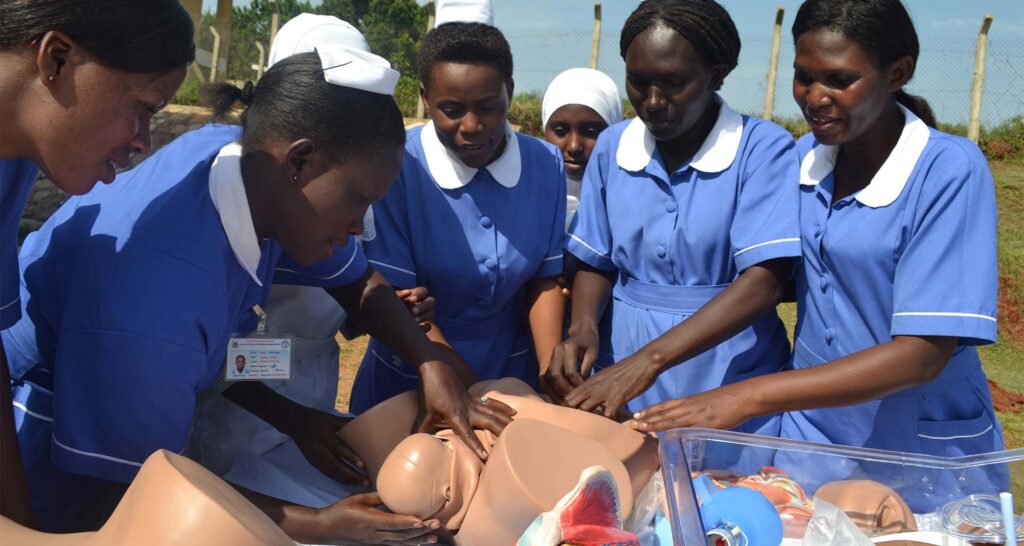
The work is so good, understandable, thanks for the great work
We are really greatfull …it has made a real difference to our research and learning…we haven’t stayed the same 🙏🙏
This is so helpful thanks for the great work
I thank this so much it has enlighten us so so much .it made us know how to approach questions 🙏🏾🙏🏾🙏🏾
Thanks alot
Thanks for your efforts
THANKS so much for the effort , I do appreciate your work
Thanks so so much 🙏🙏
Am very glad that you have shown me the way of getting access to your platform.
This is a comprehensive guide I wish I can get a pdf to always read through