
Module Unit CN-111: Anatomy and Physiology (I)
Contact Hours: 60
Module Unit Description: Introduces students to the anatomy and physiology of the human body, covering the structure and function of different body parts and systems, specifically skeletal, muscular, circulatory, and digestive systems.
By the end of this unit, the student shall be able to:
- Identify various parts of the human body and their functions.
- Differentiate the normal structure and functioning of various systems from that of abnormal conditions of the skeletal, muscular, cardiovascular and digestive systems.
Topic: Introduction to Anatomy and Physiology (Part 1)
Welcome to the study of the human body. In this module, we will learn about the different parts of the body and how they work together to keep us healthy. Understanding the normal structure and function of the body is essential for recognizing what happens when something goes wrong (illness or disease).
We will cover the foundational concepts in anatomy and physiology and then look specifically at the skeletal, muscular, cardiovascular, and digestive systems.
Think of the human body like a highly advanced car. Anatomy is the study of the car's parts: the engine, the steering wheel, the tires, and where they are located. Physiology is the study of how those parts work together to make the car drive. Pathology is the study of what happens when the engine breaks down or a tire pops!
Common Terms in Anatomy and Physiology
To begin our study, let's define some important terms that are like the basic language of this subject:
- Epithelial Tissue (Covering/Lining)
- Connective Tissue (Support)
- Nervous Tissue (Control)
- Muscle Tissue (Movement)
Anatomical and Physiological Concepts
Terms commonly used in Anatomy will be understood after these foundational concepts and abbreviations are mastered, since they will be used occasionally in your practice:
- Human Anatomy: The study of the body's structures and how they relate to each other. This is essential for nurses to understand where organs are located and how they function.
- Human Physiology: The study of how different body systems work together to keep us alive and healthy. Nurses use this knowledge to assess patients, interpret lab results, and understand how diseases or injuries affect the body.
- Homeostasis: The body's ability to maintain a stable internal environment despite external changes. This is crucial for nurses to monitor, as imbalances can indicate illness or disease.
- Pathology: The study of diseases and how they affect the body's normal functions. Nurses use this knowledge to recognize signs and symptoms of disease and understand how treatments work.
- Anatomical Position: A standard reference position used to describe the location of body parts. This ensures consistent communication among healthcare professionals.
- Planes of the body / Anatomical Planes: Imaginary lines that divide the body into sections, making it easier to describe locations of injuries or specific areas of pain.
- Directional Terms: Words used to describe the relative position of one body part to another. These help nurses accurately document and communicate findings during assessments.
- Homeostatic Imbalance: When the body is unable to maintain a stable internal environment, leading to potential health problems. Nurses monitor for these imbalances and intervene as needed to restore balance.
- Body Cavities: Hollow spaces within the body that contain organs and protect them. Nurses need to understand the location of these cavities for assessments and procedures.
Commonly Used Abbreviations
Ach: Acetylcholine
ACTH: Adrenal Corticotrophic Hormone
ADH: Anti-diuretic Hormone
ANS: Autonomic Nervous System
ATP: Adenosine Triphosphate (The energy currency of the cell)
C: Cervical, cervical vertebrae (i.e., C4 = cervical vertebra 4)
cm: Centimeter
CNS: Central Nervous System
CRH: Corticotropin Releasing Hormone
CSF: Cerebrospinal Fluid
DNA: Deoxyribonucleic Acid
/d: Per day
FSH: Follicular Stimulating Hormone
GHRH: Growth Hormone Releasing Hormone
GI: Gastrointestinal
GnRH: Gonadotrophin Releasing Hormone
HCG: Human Chorionic Gonadotrophin hormone (The pregnancy hormone)
HCl: Hydrochloric acid
GH: Growth Hormone
ICSH: Interstitial Cell Stimulating Hormone
IGF: Insulin Growth Factors
IUD: Intra Uterine Device
L: Lumbar, lumbar vertebrae (i.e., L3 = lumbar vertebra 3)
LH: Luteinizing Hormone
PNS: Peripheral Nervous System
PRH: Prolactin Releasing Hormone
PTH: Parathyroid Hormone
RNA: Ribonucleic Acid
rRNA: Ribosomal Ribonucleic Acid
T: Thoracic, thoracic vertebrae (i.e., T1 = thoracic vertebra 1)
T3: Triiodothyronine
T4: Thyroxine
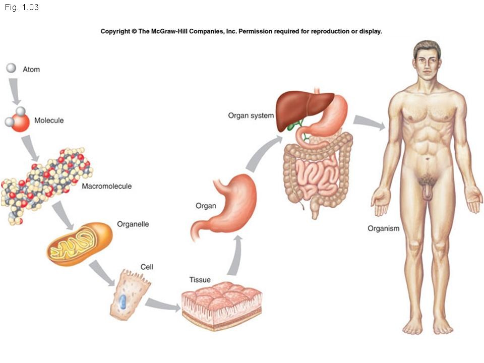
Human Body Organization
The human body is built up in layers of complexity, like building something from the ground up. Each level works with the others. Memeory Hook: Letters make words, words make sentences, sentences make paragraphs, and paragraphs make a book.
- Chemical level: This is the starting point – the very tiny non-living building blocks. It involves atoms combining through chemical bonds to form molecules. These are the chemical ingredients of life (e.g., DNA, glucose, water).
- Cellular level: The molecules come together in specific ways to create cells. Cells are the basic living units of the body. There are many different types of cells, each with a specialized job (e.g., muscle cells for contracting, nerve cells for sending signals).
- Tissue level: When many similar types of cells group together and work as a team to perform a particular job, they form a tissue.
- Organ level: Different types of tissues are organized together to form an organ. An organ is a distinct structure with a specific function (e.g., Heart, Brain, Liver).
- System level: A group of organs that work together to perform a major function essential for the body's survival is called a system (e.g., The cardiovascular system includes the heart and blood vessels).
- Organism level: All the body systems work together in a coordinated way to make a complete human being (the organism). The health of the whole person depends on all the systems working together effectively.
Anatomical Positions & Relative Directional Terms
Anatomical Position
Anatomical positions are accepted universally as the starting points for positional references to the body. This prevents dangerous errors in medicine (like operating on the wrong leg!).
In the anatomical position, the subject (body of patient or client to be observed) is standing erect and facing the observer (the medical examiner), the feet are together, and the arms are hanging at the sides with the palms facing forward.
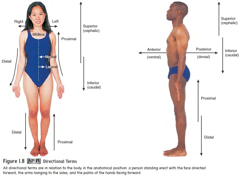
Relative Directional Terms
Standard terms of reference are used when anatomists or medical examiners describe the location of a certain body part. "Relative" means the location of one body part is always described in relation to another body part of the same human body.
| Term | Definition | Example |
|---|---|---|
| Superior (cranial) | Means towards the head. | The leg is superior to the foot. (The chest is superior to the pelvis). |
| Inferior (caudal) | Toward the feet. | The foot is inferior to the leg. (The stomach is inferior to the heart). |
| Anterior (ventral) | Toward the front part of the body. | The nose is anterior to the ears. (The breastbone/sternum is anterior to the spine). |
| Posterior (dorsal) | Towards the back of the body. | The ears are posterior to the nose. (The shoulder blades are posterior to the ribs). |
| Medial | Towards the midline of the body. | The nose is medial to the eyes. |
| Lateral | Away from the midline of the body. | The eyes are lateral to the nose. (The arms are lateral to the chest). |
| Proximal | Toward (nearer) the trunk of the body or the attached end of a limb. | The shoulder is proximal to the wrist. (The knee is proximal to the ankle). |
| Distal | Away (further) from the trunk of the body or the attached end of a limb. | The wrist is distal to the forearm. (The fingers are distal to the palm). |
| Superficial | Nearer to the surface of the body. | The ribs are superficial to the heart. (The skin is superficial to the muscles). |
| Deep | Further from the surface of the body. | The heart is deeper to the ribs. (The bones are deep to the skin). |
| Peripheral | Away from the central axis of the body. | Peripheral nerves radiate away from the brain and spinal cord. |
Body Regions, Planes and Sections

Body Parts Regions
The body parts regions are divided into two main categories:
- Axial: This is the part of the body that is near the axis (center line) of the body. This includes the head, neck, thorax (chest), abdomen, and pelvis.
- Appendicular body part: This is the part of the body out of the axis line. This includes the upper and lower extremities (arms and legs, or appendages).
The abdomen is divided into nine regions, or more easily divided into four quadrants. Health professionals use these to locate pain and diagnose issues:
- Right Upper Quadrant (RUQ): Contains the liver and gallbladder.
- Left Upper Quadrant (LUQ): Contains the stomach and spleen.
- Right Lower Quadrant (RLQ): Contains the appendix. (Pain here often indicates appendicitis!)
- Left Lower Quadrant (LLQ): Contains parts of the descending colon.

Body Planes and Sections
Body planes are imaginary flat surfaces (like sheets of glass) that divide the body into sections. This helps for further identification of specific areas, especially in medical imaging like CT scans and MRIs.
- Mid-sagittal plane: Divides the body into two equal left and right halves (right down the exact middle).
- Para-sagittal plane: Divides the body into two unequal left and right portions.
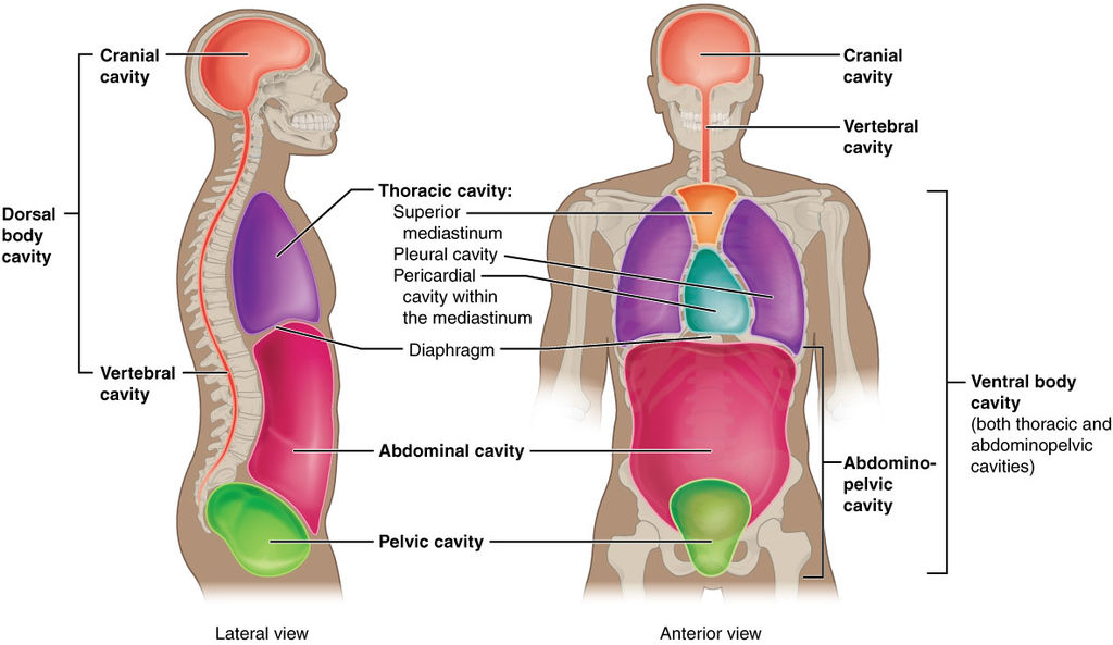
Body Cavities: Protective Spaces for Vital Organs
Body cavities are hollow, fluid-filled spaces within the human body that house, protect, and support internal organs. They act as internal "vaults" that shield delicate tissues from accidental shocks and allow organs to change size and shape dynamically (for example, allowing the lungs to expand or the stomach to stretch without compressing surrounding tissues).
Understanding their precise location and contents is absolutely essential for nurses during physical assessment, diagnosis, interpreting diagnostic imaging (like X-rays), and treating various conditions.
1. Classification by Primary Location
The body is fundamentally divided into two major continuous cavities: the Dorsal (back) and the Ventral (front).
- Dorsal (Posterior) Cavity: Located towards the back of the body. It is completely encased in bone to protect the fragile central nervous system.
- Cranial Cavity: Encloses the brain.
- Spinal (Vertebral) Cavity: Houses and protects the delicate spinal cord.
- Ventral (Anterior) Cavity: Located towards the front of the body. It is much larger and houses the visceral organs (the internal organs of the chest and belly).
- Thoracic Cavity: Contains the heart, lungs, and major vessels.
- Abdominal Cavity: Holds the digestive organs (stomach, intestines, liver).
- Pelvic Cavity: Contains the bladder and reproductive organs.
Knowledge of these anatomical spaces is foundational. If a patient presents with a penetrating stab wound to the "Ventral Cavity" above the diaphragm, the nurse instantly knows to assess the Thoracic cavity structures (heart and lungs) for life-threatening emergencies like a collapsed lung (pneumothorax).
2. The Major Body Cavities & Nursing Relevance
Major cavities contain the large organs essential for vital daily functions. Here is a detailed breakdown of their clinical significance:
Cranial Cavity
- Location: Enclosed strictly by the rigid skull bones.
- Contains: The brain (the master control center for all bodily functions).
- Nursing Relevance: Because the skull cannot expand, any bleeding or swelling here is an emergency. Vital for assessing neurological status, identifying traumatic head injuries, and monitoring Intracranial Pressure (ICP).
Spinal Cavity
- Location: Runs through the center of the vertebral column.
- Contains: The spinal cord.
- Nursing Relevance: Essential for nerve transmission. Relevant for assessing loss of motor/sensory function, managing spinal cord injuries, administering epidural anesthesia, and complex pain management.
Thoracic Cavity
- Location: Within the protective rib cage, resting above the diaphragm muscle.
- Contains:
- Pleural Cavities: Two separate lateral spaces, each housing a lung.
- Mediastinum: The central compartment between the lungs. It contains the heart (inside its own pericardial cavity), major blood vessels (aorta, superior/inferior vena cava), the esophagus, trachea, and primary bronchi.
- Nursing Relevance: Crucial for assessing respiratory and cardiovascular function, auscultating (listening to) heart and lung sounds, identifying chest trauma, and understanding how mediastinal tumors can compress the airway.
Abdominal Cavity
- Location: Below the diaphragm and above the imaginary line of the pelvic brim.
- Contains: Stomach, small/large intestines, liver, spleen, pancreas, kidneys, and adrenal glands.
- Nursing Relevance: Important for assessing digestive, urinary, and endocrine functions. Essential for palpating the abdomen to identify the source of severe pain (e.g., appendicitis, gallstones, or pancreatitis).
Pelvic Cavity
- Location: Below the abdominal cavity, securely cradled within the bony basin of the pelvic bones.
- Contains: Urinary bladder, internal reproductive organs (uterus, ovaries in females; prostate gland in males), and the terminal part of the large intestine (rectum).
- Nursing Relevance: Essential for assessing urinary retention (palpating a full bladder), identifying gynecological/urological conditions, and managing labor and pregnancy-related complications.
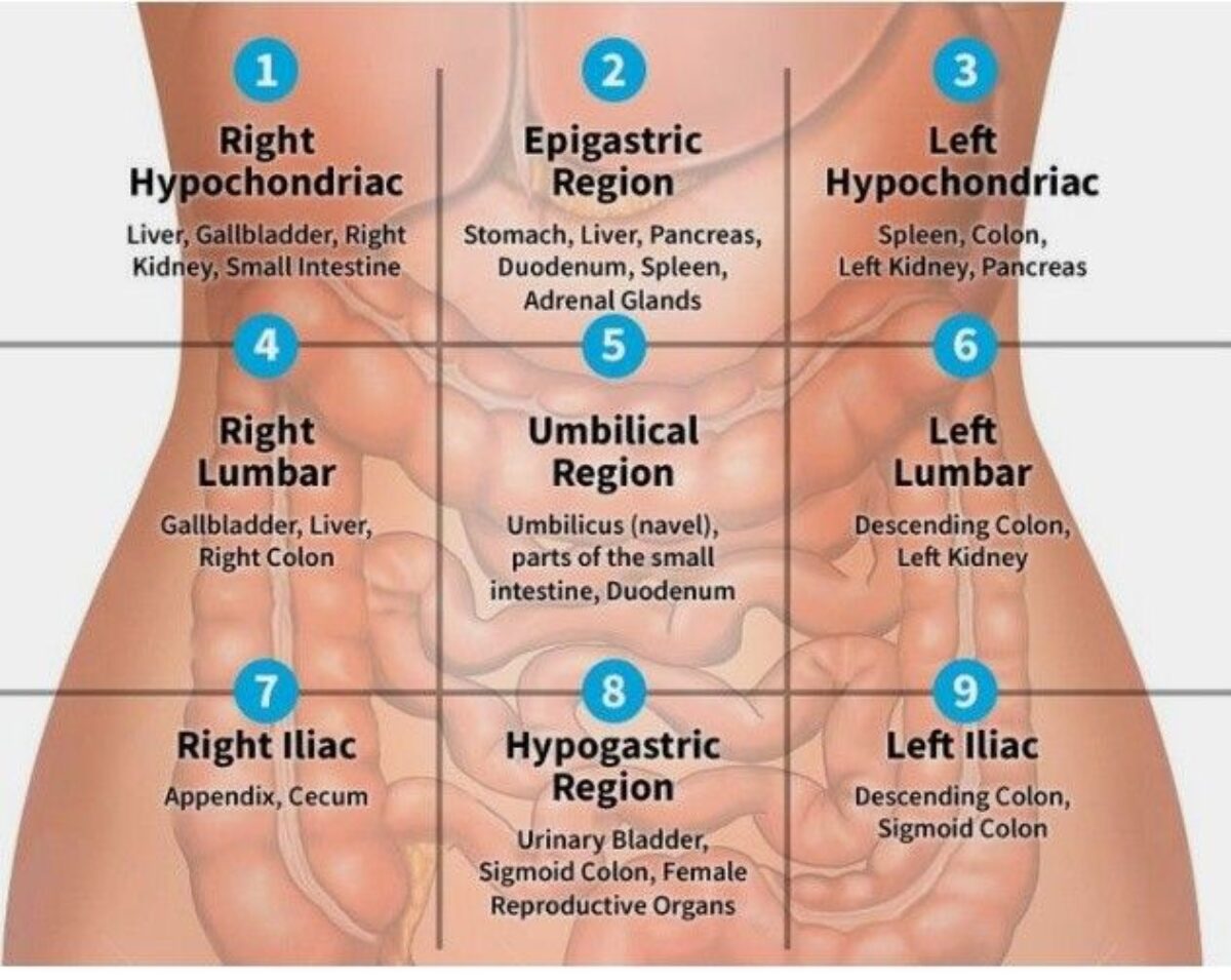
3. The 9 Abdominopelvic Regions
Because the abdominal cavity is so massive and contains so many organs, healthcare providers universally divide it into a grid of nine specific regions to accurately pinpoint and document pain, surgical incisions, and organ locations.
| Region Name | Specific Organs Contained | Clinical Example |
|---|---|---|
| Right Hypochondriac | Right portion of the liver, gallbladder, right kidney, parts of the small intestine. | Pain here often strongly suggests gallstones (cholecystitis) or liver inflammation. |
| Epigastric | Majority of the stomach, part of the liver, part of the pancreas, part of the duodenum, part of the spleen, adrenal glands. | Acid reflux, stomach ulcers, or acute pancreatitis often present as severe epigastric burning pain. |
| Left Hypochondriac | Part of the spleen, left kidney, part of the stomach, tail of the pancreas, parts of the colon. | Blunt trauma to this area can result in a ruptured, life-threateningly bleeding spleen. |
| Right Lumbar | Gallbladder, right kidney, part of the liver, ascending colon. | Pain radiating to the back here often indicates a right kidney infection or kidney stone. |
| Umbilical | The umbilicus (navel), many parts of the small intestine (duodenum, jejunum, ileum), transverse colon, bottom portions of both kidneys. | Early appendicitis often starts as a vague, dull ache squarely in the umbilical region. |
| Left Lumbar | Descending colon, left kidney, part of the spleen. | Tenderness here is assessed for left kidney issues or descending colon spasms. |
| Right Iliac (Inguinal) | Appendix, cecum, right iliac fossa. | Sharp, rebounding pain in this exact region (specifically at McBurney's Point) is the classic hallmark of acute appendicitis. |
| Hypogastric (Pubic) | Urinary bladder, part of the sigmoid colon, anus, reproductive organs (uterus/ovaries in females; prostate in males). | A distended, unemptied bladder or severe menstrual cramping will present as swelling/pain in this pubic region. |
| Left Iliac (Inguinal) | Part of the descending colon, sigmoid colon, left iliac fossa. | Pain here is highly characteristic of diverticulitis (inflammation of small pouches in the colon). |
4. Minor Cavities of Importance in Nursing
While smaller than the major cavities, these specific hollow spaces house organs with highly specialized functions and are frequently assessed during a routine nursing physical exam.
- Oral (Buccal) Cavity:
- Contains: The tongue, teeth, and salivary glands.
- Nursing Relevance: Critical for assessing a patient's overall oral hygiene, identifying swallowing difficulties (dysphagia), assessing mucous membrane hydration, and evaluating nutritional intake capability.
- Nasal Cavity:
- Contains: Nasal conchae, olfactory receptors, and mucous membranes. It acts as the body's air conditioning unit (filtering, warming, and humidifying inhaled air).
- Nursing Relevance: Essential when assessing baseline respiratory function, checking for nasal obstructions, inserting a Nasogastric (NG) tube, or managing severe sinus infections.
- Orbital (Eye Socket) Cavity:
- Contains: The eyes, optic nerves, and associated protective structures (fat pads, muscles).
- Nursing Relevance: Vital for assessing vision, evaluating direct eye injuries, and checking pupil dilation (PERRLA) as an indicator of deep neurological or brain stem function.
- Medullary Cavity:
- Contains: The hollow core found deep within the shaft of long bones (like the femur). It houses the red and yellow bone marrow, which is exclusively responsible for blood cell production (hematopoiesis).
- Nursing Relevance: Highly relevant for understanding the root cause of blood disorders (like leukemia or severe anemia) and for caring for patients undergoing invasive bone marrow biopsies.
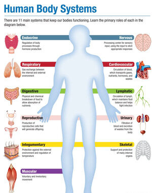
Essential Body Systems
The human body relies on interconnected systems to maintain life. Below is a summary of their core components and functions.
a. Cardiovascular System
- Components: Heart, blood vessels (arteries, veins, capillaries), blood.
- Functions: Transports oxygen, nutrients, and hormones to cells; removes waste products (like carbon dioxide) from cells; regulates body temperature and pH balance.
b. Lymphatic System
- Components: Lymph nodes, lymphatic vessels, spleen, thymus, tonsils.
- Functions: Returns excess tissue fluid to the bloodstream; serves as a major component of the immune system to fight infection and disease.
a. Nervous System
- Components: Brain, spinal cord, nerves, sensory organs.
- Functions: The body's fast-acting control system. It controls and coordinates body functions, processes sensory information, and enables thought, memory, and learning.
b. Endocrine System
- Components: Glands that produce hormones (e.g., pituitary, thyroid, pancreas, adrenal glands).
- Functions: The body's slow-acting control system. It regulates body functions through hormone secretion, controlling growth, metabolism, reproduction, and the stress response.
a. Respiratory System
- Components: Lungs, airways (trachea, bronchi, bronchioles).
- Functions: Takes in oxygen from the environment and releases carbon dioxide waste; helps regulate the body's acid-base balance.
b. Digestive System
- Components: Mouth, esophagus, stomach, small intestine, large intestine, liver, pancreas, gallbladder.
- Functions: Breaks down food physically and chemically into nutrients the body can absorb into the blood; eliminates indigestible solid waste.
c. Urinary System
- Components: Kidneys, ureters, bladder, urethra.
- Functions: Filters waste products and excess fluid from the blood to produce urine; regulates fluid balance and electrolytes; helps maintain acid-base balance.
a. Integumentary System
- Components: Skin, hair, nails, sweat glands, sebaceous glands.
- Functions: The primary physical barrier. Protects the body from injury, dehydration, and infection; regulates body temperature; senses touch, pressure, and pain; synthesizes vitamin D.
b. Lymphatic System (Immunity)
- Components: Lymph nodes, lymphatic vessels, spleen, thymus, tonsils.
- Functions: Works alongside the integumentary system to provide internal protection by fighting off invading pathogens.
a. Musculoskeletal System
- Components: Bones, joints, muscles, tendons, ligaments.
- Functions: Provides structural support for the entire body; enables movement through muscle contraction; protects vital internal organs (like the skull protecting the brain); stores vital minerals (calcium); produces blood cells within the bone marrow.
a. Reproductive System
- Components: Female: Ovaries, fallopian tubes, uterus, vagina. Male: Testes, epididymis, vas deferens, seminal vesicles, prostate gland, penis.
- Functions: Produces sex hormones (estrogen, testosterone) to drive development and behavior; produces gametes (sperm and eggs) to enable human reproduction and the continuation of the species.
Revision Questions for Page 1 (Part 1)
- Define the following terms in your own words: Anatomy, Physiology, Homeostasis, Pathology, Pathophysiology.
- List the six levels of structural organization in the human body from simplest to most complex.
- Describe the standard anatomical position.
- Use directional terms to describe the location of the nose relative to the ears, and the elbow relative to the wrist.
- What is the difference between the axial and appendicular regions of the body?
- Differentiate between the sagittal, frontal, and transverse body planes.
References (from Curriculum for CN-111)
Below are the references listed in the curriculum for Module CN-1102. Refer to the original document for full details.
- Cohen, JB and Hull, L.K (2016) Memmlers – The Human body in Health and diseases 13th Edition, Wolters, Kluwer. (Core Reference)
- Cohen, J.B and Hull, L.K (2016) Memmler's Structure and Function of the Human Body. 11th Edition. Wolters Kluwer, China
- Kumar, M and Anand, M (2010) Human Anatomy and Physiology for Nursing and Allied Sciences. 2nd Edition. Jaypee Brothers Medical Publishers Ltd.
- Scott, N.W. (2011) Anatomy and Physiology made incredibly easy. 1st Edition. Wolwers Kluwers, Lippincotts Williams and Wilkins.
- Moore, L. K, Agur, M.R.A and Dailey, F.A. (2015) Essential Clinical Anatomy. 15th Edition. Wolters Kluwer.
- Snell, S. R. (2012) Clinical Anatomy by Regions. 9th Edition. Wolters Kluwer, Lippincott Williams and Wilkins, China
- Wingerd, B, (2014) The Human Body-Concepts of Anatomy and Physiology. 3rd Edition Lippincott Williams and Wilkins and Wolters Kluwer.
- Rohen, Y.H-Orecoll. (2015) Anatomy. A Photographic Atlas 8th Edition. Lippincott Williams & Wilkins
- Waugh, A., & Grant, A. (2014). Ross and Wilson Anatomy & Physiology in Health and Illness (12th ed.). Churchill Livingstone Elsevier. (Added as per user's reference)