
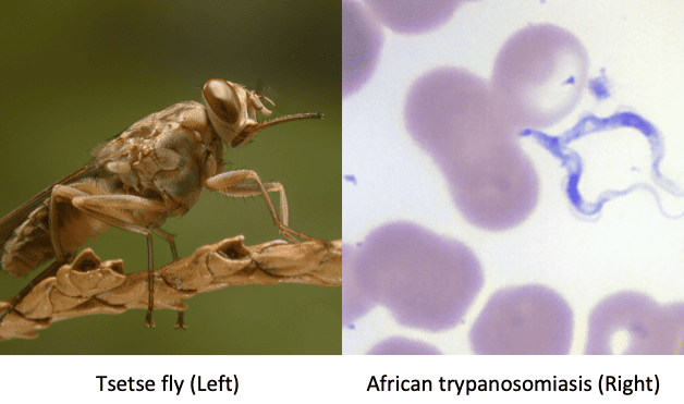
Human African Trypanosomiasis (Sleeping Sickness): Comprehensive Clinical, Parasitological, and Nursing Guide
What is Human African Trypanosomiasis (HAT)?
Human African Trypanosomiasis (HAT), clinically known as sleeping sickness, is a severe, vector-borne, neglected tropical disease (NTD) caused by protozoan hemoflagellates belonging to the genus Trypanosoma, transmitted to humans through the painful bite of blood-sucking tsetse flies (genus Glossina).
The disease progresses through two distinct clinical stages: an initial systemic hemolymphatic phase characterized by undulating fevers, lymphadenopathy, and organomegaly, followed inexorably by an advanced meningoencephalitic phase when parasites traverse the blood-brain barrier (BBB), precipitating severe neuroinflammation, cognitive decay, motor disturbances, circadian rhythm inversion (daytime somnolence and nocturnal insomnia), stupor, coma, and death if untreated.
The Two Pathogenic Subspecies
Human African Trypanosomiasis is caused exclusively by two morphologically identical morphological subspecies of Trypanosoma brucei, which differ markedly in their geographic distribution, reservoir hosts, clinical course, and therapeutic responsiveness:
| Feature | Trypanosoma brucei rhodesiense (T.b.r.) | Trypanosoma brucei gambiense (T.b.g.) |
|---|---|---|
| Disease Form | Acute, virulent, fulminant sleeping sickness. | Chronic, slowly progressive, debilitating sleeping sickness. |
| Geographic Distribution | Eastern and Southern Africa (Uganda, Tanzania, Kenya, Malawi, Zambia, Zimbabwe). | Western and Central Africa (Democratic Republic of Congo, Central African Republic, South Sudan, northwestern Uganda). |
| Primary Vectors | Glossina morsitans group (savannah/game park tsetse flies). | Glossina palpalis group (riverine and lacustrine tsetse flies). |
| Major Reservoirs | Wild game animals (bushbuck, waterbuck, hartebeest, lions) and domestic livestock (cattle, pigs). Zoonotic cycle; humans are accidental hosts. | Humans are the primary reservoir (anthroponotic cycle). Domestic animals (pigs, dogs, goats) can serve as minor animal reservoirs. |
| Progression to CNS Stage | Rapid: occurs within a few weeks to months (typically <3 months). | Slow: occurs after months to several years (average 1–3 years). |
| Trypanosomal Chancre | Common (presents in up to 50% of non-immune patients). | Rarely observed in endemic local populations. |
| Parasitaemia Level | High density; easily identified on direct wet and thick blood films. | Extremely low density and fluctuating; often requires concentration techniques. |
| Prognosis if Untreated | Rapidly fatal within 3 to 6 months due to acute congestive heart failure, toxemia, or fulminant meningoencephalitis. | Invariably fatal within 2 to 3 years due to profound central neurodegeneration, secondary infections, or cachexia. |
Uganda's Unique Epidemiological Position:
Uganda is the only country in the world that is co-endemic for both forms of sleeping sickness. Historically, T. b. gambiense has been localized to the northwestern region (West Nile: Arua, Maracha, Yumbe, Moyo), while T. b. rhodesiense has affected eastern and central districts (Busoga, Tororo, Soroti, Alebtong, Dokolo, Kaberamaido). The major national public health priority is preventing the geographic overlap or "merger" of these two forms, which would severely complicate diagnosis and empirical treatment.
I. Vector Ecology (The Tsetse Fly)
Table of Contents
Toggle- Taxonomy: Genus Glossina. Both male and female adult tsetse flies are obligate blood-feeders (hematophagous) and can transmit the parasite.
- Riverine Species (Glossina palpalis group): Inhabits shaded, dense riverbanks, lake shores, and gallery forests. Transmits T. b. gambiense to villagers washing clothes, fetching water, fishing, or farming near water bodies.
- Savannah Species (Glossina morsitans group): Inhabits dry, open woodlands, savannah grasslands, thickets, and national game parks. Transmits T. b. rhodesiense to cattle herdsmen, game rangers, poachers, firewood collectors, and tourists.
- Reproduction: Highly unusual reproduction called adenotrophic viviparity: female flies fertilize one egg at a time and nourish the larva inside their uterus via specialized "milk glands," depositing a fully grown third-instar larva directly onto loose, shaded, moist soil, which immediately burrows and pupates.
II. The Parasite Life Cycle
The life cycle requires sequential developmental stages alternating between the mammalian host and the insect vector:
A. The Tsetse Fly Phase (Vector Cycle / Cyclical Transmission)
- Ingestion: While taking a blood meal from an infected mammalian host, the tsetse fly ingests free-swimming, non-proliferative short stumpy bloodstream trypomastigotes.
- Midgut Transformation: Inside the fly’s midgut lumen, the parasites shed their protective variable surface glycoprotein (VSG) coat, acquire a procyclin surface coat, and transform into dividing procyclic trypomastigotes.
- Migration to Salivary Glands: After 2 to 3 weeks of intensive binary fission, parasites migrate forward through the proventriculus, up the esophagus, and into the fly's salivary glands.
- Epimastigote to Metacyclic Form: In the salivary glands, parasites attach to microvilli, transform into epimastigotes, and undergo further multiplication. Finally, they detach, re-acquire a dense VSG coat, and mature into metacyclic trypomastigotes. This is the only stage infectious to mammalian hosts. The entire extrinsic developmental cycle inside the fly takes 20 to 35 days, after which the fly remains infective for the rest of its natural lifespan.
B. The Human / Mammalian Phase
- Inoculation: During a subsequent blood meal, the infective tsetse fly probes the human dermis and inoculates several thousand metacyclic trypomastigotes into the interstitial tissue along with saliva.
- Chancre Formation: In the subcutaneous tissue, parasites rapidly transform into long slender bloodstream trypomastigotes and multiply by longitudinal binary fission, provoking an intense localized inflammatory reaction termed a trypanosomal chancre.
- Hemolymphatic Dissemination (Stage 1): Parasites escape from local interstitial spaces into draining lymphatic vessels and enter the systemic bloodstream. They survive entirely extracellularly, bathed in plasma and tissue interstitial fluids, metabolizing host glucose.
- Cerebrovascular Invasion (Stage 2): Bloodstream trypanosomes cross the capillary endothelium of the blood-brain barrier (BBB) and choroid plexus, infiltrating the virchow-robin perivascular spaces, cerebrospinal fluid (CSF), and cerebral parenchyma.
Non-Vector Modes of Transmission
- Congenital Transmission: Transplacental migration of trypomastigotes from an infected mother to the fetus, precipitating perinatal death, abortion, or neonatal sleeping sickness.
- Blood Transfusion: Transfusion of unscreened, infected donor whole blood.
- Accidental Laboratory Inoculation: Needle-stick injuries involving contaminated blood or live cultures.
I. Antigenic Variation & The VSG Coat
The primary mechanism enabling African trypanosomes to persist indefinitely in immunocompetent mammalian hosts is Variant Surface Glycoprotein (VSG) gene switching:
- The entire external surface of the bloodstream trypomastigote is covered by a dense, macromolecular coat consisting of approximately 10 million homodimers of a single VSG molecule.
- When host B-lymphocytes mount a specific, complement-fixing IgM/IgG antibody response against the dominant VSG variant, over 99% of the parasite population is destroyed via immune-mediated lysis and Kupffer cell phagocytosis. This clearance corresponds clinically to the temporary subsidence of fever.
- However, through programmed genetic recombination and transcriptional switching among a genomic repertoire of over 1,000 distinct silent VSG genes, a tiny fraction (<0.1%) of parasites switch their surface coat to an immunologically distinct VSG variant.
- These antigenically novel trypanosomes multiply unchecked, generating a new wave of parasitaemia, stimulating a new wave of antibodies, and triggering a clinical relapse. This continuous cycle generates the classic undulating or wave-like intermittent fever spikes seen in sleeping sickness.
II. Systemic Organ Pathology (Stage 1)
- Immune Complex Deposition & Hypergammaglobulinemia: Massive B-cell activation leads to high serum concentrations of non-specific, polyclonal Immunoglobulin M (IgM) (frequently 4 to 16 times normal levels). Autoantibodies (anti-DNA, anti-erythrocyte, anti-myelin) are generated. Soluble immune complexes deposit in vascular walls, activating the complement cascade, precipitating microvascular permeability, tissue edema, and urticaria.
- Lymphoid Hyperplasia & Splenomegaly: Massive reticuloendothelial stimulation causes follicular hyperplasia in the spleen and lymph nodes. Nodes become soft, mobile, enlarged, and fluid-rich.
- Severe Anemia: Triggered by red cell phagocytosis in the hypersplenic reticuloendothelial system, complement-mediated intravascular hemolysis, and bone marrow dyserythropoiesis.
- Myocarditis (Particularly in T. b. rhodesiense): Direct trypanosomal invasion of cardiac interstitium leads to severe lymphocytic and histiocytic pancarditis, pericardial effusions, conduction arrhythmias, and acute congestive heart failure.
III. Central Nervous System Invasion & Sleep Disruption (Stage 2)
Parasites traverse the blood-brain barrier via transcellular, paracellular, or Trojan-horse pathways:
- Chronic Perivascular Meningoencephalitis: Parasites, activated astrocytes, and microglia release inflammatory mediators: Interferon-gamma (IFN-γ), Tumor Necrosis Factor-alpha (TNF-α), nitric oxide, and prostaglandin D2 (PGD2). Perivascular spaces are cuffed by lymphocytes, plasma cells, and pathognomonic Mott cells (morular cells of Christie)—large, plasma cells packed with grape-like inclusions of unsecreted IgM.
- Sleep-Wake Architecture Inversion: Prostaglandin D2 (PGD2) is the body's primary endogenous somnogenic (sleep-inducing) molecule. Trypanosomes secrete substantial amounts of PGD2 isomerase. Heavy neuroinflammation within the anterior hypothalamus and optic chiasm dysregulates the suprachiasmatic nucleus (the master circadian pacemaker). This causes complete phase-reversal of sleep architecture: the patient is overcome by unstoppable, deep sleep bouts during daylight hours, while experiencing persistent insomnia, motor agitation, and hallucinations at night.
- Neuronal Degeneration & Brain Ventricle Injury: Inflammatory disruption of ependymal cells lining the ventricular system causes hydrocephalus, high intracranial pressure, focal demyelination, ataxia, tremors, and eventually terminal vegetative coma.
I. The Primary Stage: The Trypanosomal Chancre
- Appears 5 to 15 days following an infective tsetse bite.
- Presents as an indurated, firm, painful, erythematous, warm, non-purulent inflammatory nodule or plaque, ranging from 2 to 5 cm in diameter.
- Frequently surrounded by a pale halo of localized edema; regional lymph nodes enlarge rapidly.
- May desquamate, blister, or ulcerate, resembling a large carbuncle or boil. Resolves spontaneously over 2 to 3 weeks.
- Clinical Pearl: Frequent in tourists and individuals infected with T. b. rhodesiense; rarely seen in T. b. gambiense infections.
II. Stage 1: Hemolymphatic (Systemic) Stage
- Undulating Fevers: Irregular spikes of high fever accompanied by severe rigor and chills, lasting 1 to 7 days, separated by afebrile asymptomatic intervals of several days to weeks.
- Winterbottom's Sign: Marked enlargement of the posterior cervical lymph nodes (posterior triangle of the neck). Nodes are typically non-tender, discrete, soft, elastic, mobile, and walnut-sized ("rubbery"). This is the classic clinical hallmark of T. b. gambiense.
- Cutaneous Manifestations (Trypanids): Fleeting, polycyclic, erythematous, targetoid, or circinate annular erythematous patches (15–30 cm across) localized predominantly over the trunk, chest, back, and thighs. Accompanied by intense, unyielding generalized pruritus and localized facial or periorbital angioedema.
- Endocrine & Autonomic Dysfunction: Loss of libido, impotence, severe secondary amenorrhea in women of childbearing age, and high rates of spontaneous abortion.
- Hepatosplenomegaly & Serosal Effusions: Moderate, tender enlargement of the spleen and liver; painless ascites and pleural/pericardial effusions.
III. Stage 2: Meningoencephalitic (CNS) Stage
Marks the invasion of the central nervous system. Clinical features include:
1. Circadian & Sleep Disturbances:
- Inversion of the Normal Sleep Cycle: Overwhelming daytime somnolence; the patient falls asleep while talking, eating, or standing.
- Nocturnal Agitation: Complete inability to sleep at night, with severe restlessness, nocturnal wanderings, and nightmares.
- Uncontrollable sleep episodes fragment the circadian structure, progressing to progressive stupor and irreversible terminal coma.
2. Neuropsychiatric & Behavioral Manifestations:
- Profound personality alterations: apathy, emotional lability, unprovoked weeping or euphoria.
- Progressive dementia, disorientation to time and place, memory loss, and severe confusion.
- Psychotic features: acute paranoid delusions, auditory and visual hallucinations, and bouts of extreme physical aggression.
3. Motor & Neurological Dysfunction:
- Movement Disorders: Fine or coarse resting tremors of the tongue, lips, and outstretched fingers; choreiform movements; athetoid writhing of limbs.
- Motor Incoordination: Cerebellar ataxia, wide-based staggering gait, dysdiadochokinesia, and dysmetria.
- Speech & Swallowing: Slurred, scanning speech (dysarthria); progressive dysphagia leading to severe aspiration and choking.
- Pyramidal & Extrapyramidal Signs: Hyperreflexia, sustained ankle clonus, bilateral extensor plantar responses (Babinski sign), and muscular rigidity.
- Sensory Abnormalities: Kerandel’s Sign (Hyperesthesia): Severe, excruciating, delayed deep bone and muscle pain experienced upon light compression of deep soft tissues (such as pinching the calf muscles or squeezing the palm).
- Terminal Phase: Progressive severe cachexia, total incontinence of urine and feces, seizures (status epilepticus), aspiration pneumonia, or deep unarousable coma culminating in death.
I. Stepwise Diagnostic Strategy
Diagnosis of sleeping sickness follows a rigid tripartite pathway: Screening ➔ Parasitological Confirmation ➔ Staging.
A. Serological Screening (exclusively for T. b. gambiense)
- Card Agglutination Test for Trypanosomiasis (CATT): Rapid, point-of-care, 5-minute field agglutination test utilizing freeze-dried, fixed bloodstream trypomastigotes of T. b. gambiense expressing specific predominant VSG antigens (variable antigen type LiTat 1.3).
- Principle: Mixing a drop of the patient's whole blood or reconstituted plasma with the antigen on a plastic card; agglutination visible to the naked eye denotes positive anti-trypanosome antibodies.
- Limitation: CATT is purely a screening test. It cannot be used alone to initiate toxic therapies. A positive CATT mandates parasitological microscopic confirmation.
- Rapid Diagnostic Tests (mRDTs for HAT): Recombinant antigen immunochromatographic strips (e.g., SD BIOLINE HAT) used for remote active screening.
B. Parasitological Confirmation (Microscopy)
- Lymph Node Puncture & Aspirate: Puncture a swollen, mobile cervical node (Winterbottom's node) with a sterile 21–23 gauge dry needle without a syringe. Express node juice onto a slide, coverslip, and immediately examine under high dry magnification (×400). Live, motile trypanosomes appear rapidly undulating between lymphocytes. Highest diagnostic yield in T. b. gambiense.
- Direct Blood Smears:
- Wet Blood Film: Direct drop of blood under a coverslip; allows visualization of vigorous parasite motility as it pushes aside erythrocytes.
- Thick & Thin Blood Smear: Air-dried and stained with Giemsa or Leishman stain. Demonstrates characteristic slender trypomastigotes with a single nucleus, posterior kinetoplast, and an undulating membrane terminating in a free anterior flagellum.
- Concentration Techniques (Essential for T. b. gambiense):
- Microhematocrit Centrifugation Technique (mHCT / Woo Test): Centrifuging capillary tubes of anticoagulated blood; direct microscopic examination of the sealed tube focused directly on the buffy coat layer, where motile parasites concentrate.
- Mini-Anion Exchange Centrifugation Technique (mAECT): The single most sensitive parasitological method. Anticoagulated blood passes through a DEAE-cellulose anion-exchange chromatography column. Positively charged blood cells bind firmly to the resin, whereas negatively charged trypanosomes elute through, are collected, centrifuged, and identified in a special viewing chamber.
C. Disease Staging via Lumbar Puncture (LP)
Every patient with parasitologically confirmed sleeping sickness MUST undergo a Lumbar Puncture for clinical disease staging before starting medication.
Cerebrospinal Fluid (CSF) Staging Criteria:
Staging determines drug choice. Drugs that cross the blood-brain barrier are required for Stage 2, whereas less toxic, non-CNS penetrating drugs are used for Stage 1.
- Stage 1 (Hemolymphatic): White blood cell count ≤ 5 cells/μL in CSF AND complete absence of trypanosomes in CSF.
- Stage 2 (Meningoencephalitic): WBC > 5 cells/μL in CSF OR demonstration of any trypanosomes in CSF (via double centrifugation of CSF).
- Mott Cells: The presence of morular Mott plasma cells in CSF confirms Stage 2 neuroinflammation.
General Therapeutic Principles
- Treatment is strictly determined by: (1) The causative subspecies (gambiense vs. rhodesiense), and (2) The disease stage (Stage 1 vs. Stage 2).
- Modern Paradigm Shift — Fexinidazole: The World Health Organization approved Fexinidazole as the first all-oral treatment for both Stage 1 and non-severe Stage 2 T. b. gambiense, radically reducing the need for hospital-based intravenous infusions.
I. Treatment of T. b. gambiense (g-HAT)
A. Fexinidazole (Preferred First-Line Oral Therapy)
- Indications: Patients aged ≥ 6 years and body weight ≥ 20 kg with T. b. gambiense infection (effective for Stage 1 and non-severe Stage 2 with CSF WBC count ≤ 100 cells/μL). If CSF WBC > 100 cells/μL, NECT is preferred.
- Administration: Must be taken once daily with a substantial solid meal (increases systemic bioavailability by 3-fold) under strict direct observation by a healthcare worker.
- Dosing Regimen (10-Day Total Course):
- Adults / Body Weight ≥ 35 kg:
- Days 1 to 4 (Loading Dose): 1,800 mg (three 600 mg tablets) once daily.
- Days 5 to 10 (Maintenance Dose): 1,200 mg (two 600 mg tablets) once daily.
- Children / Body Weight 20 kg to <35 kg:
- Days 1 to 4: 1,200 mg (two 600 mg tablets) once daily.
- Days 5 to 10: 600 mg (one 600 mg tablet) once daily.
- Adults / Body Weight ≥ 35 kg:
- Contraindications: Severe hepatic impairment, QT-prolongation risks, pregnancy, breastfeeding, or children <20 kg.
B. Pentamidine Isethionate (Classical Stage 1 Alternative)
- Indication: Stage 1 T. b. gambiense (cannot cross the blood-brain barrier).
- Dosage: 4 mg base/kg body weight once daily for 7 consecutive days, administered by deep intramuscular (IM) injection (preferably in the upper outer quadrant of the buttock, alternating sides daily).
- Critical Nursing Precautions:
- Hypoglycemia Prevention: Pentamidine causes direct pancreatic beta-cell lysis and sudden insulin release. The patient MUST eat a carbohydrate-rich meal 1 hour prior to the injection. Keep IV 10% Dextrose immediately accessible.
- Hypotension Prevention: Drug induces peripheral vasodilation. The patient must remain in a flat, supine position during the injection and for a full 1 hour afterward. Monitor blood pressure before and at 15, 30, and 60 minutes post-dose.
- Nephrotoxicity: Monitor baseline and daily serum creatinine and urinalysis. Sterile abscesses at injection sites are common; rotate injection sites.
C. NECT: Nifurtimox-Eflornithine Combination Therapy (Stage 2 Gold Standard)
- Indication: Stage 2 (meningoencephalitic) T. b. gambiense infection, particularly in patients with CSF WBC > 100 cells/μL or those unable to take oral fexinidazole.
- Eflornithine (DFMO): 200 mg/kg body weight IV every 12 hours for 7 consecutive days (total 14 infusions).
- Infusion Method: Dilute each 200 mg/kg dose in 250 mL of sterile water for injection or Normal Saline. Infuse slowly via an intravenous cannula over a minimum duration of 2 hours (rate: approximately 50 drops/minute).
- Nifurtimox: 5 mg/kg body weight orally every 8 hours for 10 consecutive days (total daily dose 15 mg/kg, starting on Day 1 of eflornithine). Must be taken with food.
- Pediatric Eflornithine Monotherapy (Children ≤ 12 years and <35 kg):
- Dose: 150 mg/kg body weight IV every 6 hours for 14 full days (total 600 mg/kg/day). Dilute in 100 mL sterile water and infuse over 2 hours (rate: 20 drops/minute).
II. Treatment of T. b. rhodesiense (r-HAT)
A. Suramin Sodium (Stage 1 Standard)
- Indication: First-line treatment for Stage 1 T. b. rhodesiense. Does not penetrate into the CSF.
- Reconstitution: Reconstitute the 1.0 g vial in 10 mL of sterile water for injection to produce a 10% solution (100 mg/mL). Must be freshly prepared and injected immediately; discard unconsumed solution.
- Schedule & Dosing:
- Day 0 (Mandatory Test Dose): 5 mg/kg IV slowly (maximum 100–200 mg) to test for idiosyncratic hypersensitivity and circulatory collapse. Observe vital signs for 24 hours.
- DANGER: If anaphylaxis, prostration, or severe circulatory shock occurs after the test dose, SURAMIN IS STRICTLY PROHIBITED.
- Days 3, 8, 13, 18, and 23: 20 mg/kg IV slowly over 10 minutes (maximum single dose: 1,000 mg per infusion). A total of 5 full therapeutic doses are given every 5 days, totaling 100 mg/kg over 23 days.
- Toxicity & Monitoring: Severe nephrotoxicity (albuminuria, hematuria, granular casts). Check urinalysis prior to every single injection. If heavy proteinuria (>2+) or casts appear, reduce the dose or suspend treatment. Peripheral neuropathy and exfoliative dermatitis may occur.
- Onchocerciasis Warning: Do NOT administer Suramin in areas co-endemic for Onchocerciasis (River Blindness). Rapid killing of microfilariae in the anterior chamber of the eye precipitates intense intraocular inflammation, leading to permanent blindness.
B. Melarsoprol / Mel B (Stage 2 Treatment)
- Indication: Late-stage (CNS) T. b. rhodesiense infection. (Historically used for gambiense, now restricted to rhodesiense due to high toxicity).
- Chemistry: Trivalent organic arsenical derivative dissolved in propylene glycol. Highly irritating, caustic, and sclerosing.
- Standard 10-Day Regimen: 2.2 mg/kg body weight IV once daily for 10 consecutive days.
- Administration Rules: Administer via slow, careful intravenous injection through a fine needle (23 gauge). Avoid any extravasation (causes excruciating pain, tissue necrosis, and chemical thrombophlebitis).
- Reactive Arsenical Encephalopathy (Emergency Protocol):
- Melarsoprol causes a devastating post-treatment reactive encephalopathy (PTRE) in 5% to 10% of patients, carrying a 50% mortality rate.
- Typically occurs between Day 5 and Day 10 of treatment, presenting as sudden fever, convulsions, status epilepticus, pulmonary edema, coma, and rapid herniation.
- Prophylaxis with Corticosteroids: All patients receiving melarsoprol must receive concurrent Oral Prednisolone 1 mg/kg/day (maximum 40 mg daily) starting 1 day before the first melarsoprol dose, continuing throughout the 10-day cycle, and then tapered.
- Hypoadrenalism & Hydrocortisone: Patients with late-stage sleeping sickness frequently have severe hypoadrenalism (adrenocortical exhaustion). Intravenous hydrocortisone can be administered, but hydrocortisone should not be continued after Day 24. Prednisolone provides equivalent anti-inflammatory suppression of encephalopathy, but does not substitute for mineralocorticoid replacement if true Addisonian adrenal crisis is present.
- Relapses: Relapses following initial therapy are retreated with Melarsoprol at 2.2 mg/kg daily for 10 days under continuous neuro-intensive care.
| Nursing Diagnosis | Assessment Findings | Desired Outcomes | Nursing Interventions | Scientific Rationale | Evaluation |
|---|---|---|---|---|---|
| Disrupted Sleep Pattern related to neuroinflammation, PGD2 elevation, and suprachiasmatic nucleus dysregulation. |
- Uncontrollable daytime sleepiness - Nocturnal wakefulness & wandering - Confusion upon awakening - Constant yawning and stupor | Patient will exhibit improved circadian balance, remaining awake during scheduled daytime meals and sleeping >5 hours at night. |
1. Create a quiet, dimly lit, secure environment at night. 2. Maintain daytime stimulation: keep curtains open, engage in conversations, schedule physical activity. 3. Cluster nursing care and diagnostic procedures during daytime wake periods. 4. Administer prescribed Stage 2 antimalarials/trypanocidals promptly. |
1. Reducing sensory stimulation facilitates nocturnal rest. 2. Daytime light exposure suppresses aberrant melatonin production and aids circadian retraining. 3. Eradication of CNS trypanosomes halts inflammatory PGD2 production. | Goal Met: Patient remains awake during morning and afternoon meals; nighttime wandering episodes cease. |
| Risk for Injury & Aspiration related to tremors, cerebellar ataxia, seizures, and bulbar palsy (dysphagia). |
- Staggering, wide-based gait - Dysarthric, slurred speech - Choking when taking liquids - Choreoathetoid movements | Patient will experience zero traumatic falls, accidental injuries, or aspiration pneumonitis episodes. |
1. Elevate padded bed rails; keep bed in lowest position. 2. Assess gag and swallow reflexes before any oral feeding. 3. Position patient upright (90 degrees) during meals; use thickened fluids; consider NG tube if dysphagia is severe. 4. Keep suction unit and IV Diazepam at bedside. 5. Assist with all ambulation. |
1. Protects patient from head trauma during unpredictable choreic spasms or sudden sleep attacks. 2. Loss of bulbar control permits food entry into trachea. 3. Preparedness ensures rapid airway clearance during seizures. | Goal Met: Patient maintained normal breath sounds; no choking spells recorded; ambulates safely with staff assistance. |
| Inadequate protein energy intake related to daytime somnolence, anorexia, dysphagia, and parasite hypermetabolism. |
- Weight loss > 10% of baseline - Muscle wasting & sunken eyes - Skipping meals due to sleeping - Serum albumin < 3.0 g/dL | Patient will achieve stabilized body weight and consume >80% of daily caloric requirements. |
1. Weigh patient daily at the same time on a calibrated scale. 2. Schedule nutrient-dense meals during peak wakefulness windows. 3. Provide high-protein, high-calorie small, frequent feedings. 4. Insert a fine-bore nasogastric tube (NGT) if oral intake remains <50% of requirements. 5. Provide meticulous oral hygiene after feedings. |
1. Detects progressive cachexia. 2. Coordinates nutritional delivery with the patient's inverted circadian sleep cycles. 3. Enteral tube feeding ensures metabolic sustenance while protecting against aspiration in stuporous states. | Goal Met: Weight stabilized over 14 days of NECT therapy; oral intake improved to 1,800 kcal/day. |
| Risk for Impaired Fluid Volume balance related to pentamidine-induced hypotension/hypoglycemia, fever diaphoresis, or suramin toxicity. |
- Orthostatic hypotension - Blood glucose < 3.5 mmol/L - Tachycardia & dry tongue - Poor skin turgor | Patient will maintain systolic BP > 90 mmHg, normoglycemia (4.0–7.0 mmol/L), and urine output > 0.5 mL/kg/hr. |
1. Give high-carbohydrate meal 1 hour prior to Pentamidine. 2. Enforce flat supine positioning during and for 1 hour after Pentamidine injection. 3. Monitor blood pressure and capillary blood glucose 15, 30, and 60 minutes post-injection. 4. Maintain IV patency; keep 10% Dextrose and IV Normal Saline at bedside. 5. Record strict intake and output chart. |
1. Prevents profound beta-cell-mediated hyperinsulinemic hypoglycemia. 2. Eliminates venous pooling and prevents sudden orthostatic syncope. 3. Identifies early volume depletion and drug-induced hemodynamic collapse. | Goal Met: Patient exhibited stable blood pressure (115/75 mmHg) and blood glucose (5.8 mmol/L) post-injection. |
| Acute Confusion / Impaired Thought Processes related to meningoencephalitis, cerebral edema, or arsenical encephalopathy. |
- Hallucinations & paranoia - Disorientation to time and person - Aggressive outbursts - Clouded sensorium | Patient will demonstrate reduced agitation, maintain physical orientation, and experience resolution of psychotic symptoms. |
1. Orient patient to date, time, and hospital surroundings frequently using calm, simple language. 2. Provide a constant, familiar caregiver or family member at bedside. 3. Monitor for warning signs of reactive encephalopathy (fever spike, headache, twitching, rapid neurological worsening). 4. Administer prescribed Prednisolone concurrently with Melarsoprol. 5. Avoid physical restraints; utilize environmental de-escalation. |
1. Reorientation decreases terrifying hallucinatory experiences. 2. Familiar faces decrease paranoid agitation. 3. Early recognition of arsenical encephalopathy allows prompt emergency intervention before brain herniation occurs. | Goal Met: Patient calmed; hallucinations subsided by Day 6 of treatment; oriented to name and location. |
Medical & Neurological Complications
- Reactive Arsenical Encephalopathy (PTRE): The most dreaded iatrogenic complication of Melarsoprol therapy, occurring in up to 10% of treated patients. Represents an acute immune-allergic cerebral vasculitis characterized by brain edema, coma, and intractable seizures, carrying high case fatality.
- Congestive Heart Failure & Arrhythmias: Severe, acute pancarditis in T. b. rhodesiense, resulting in pulmonary edema, cardiogenic shock, and ventricular fibrillations.
- Aspiration Pneumonitis: Consequent to pseudobulbar palsy, impaired swallowing reflexes, and daytime somnolence during meals.
- Secondary Bacterial Sepsis: Indolent decubitus ulcers, suppurative parotitis, and catheter-associated urinary tract infections in bedridden, comatose patients.
- Permanent Neuropsychiatric Sequelae: Persistent cognitive impairment, parkinsonian tremors, ataxia, behavioral changes, and emotional blunting persisting long after parasitological cure.
I. Integrated Vector Management (Tsetse Control)
- Odor-Bait Insecticide-Impregnated Traps (Biconical & Pyramidal Traps): Tsetse flies are visual and olfactory hunters. Blue and black cloths visually attract the flies, which fly into conical funnels where they are trapped and perish in collection cages. Traps placed along riverbanks and gallery forests drastically reduce Glossina palpalis densities.
- Tiny Targets: Small (50 cm × 25 cm) insecticide-treated blue and black screens deployed systematically along riverbanks in West Nile. Extremely cost-effective; reduces riverine fly populations by >80%.
- Live Bait Technique (Insecticide Cattle Dipping): Treating domestic cattle with synthetic pyrethroid "pour-on" insecticides. When zoophilic Glossina morsitans attempt to feed on livestock, they absorb a lethal dose of pyrethroid and die. This is the primary vector control strategy for T. b. rhodesiense in eastern Uganda.
- Bush Clearing: Selective clearing of low woody vegetation around river crossings, ferry landings, washing spots, and homestead perimeters to destroy tsetse resting sites.
- Sterile Insect Technique (SIT): Rearing millions of male tsetse flies in laboratory bio-factories, sterilizing them with gamma irradiation, and releasing them via aircraft. When sterile males mate with wild virgin females, no offspring are produced, eventually collapsing the fly population (used successfully to eradicate Glossina austeni on Zanzibar Island).
II. Active Population Screening & Case Surveillance
Mobile health teams travel to endemic villages systematically screening the entire resident population using the CATT test and mobile microscopy units. Treating human carriers eliminates the biological parasite reservoir, interrupting transmission to tsetse flies.
III. Personal Protection & Community Education Counseling Points
- Avoid Wearing Dark or Vivid Blue Clothes: Tsetse flies are strongly attracted to bright blue, black, and very dark colors. Travelers, farmers, and rangers should wear dull, neutral, wrist-and-ankle-covering khaki, olive, or beige clothing made of heavy fabric (tsetse flies can bite through thin cotton fabrics).
- Avoid Moving Vehicles with Open Windows: Tsetse flies are strongly attracted to the movement and heat of motor vehicles and dust trails. Keep safari and transport windows closed.
- Use Insect Repellents: While standard DEET sprays have variable efficacy against tsetse flies compared to mosquitoes, concentrated permethrin garment sprays offer supplemental defense.
- Inspect Bushes and Avoid Dense Riverbanks: Avoid collecting firewood or bathing in shaded, dense bush during hot daylight hours when flies are actively resting in brush.
- Promptly Report Any Fly Bites Developing a Chancre: Seek immediate testing at designated health facilities if a painful boil appears days after visiting an endemic area or game park.
References
- World Health Organization. WHO Interim Guidelines for the Treatment of Human African Trypanosomiasis. Geneva: World Health Organization.
- Doctors Revision Uganda. Clinical Guide: Human African Trypanosomiasis (Sleeping Sickness) Pathology, Staging and Protocols.
- Ministry of Health Uganda. National Guidelines for the Management of Neglected Tropical Diseases: Human African Trypanosomiasis Control Policy. Kampala, Uganda.
- Franco JR, et al. Monitoring the elimination of human African trypanosomiasis: Update to 2024. PLoS Neglected Tropical Diseases.
- Brun R, et al. Human African trypanosomiasis. The Lancet, 375(9710), 148-159.